Article

Physiological Characteristics and Transcriptomic Analysis of Young Stems Differentiation in Adventitious Bud and Root Formation in Cinnamomum parthenoxylon

1
Camphor Engineering and Technology Research Centre of National Forestry and Grassland Administration, Jiangxi Academy of Forestry, Nanchang 330032, China
2
Co-Innovation Center for Sustainable Forestry in Southern China, Nanjing Forestry University, Nanjing 210037, China
3
Jiangxi Provincial Key Laboratory of Improved Variety Breeding and Efficient Utilization of Native Tree Species, Jiangxi Academy of Forestry, Nanchang 330032, China
*
Authors to whom correspondence should be addressed.
These authors contributed equally to this work.
Forests 2025, 16(7), 1049; https://doi.org/10.3390/f16071049
Submission received: 29 April 2025 / Revised: 18 June 2025 / Accepted: 18 June 2025 / Published: 24 June 2025
(This article belongs to the Section Forest Ecophysiology and Biology)

Abstract

Cinnamomum parthenoxylon (Jack) Meisner is an important spice tree species in southern China. In in vitro cultures of C. parthenoxylon, the young stem explants can differentiate into adventitious buds and roots under different exogenous growth regulator conditions. However, the underlying regulatory mechanisms governing this differentiation process remain unclear. In this study, physiological and biochemical characteristics were measured, and transcriptomic sequencing was performed in different differentiation processes. Significant changes in physiological and biochemical parameters were observed during the differentiation of the young stems. Soluble sugars, soluble proteins, malondialdehyde (MDA), zeatin riboside (ZR), abscisic acid (ABA), gibberellin (GA) content, the (IAA + GA + ZR)/ABA ratio, and polyphenol oxidase (PPO) activity displayed contrasting expression patterns during the formation of adventitious buds and roots. The RNA-seq result revealed that the differentiation direction of young stems is regulated by the synthesis of endogenous hormones and associated signaling pathways. At the same time, phenylpropanoid metabolism and glucose metabolism pathways acted as auxiliary pathways, facilitating the formation of adventitious buds and roots. Furthermore, quantitative real-time PCR (qRT-PCR) results were highly consistent with transcriptome sequencing results. This study lays the foundation for exploring the directional differentiation of young stems in C. parthenoxylon.

1. Introduction

Cinnamomum parthenoxylon Jack is an important raw material in the woody plant-based spice industry. The leaves of C. parthenoxylon are rich in essential oils [1], which contain 114 components, such as eucalyptol, camphor, linalool, and elemol, and are widely utilized and applied in diverse fields, including the food industry [2], the pharmaceutical industry [3], and the spice industry [4]. The biomass of the leaves can be harvested annually through coppicing, allowing for the extraction of essential oils, which contributes to resource development and sustainable utilization [5]. C. parthenoxylon has a high content and high-quality production, and a utilization time of more than 30 years after planting [6]. As such, C. parthenoxylon is an economically significant tree species in southern China. The composition of C. parthenoxylon essential oils is complex, with notable variations between individual plants, which are further influenced by geographical factors [1]. Consequently, superior cultivars can only be selected and propagated on an individual basis. Seed propagation results in considerable variability in essential oil quality, making cutting propagation the preferred method for producing seedlings with stable essential oil characteristics and superior clones [7]. Semi-wood scion with terminal bud and two to three leaves was beneficial for cutting of C. parthenoxylon, and the rooting rate was more than 90% [8]. However, the large-scale propagation and promotion of superior clones through cutting propagation are heavily dependent on the quality of the cutting stock plantations [9]. Unfortunately, almost all major production regions lack standardized and large-scale plantations of high-quality C. parthenoxylon chemotypes, resulting in an insufficient supply of high-quality cuttings. Moreover, the lengthy propagation cycle of cuttings significantly hinders the promotion and application of superior varieties, thereby limiting the development of the spice industry. Thus, research into tissue culture techniques is crucial for the breeding and industrial production of high-quality cultivars. Tissue culture offers several advantages, including a shorter growth cycle and high propagation coefficient. Additionally, clonal propagation through tissue culture ensures that offspring retain the desirable traits of the mother plant, thereby guaranteeing uniformity in the quality and yield of essential oils [10]. Therefore, advancing tissue culture techniques is essential for the breeding and large-scale production of C. parthenoxylon.
De novo organogenesis is a key method in plant tissue culture, involving the regeneration of adventitious buds (ABs) or roots (ARs) from wounded or excised plant tissues [11]. The formation of adventitious buds and roots is critical and challenging in tissue culture, as ABs formation determines the propagation coefficient and production cost, while ARs formation ensures survival and propagation success. Nearly all plant cells possess totipotency; however, most plant regeneration occurs through cellular pluripotency, except for somatic embryogenesis [12]. Pluripotent callus tissues have the capacity to regenerate both buds and roots, and auxins and cytokinins (CTKs) play key roles in the formation of ABs and ARs [13,14]. Previous evidence has shown that in plant tissue culture, callus tissues differentiate into ARs when auxins are added to the culture medium, while callus tissues differentiate into ABs when CTKs are added to the culture medium [15]. This differentiation process also occurs in vascular cells [16,17]. The vascular cambium, a meristematic tissue with the ability to continuously divide and differentiate, is crucial for the amplification of polarized cells that give rise to various tissues, including root and bud meristems. This process ultimately leads to morphogenesis, forming the cellular foundation for the development of plant tissues [18,19].
Currently, direct organogenesis remains the primary regeneration pathway in the in vitro culture system of Cinnamomum, including C. parthenoxylon [20], C. camphora Nees [21] and C. bodinieri Levl. [22]. Adventitious buds and roots form at the base of the cultured young stems in contact with the culture medium, with a multiplication coefficient of 3.78 and a rooting rate of 85.56% [20]. Previous research by our group revealed that the vascular cambium at the base of C. parthenoxylon young stems differentiated into adventitious buds or roots under the influence of two different exogenous hormone analogs. Specifically, 6-benzylaminopurine (6-BA) and 1-naphthaleneacetic acid (NAA) induced ABs formation, while 3-indolebutyric acid (IBA) and NAA induced ARs formation. Although a direct organogenesis system for C. parthenoxylon has been established, the physiological and biochemical changes, as well as the gene expression dynamics during organ differentiation, remain unclear. Furthermore, the regulatory mechanisms by which different hormone analogs influence organogenesis have yet to be reported, and the similarities and differences between the two types of organ morphogenesis are not clear. Among them, auxin and cytokinin play a dominant role, especially in the formation and maintenance of meristematic tissues [23]. Therefore, research in this area is highly necessary. To investigate the regulatory mechanisms underlying ABs and ARs formation and to better understand the differentiation of young stems in C. parthenoxylon, this study aims to clarify the similarities and differences between the two types of organ morphogenesis through measuring the endogenous nutrient, hormone, and enzyme activity and gene expression profiles at the base of C. parthenoxylon young stems cultured under different hormone conditions, and analyze the important metabolite change regularity and morphobia mechanism of the two types of organ morphogenesis. By analyzing the patterns of change under various treatments, this study seeks to elucidate the mechanisms regulating young stems differentiation, providing a theoretical basis for the efficient propagation of C. parthenoxylon.

2. Materials and Methods

2.1. Experimental Materials

Tissue culture seedlings of C. parthenoxylon from the Modern Plant Tissue Culture and Breeding Center of the Jiangxi Academy of Forestry (115°48′47″ E, 28°44′41″ N) were used as the experimental material. Explants from two-year-old seedlings with good growth were cultured in a Murashige and Skoog (MS) [24] medium to induce early adventitious shoot clusters (Figure S1) [20]. A part of the young stems from the early adventitious shoot clusters was cultured in a MS medium containing 2.0 mg/L 6-BA (a CTK analog) and 0.5 mg/L 1-naphthaleneacetic acid (NAA, an auxin analog), which induced the formation of adventitious buds, and after 10 days of cultivation, adventitious buds started to emerge at the base of the stems (Figure 1A). Additionally, another part of the young stem was cultured for 13 days in a MS medium containing 0.5 mg/L IBA (an auxin analog) and 0.5 mg/L NAA, resulting in the formation of adventitious roots at the base of the stems after 13 d (Figure 1C). Young stem explants were inserted 1 cm into the medium. The culture temperature was (25 ± 2) °C, the light intensity was 2500 lx, and the illumination time was 14 h/d.
Sampling times were determined based on observations of the anatomical structure (Figure 1D–F), and stem segments of approximately 1 cm from the basal cut region of the young stems, where adventitious buds and roots mainly formed, were collected at three stages: pre-induction (PK, 0 days), adventitious buds formation (PB, 7 days of ABs induction in medium (MS + 0.5 mg/L 6-BA + 0.5 mg/L NAA)), and adventitious roots formation (PR, 9 days of ARs induction in medium (MS + 0.5 mg/L NAA + 0.5 mg/L IBA)). Samples were collected at 15:00 on the day of the experiment under a constant temperature environment of (25 ± 2) °C. Immediately after collection, they were immersed in liquid nitrogen for 10 min to preserve their physiological states, then transferred into a −80 °C ultra-low-temperature freezer for storage. The enlarged basal segments of seedings were excised for anatomical structure observation, subsequent physiological index measurement, and transcriptomic sequencing. Three biological replicates were performed for each group and at least a 2.5 g (n = 600) sample was taken for each biological replicate. The anatomical structure was examined using the conventional paraffin sectioning technique. Staining was performed using Safranin-O and Fast Green. Observations and photographic documentation of the organ formation process were conducted using a DFC 500 optical microscope (Leica Microsystems, Wetzlar, Germany) [25].

2.2. Measurement of Endogenous Nutrient, Hormone, and Enzyme Activity

The soluble sugar [26] content was measured using the anthrone method [27], and the soluble protein [28] content was determined using the Coomassie brilliant blue method [29], with 0.2 g of the sample taken each time. The endogenous hormone 3-indoleacetic acid (IAA) [30], gibberellin (GA) [31], abscisic acid (ABA) [32], and zeatin riboside (ZR) [33] levels in the phloem of the basal stem segments were measured utilizing high-performance liquid chromatography–mass spectrometry (HPLC-MS/MS) [34], with 1 g of the sample taken each time. The malondialdehyde (MDA) [35] content was estimated using the thiobarbituric acid (TBA) method coupled with HPLC [36], with at least 0.1 g of the sample taken each time. The activities of IAA oxidase (IAAO) [37], superoxide dismutase (SOD) [38], polyphenol oxidase (PPO) [39], and peroxidase (POD) [40] were assessed using colorimetric methods, and for each indicator, at least 0.1 g of the sample was used per measurement. Six biological replicates were performed for each sample, including two technical replicates and three biological replicates.

2.3. Transcriptome sequencing and Data Analysis

2.3.1. RNA Extraction and Transcriptome Sequencing

Total RNA was extracted from the samples utilizing the RNA Plus Reagent kit (Takara). Three biological replicates were performed for each sample, with 0.5 g taken each time. After RNA extraction, the concentration and quality of the RNA were assessed through RNase-free agarose gel electrophoresis, using a NanoDrop One spectrophotometer (Thermo Fisher Scientific, Waltham, MA, USA) and a Qubit 3.0 Fluorometer Thermo Fisher Scientific, Waltham, MA, USA). The extracted mRNA was fragmented, and single-strand cDNA and double-strand cDNA were synthesized according to standard protocols. The library preparation was completed with steps such as end-repair and poly-A tailing. The fragment size and concentration of the library were detected using an Agilent 2100 Bioanalyzer (Agilent, Santa Clara, CA, USA). After passing quality control, the library was sequenced utilizing the BGI high-throughput sequencing platform DNBSEQ-T7. Low-quality reads with more than 5% of N base content or adapter pollution were removed from the raw sequencing data to obtain clean reads. Quality control of the filtered data was performed with the help of FastQC [41].

2.3.2. Transcriptome Assembly and Functional Annotation

Transcriptome de novo assembly was carried out with short reads assembling program–Trinity [42]. The quality of assembly results is commonly evaluated by metrics such as the N50 value and overall sequence length, and BUSCO (Benchmarking Universal Single-Copy Orthologs) is employed to assess the transcript. CD-HIT [43] was employed to further cluster the transcripts derived from the aforementioned analysis, aiming to obtain the unigene sequences. The integrity of unigene was assessed using single-copy orthologous genes, and the coding sequence (CDS) of unigene was predicted. The unigene sequences were aligned against protein databases (NR, Swissprot, KEGG, and COG/KOG) using BLASTX with an E-value threshold of <0.00001, enabling the acquisition of protein function annotation information for the UniGenes.

2.3.3. Identification and Analysis of Differentially Expressed Genes (DEGs)

Unigenes were obtained using transcript assembly using Cluster Database at High Identity with Tolerance (CD-HIT). The expression level of each unigene in the samples was quantified using fragments per kilobase of transcript per million mapped reads (FPKM). DESeq was employed to identify DEGs between samples with biological replicates. Genes were considered differentially expressed if they met the threshold criteria of P.adj < 0.05 and |log2FoldChange| > 1. GO classification and KEGG pathway analysis were subsequently performed on the identified DEGs.

2.3.4. Validation of RNA-Seq by qRT-PCR

The 16 differentially expressed genes related to adventitious buds and roots were validated using real-time PCR. The specific primers for these genes were designed by using Primer premier 5, then were handed over to Ruibiotech (Beijing, China) for primer synthesis (Table S1). Actin was used as an internal reference gene. The extracted RNA was reverse-transcribed using the PrimeScript™ RT reagent kit (TaKaRa, Kyoto, Japan), and the PCR reaction solution was prepared with TB Green Premix Ex Taq™ II (TaKaRa, Kyoto, Japan). The qRT-PCR amplification protocol was set as follows: 95 °C for 1 min; followed by 40 cycles of 95 °C for 15 s, 60 °C for 15 s, and 72 °C for 30 s. The relative expression of the genes was quantified using the 2−ΔΔCT method. Three biological replicates were performed.

2.3.5. Data Processing

Data were organized using Excel 2016. The physiological indexes are presented as the mean ± SD, and analyzed by one-way ANOVA and multiple comparisons by LSD utilizing SPSS 26. If the p-value for Levene’s test of homogeneity of variance between groups is greater than 0.05, use the ANOVA results; otherwise, use the Jonckheere–Terpstra test in nonparametric statistics. Differences in the means between stages were considered significant if the p-values were less than 0.05. The bar charts and combined line and column charts were generated using GraphPad Prism 8.4.3. The volcano plots and Venn diagram of differentially expressed genes among different comparisons were plotted using https://www.bioinformatics.com.cn (last accessed on 1 April 2025), an online platform for data analysis and visualization. The GO and KEGG enrichment analysis of differentially expressed genes was conducted in R 4.3.2 with the ClusterProfiler package, or in Tbtools-II [44]. The annotation and color mark images of differentially expressed genes in different KEGG pathways were plotted by https://www.genome.jp/kegg/ (accessed on 5 October 2024). The heatmap that represents the expression levels of differentially expressed genes was conducted in R 4.3.2 with pheatmap package.

3. Results

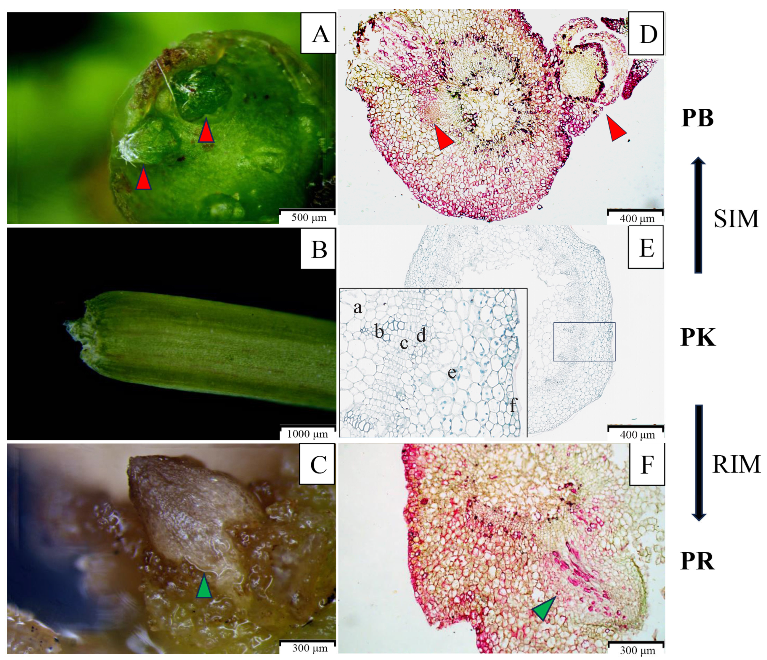
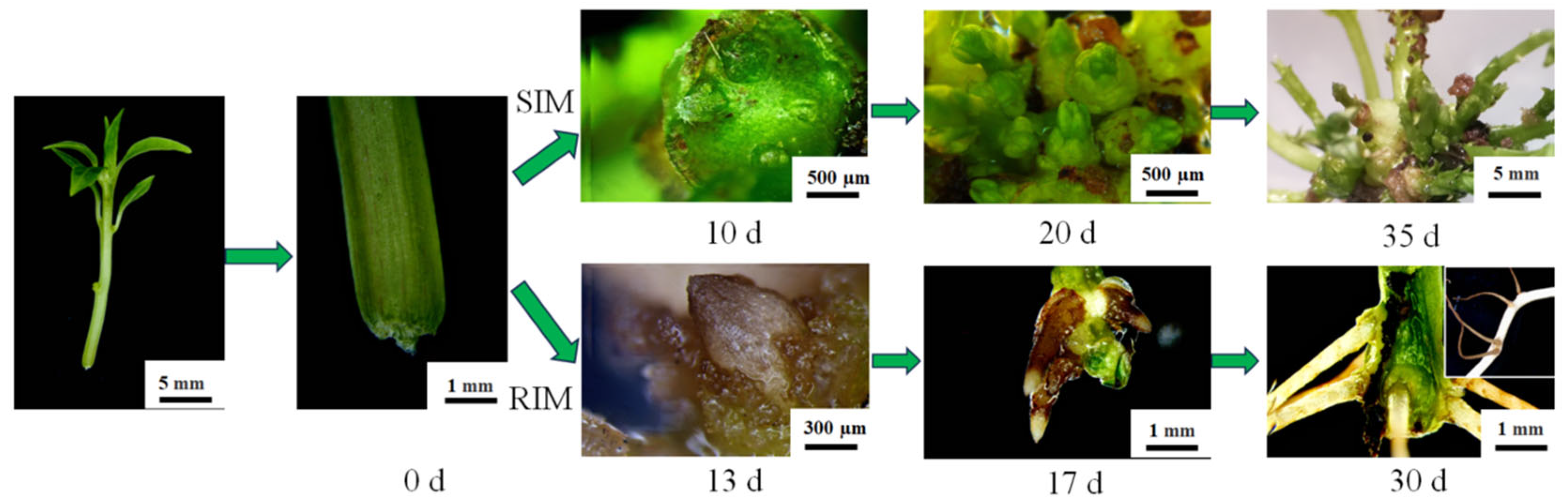
3.1. Analysis of Phenotype Characteristics of Explants at the Early Stage of In Vitro Regeneration

In this study, tissue-cultured seedlings were inoculated into different culture media to investigate the effects of various hormone combinations on the early phenotypic characteristics of in vitro regeneration. The results demonstrated that the induction of adventitious buds and roots predominantly occurred at the base of tissue-cultured seedlings (Figure 2). Specifically, in the treatment with a combination of 6-BA and NAA, bud primordia started to emerge at the base of tissue-cultured seedlings on the 10th day of culture. The number of bud primordia significantly increased and continued to develop on the 20th day, forming a cluster of adventitious buds by the 35th day. In contrast, upon induction with IBA and NAA, adventitious roots emerged at the base of tissue-cultured seedlings on the 13th day. A large number of brown adventitious roots grew densely on the 17th day, and secondary roots developed in the root system by the 30th day. These results indicate that different hormones regulate the morphogenesis of distinct organs.

3.2. Changes in Nutrient Content and Enzyme Activity

Figure 3 presents the nutrient content and enzyme activity of C. parthenoxylon samples at different stages. Significant differences (p < 0.05) were observed in the soluble sugar and soluble protein content at different stages, ordered as adventitious buds formation > pre-induction > adventitious roots formation (Figure 3A,B). Similarly to nutrient content, MDA levels and PPO activity were elevated during ARs formation, while they significantly decreased during ABs formation (Figure 3C,D). These results indicate that the elevation in soluble sugar, soluble protein, MDA content, and PPO activity may facilitate the induction of adventitious buds, while inhibiting the formation of adventitious roots. The activities of POD, SOD, and IAAO decreased during the morphogenesis of adventitious buds and roots. These findings suggest that POD, SOD, and IAAO enzymes may play an inhibitory role in organ differentiation. Specifically, POD and SOD activities in ARs formation were distinctly lower than those in ABs formation, whereas IAAO activity in ARs formation was higher than in ABs formation (Figure 3E,G).

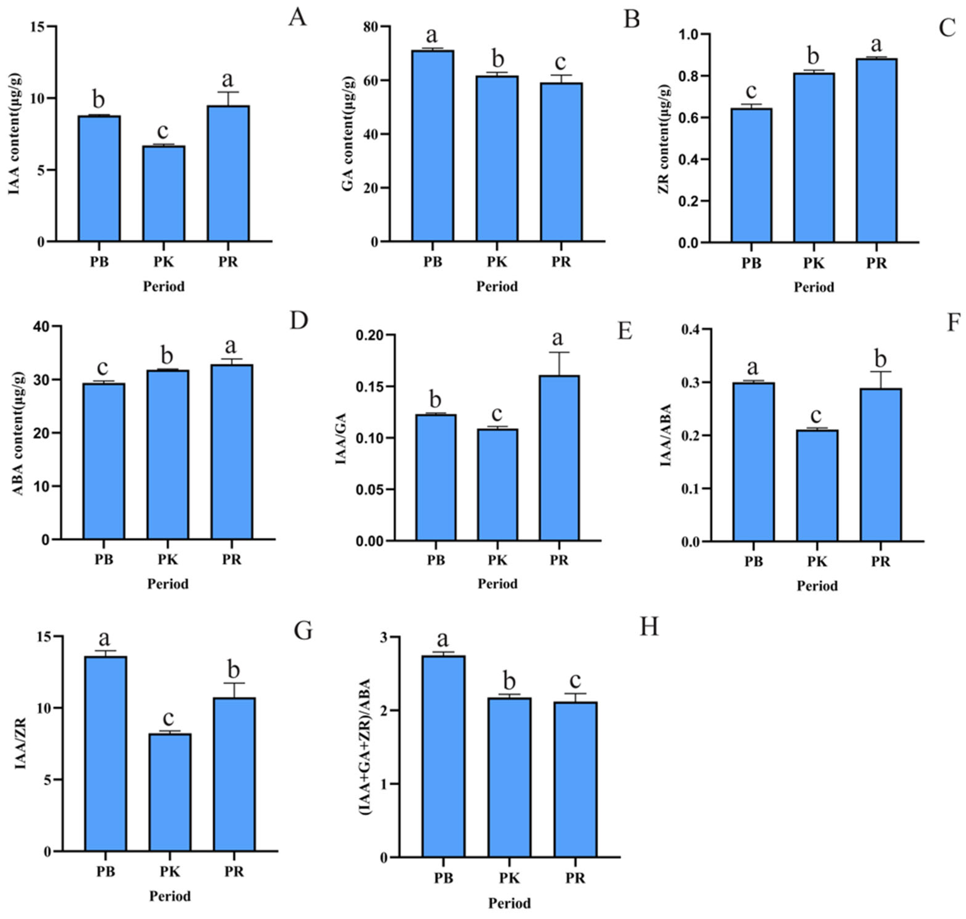
3.3. Changes in Endogenous Hormone Levels

As illustrated in Figure 4, the IAA content in the stem segments was higher in morphogenesis compared to pre-induction, with adventitious roots formation exhibiting significantly higher levels than adventitious buds formation (Figure 4A) (p < 0.05). The gradual increase in IAA concentration may be more conducive to organ morphogenesis, especially for adventitious roots. Compared to pre-induction, GA content increased in ABs formation, but decreased in ARs formation (Figure 4B), indicating that gibberellin may play a crucial role in regulating the differentiation direction of plant organs. The trends for ZR and ABA levels were opposite to that of GA, with levels in the order of adventitious roots formation > pre-induction > adventitious buds formation (Figure 4C,D). The ratios of IAA/GA, IAA/ABA, and IAA/ZR all increased significantly during the adventitious buds and roots morphogenesis, with IAA/GA being notably higher in ARs formation compared to ABs formation, while IAA/ABA and IAA/ZR showed the opposite trend (Figure 4E,G). Additionally, the combined ratio of (IAA + GA + ZR)/ABA was highest in ABs formation and lowest in ARs formation (Figure 4H).

3.4. Transcriptome and Differentially Expressed Genes Analysis

The correlation between biological replicates at the same stage was strong, while the correlation between samples from different stages was lower. The RNA sample demonstrated qualified quality (e.g., concentration ≥ 200 ng/μL, A260/A280 and A260/A230 > 2.0) and meets the specifications for second-generation library preparation (Table S2 and Figure S2). After filtering and quality control, the transcriptomic data from nine samples produced at least 4.3 million clean reads per sample, with Q20 values exceeding 98.5%. The GC content of guanine (G) and cytosine (C) nucleotides ranged from 46.18% to 46.49%, and the N50 of the transcripts was 2158 (Tables S3 and S4). These results indicate high-quality transcriptome sequencing. Functional annotation of the unigene sequences was performed using seven databases, with the highest annotation rate (25.79%) observed in the Uniprot database, followed by 18.98% in the GO database and 10.19% in the KEGG database. After aligning the unigene sequences with the Nr database, the highest number of homologous sequences was identified for the Cinnamomum micranthum (Figure S5).
Using pre-induction stage as control, 4511 differentially expressed genes (DEGs) were identified during adventitious buds formation, with 1960 downregulated and 2551 upregulated genes (Figure 5A). In adventitious roots formation period, 12,211 DEGs were identified, of which 5028 were downregulated and 7183 were upregulated (Figure 5B). A Venn diagram was utilized to compare the DEGs between the two treatment groups, revealing 13,549 DEGs, including 1338 genes unique to ABs formation, 9238 genes unique to ARs formation, and 2973 shared DEGs (Figure 5C). Among the shared differentially expressed genes, approximately 75% exhibited the same expression trends in the morphogenesis of adventitious buds and roots, while 25% showed opposite expression trends (Figure 5D). The differentiation of the young stems involves the participation of numerous genes, and there may be a common regulatory network involved in the differentiation of both different organ morphogenesis processes.

3.5. Functional Enrichment Analysis of DEGs

To investigate the biological functions of differentially expressed genes involved in young stems differentiation of C. parthenoxylon, GO functional enrichment analysis was conducted. Based on p-values, the top 20 most significantly enriched GO terms in the biological process (BP), cellular component (CC), and molecular function (MF) categories were selected for visualization (Figure 6). A total of 434 terms were annotated for the DEGs involved in the differentiation of young stems, with 354 terms annotated during adventitious buds morphogenesis. In the BP category, apart from carbohydrate metabolic processes, the DEGs were mainly participated in cell division and differentiation, including changes in cell wall components. In the CC category, the genes were mainly enriched in extracellular regions, plasma membranes, vascular tissues, and cell walls. In the MF category, the most enriched terms included DNA-binding transcription factor activity, iron ion binding, and monooxygenase activity (Figure 6A). For the adventitious roots formation period, a total of 432 terms were annotated, with the most enriched BP being carbohydrate metabolic processes, which had significantly more associated genes than any other term. Other enriched processes included responses to oxidative stress and hydrogen peroxide catabolism. In the CC category, the genes were primarily enriched in extracellular regions, plasma membranes, and cell walls. In the MF category, the most enriched terms were DNA-binding transcription factor activity, iron ion binding, and oxidoreductase activity (Figure 6B).
To further understand the metabolic activities associated with the identified differentially expressed genes, KEGG annotation was performed, and the top 20 enriched pathways were selected for display (Figure 7). A total of 115 metabolic pathways were involved in adventitious buds and roots organ morphogenesis. Of these, 106 metabolic pathways participated in ABs formation, with plant hormone signal transduction, phenylpropanoid biosynthesis, MAPK signaling, and starch and sucrose metabolism pathways being the most enriched, containing 52, 34, 28, and 26 DEGs, respectively (Figure 7A). ARs formation involved 115 metabolic pathways, with carbon metabolism, plant hormone signal transduction, phenylpropanoid biosynthesis, and amino acid biosynthesis pathways being significantly enriched, with up to 100 DEGs involved (Figure 7B).

3.6. Analysis of DEGs in Major Enriched Pathways

Based on the expression patterns of genes during organ formation, the differentially expressed genes involved in the differentiation of the stem could be divided into two categories: one group showed significant changes in expression during both organ formation processes, indicating their involvement in the differentiation of the young stems, while the other group displayed differential expression specific to adventitious buds or roots differentiation, suggesting their role in regulating the direction of the differentiation of the young stems and organ morphogenesis. To explore their function in the differentiation of the young stems, we analyzed their expression in key enriched pathways.

3.6.1. Plant Hormone Signal Transduction Pathway

Plant hormone signal transduction plays a crucial role in de novo organogenesis in plants. The enrichment and annotation of differentially expressed genes in this pathway are depicted in Figure S3 and Tables S5 and S6. In C. parthenoxylon, this pathway was involved during both organ morphogenesis. We identified 52 DEGs during adventitious buds induction and 112 during roots induction, indicating a higher DEGs involvement in ARs formation. In the ARs formation period, the proteins regulated by DEGs in ethylene (ET) and brassinosteroid (BR) signaling pathways were enriched in the roots formation period, suggesting their primary involvement in adventitious roots formation.
Among the 128 DEGs associated with this pathway, 36 were differentially expressed in both adventitious buds and roots morphogenesis stages. These genes were mainly enriched in auxin (11 genes), CTK (9 genes), and BR (6 genes) signal transduction pathways, with additional enrichment in ABA, ET, salicylic acid (SA), and GA signaling pathways. Furthermore, 31 of the DEGs exhibited similar expression trends during both adventitious buds and roots formation, with most showing upregulation, indicating their significant role in promoting stem base differentiation. In the auxin signal transduction pathway, eight genes encoding proteins such as ARF, AUX/IAA, AUX1, and GH3 were upregulated during both morphogenetic processes, with higher expression levels observed in roots compared to buds. Conversely, two genes encoding SAUR exhibited downregulation. The differentially expressed genes involved in CTK and gibberellin signal transduction showed similar expression patterns to auxin-related differentially expressed genes during organ differentiation. DEGs in the BR and SA signaling pathways were downregulated. Additionally, certain DEGs showed opposite expression patterns during the two morphogenetic processes, including genes encoding GH3 (unigene11161), B-ARR (unigene4267), TCH4 (unigene47170), JA (unigene19699), and ABF (unigene10271), indicating their crucial role in regulating the direction of young stems differentiation (Figure 8 and Tables S5 and S6).
Furthermore, 16 DEGs were uniquely identified in the adventitious buds formation period, with upregulated expression mainly being enriched in the auxin signal transduction pathway (10 genes), including AUX/IAA, GH3, and SAUR. In contrast, 76 DEGs were identified only in the roots formation period, primarily related to auxin signal transduction (22 genes), followed by BR (12 genes), ABA (10 genes), ET (10 genes), and SA (9 genes). And genes encoding proteins such as TIR1 and GID1 exhibited differential expression, with TIR1 upregulated and GID1 downregulated, while these changes were not evident during buds formation. Additionally, genes encoding B-ARR proteins, such as unigene7472 and unigene4436, were downregulated during roots formation. The differentially expressed genes related to ET and BR signal transduction pathways were also downregulated in roots formation (Figure 8).

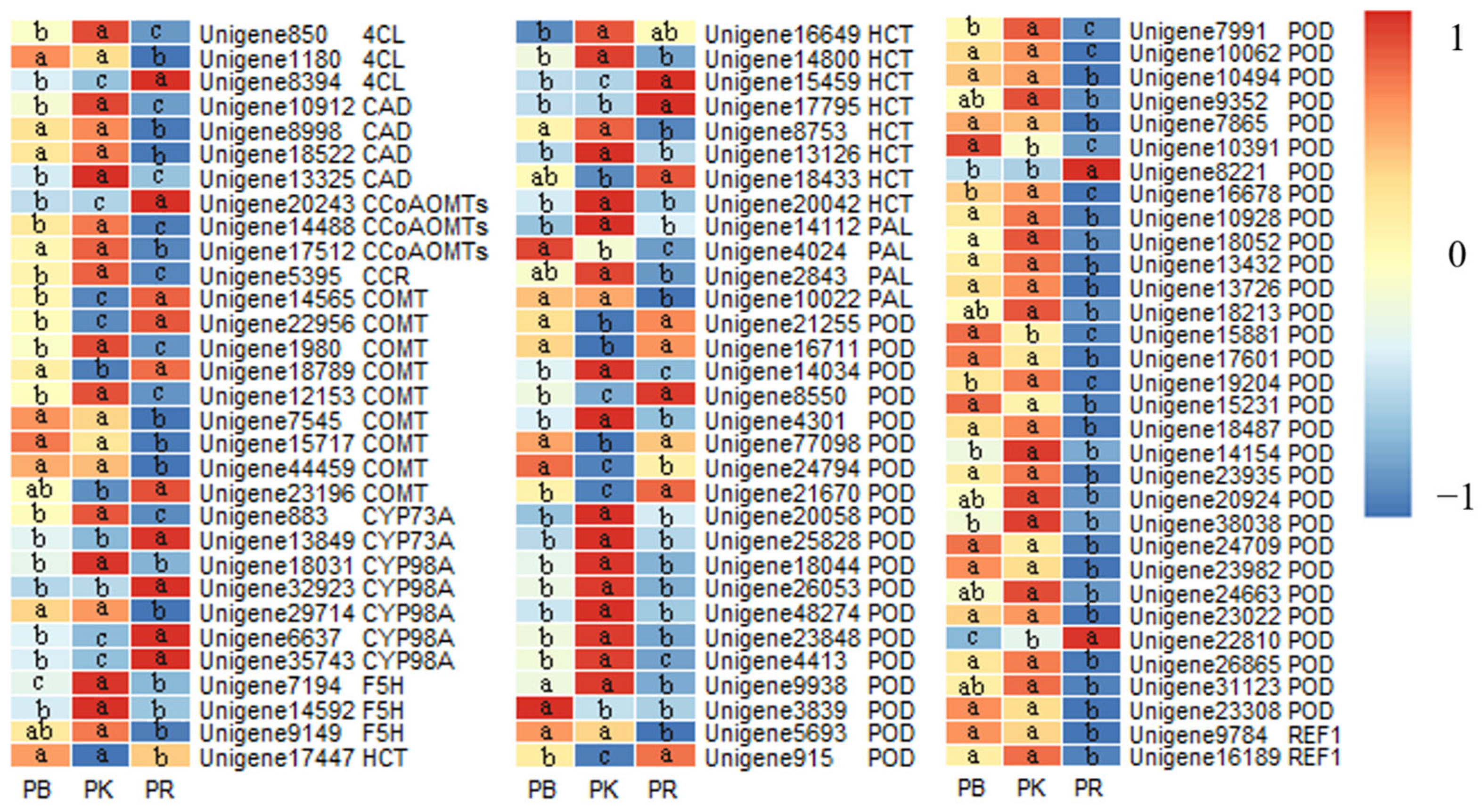
3.6.2. Phenylpropanoid Biosynthesis Pathway

In this study, more differentially expressed genes were involved in adventitious roots formation compared to buds formation, though roots formation had only three additional DEGs encoding proteins involved in the phenylpropanoid biosynthesis pathway, namely CCR, CYP73A, and REF1 (Figure S4 and Tables S7 and S8). Further analysis revealed that 31 common DEGs were enriched in this pathway during the stem differentiation of C. parthenoxylon. These included genes encoding key enzymes (PAL and 4CL) involved in the plant phenylpropanoid metabolic pathway, which are primarily responsible for regulating lignin biosynthesis. Of these, 16 genes were related to POD activity. Similarly to auxin, the common DEGs enriched in the phenylpropanoid biosynthesis pathway exhibited similar downregulation patterns in both periods, including PAL, 4CL, F5H, and others. Only one gene encoding POD showed differential expression in buds formation, while roots formation had a large number of DEGs, with most encoding the same proteins as the common DEGs, particularly those encoding POD, which were mostly downregulated. Additionally, three DEGs encoding CCR and CYP73A proteins were found, but this was only during roots formation (Figure 9).

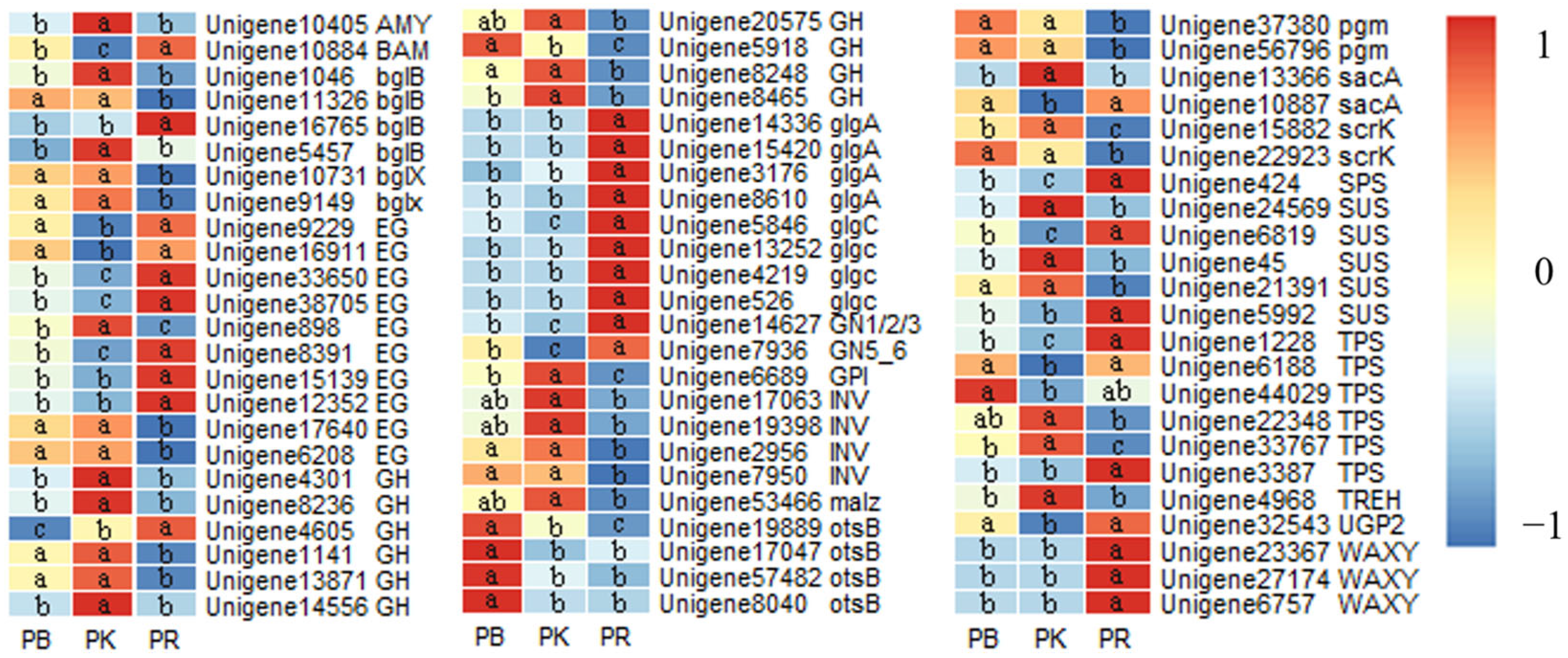
3.6.3. Starch and Sugar Metabolism Pathway

Sugars are not only essential components for plant life processes, but also the primary energy source for plants. In the starch and sugar metabolism pathway, 26 differentially expressed genes were involved in adventitious buds formation, mainly participating in the metabolism of sucrose, glucose, and trehalose (Figure S5A and Table S9). In the ARs formation period, 66 DEGs were involved, significantly more than in buds formation, with additional involvement in starch synthesis (Figure S5B and Table S10). During the differentiation of the young stems, 21 common DEGs were enriched in the starch and sugar metabolism pathway, primarily involved in the metabolism of sucrose, glucose, trehalose, and cellulose. In this pathway, four DEGs were involved in sucrose synthesis, two in sucrose decomposition, three in trehalose synthesis, nine in cellulose degradation to glucose, and one each in starch and maltose synthesis. Apart from the gene encoding trehalose-6-phosphate phosphatase, the expression trends of these genes were consistent in both stages. Further analysis of the DEGs involved in the starch and sugar metabolism pathway revealed five differentially expressed genes uniquely expressed in buds formation, primarily related to enzymes involved in trehalose synthesis, with upregulated expression (Figure S5A and Figure 10). In contrast, 45 DEGs were only differentially expressed in roots formation, participating not only in the metabolism of sucrose, glucose, trehalose, and cellulose, but also in the synthesis and metabolism of starch and maltose (Figure S5B and Figure 10).

3.7. Gene Expression Involved in the Metabolism of Related Substances

The expression levels of key genes involved in the metabolism of sucrose and related enzymes were analyzed using heat maps (Figure 11 and Table S11). SPS and SS are key enzymes involved in sucrose metabolism and accumulation. During the differentiation of the stem in C. parthenoxylon, most of the genes encoding SPS, except for Unigene34058, were downregulated during adventitious buds formation and upregulated during roots formation, while the expression trend of SS-encoding genes was opposite. Genes related to PPO were upregulated during buds formation and downregulated during roots formation. RBOHD, a gene involved in reactive oxygen species (ROS) synthesis, was downregulated during buds formation and upregulated during roots formation. Aside from Unigene26963, genes encoding SOD were downregulated during roots formation. In contrast to SOD-encoding genes, most catalase (CAT)-encoding genes were upregulated during adventitious root formation, whereas APX-encoding genes exhibited inconsistent expression patterns. Notably, the expression levels of Unigene1117 and Unigene5637 were significantly higher than those of other genes across all three developmental stages. During the differentiation of the stem, the YUC gene, a key regulator of auxin synthesis, was upregulated, with higher expression levels observed during roots formation compared to buds formation. The differential expression of CKX-encoding genes, which are involved in CTK degradation, showed upregulation during buds formation and downregulation during roots formation. Similarly, the differential expression of genes involved in the conversion of active GA was upregulated during buds formation and downregulated during roots formation. Furthermore, genes involved in ABA synthesis (NCED) and degradation (CYP707A) showed opposite expression trends during buds and roots formation, with increased synthesis and reduced degradation during roots formation.

3.8. Validation of Gene Expression

To verify the results of transcriptome sequencing, sixteen genes closely associated with the morphogenesis of adventitious buds and roots were selected from the DEGs for validation via qRT-PCR. These genes include CpGH3, CpGA2ox, CpSPS, and others. The expression trends observed were largely consistent with those identified by transcriptome sequencing, affirming the reliability of the RNA-seq results (Figure 12).

4. Discussion

Currently, plant regeneration in vitro is largely achieved under artificial control, relying on external stimuli (such as plant hormone analogs) and the plant’s response to these stimuli. In stem segments, cells with regenerative capacity are mainly located in the procambium and adjacent parenchyma cells, which serve as the cellular source for callus formation, adventitious buds, and adventitious roots during organ regeneration [16]. Stem differentiation in plants involves a series of physiological and biochemical reactions, with various nutrients, enzymes, and genes playing essential roles in this process. To investigate the intrinsic regulatory mechanisms of adventitious bud and root formation from the stems in C. parthenoxylon, this study analyzed the physiological, biochemical, and transcriptomic changes during the differentiation of the young stems, aiming to provide a theoretical foundation for directed organogenesis and efficient regeneration in C. parthenoxylon.

4.1. Physiological and Biochemical Changes During the Differentiation of the Young Stems

In this study, the contents of soluble sugars and proteins increased significantly in adventitious buds formation compared to pre-induction, but both decreased significantly during rooting. It is likely that 6-BA activates the sugar signaling pathway (INV) and induces ribosomal gene expression, thereby promoting glucose production and protein synthesis. In contrast, roots lack photosynthetic capacity, leading to nutrient consumption exceeding accumulation. A brief immersion in a high sucrose concentration solution was shown to promote ABs formation in Phoenix dactylifera L., leading to the development of bud clusters [45]. Conversely, in Morus alba Linn. cuttings, the soluble sugar content showed a decreasing trend [46]. Malondialdehyde (MDA) is a final decomposition product of lipid peroxidation and serves as an indicator of oxidative stress in cell membranes. During adventitious roots formation in C. parthenoxylon, MDA levels decreased significantly, suggesting that the formation of roots may require a low-concentration MDA environment. In Cucumis sativus L., inhibiting MDA accumulation increased the number of roots, root length, and root weight during ARs formation [47]. This study also observed a significant increase in MDA content during adventitious buds formation in C. parthenoxylon, indicating that reactive oxygen species (ROS) may play a role in regulating stem base differentiation towards buds, as the gene CpRBOHD, which regulates ROS synthesis, and the activity of SOD and related genes were significantly downregulated during this process. The studies found that the plant RBOHD mediates rapid systemic signal transduction to wounding, thereby inducing de novo organogenesis [48,49]. In callus tissues of Mesembryanthemum crystallinum L., high H2O2 concentrations promoted the regeneration potential for adventitious buds formation [50]. Enzymes such as polyphenol oxidase (PPO), peroxidase (POD), superoxide dismutase (SOD), and indoleacetic acid oxidase (IAAO) play crucial roles in plant morphogenesis and are positively regulated by auxin [40,51]. POD is involved in lignin synthesis, affecting cell wall formation and root lignification [52]. 6-BA may promote adventitious bud formation by regulating POD activity to modulate cell wall lignification in this study. In this study, SOD activity decreased after stems differentiation, possibly because the cultured young stems were derived from callus-bearing clusters before induction. However, their activity in adventitious roots was significantly lower than in buds, and the unigene1273 encoding SOD1, unigene19052 and unigene7798 encoding SOD2 were significantly downregulated in rooting. This difference in enzyme activity might be due to varying ROS environments required for the formation of different organs. Furthermore, significant changes in IAAO activity were observed, corresponding to changes in 3-indoleacetic acid (IAA) content. In summary, nutrients and oxidative enzymes may be regulated by endogenous hormones and contribute to organogenesis.
The levels of IAA, a key endogenous auxin hormone, significantly increased in both the organ morphogenesis stages of C. parthenoxylon, demonstrating that auxin is also implicated in the formation of adventitious buds [23]. Studies have demonstrated that defects in auxin synthesis or transport significantly impair bud regeneration [53]. In addition, it has also been demonstrated that auxin induces ROS production to regulate the cell cycle and ROS homeostasis. Conversely, ROS inhibits auxin signaling and activates oxidative stress signaling by triggering mitogen-activated protein kinase (MAPK) signaling [54]. Transcriptome analysis also revealed that the key gene CpYUC, which is involved in auxin synthesis, was significantly upregulated during the differentiation of the young stems, indicating that a large amount of auxin is required for this process. In Arabidopsis thaliana, following physical injury, an auxin synthesis source forms near the wound site, synthesizing large quantities of auxin to promote regeneration [30]. However, the auxin content was higher during ARs formation compared to ABs formation, suggesting that root morphogenesis requires higher concentrations of auxin. Plants regulate adventitious roots formation through the specific accumulation of auxin at the site, with various regulatory modules mediated by ARF in auxin signal transduction being critical to this biological process [30,55]. Cytokinin plays an important role in the induction of adventitious buds [56]. In this study, 6-BA was used to promote the formation of adventitious buds, but the endogenous ZR showed a downward trend, suggesting that it is possible that exogenous cytokinin can not only replace endogenous cytokinin, but also inhibit the synthesis and metabolism of endogenous cytokinin. Similar phenomena had also been found in other studies [57,58]. During adventitious buds formation in this study, the content of gibberellic acid (GA) also increased, while the content of abscisic acid (ABA) decreased, indicating that high GA levels and low ABA levels promote buds formation. In Saussurea involucrata, the increase in endogenous GA3 levels is a fundamental factor promoting bud morphogenesis [59]. Generally, GA levels are negatively correlated with rooting rates. High concentrations of GA in the stem not only promote the polar transport of auxin, but also interact with auxin to inhibit the differentiation of young stems into root primordial [60,61]. In this study, CpGA2OX, a gene regulating GA content, was significantly downregulated during adventitious roots formation, and GA levels also decreased significantly. In Capsicum annuum L., materials with a high induction rate and a high quantity of adventitious buds have lower endogenous ABA content [62]. ABA plays a dose-dependent role in adventitious roots formation in Cucumis sativus, participating in glucose-induced root formation [62]. These findings indicated that a high concentration of IAA is necessary for the differentiation of the young stems, with increased GA levels favoring ABs formation and inhibiting ARs formation, while ZR and ABA have opposing roles in regulating the differentiation of the adventitious buds and roots. The higher the IAA/ABA and CTK/ABA ratios, the more adventitious buds formation is promoted [63]. Similarly, this study found that the IAA/ABA and (IAA + GA + ZR)/ABA ratios significantly increased during adventitious buds differentiation. In summary, the levels of plant hormones and the balance between them are crucial for regulating the direction of young stems differentiation, and it is regulated by plant growth regulators. This process is similar to the differentiation of pluripotent callus into buds and roots [64].

4.2. Gene Expression During the Differentiation of the Young Stems

The low GO and KEGG annotation rates of unigenes in this study are likely due to the evolutionary distance between the target species and its relatives in public databases, resulting in suboptimal sequence matching efficiency. This phenomenon is prevalent in non-model species lacking a reference genome, particularly within the genus Cinnamomum, such as Cinnamomum bodinieri [65] and Cinnamomum camphora [66]. Transcriptome analysis showed that the differentiation of the young stems in C. parthenoxylon involves the expression of a large number of genes, with more differentially expressed genes involved in roots differentiation than in buds. This suggests that a more complex regulatory network is required for adventitious roots morphogenesis. Among the common DEGs in adventitious buds and roots morphogenesis, genes with opposite expression trends may play critical roles in young stems differentiation. Studies have shown that the same gene can function differently in the morphogenesis of different organs. For example, in Malus pumila, disrupting MdWOX11 promotes ABs regeneration, while overexpressing MdWOX11 enhances ARs primordium formation [57,67]. The young stems differentiation and organ morphogenesis involve cell division and differentiation [68]. Research has shown that the cell wall is a key structure in sensing wounds and promoting tissue regeneration in plants. Mechanical stress induced by cell-wall-loosening enzymes can fix microtubules, adjust the direction of cell division planes and tissue expansion, and activate de novo regeneration of buds in Arabidopsis [69,70]. During the differentiation of the stems in C. parthenoxylon, DEGs were mainly associated with microtubules, cell walls, transcription factor regulation, and enzyme activity, participating in carbohydrate metabolism and cell division. Major metabolic pathways involved in young stems differentiation include plant hormone signal transduction, sugar metabolism, and phenylpropanoid metabolism. These metabolic pathways work together to promote the differentiation of the young stems. These pathways and processes collectively regulate young stems differentiation and organ morphogenesis in C. parthenoxylon.
Plant hormone signal transduction plays a critical role in plant development and morphogenesis. In the auxin signal transduction pathway, AtTIR1 and AtARF are positive regulators of the adventitious roots formation in Arabidopsis [71], and their upregulation was also observed during roots formation in this study. The gene CpTIR1 was only differentially expressed during ARs formation. In addition, CTK signal transduction is also involved in regulating different organ morphogenesis in plants. For example, the members of the B-type ARR family in Arabidopsis have multiple functions and antagonistic roles in shoot regeneration, ARR1 acts as an inhibitor of ABs regeneration, while ARR12 promotes it. ARR1 inhibits shoot regeneration by competing with ARR12 to bind to the WUS promoter [72]. In C. parthenoxylon, CpARR was significantly upregulated during adventitious buds formation but downregulated during roots formation, suggesting its role in promoting bud formation and inhibiting root formation. The IBA treatment can promote SA increase during adventitious root formation in Robinia pseudoacacia L. [73]. In Arabidopsis, the salicylic acid (SA) receptor NPR1 promotes the degradation of the gibberellin (GA) receptor GID1, enhancing the stability of DELLA proteins and negatively regulating GA signal transduction [74]. In this study, the differential expression of CpNPR1 and CpGID1 was only observed during adventitious roots formation. In the SA signal transduction pathway, CpNPR1 and CpDELLA were upregulated, while CpGID1 was downregulated in the GA signal transduction pathway, suggesting that roots formation may be regulated by the negative modulation of GA signal transduction through the SA pathway in young stems differentiation. Ethylene (ET), a small molecule, participates in the formation and development of adventitious roots [75]. Research has shown that ethylene (ET) can promote ARs formation through its interaction with auxin, acting as a wound signal [74]. The addition of the ET-precursor ACC inhibited adventitious roots formation in Prunus persica L., and genes involved in ET biosynthesis and signaling pathway were downregulated in cuttings with a high rooting rate [76]. A similar downregulation was observed during roots formation in C. parthenoxylon, such as the genes encoding S-adenosylmethionine synthetase (SAMs) and ERF1/2. ERF1/2 is a member of the AP2/ERF transcription factor family. The AP2/ERF family of transcription factors is involved in plant morphogenesis, hormone signal transduction, and metabolite regulation [77]. In plant-wound signal transduction, ERF109, an important wound-responsive gene from the Arabidopsis AP2/ERF family, participates in wound signal transduction, promotes auxin biosynthesis, and reprograms stem cells, thus contributing to plant regeneration [78]. Furthermore, the brassinosteroids (BR) signaling was also involved in adventitious roots formation [79]. In a study on BR regulation of lateral root development, Xu et al. [80] found that BR signaling promotes root development by activating the expression of key enzymes involved in plant cell wall remodeling, leading to cell wall loosening around root primordia. This mechanism may also be present during adventitious roots formation in C. parthenoxylon. Plant hormone signal transduction played an important role in the process of adventitious buds and roots morphogenesis of C. parthenoxylon, and different hormones may be involved in the regulation of the differentiation direction of young stems.
The process of phenylpropane metabolism is involved in the differentiation process of the young stems in C. parthenoxylon. Flavonols, which act as endogenous inhibitors of auxin polar transport, play a role in the phenylpropanoid metabolism process during the morphogenesis of adventitious buds and roots in C. parthenoxylon. Overexpression of the flavonol-encoding gene Ok4CL11 can lead to the upregulation of genes involved in flavonoid biosynthesis and related glycosyltransferases, disrupting cofactor transport/signaling and inhibiting adventitious roots formation [81,82]. In this study, Cp4CL was significantly downregulated during roots formation, suggesting that a decrease in flavonol content promotes the occurrence of roots in C. parthenoxylon. Moreover, the phenylpropanoid pathway regulates lignin synthesis, which influences organ differentiation, with POD being a key enzyme. Previous studies have shown that the progressive lignification of callus tissues reduces the rooting rate of Platycladus orientalis L. cuttings, and that breaking the mechanical barrier of lignification can significantly improve rooting results [83]. During the differentiation of the young stems in C. parthenoxylon, genes related to lignin synthesis were significantly downregulated, promoting the formation of roots and buds. Beyond their role as an energy source, carbohydrates are involved in regulating flavonoid synthesis, lignin deposition in the phenylpropanoid pathway [84], and plant hormone signal transduction [85]. In Cucumis sativus, glucose promotes adventitious roots formation by enhancing the expression of ABA signal transduction genes like CsPYR1/2/8 [62], and in C. parthenoxylon, the upregulation of CpPYR was also observed during roots formation. Additionally, sucrose can induce MED17 transcription, occupying the promoter of the ARF7 gene, thus promoting its expression, activating auxin signaling, and enhancing root development [86]. Treatment with trehalose significantly inhibited MDA and ROS accumulation in Cucumis sativus, increased starch and trehalose levels, reduced glucose levels, and improved adventitious roots formation [47]. A similar phenomenon was observed during roots formation in C. parthenoxylon.
Exogenous growth regulators regulate the differentiation direction of organs, which involves a complex regulatory network, in which many endogenous secondary metabolites and genes are involved. Among all substances, plant hormones play a central regulatory role in the differentiation of adventitious roots and buds in C. parthenoxylon, precisely governing organ differentiation direction by mediating nutrient allocation and dynamic enzyme activity changes. Soluble sugars, soluble proteins, MDA, ZR, ABA, GA contents, the (IAA + GA + ZR)/ABA ratio, and PPO activity displayed contrasting expression patterns during the formation of adventitious buds and roots, it could be used as an indicator of the differentiation direction of young stems. Through further research and analysis of differentially expressed genes, it was found that there may be a common regulatory network in the formation of adventitious buds and roots, which are mainly involved in cell division and differentiation, including cell walls, enzymes, and other important substances. On this basis, the differentiation direction of young stems is regulated through the synthesis of endogenous hormones and signaling pathways. At the same time, phenylpropanoid metabolism and glucose metabolism pathways, as auxiliary pathways, promote the formation of adventitious buds and roots. In the future, important secondary metabolites and genes regulating the differentiation of adventitious buds and roots in C. parthenoxylon need be further screened and studied. Through the study and comparison of adventitious bud and root differentiation, this research aims to gain insights into the factors governing plant differentiation and uncover the regulatory mechanisms underlying plant morphogenesis during this process. Analyzing the changes in critical substances and examining the patterns and impacts associated with the changes will offer insights and references for optimizing vitro regeneration conditions.

5. Conclusions

Based on the same origin of ABs and ARs, the physiological substances and gene expression of cultured young stems under different exogenous growth regulator treatments were studied in this study. Adventitious buds and roots morphogenesis is governed by intricate regulatory networks, during which notable changes occur in nutrients, related enzymes, plant hormones, and gene expression. Soluble sugars, soluble proteins, MDA, ABA, GA contents, the (IAA + GA + ZR)/ABA ratio, and PPO activity displayed contrasting expression patterns during the formation of buds and roots, suggesting that they could serve as indicators for predicting the differentiation direction of stems. KEGG pathway analysis identified plant hormones, and signal transduction played a major role in the regulation of direction differentiation. Auxin emerged as a key hormone promoting the formation of both adventitious buds and roots. CTK and GA may act synergistically with auxin to modulate adventitious bud development, while ET, BR, SA, and ABA might collaborate with auxin to regulate ARs formation. In conclusion, this study explored the regulatory mechanisms of different hormones on the development of ABs and ARs, and compared the morphological processes of buds and roots on multiple levels. This research elucidated the regulatory mechanisms of different exogenous growth regulators on ABs and ARs development and compared the morphological processes of bud and root formation across multiple levels. These results not only enrich the gene resources for molecular mechanism research on organ morphogenesis, but also provide theoretical underpinnings for guiding the differentiation direction of adventitious buds and roots.

Supplementary Materials

The following supporting information can be downloaded at: https://www.mdpi.com/article/10.3390/f16071049/s1, Figure S1: The cultivation process of C. parthenoxylon; Figure S2: Agarose gel electrophoresis (1% agarose gel); Figure S3: Annotation of differentially expressed genes (DEGs) in the plant hormone signaling pathway; Figure S4: Annotation of differentially expressed genes (DEGs) in the phenylpropanoid biosynthesis pathway; Figure S5: Annotation of differentially expressed genes (DEGs) in the starch and sugar metabolism pathway; Table S1: List of primers; Table S2: RNA detection results; Table S3: Summary of clean reads; Table S4: Transcript Assembly Statistics Table; Table S5: Statistics of unigene Nr annotation results; Table S6: List of differentially expressed genes (DEGs) in the plant hormone signaling pathway during adventitious buds formation; Table S7: List of differentially expressed genes (DEGs) in the plant hormone signaling pathway during adventitious roots formation; Table S8: List of differentially expressed genes (DEGs) in the phenylpropanoid biosynthesis pathway during adventitious buds formation; Table S9: List of differentially expressed genes (DEGs) in the phenylpropanoid biosynthesis pathway during adventitious roots formation; Table S10: List of differentially expressed genes (DEGs) in the phenylpropanoid biosynthesis pathway during adventitious buds formation; Table S11: List of differentially expressed genes (DEGs) in the phenylpropanoid biosynthesis pathway during adventitious roots formation; Table S12: List of differentially expressed genes (DEGs) involved in nutrient metabolism, related enzymes, and hormone metabolism during the differentiation of the vascular cambium; Table S13: The FPKM of five actin-encoding genes.

Author Contributions

Conceptualization, X.L. and X.Z.; software, C.L.; formal analysis, C.L., X.D., T.Z. and Y.Z. (Yueting Zhang); investigation, C.L., X.D., Y.Z. (Yueting Zhang), T.Z., X.Z. and X.L.; resources, T.Z., X.D., Y.Z. (Yongjie Zheng) and X.Z.; writing—original draft preparation, C.L. and T.Z.; writing—review and editing, C.L., X.D. and T.Z.; visualization, C.L.; supervision, X.L. and X.Z.; project administration, X.L. and X.Z.; funding acquisition, X.L. All authors have read and agreed to the published version of the manuscript.

Funding

This research was funded by the Natural Science Foundation of Jiangxi Province (Project No. 20224BAB205030), Basic Research and Talent Scientific Research Special Project of Jiangxi Academy of Forestry (Project No. 2025522001) and the Forestry Science and Technology Innovation Project of Jiangxi Forestry Bureau (Project No. 202205).

Data Availability Statement

The raw sequence data reported in this paper have been deposited in the Genome Sequence Archive (Genomics, Proteomics & Bioinformatics 2021) in the National Genomics Data Center (Nucleic Acids Res 2024), China National Center for Bioinformation/Beijing Institute of Genomics, Chinese Academy of Sciences (GSA: CRA019666), which are publicly accessible at https://bigd.big.ac.cn/gsa/browse/CRA019666, accessed on 1 November 2024.

Acknowledgments

We thank the reviewers for their critical reviews of the manuscript.

Conflicts of Interest

The authors declare no conflicts of interest.

Abbreviations

The following abbreviations are used in this manuscript:
ABsAdventitious buds
ARsAdventitious roots
PB7 days of ABs induction
PKPre-induction
PR9 days of ARs induction
DEGsDifferentially expressed genes

References

  1. Qiu, F.; Yang, H.; Fu, C.; Zhou, S.; Sheng, Y.; Zhang, T. Essential oil diversity of Cinnamomum parthenoxylon (Jack) Meisner from China. S. Afr. J. Bot. 2023, 158, 452–460. [Google Scholar] [CrossRef]
  2. Alencar-Luciano, W.; Magnani, M.; Martín-Belloso, O.; Salvia-Trujillo, L. Effect of digestible versus non-digestible citral nanoemulsions on human gut microorganisms: An in vitro digestion study. Food Res. Int. 2023, 173, 113313. [Google Scholar] [CrossRef] [PubMed]
  3. Wang, J.; Su, B.; Jiang, H.; Cui, N.; Yu, Z.; Yang, Y.; Sun, Y. Traditional uses, phytochemistry and pharmacological activities of the genus Cinnamomum (Lauraceae): A review. Fitoterapia 2020, 146, 104675. [Google Scholar] [CrossRef]
  4. Dallay, C.; Malhiac, C.; Duchemin, B.; Savary, G.; Picard, C. Effect of linalool on lamellar-structured emulsions: From molecular organization to organoleptic properties. Food Hydrocoll. 2024, 149, 109575. [Google Scholar] [CrossRef]
  5. Zhou, Y.; Yan, W. Conservation and applications of camphor tree (Cinnamomum camphora) in China: Ethnobotany and genetic resources. Genet. Resour. Crop Evol. 2016, 63, 1049–1061. [Google Scholar] [CrossRef]
  6. Zhou, S.; Zhang, T.; Sheng, Y.; Wang, W.; Wen, S.; Qiu, F. Changes in essential oil contents and principal components of Cinnamomum parthenoxylon at different ages. South China For. Sci. 2023, 51, 5–10. (In Chinese) [Google Scholar]
  7. Costa Junior, E.D.S.; Barbosa, M.S.d.M.; Silva, C.M.d.; Silva, R.V.C.d.; Kiill, L.H.P.; Beckmann-Cavalcante, M.Z. Vegetative propagation of Rhaphiodon echinus Schauer (Lamiaceae): Effects of the period of cutting in rooting, cuttings arrangement and IBA concentrations for seedlings production. Ornam. Hortic. 2018, 3, 238–247. [Google Scholar] [CrossRef]
  8. Qiu, F.; Zhong, Y.; Zhang, T.; Liu, X.; Li, J. Effect of scion types and leaves number on cutting propagation of Cinnamomum porrectum. South China For. Sci. 2018, 46, 25–28. (In Chinese) [Google Scholar]
  9. Tate, H.; Page, T. Cutting propagation of Santalum austrocaledonicum: The effect of genotype, cutting source, cutting size, propagation medium, IBA and irradiance. New For. 2018, 49, 551–570. [Google Scholar] [CrossRef]
  10. Gautam, R.D.; Kumar, A.M.; Kumar, R.; Chauhan, R.; Singh, S.; Kumar, M.; Kumar, D.; Kumar, A.; Singh, S. Clonal propagation of Valeriana jatamansi retains the essential oil profile of mother plants: An approach toward generating homogenous grade of essential oil for industrial use. Front. Plant Sci. 2021, 12, 438247. [Google Scholar] [CrossRef]
  11. Sang, Y.L.; Cheng, Z.J.; Zhang, X.S. Plant stem cells and de novo organogenesis. New Phytol. 2018, 218, 1334–1339. [Google Scholar] [CrossRef]
  12. Verdeil, J.-L.; Alemanno, L.; Niemenak, N.; Tranbarger, T.J. Pluripotent versus totipotent plant stem cells: Dependence versus autonomy? Trends Plant Sci. 2007, 12, 245–252. [Google Scholar] [CrossRef] [PubMed]
  13. Pokimica, N.; Ćosić, T.; Uzelac, B.; Ninković, S.; Raspor, M. Dissecting the roles of the cytokinin signaling network: The case of de novo shoot apical meristem formation. Biomolecules 2024, 14, 318. [Google Scholar] [CrossRef] [PubMed]
  14. Moreno-García, I.; Rodríguez, V.; García-Sogo, B.; Ventura, C.; Moreno, V.; Pineda, B. The first protocol for in vitro propagation of kalanchoe beharensis through adventitious shoots, a preliminary study. Horticulturae 2024, 10, 1379. [Google Scholar] [CrossRef]
  15. Zhai, N.; Xu, L. Pluripotency acquisition in the middle cell layer of callus is required for organ regeneration. Nat. Plants 2021, 7, 1453–1460. [Google Scholar] [CrossRef]
  16. Liu, J.; Sheng, L.; Xu, Y.; Li, J.; Yang, Z.; Huang, H.; Xu, L. WOX11 and 12 are involved in the first-step cell fate transition during de novo root organogenesis in Arabidopsis. Plant Cell 2014, 26, 1081–1093. [Google Scholar] [CrossRef]
  17. Pan, R.; Liu, Y.; Buitrago, S.; Jiang, W.; Gao, H.; Han, H.; Wu, C.; Wang, Y.; Zhang, W.; Yang, X. Adventitious root formation is dynamically regulated by various hormones in leaf-vegetable sweetpotato cuttings. J. Plant Physiol. 2020, 253, 153267. [Google Scholar] [CrossRef] [PubMed]
  18. Zhang, Y.; Dong, J. Cell polarity: Compassing cell division and differentiation in plants. Curr. Opin. Plant Biol. 2018, 45, 127–135. [Google Scholar] [CrossRef]
  19. Ruan, J.; Lai, L.; Ou, H.; Yi, P. Two subtypes of GTPase-activating proteins coordinate tip growth and cell size regulation in Physcomitrium patens. Nat. Commun. 2023, 32, 2271. [Google Scholar] [CrossRef]
  20. Dai, X.; Liu, X.; Qiu, F.; Zhang, T.; Li, J. Establishment of regeneration system for stems of Cinnamomum porrectum. J. Cent. South Univ. For. Technol. 2018, 38, 20–25+33. (In Chinese) [Google Scholar]
  21. Xiao, Z.; Zheng, L.; Zhang, B.; Wang, Y.; Li, F.; Jiang, R.; Peng, Y.; Jin, Z. Study on tissue culture technology of stem segment of Cinnamomum camphora ‘Ganfang1’. J. Nanchang Inst. Technol. 2022, 41, 71–77. (In Chinese) [Google Scholar]
  22. Xiang, Z.; Wang, L.; Cao, L.; Liao, W.; Jin, Z.; Li, F.; Lv, X.; Zhang, H.; Zhao, J. Tissue Culture Technology of Stem Segment of Cinnamomum bodinieri var. citralifera. Bull. Bot. Res. 2020, 40, 196–201. (In Chinese) [Google Scholar]
  23. Su, Y.-H.; Liu, Y.-B.; Zhang, X.-S. Auxin–Cytokinin Interaction Regulates Meristem Development. Mol. Plant 2011, 4, 616–625. [Google Scholar] [CrossRef] [PubMed]
  24. Murashige, T.; Skoog, F.K. A revised medium for rapid growth and bio assays with tobacco tissue cultures. Physiol. Plant. 1962, 15, 473–497. [Google Scholar] [CrossRef]
  25. Zhanmu, O.; Yang, X.; Gong, H.; Li, X. Paraffin-embedding for large volume bio-tissue. Sci. Rep. 2020, 10, 12639. [Google Scholar] [CrossRef]
  26. Alagbe, E.; Amlabu, Y.; Daniel, E.; Ojewumi, M. Effect of varying drying temperature on the soluble sugar and nutritional content of banana. Open Chem. Eng. J. 2020, 14, 11–16. [Google Scholar] [CrossRef]
  27. Yemm, E.W.; Willis, A.J. The estimation of carbohydrates in plant extracts by anthrone. Biochem. J. 1954, 57, 508–514. [Google Scholar] [CrossRef] [PubMed]
  28. Li, D.; Li, C.; Sun, H.; Wang, W.; Liu, L.; Zhang, Y. Effects of drought on soluble protein content and protective enzyme system in cotton leaves. Front. Agric. China 2010, 4, 56–62. [Google Scholar] [CrossRef]
  29. Pierce, J.; Suelter, C. An evaluation of the Coomassie brilliant blue G-250 dye-binding method for quantitative protein determination. Anal. Biochem. 1977, 81, 478–480. [Google Scholar] [CrossRef]
  30. Brumós, J.; Robles, L.; Yun, J.; Vu, T.-C.; Jackson, S.; Alonso, J.M.; Stepanova, A.N. Local auxin biosynthesis is a key regulator of plant development. Dev. Cell 2018, 47, 306–318.e305. [Google Scholar] [CrossRef]
  31. Hedden, P.; Sponsel, V. A century of gibberellin research. J. Plant Growth Regul. 2015, 34, 740–760. [Google Scholar] [PubMed]
  32. Finkelstein, R. Abscisic acid synthesis and response. Arab. Book/Am. Soc. Plant Biol. 2013, 11, e0166. [Google Scholar] [CrossRef]
  33. Li, S.-M.; Zheng, H.-X.; Zhang, X.-S.; Sui, N. Cytokinins as central regulators during plant growth and stress response. Plant Cell Rep. 2021, 40, 271–282. [Google Scholar] [CrossRef]
  34. Niu, Q.; Zong, Y.; Qian, M.; Yang, F.; Teng, Y. Simultaneous quantitative determination of major plant hormones in pear flowers and fruit by UPLC/ESI-MS/MS. Anal. Methods 2014, 6, 1766–1773. [Google Scholar] [CrossRef]
  35. Morales, M.; Munné-Bosch, S. Malondialdehyde: Facts and Artifacts. Plant Physiol. 2019, 180, 1246–1250. [Google Scholar] [CrossRef] [PubMed]
  36. Draper, H.; Squires, E.; Mahmoodi, H.; Wu, J.; Agarwal, S.; Hadley, M. A comparative evaluation of thiobarbituric acid methods for the determination of malondialdehyde in biological materials. Free Radic. Biol. Med. 1993, 15, 353–363. [Google Scholar] [CrossRef] [PubMed]
  37. Gebhardt, K. Activation of indole-3-acetic acid oxidase from horseradish and Prunus by phenols and H2O2. Plant Growth Regul. 1982, 1, 73–84. [Google Scholar] [CrossRef]
  38. Bafana, A.; Dutt, S.; Kumar, A.; Kumar, S.; Ahuja, P.S. The basic and applied aspects of superoxide dismutase. J. Mol. Catal. B Enzym. 2011, 68, 129–138. [Google Scholar] [CrossRef]
  39. Yoruk, R.; Marshall, M.R. Physicochemical properties and function of plant polyphenol oxidase: A review 1. J. Food Biochem. 2003, 27, 361–422. [Google Scholar] [CrossRef]
  40. Tang, W.; Newton, R.J. Peroxidase and catalase activities are involved in direct adventitious shoot formation induced by thidiazuron in eastern white pine (Pinus strobus L.) zygotic embryos. Plant Physiol. Biochem. 2005, 43, 760–769. [Google Scholar] [CrossRef]
  41. Chen, S.; Zhou, Y.; Chen, Y.; Gu, J. Fastp: An ultra-fast all-in-one FASTQ preprocessor. Bioinformatics 2018, 34, 884–890. [Google Scholar] [CrossRef] [PubMed]
  42. Grabherr, M.G.; Haas, B.J.; Yassour, M.; Levin, J.Z.; Thompson, D.A.; Amit, I.; Adiconis, X.; Fan, L.; Raychowdhury, R.; Zeng, Q. Full-length transcriptome assembly from RNA-Seq data without a reference genome. Nat. Biotechnol. 2011, 29, 644–652. [Google Scholar] [CrossRef] [PubMed]
  43. Fu, L.; Niu, B.; Zhu, Z.; Wu, S.; Li, W. CD-HIT: Accelerated for clustering the next-generation sequencing data. Bioinformatics 2012, 28, 3150–3152. [Google Scholar] [CrossRef]
  44. Chen, C.; Chen, H.; Zhang, Y.; Thomas, H.R.; Frank, M.H.; He, Y.; Xia, R. TBtools: An integrative toolkit developed for interactive analyses of big biological data. Mol. Plant 2020, 13, 1194–1202. [Google Scholar] [CrossRef]
  45. Fki, L.; Bouaziz, N.; Kriaâ, W.; Benjemaa-Masmoudi, R.; Gargouri-Bouzid, R.; Rival, A.; Drira, N. Multiple bud cultures of ‘Barhee’ date palm (Phoenix dactylifera) and physiological status of regenerated plants. J. Plant Physiol. 2011, 168, 1694–1700. [Google Scholar] [CrossRef] [PubMed]
  46. Shang, C.; Yang, H.; Ma, S.; Shen, Q.; Liu, L.; Hou, C.; Cao, X.; Cheng, J. Physiological and transcriptomic changes during the early phases of adventitious root formation in mulberry stem hardwood cuttings. Int. J. Mol. Sci. 2019, 20, 3707. [Google Scholar] [CrossRef]
  47. Li, C.; Lu, X.; Liu, Y.; Xu, J.; Yu, W. Trehalose alleviates the inhibition of adventitious root formation caused by drought stress in cucumber through regulating ROS metabolism and activating trehalose and plant hormone biosynthesis. Plant Physiol. Biochem. 2023, 205, 108159. [Google Scholar] [CrossRef]
  48. Miller, G.; Schlauch, K.A.; Tam, R.C.Y.; Cortes, D.F.; Torres, M.A.; Shulaev, V.; Dangl, J.L.; Mittler, R. The plant NADPH oxidase RBOHD mediates rapid systemic signaling in response to diverse stimuli. Sci. Signal. 2009, 2, ra45. [Google Scholar] [CrossRef]
  49. Shin, S.Y.; Park, S.-J.; Kim, H.-S.; Jeon, J.-H.; Lee, H.-J. Wound-induced signals regulate root organogenesis in Arabidopsis explants. BMC Plant Biol. 2022, 22, 133. [Google Scholar] [CrossRef]
  50. Libik, M.; Konieczny, R.; Pater, B.; Ślesak, I.; Miszalski, Z. Differences in the activities of some antioxidant enzymes and in H2O2 content during rhizogenesis and somatic embryogenesis in callus cultures of the ice plant. Plant Cell Rep. 2005, 23, 834–841. [Google Scholar] [CrossRef]
  51. Bashri, G.; Prasad, S.M. Exogenous IAA differentially affects growth, oxidative stress and antioxidants system in Cd stressed Trigonella foenum-graecum L. seedlings: Toxicity alleviation by up-regulation of ascorbate-glutathione cycle. Ecotoxicol. Environ. Saf. 2016, 132, 329–338. [Google Scholar] [CrossRef]
  52. Zhai, X.; Yan, X.; Zenda, T.; Wang, N.; Dong, A.; Yang, Q.; Zhong, Y.; Xing, Y.; Duan, H. Overexpression of the peroxidase gene ZmPRX1 increases maize seedling drought tolerance by promoting root development and lignification. Crop J. 2024, 12, 753–765. [Google Scholar] [CrossRef]
  53. Rosspopoff, O.; Chelysheva, L.; Saffar, J.; Lecorgne, L.; Gey, D.; Caillieux, E.; Colot, V.; Roudier, F.; Hilson, P.; Berthomé, R.; et al. Direct conversion of root primordium into shoot meristem relies on timing of stem cell niche development. Development 2017, 144, 1187–1200. [Google Scholar] [CrossRef] [PubMed]
  54. Tognetti, V.B.; Mühlenbock, P.; Van Breusegem, F. Stress homeostasis–the redox and auxin perspective. Plant Cell Environ. 2012, 35, 321–333. [Google Scholar] [CrossRef]
  55. Zhang, T.; Ge, Y.; Cai, G.; Pan, X.; Xu, L. WOX-ARF modules initiate different types of roots. Cell Rep. 2023, 42, 112966. [Google Scholar] [CrossRef] [PubMed]
  56. Shin, J.; Bae, S.; Seo, P.J. De novo shoot organogenesis during plant regeneration. J. Exp. Bot. 2020, 71, 63–72. [Google Scholar] [CrossRef]
  57. Mao, J.; Ma, D.; Niu, C.; Ma, X.; Li, K.; Tahir, M.M.; Chen, S.; Liu, X.; Zhang, D. Transcriptome analysis reveals the regulatory mechanism by which MdWOX11 suppresses adventitious shoot formation in apple. Hortic. Res. 2022, 9, uhac080. [Google Scholar] [CrossRef] [PubMed]
  58. Peng, L.; Li, H.; Song, J.; Xie, W.; Zhang, L.; Li, S.-f.; Cai, Y.; Zhao, Z.-x. Morphological and transcriptome analyses reveal mechanism for efficient regeneration of adventitious buds from in vitro leaves of Rhododendron delavayi regulated by exogenous TDZ. Vitr. Cell. Dev. Biol. Plant 2022, 58, 1025–1037. [Google Scholar] [CrossRef]
  59. Guo, B.; He, W.; Zhao, Y.; Wu, Y.; Fu, Y.; Guo, J.; Wei, Y. Changes in endogenous hormones and H2O2 burst during shoot organogenesis in TDZ-treated Saussurea involucrate explants. Plant Cell Tissue Organ Cult. (PCTOC) 2016, 128, 1–8. [Google Scholar] [CrossRef]
  60. Mäkilä, R.; Wybouw, B.; Smetana, O.; Vainio, L.; Solé-Gil, A.; Lyu, M.; Ye, L.; Wang, X.; Siligato, R.; Jenness, M.K.; et al. Gibberellins promote polar auxin transport to regulate stem cell fate decisions in cambium. Nat. Plants 2023, 9, 631–644. [Google Scholar] [CrossRef]
  61. Mauriat, M.; Petterle, A.; Bellini, C.; Moritz, T. Gibberellins inhibit adventitious rooting in hybrid aspen and Arabidopsis by affecting auxin transport. Plant J. Cell Mol. Biol. 2014, 78, 372–384. [Google Scholar] [CrossRef] [PubMed]
  62. Li, X.; Mushtaq, N.; Xing, N.; Wu, S.; Liu, J.; Wang, Z. Efficient In vitro regeneration system and comparative transcriptome analysis offer insight into the early development characteristics of explants from cotyledon with partial petiole in small-fruited pepper (Capsicum annuum). Int. J. Mol. Sci. 2024, 25, 7547. [Google Scholar] [CrossRef] [PubMed]
  63. Li, C.; Hou, X.; Mou, K.; Liu, H.; Zhao, Z.; Liao, W. The involvement of abscisic acid in glucose promoted adventitious root development in cucumber. Sci. Hortic. 2022, 295, 110816. [Google Scholar] [CrossRef]
  64. Zhai, N.; Pan, X.; Zeng, M.; Xu, L. Developmental trajectory of pluripotent stem cell establishment in Arabidopsis callus guided by a quiescent center-related gene network. Development 2023, 150, 200879. [Google Scholar] [CrossRef] [PubMed]
  65. Yu, W.; Li, Y.; Zhang, Y.; Luo, C.; Zheng, Y.; Zhang, T.; Fu, C.; Liu, X. Integrated Physiological and Transcriptomic Analyses Reveal Mechanisms Regulating Endogenous Phytohormones in Adventitious Root Formation During Cinnamomum bodinieri Cutting Propagation. Forests 2025, 16, 509. [Google Scholar] [CrossRef]
  66. Chen, C.; Zheng, Y.; Zhong, Y.; Wu, Y.; Li, Z.; Xu, L.; Xu, M. Transcriptome analysis and identification of genes related to terpenoid biosynthesis in Cinnamomum camphora. BMC Genom. 2018, 19, 550. [Google Scholar] [CrossRef] [PubMed]
  67. Mao, J.; Niu, C.; Li, K.; Fan, L.; Liu, Z.; Li, S.; Ma, D.; Tahir, M.M.; Xing, L.; Zhao, C.; et al. Cytokinin-responsive MdTCP17 interacts with MdWOX11 to repress adventitious root primordium formation in apple rootstocks. Plant Cell 2022, 35, 1202–1221. [Google Scholar] [CrossRef]
  68. Fox, S.; Southam, P.; Pantin, F.; Kennaway, R.; Robinson, S.; Castorina, G.; Sánchez-Corrales, Y.E.; Sablowski, R.; Chan, J.; Grieneisen, V. Spatiotemporal coordination of cell division and growth during organ morphogenesis. PLoS Biol. 2018, 16, e2005952. [Google Scholar] [CrossRef]
  69. Varapparambath, V.; Mathew, M.M.; Shanmukhan, A.P.; Radhakrishnan, D.; Kareem, A.; Verma, S.; Ramalho, J.J.; Manoj, B.; Vellandath, A.R.; Aiyaz, M.; et al. Mechanical conflict caused by a cell-wall-loosening enzyme activates de novo shoot regeneration. Dev. Cell 2022, 57, 2063–2080. [Google Scholar] [CrossRef]
  70. Hoermayer, L.; Montesinos, J.; Trozzi, N.; Spona, L.; Yoshida, S.; Marhava, P.; Caballero-Mancebo, S.; Benková, E.; Heisenberg, C.-P.; Dagdas, Y.; et al. Mechanical forces in plant tissue matrix orient cell divisions via microtubule stabilization. Dev. Cell 2024, 59, 1333–1344. [Google Scholar] [CrossRef]
  71. Lakehal, A.; Chaabouni, S.; Cavel, E.; Le Hir, R.; Ranjan, A.; Raneshan, Z.; Novák, O.; Păcurar, D.I.; Perrone, I.; Jobert, F.; et al. A molecular framework for the control of adventitious rooting by TIR1/AFB2-Aux/IAA-Dependent auxin signaling in Arabidopsis. Mol. Plant 2019, 12, 1499–1514. [Google Scholar] [CrossRef]
  72. Liu, Z.; Dai, X.; Li, J.; Liu, N.; Liu, X.; Li, S.; Xiang, F. The Type-B cytokinin response regulator ARR1 inhibits shoot regeneration in an ARR12-dependent manner in Arabidopsis. Plant Cell 2020, 32, 2271–2291. [Google Scholar] [CrossRef] [PubMed]
  73. Uddin, S.; Munir, M.Z.; Larriba, E.; Pérez-Pérez, J.M.; Gull, S.; Pervaiz, T.; Mahmood, U.; Mahmood, Z.; Sun, Y.; Li, Y. Temporal profiling of physiological, histological, and transcriptomic dissection during auxin-induced adventitious root formation in tetraploid Robinia pseudoacacia micro-cuttings. Planta 2024, 259, 66. [Google Scholar] [CrossRef] [PubMed]
  74. Yu, X.; Cui, X.; Wu, C.; Shi, S.; Yan, S. Salicylic acid inhibits gibberellin signaling through receptor interactions. Mol. Plant 2022, 15, 1759–1771. [Google Scholar] [CrossRef]
  75. Deng, Y.; Wang, C.; Wang, N.; Wei, L.; Li, W.; Yao, Y.; Liao, W. Roles of small-molecule compounds in plant adventitious root development. Biomolecules 2019, 9, 420. [Google Scholar] [CrossRef]
  76. Park, S.-H.; Elhiti, M.; Wang, H.; Xu, A.; Brown, D.; Wang, A. Adventitious root formation of in vitro peach shoots is regulated by auxin and ethylene. Sci. Hortic. 2017, 226, 250–260. [Google Scholar] [CrossRef]
  77. Feng, K.; Hou, X.; Xing, G.; Liu, J.; Duan, A.; Xu, Z.; Li, M.; Zhuang, J.; Xiong, A. Advances in AP2/ERF super-family transcription factors in plant. Crit. Rev. Biotechnol. 2020, 40, 750–776. [Google Scholar] [CrossRef]
  78. Zhang, G.; Zhao, F.; Chen, L.; Pan, Y.; Sun, L.; Bao, N.; Zhang, T.; Cui, C.-X.; Qiu, Z.; Zhang, Y.; et al. Jasmonate-mediated wound signalling promotes plant regeneration. Nat. Plants 2019, 5, 491–497. [Google Scholar] [CrossRef] [PubMed]
  79. Wen, Z.; Chen, Z.-f.; Liu, X.; Sun, J.; Zhang, F.; Zhang, M.; Dong, C. 24-epibrassinolide facilitates adventitious root formation by coordinating cell-wall polyamine oxidase- and plasma membrane respiratory burst oxidase homologue-derived reactive oxygen species in Capsicum annuum L. Antioxidants 2023, 12, 1425. [Google Scholar] [CrossRef]
  80. Xu, P.; Fang, S.; Chen, H.; Cai, W. The brassinosteroid (BR) responsive xyloglucan endotransglucosylase/hydrolase 19 (XTH19) and XTH23 genes are involved in lateral root development under salt stress in Arabidopsis. Plant J. Cell Mol. Biol. 2020, 104, 59–75. [Google Scholar] [CrossRef]
  81. Tan, H.; Man, C.; Xie, Y.; Yan, J.; Chu, J.; Huang, J. A crucial role of ga-regulated flavonol biosynthesis in root growth of Arabidopsis. Mol. Plant 2019, 12, 521–537. [Google Scholar] [CrossRef] [PubMed]
  82. Lavhale, S.G.; Kondhare, K.R.; Sinthadurai, V.S.; Barvkar, V.T.; Kale, R.S.; Joshi, R.S.; Giri, A.P. Ocimum kilimandscharicum 4CL11 negatively regulates adventitious root development via accumulation of flavonoid glycosides. Plant J. Cell Mol. Biol. 2024, 119, 176–196. [Google Scholar] [CrossRef] [PubMed]
  83. Chang, E.; Guo, W.; Dong, Y.; Jia, Z.; Zhao, X.; Jiang, Z.; Zhang, L.; Zhang, J.; Liu, J. Metabolic profiling reveals key metabolites regulating adventitious root formation in ancient Platycladus orientalis cuttings. Front. Plant Sci. 2023, 14, 1192371. [Google Scholar] [CrossRef] [PubMed]
  84. Dong, N.; Lin, H. Contribution of phenylpropanoid metabolism to plant development and plant–environment interactions. J. Integr. Plant Biol. 2021, 63, 180–209. [Google Scholar] [CrossRef] [PubMed]
  85. Mao, J.; Li, W.; Mi, B.; Ma, Z.; Dawuda, M.M.; Zuo, C.; Zhang, Y.; Jiang, X.; Chen, B. Transcriptome analysis revealed glucose application affects plant hormone signal transduction pathway in “Red Globe” grape plantlets. Plant Growth Regul. 2018, 84, 45–56. [Google Scholar] [CrossRef]
  86. Agrawal, R.; Singh, A.; Giri, J.; Magyar, Z.; Thakur, J.K. MEDIATOR SUBUNIT17 is required for transcriptional optimization of root system architecture in Arabidopsis. Plant Physiol. 2023, 192, 1548–1568. [Google Scholar] [CrossRef]
Figure 1. Differentiation of the young stems into adventitious buds and roots under different hormonal treatments. (AC) Morphological observations of the basal stem segments of cultured young stems at different stages. (A) After 10 days of adventitious buds induction, protrusions of ABs appeared on the stem surface; (B) before induction, the stem surface was smooth; (C) after 13 days of adventitious roots induction, protrusions of ARs formed on the stem surface. (DF) Anatomical observations of the basal stem segments of cultured young stems at different stages. (D) After 7 days of adventitious buds induction, initial bud organs appeared on the stem surface and there were meristem cell clusters in stem segments; (E) before induction, no buds or root primordia were observed. a, pith, b, xylem, c, vascular cambium. d, phloem. e, cortex. f, epidermis; (F) after 9 days of adventitious roots induction, root primordial cells continued to divide, and the root primordium elongated. PB: 7 days of ABs induction, PK: pre-induction, PR: 9 days of ARs induction, SIM: adventitious buds induction medium, RIM: adventitious roots induction medium. Red arrows indicate ABs and green arrows indicate ARs.
Forests 16 01049 g001
Figure 2. The morphogenesis of adventitious root and adventitious bud. SIM: adventitious bud induction medium (MS + 2.0 mg/L 6-BA + 0.5 mg/L NAA). RIM: adventitious root induction medium (MS + 0.5 mg/L NAA + 0.5 mg/L IBA).
Forests 16 01049 g002
Figure 3. Changes in nutrient content and enzyme activity in morphogenesis of adventitious buds and roots. (A) Soluble sugar. (B) Soluble protein. (C) MDA. (D) PPO. (E) POD. (F) IAAO. (G) SOD. The data represent mean ± SD (n = 6). Different lowercase letters (a, b, and c) indicate significant differences in physiological indices between different samples at the 0.05 level based on one-way ANOVA and LSD multiple comparison. PB: 7 days of ABs induction in medium (MS + 2.0 mg/L 6-BA + 0.5 mg/L NAA); PK: pre-induction; PR: 9 days of ARs induction in medium (MS + 0.5 mg/L IBA + 0.5 mg/L NAA).
Forests 16 01049 g003
Figure 4. Changes in endogenous hormone levels in morphogenesis of two organs. (A) IAA. (B) GA. (C) ZR. (D) ABA. (E) IAA/GA. (F) IAA/ABA. (G) IAA/ZR. (H) (IAA + GA + ZR)/ABA. The data represent mean ± SD (n = 6). Different lowercase letters (a, b, and c) indicate significant differences in physiological indices between different samples at the 0.05 level based on one-way ANOVA and LSD multiple comparison. PB: 7 days of ABs induction in medium (MS + 2.0 mg/L 6-BA + 0.5 mg/L NAA); PK: pre-induction; PR: 9 days of ARs induction in medium (MS + 0.5 mg/L IBA + 0.5 mg/L NAA).
Forests 16 01049 g004
Figure 5. Analysis of differentially expressed genes in morphogenesis of adventitious buds and roots. (A,B) Volcano plots of differentially expressed genes. (A) PB vs. PK, adventitious buds induction process; (B) PR vs. PK, adventitious roots induction process. (C) Venn diagram of DEGs. (D) Pie chart showing the expression trends of common DEGs involved in both morphogenetic processes. Red indicates DEGs with the same expression trend in both processes, and blue indicates DEGs with opposite expression trends in both processes.
Forests 16 01049 g005
Figure 6. GO enrichment analysis of differentially expressed genes in morphogenesis of adventitious buds and roots. (A) GO enrichment of differentially expressed genes during adventitious buds formation. (B) GO enrichment of differentially expressed genes during adventitious roots formation.
Forests 16 01049 g006
Figure 7. KEGG enrichment analysis of differentially expressed genes in morphogenesis of adventitious buds and roots. (A) KEGG enrichment of DEGs during ABs formation. (B) KEGG enrichment of DEGs during ARs formation.
Forests 16 01049 g007aForests 16 01049 g007b
Figure 8. Heat map analysis of differentially expressed genes related to hormone signaling pathway in morphogenesis of adventitious buds and roots. Colored squares represent the average expression levels of DEGs across three biological replicates. Each row represents a gene, and each square represents a stage. Red indicates high expression, and blue indicates low expression. PB: adventitious buds formation; PK: pre-induction; PR: adventitious roots formation. Different lowercase letters (a, b, and c) indicate significant differences in physiological indices between different samples at the 0.05 level based on one-way ANOVA and LSD multiple comparison.
Forests 16 01049 g008
Figure 9. Heat map analysis of differentially expressed genes related to the phenylpropanoid biosynthesis pathway in morphogenesis of adventitious buds and roots. Colored squares represent the average expression levels of DEGs across three biological replicates. Each row represents a gene, and each square represents a stage. Red indicates high expression, and blue indicates low expression. PB: adventitious buds formation; PK: pre-induction; PR: adventitious roots formation. Different lowercase letters (a, b, and c) indicate significant differences in physiological indices between different samples at the 0.05 level based on one-way ANOVA and LSD multiple comparison.
Forests 16 01049 g009
Figure 10. Heat map analysis of differentially expressed genes related to the starch and sugar metabolism pathway in morphogenesis of adventitious buds and roots. Colored squares represent the average expression levels of DEGs across three biological replicates. Each row represents a gene, and each square represents a stage. Red indicates high expression, and blue indicates low expression. PB: adventitious buds formation; PK: pre-induction; PR: adventitious roots formation. Different lowercase letters (a, b, and c) indicate significant differences in physiological indices between different samples at the 0.05 level based on one-way ANOVA and LSD multiple comparison.
Forests 16 01049 g010
Figure 11. Heat map analysis of differentially expressed genes involved in nutrient metabolism, related enzymes, and hormone metabolism in morphogenesis of adventitious buds and roots. Colored squares represent the expression levels of genes. Each row represents a gene, and each square represents a stage. Red indicates high expression, and blue indicates low expression. PB: adventitious buds formation; PK: pre-induction; PR: adventitious roots formation. Different lowercase letters (a, b, and c) indicate significant differences in physiological indices between different samples at the 0.05 level based on one-way ANOVA and LSD multiple comparison.
Forests 16 01049 g011
Figure 12. The qRT-PCR validation of genes in RNA-seq results. Each cell includes the values at different time points. Lines graphs show the relative expression levels by qRT-PCR. Blue bars represent the FPKM of RNA-seq. The data represent mean ± SD (n = 3). PB: adventitious buds formation; PK: pre-induction; PR: adventitious roots formation.
Forests 16 01049 g012
Disclaimer/Publisher’s Note: The statements, opinions and data contained in all publications are solely those of the individual author(s) and contributor(s) and not of MDPI and/or the editor(s). MDPI and/or the editor(s) disclaim responsibility for any injury to people or property resulting from any ideas, methods, instructions or products referred to in the content.

