Article

New Insights into the Role of Cytokinin in Regulating Anthocyanin Biosynthesis and Leaf Expansion: An Integrated Transcriptomic, Metabolomic, and Physiological Analysis of Hypericum monogynum

1
Guangxi Key Laboratory of Forest Ecology and Conservation, College of Forestry, Guangxi University, Nanning 530004, China
2
State Key Laboratory for Conservation and Utilization of Subtropical Agro-Bioresources, College of Forestry, Guangxi University, Nanning 530004, China
*
Author to whom correspondence should be addressed.
Forests 2025, 16(3), 465; https://doi.org/10.3390/f16030465
Submission received: 4 February 2025 / Revised: 19 February 2025 / Accepted: 4 March 2025 / Published: 6 March 2025
(This article belongs to the Section Forest Ecophysiology and Biology)

Abstract

:
Hypericum monogynum is a valuable perennial species with multiple uses, one of which is its ornamental value. In this study, we found that cytokinin treatment not only efficiently induced anthocyanin biosynthesis but also promoted leaf expansion in H. monogynum, both of which could potentially enhance its ornamental qualities. To investigate the molecular regulatory network underlying these processes, time-resolved transcriptomic sequencing and widely targeted metabolomic analysis were conducted. The results revealed 752, 385, and 1009 differentially expressed genes (DEGs) at 6, 12, and 24 h after 6-BA (6-benzylaminopurine) treatment, respectively. A total of 101 DEGs were co-regulated at all three time points, including key components of cytokinin metabolism and signaling. KEGG analysis identified metabolic pathways, biosynthesis of secondary metabolites, and plant hormone signal transduction as the most significantly enriched pathways. Key DEGs associated with the MYB and bHLH families, involved in flavonoid biosynthesis and cell proliferation, were also identified. Specifically, four MYB113 genes were found to be cytokinin-responsive and upregulated by 6-BA treatment at various time points. Several genes in the anthocyanin biosynthesis pathway, such as CHS, F3H, and F3′H, were upregulated by 6-BA treatment. Additionally, many DEGs related to nutrient transport, sugar metabolism, cell cycle, and cell expansion were identified, most of which were upregulated by 6-BA treatment, supporting cytokinin’s role in promoting leaf growth and expansion. Furthermore, metabolomic analysis revealed key differentially accumulated metabolites in the flavonoid biosynthesis pathway, with major anthocyanins being identified. In conclusion, this study suggests that cytokinin application is an effective strategy for enhancing the ornamental value of H. monogynum and provides new insights into its role in regulating anthocyanin biosynthesis and leaf expansion in tree species.

1. Introduction

Hypericum monogynum is a small, semi-evergreen tree or shrub with distinctive ornamental value and is widely distributed across tropical and subtropical regions. Species of the Hypericum genus are also rich in secondary metabolites with various medicinal properties, including antineoplastic, antimicrobial, anti-inflammatory, and antioxidant activities [1,2]. Previous studies have shown that Hypericum is particularly abundant in flavonoid glycosides, especially in its flowers, where quercitrin and hyperoside are the most abundant compounds [3]. As secondary metabolites, particularly flavonoids, play a crucial role in the medicinal and ornamental value of Hypericum, the study of their metabolic profiles and the regulation of their biosynthesis in medicinal and ornamental plants has become a central focus in horticultural research.
Anthocyanins are flavonoids formed by the metabolism of phenylpropanes from phenylalanine, playing important roles in both plants and animals [4]. As water-soluble natural pigments, they are synthesized in the cytoplasm and then transported to the vesicles [5,6]. Plants produce anthocyanins to attract pollinators and dispersers and also to protect the photosynthetic tissues from the oxidative damage caused by various environmental stresses, such as high light, cold, drought, or diseases [7]. Anthocyanin biosynthesis pathway, which involves the structural and regulatory genes, is conserved in so far studied plant species [8,9,10,11]. The first step of anthocyanin biosynthesis is the conversion of the initial substrate phenylalanine into the 4-coumaroyl-CoA by the activities of phenylalanine lyase (PAL), cinnamate 4-hydroxylase (C4H), and 4-coumarate-CoA ligase (4CL). Then, 4-coumaroyl-CoA is converted into dihydroflavonols with the involvement of chalcone synthase (CHS), chalcone isomerase (CHI), flavanone 3-hydroxylase (F3H) [12,13]. The third step is regulated by dihydroflavonol reductase (DFR) and anthocyanin synthase (ANS) [14], with the conversion of dihydroflavonols to form a variety of colorless basic anthocyanins [15]. After glycosylation with different ligands, different types of anthocyanins were formed. The synthesis and modification of anthocyanins are completed in the cytoplasm and endoplasmic reticulum membrane and then transported to the vacuoles for storage.
Phytohormones are a class of trace organic compounds that play a crucial role in various aspects of plant growth and development, including the regulation of anthocyanin biosynthesis [16]. Auxins and gibberellins (GAs) primarily exert a negative effect, while cytokinins (CKs), abscisic acid (ABA), ethylene (ETH), and jasmonic acid (JA) positively regulate anthocyanin biosynthesis [16]. CKs primarily serve as growth-promoting hormones and also play a role in regulating stress responses [17]. However, the relationship between their traditional role and stress defense (e.g., anthocyanin biosynthesis) remains unclear. In Eucalyptus, cytokinin could modulate the sugar transportation to activate anthocyanin biosynthesis [18]. In Arabidopsis, sugar-induced anthocyanin biosynthesis can be enhanced by cytokinin [19]. Hormonal regulation of anthocyanin biosynthesis appears to be influenced by sugar status, positioning sugar as a key mediator. In Arabidopsis, the promotion of anthocyanin biosynthesis by hormones depends on co-application with sugar [20,21]. These findings suggest that the regulation of anthocyanin biosynthesis is governed by a complex hormone–sugar regulatory network. Plants can produce anthocyanins in vegetative organs under environmental stresses such as salinity [22], high light [23], cold [24], and drought [25]. This process serves as an efficient mechanism to counteract stress-induced reactive oxygen species (ROS) and protect the photosynthetic system, thereby helping plants adapt to changing environments. One key mechanism for coordinating rapid growth and stress defense under various environmental conditions is the regulation of endogenous phytohormone homeostasis. For instance, in Eucalyptus, stresses likely inhibit gibberellin (GA) biosynthesis, which negatively impacts anthocyanin biosynthesis induced by high light and nutrient deficiency [26,27]. In most plant species, anthocyanin biosynthesis is regulated by a core system comprising a ternary complex of MYB–bHLH–WDR (MBW) transcription factors [28]. The MYB family, one of the largest gene families in plants, plays a crucial role in regulating cellular metabolism, including flavonoid and lignin biosynthesis [29,30]. Environmental or hormonal signals can directly target the expression of key MYB genes to initiate or suppress anthocyanin biosynthesis [31]. In Eucalyptus, EgrMYB113 is pivotal in regulating anthocyanin biosynthesis in response to cytokinin, high light, ROS, and nitrogen deficiency [18,26,27]. In Acer rubrum, the expression of ArMYB89 is closely linked to leaf pigmentation in autumn [32]. In Arabidopsis, MYB75, MYB90, MYB113, and MYB114 are master regulators of anthocyanin biosynthesis in response to various environmental signals [33]. Despite the substantial knowledge of the MBW regulatory module and its gene targets in the control of anthocyanin biosynthesis across different plant species, how environmental and hormonal signals coordinately regulate the MBW members remains largely unknown.
In this study, we discovered that cytokinin treatment alone could effectively induce anthocyanin biosynthesis and leaf expansion in H. monogynum, potentially enhancing its ornamental value as an important landscape plant. Despite its high ornamental and medicinal value, molecular and multi-omics studies on H. monogynum, particularly those related to the modification of ornamental traits, remain limited. To further investigate the cytokinin-mediated regulatory network underlying anthocyanin biosynthesis and leaf growth, we conducted high-throughput transcriptome and widely targeted metabolome analyses. This study identified key secondary metabolites in H. monogynum leaves, including specific anthocyanins, flavonoids, and other cytokinin-responsive medicinal metabolites, and constructed gene regulatory networks involving cytokinin-responsive signaling components, MBW members, and anthocyanin biosynthesis pathway (ABP) genes. Our findings not only propose a novel and efficient strategy to enhance the ornamental value of H. monogynum but also provide new insights into the hormonal regulation of anthocyanin biosynthesis in this species.

2. Materials and Methods

2.1. Plant Materials and Treatments

The first fully expanded young leaves at the shoot tip of H. monogynum were used for all experiments in this study. A 6-benzylaminopurine (6-BA) stock solution (50 mM) was prepared by dissolving the powder in 2 mL of 1 M sodium hydroxide, followed by dilution to a final volume of 10 mL with distilled water, and stored at −20 °C for long-term use. The working solutions (50, 200, and 500 μM) were prepared by directly diluting the stock solution with distilled water. All working solutions contained the same solvent concentration and 0.5% Tween-20. Both sides of the leaf blade were treated with the 6-BA solution using a soft brush. H. monogynum and Eucalyptus seedlings were planted in a growth chamber (25 °C temperature,100 μmol m−2 s−1 irradiance, 16 h light/8 h dark photoperiod) and were fertilized every week with 100% Hoagland solution (Hopebio, Qingdao, China). Arabidopsis plants were cultured under 22 °C.

2.2. Agrobacterium-Mediated Transformation on Arabidopsis and Eucalyptus

The function of genes isolated from H. monogynum was validated in both Arabidopsis and Eucalyptus grandis. The full-length HmMYB113 gene was cloned into the plant expression vectors pOCA30 and pXCG41, as previously described [27]. A. tumefaciens EHA105 and A. rhizogenes K599 were used for the transformation of Arabidopsis and Eucalyptus, respectively, following the methods outlined in previous studies [18,27]. More than ten independent transgenic Arabidopsis lines overexpressing HmMYB113 were obtained and confirmed by quantitative real-time PCR (qRT-PCR) (Figure S1). Additionally, over twenty transgenic E. grandis seedlings with anthocyanin-overproducing hairy roots were generated. The Eucalyptus seedlings were then transplanted into the peat soil and fertilized weekly with liquid Hoagland solution. The expression level of HmMYB113 (Figure S2) and the ABP genes in the transgenic hairy roots were analyzed by qRT-PCR.

2.3. RNA Extraction and qRT-PCR

Fresh leaves were collected and immediately frozen in liquid nitrogen. Total RNA was extracted using the Plant Total RNA Extraction Kit (Omega, Shanghai, China). The quality and quantity of the RNA were assessed by electrophoresis and measured using a NanoDrop2000c spectrophotometer (Eppendorf, Framingham, MA, USA). Complementary DNA (cDNA) was synthesized using the One-Step gDNA Removal and cDNA Synthesis SuperMix (Transgen, Beijing, China). Quantitative real-time PCR (qRT-PCR) was performed on a LightCycler 96 instrument (Roche, Basel, Switzerland) using the ChamQ Universal SYBR qPCR Master Mix Kit (Novozymes, Shanghai, China). The PCR program consisted of one cycle at 95 °C for 5 min, followed by 45 cycles of 95 °C for 5 s, 60 °C for 10 s, and 72 °C for 10 s. Gene expression levels relative to the reference gene ACT2 were calculated using the 2−∆∆CT method. Information about all the primers used in this study is provided in Table S1.

2.4. Determination of Chlorophyll and Anthocyanin Content

The total chlorophyll and anthocyanin content were determined following previously described methods [18]. Briefly, approximately 0.08 g of leaf samples were extracted in 80% acetone at 25 °C in the dark for 12 h. The supernatant was then collected by centrifugation at 12,000 rpm for 5 min. Total chlorophyll content was determined by measuring absorbance at wavelengths of 663 nm and 645 nm using a spectrophotometer (Infinite M200 Pro, TECAN, Männedorf, Switzerland), according to the following formula: total chlorophyll (mg/g) = (20.19 × A645 + 8.04 × A663) × V / (1000 × m). For anthocyanin content determination, fresh leaf samples (approximately 0.08 g) were extracted in methanol containing 1% HCl for 12 h. After centrifugation at 10,000 rpm for 10 min, the absorbance of the supernatant was measured at 535 nm. Total anthocyanin content is expressed as A535 g−1 (fresh weight), as described previously [26].

2.5. Analysis of the Soluble Sugars, Starch, Free Amino Acid, and Total Flavonoid

The leaf blades were collected for biochemical analysis. Soluble sugar content in the plant was measured using a Plant Soluble Sugar Test Kit (Nanjing Jiancheng Bioengineering Institute, Nanjing, China), following the manufacturer’s instructions. Starch content was determined using a Starch Assay Kit (Jiancheng Bioengineering Institute, Nanjing, China), and detailed methods are provided in a previous study [18]. The content of free amino acids was determined using the Total Amino Acid Assay Kit (Jiancheng Bioengineering Institute, Nanjing, China), as described previously [18]. The total flavonoid content in different leaf samples was measured using the Flavonoid Assay Kit (Jiancheng Bioengineering Institute, Nanjing, China). Briefly, approximately 0.05 g of leaf samples were accurately weighed, ground into fine powder in liquid nitrogen, and extracted in 60% ethanol for 2 h. After centrifugation at 12,000 rpm for 10 min, the absorbance at 502 nm was measured using a spectrophotometer (Infinite M200 Pro, TECAN, Switzerland). A standard curve was prepared based on the measured absorbance values (A502 nm) of standard samples. The flavonoid content in the plant tissues was calculated using the following formula: total flavonoid (mg/g) = (A502 − 0.0127) / (9.1453 × 2 × m).

2.6. Transcriptome Analysis

The leaves of H. monogynum were collected at 6, 12, and 24 h following mock or 6-BA (200 µM) treatment and immediately frozen in liquid nitrogen. After RNA isolation and quality assessment, 18 RNA-seq libraries (three biological replicates per treatment) were constructed and sequenced on the Illumina HiSeq 4000 platform by Metware (Wuhan, China). The raw data are available in GenBank under BioProject PRJNA1166705. Gene expression levels were quantified as fragments per kilobase of transcript per million mapped reads (FPKM). Differentially expressed genes (DEGs) were identified based on the following thresholds: |Log2(FoldChange)| ≥ 1 and Padj < 0.05. The DEGs at 6, 12, and 24 h after mock and 6-BA treatment are listed in Tables S2–S4. The R packages GOSeq (v3.20) and KOBAS (v3.0) were used for Gene Ontology (GO) and Kyoto Encyclopedia of Genes and Genomes (KEGG) analysis, respectively, as previously described [18].

2.7. Metabolome Analysis

Leaf samples of H. monogynum were collected for metabolomic analysis at 2 days after treatment, with three biological replicates per treatment. The samples were crushed into a fine powder in liquid nitrogen, and 50 mg of the powdered sample was resuspended and extracted in 1.2 mL of 70% methanol solution. After centrifugation and filtration, the samples were analyzed using a UPLC—MS/MS system (UPLC, ExionLC AD; MS, Applied Biosystems 6500 Q TRAP), as previously described [27]. The mass spectrometry data were analyzed using the mass spectrometry databases, such as MassBank (http://www.massbank.jp, accessed on 8 October 2024) and MWDB V.20 (Metware Biotechnology Co., Ltd. Wuhan, China), as described previously [18]. Metabolites were quantified in multiple reaction monitoring (MRM) mode using triple quadrupole mass spectrometry. Differentially accumulated metabolites (DAMs) were identified based on the following thresholds: |log2(FoldChange)| ≥ 1 and variable importance in projection (VIP) ≥ 1 [34]. VIP values were extracted from the orthogonal projections to latent structures discriminant analysis (OPLS-DA) results, as previously described [35]. These analyses were conducted using the R package MetaboAnalystR (version 2.0) [36].

2.8. Statistical Analysis

Multiple comparisons were carried out by ANOVA followed by Tukey’s test in R version 4.4.2. The R packages pheatmap (v1.0.12) and ggplot2 (v3.5.1), along with Metware cloud online software, were used to generate heatmaps, bar graphs, and Venn diagrams.

3. Results

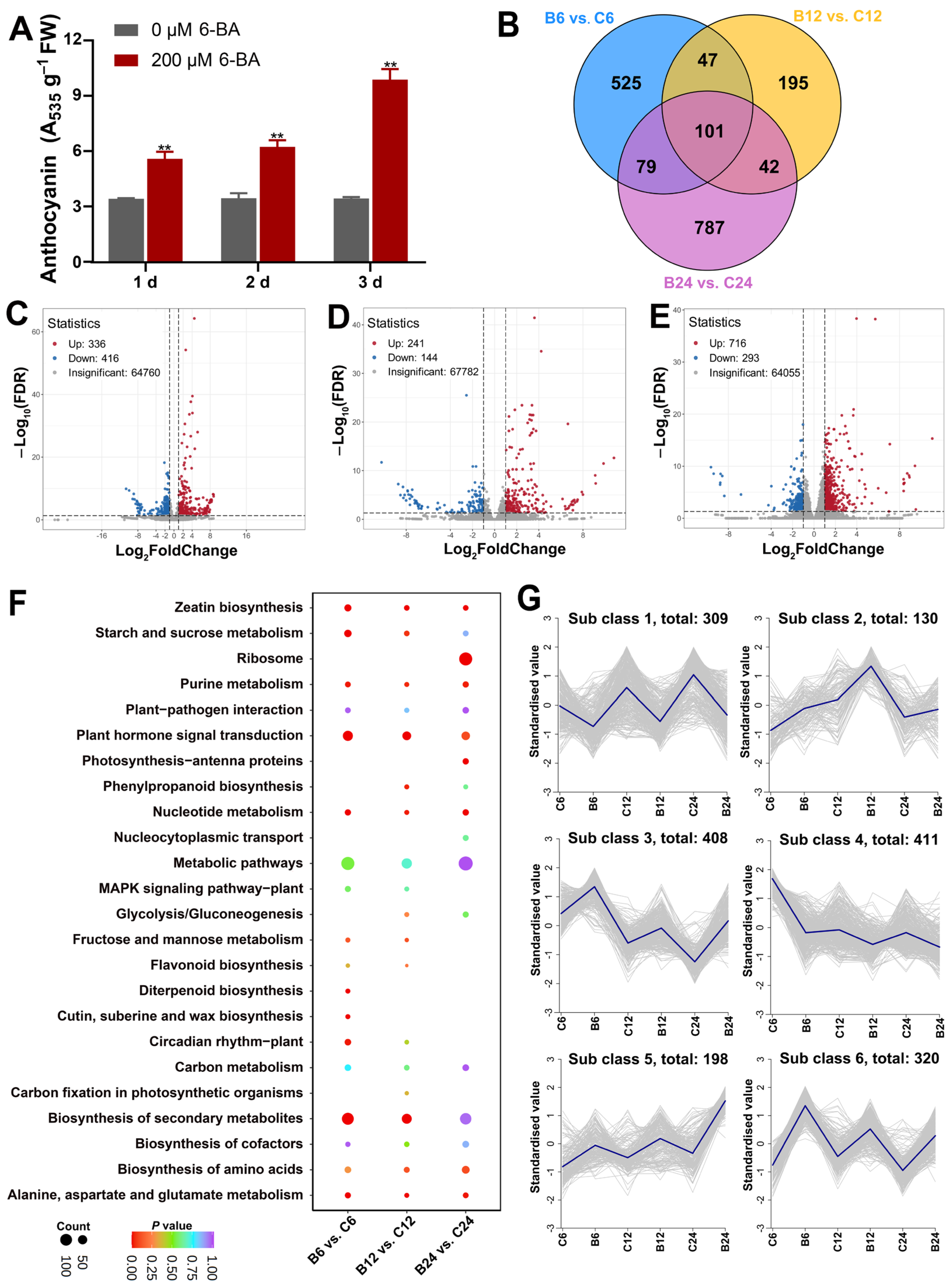
3.1. Anthocyanin Biosynthesis in the Leaves of H. monogynum Can Be Effectively Induced by Cytokinin Treatment

Anthocyanins in the leaves are crucial for ornamental landscape plants. In this study, we found that exogenous cytokinin application effectively induces anthocyanin biosynthesis in the leaves of H. monogynum (Figure 1A). First, different concentrations of 6-benzylaminopurine (6-BA) were applied to the leaves of H. monogynum. The results demonstrated that low concentrations of 6-BA could promote anthocyanin accumulation within days after treatment (Figure 1B). A substantial increase in anthocyanin content was observed one week after treatment, followed by a decrease at two weeks (Figure 1C). Meanwhile, chlorophyll content decreased at one week and increased at two weeks (Figure 1D), indicating the differential roles of cytokinin in regulating anthocyanin and chlorophyll biosynthesis in H. monogynum. Additionally, total flavonoid content increased 3 days post-treatment and reached significantly higher levels at 7 days (Figure 1E). Previous studies have shown that exogenous cytokinin treatment can increase sugar levels in cytokinin-treated leaves [18]. Our results also indicated that 6-BA treatment elevated the levels of both soluble sugars and starch in the cytokinin-treated H. monogynum leaves (Figure 1F,G). Interestingly, in contrast to findings in Eucalyptus, we observed a significant increase in amino acid content following 6-BA treatment (Figure 1H). Collectively, these results demonstrate that cytokinin alone could effectively induce anthocyanin biosynthesis, and the color change in H. monogynum leaves induced by 6-BA treatment is the result of increased anthocyanin levels and decreased chlorophyll content.

3.2. Transcriptome Analysis of H. monogynum Leaves After Cytokinin Treatment

Cytokinin not only induces anthocyanin biosynthesis but also promotes leaf expansion in H. monogynum (Figure 1A). To further investigate the role of cytokinin in regulating both biological processes, comprehensive transcriptome and metabolome analyses were conducted on the leaves of H. monogynum. The results indicated that 6-BA treatment effectively induced anthocyanin biosynthesis within 1–2 days post-treatment (Figure 2A). To identify early cytokinin-responsive genes, leaf samples were collected at 6, 12, and 24 h after mock and 6-BA (200 µM) treatment for transcriptome sequencing. A total of 18 RNA-seq libraries were constructed, and the transcriptome sequencing data are summarized in Tables S2–S4. Pairwise comparisons between 6-BA and mock treatments at different time points were performed to identify differentially expressed genes (DEGs). The results revealed 752, 385, and 1009 DEGs at 6, 12, and 24 h, respectively (Figure 2B–E). In total, 336 upregulated and 416 downregulated DEGs were found in B6 vs. C6, 241 upregulated and 144 downregulated DEGs in B12 vs. C12, and 716 upregulated and 293 downregulated DEGs in B24 vs. C24 (Figure 2B–E). A total of 101 DEGs were co-regulated at different time points (Figure 2B). Additionally, the expression levels of nine DEGs were validated by qRT-PCR, and these results were consistent with the RNA-seq data (Figure S3), confirming the reliability of the transcriptome. KEGG analysis indicated that the top three enriched signaling pathways were related to metabolic pathways, biosynthesis of secondary metabolites, and plant hormone signal transduction (Figure 2F). Expression pattern analysis of all DEGs in the different sample groups revealed six subclasses (Figure 2G). Subclass 1 contained 309 DEGs, which exhibited relatively low expression in 6-BA-treated samples, while subclass 6 contained 320 DEGs, which showed higher expression after 6-BA treatment (Figure 2G). The DEGs consistently regulated across time points are likely involved in cytokinin signaling and cellular metabolism, including cytokinin signal perception, signaling, and anthocyanin biosynthesis. These findings suggest that cytokinin plays a crucial role in H. monogynum leaf metabolism, which may be linked to anthocyanin biosynthesis, as well as cell growth and proliferation.

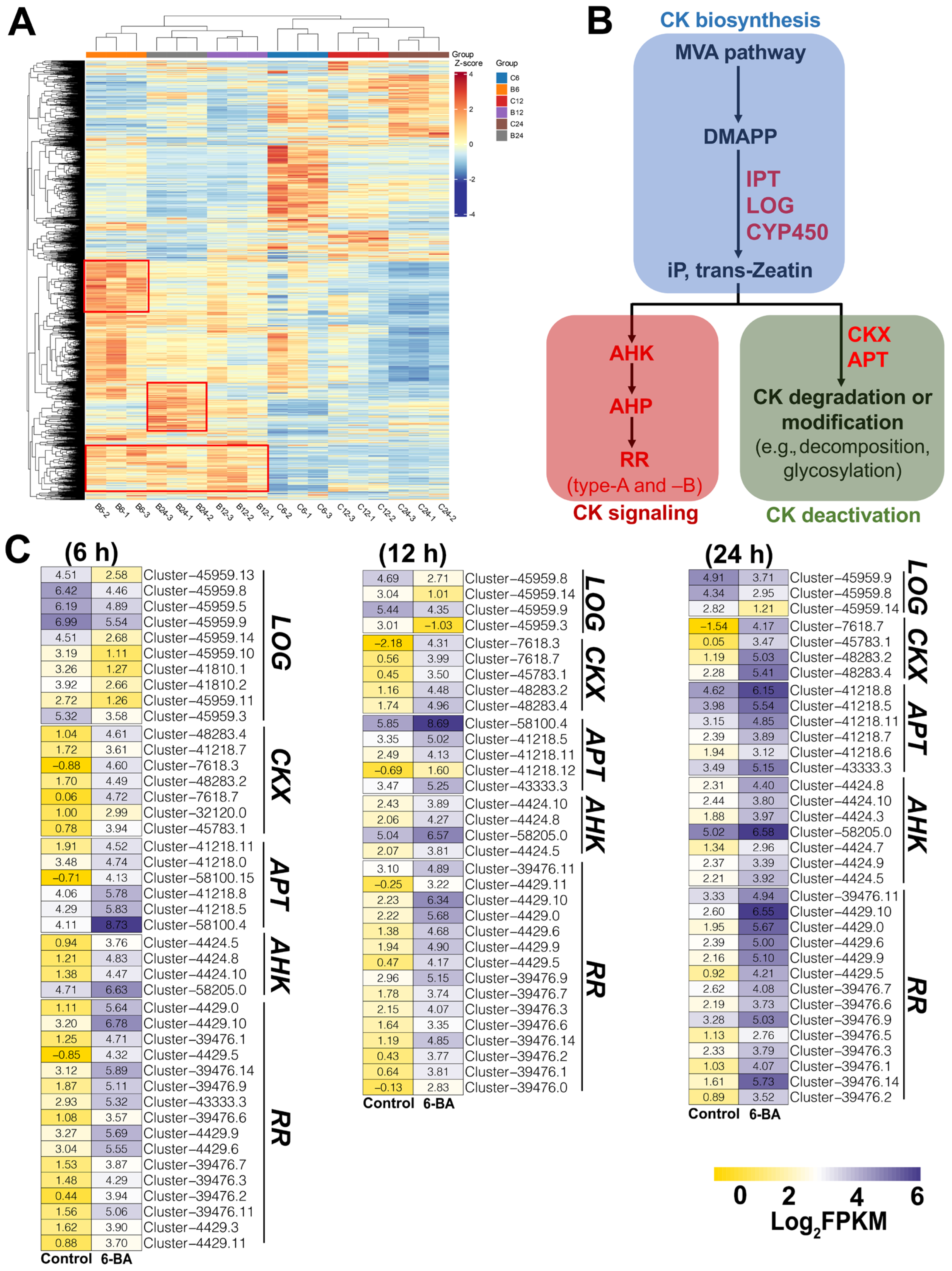
3.3. Identification of Key DEGs Related to CK Biosynthesis, Metabolism, and Signaling

The clustering of the DEGs across various groups revealed several regions with consistently higher expression levels in the BA-treated samples (Figure 3A). Further analysis of these DEGs identified key genes involved in regulating CK metabolism, perception, and signaling in response to BA treatment on H. monogynum leaves (Figure 3B). The lonely guy (LOG) gene encodes key catalytic enzymes in CK biosynthesis. The results showed that ten, four, and three LOG genes were downregulated at 6, 12, and 24 h after 6-BA treatment, respectively (Figure 3C). Cytokinin oxidases (CKXs) are the primary enzymes involved in CK degradation, while adenosine phosphate transferases (APTs) deactivate bioactive CKs. The results also indicated that seven, five, and four CKX genes were upregulated at 6, 12, and 24 h post-treatment, respectively (Figure 3C), and several APT genes were upregulated at different time points following 6-BA treatment (Figure 3C). These findings suggest a strong feedback regulation of CK homeostasis in H. monogynum leaves after 6-BA treatment. More importantly, key CK signaling genes, such as AHK and response regulator (RR), were also identified. RRs are important components of CK signaling, some of which act as key transcription factors mediating CK signal transduction. The results demonstrated that 6-BA treatment activated the expression of several RRs in the leaves (Figure 3C). In conclusion, the identification of key CK-responsive RRs is crucial for further elucidating the CK signaling pathways in H. monogynum.

3.4. Identification of Key DEGs Related to Leaf Growth and Expansion

Cytokinin treatment induced significant leaf expansion in H. monogynum (Figure 1A), which might enhance its ornamental value as a landscape plant. Leaf width and length were measured at various time points following control and 200 µM 6-BA treatments. The results indicated that 6-BA treatment significantly increased both leaf width and length, with notable changes observed as early as the second day after treatment (Figure 4A,B). Additionally, total leaf biomass increased following 6-BA treatment (Figure 4C,D). This extensive leaf expansion is likely linked to enhanced nutrient supply, as well as cell division and growth. Transcriptomic analysis revealed that many DEGs related to nutrient transport (e.g., sugar and nitrate) and sugar metabolism were differentially regulated by 6-BA treatment (Figure 4E). Expansins, key modulators of cell expansion and division, were among the genes showing increased expression following 6-BA treatment (Figure 4E). These findings suggest that exogenous cytokinin promotes leaf expansion in H. monogynum by regulating nutrient transport, metabolism, and the expression of genes involved in cell cycle and expansion processes.

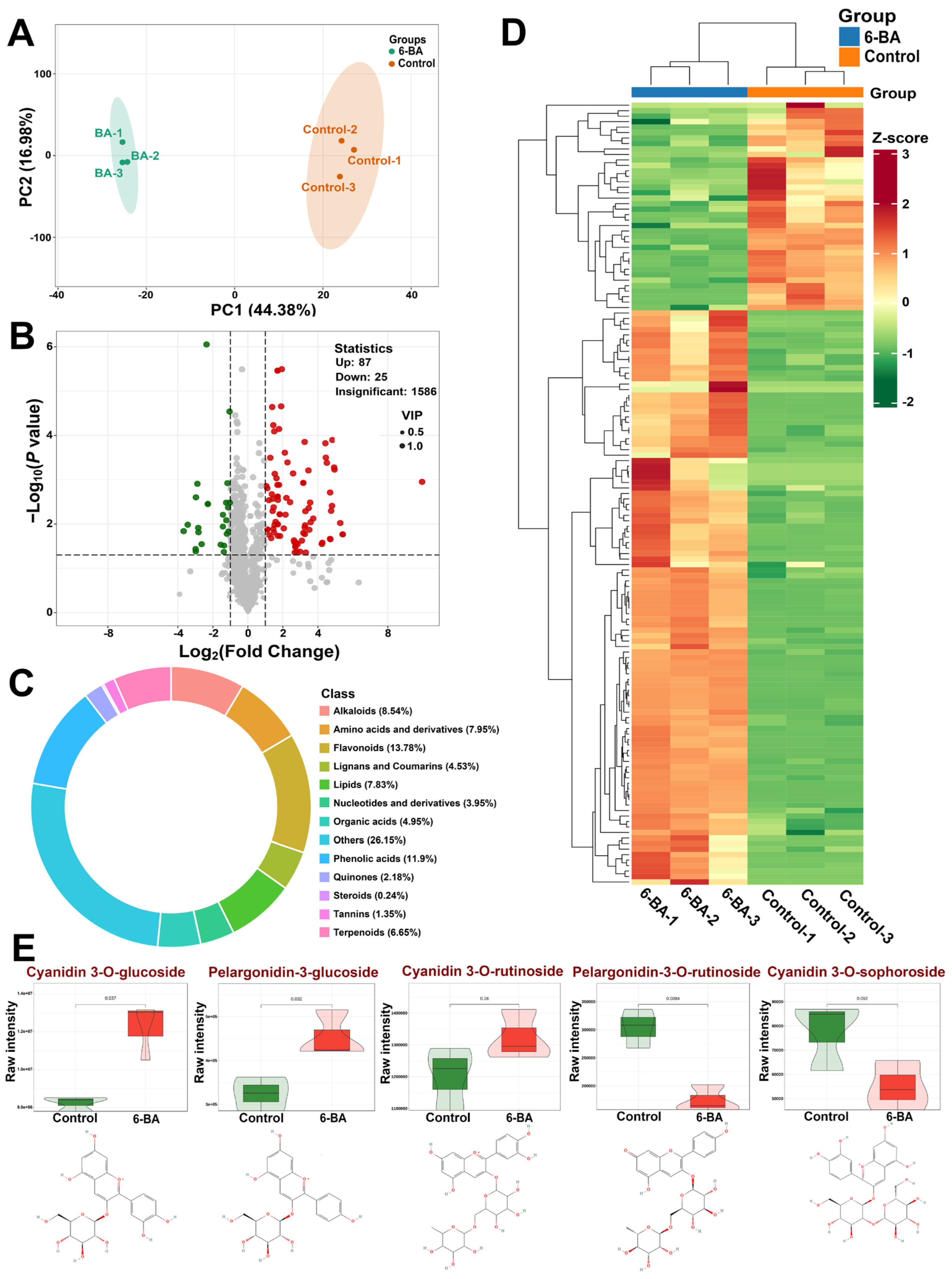
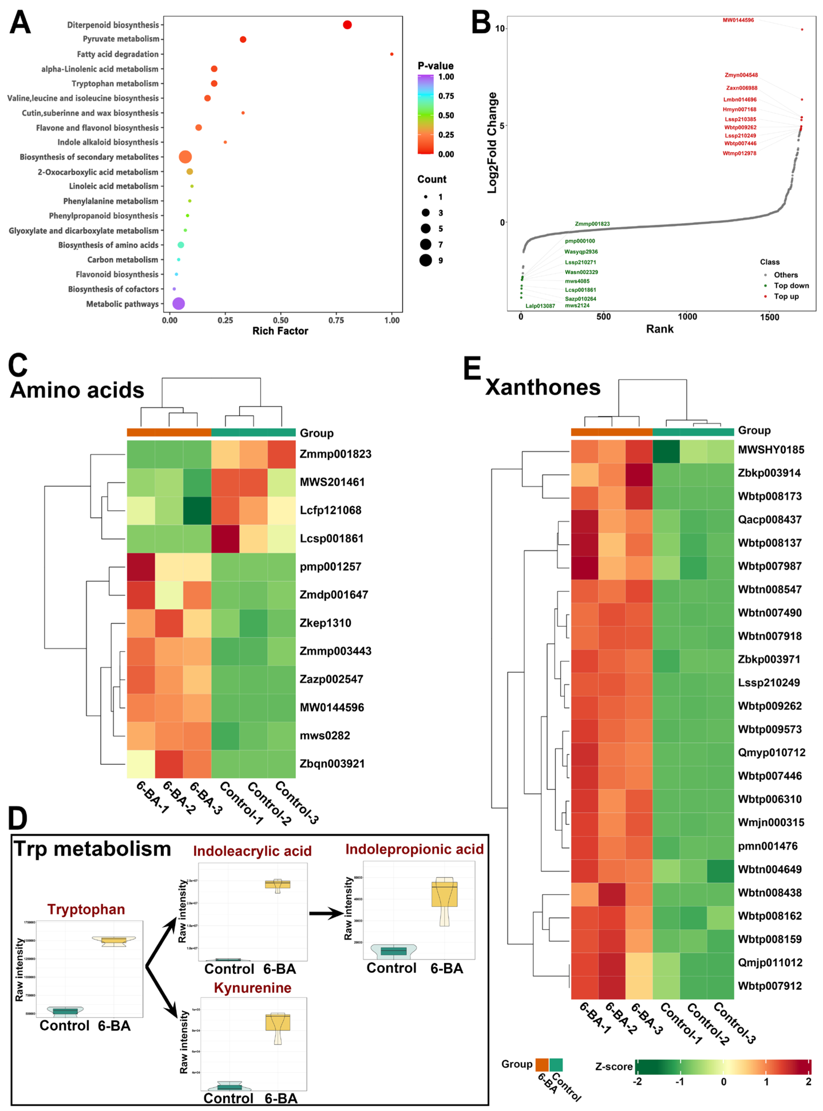
3.5. Widely Targeted Metabolome Analysis of H. monogynum Leaves by Cytokinin Treatment

H. monogynum is valued both as an ornamental and medicinal plant; however, its metabolite profile remains poorly understood. To identify the primary chemical components of this plant and investigate how cytokinin affects metabolism in H. monogynum leaves, particularly those metabolites associated with leaf coloration and expansion, a widely targeted metabolomic analysis was performed on leaves treated with control and 6-BA. Significant anthocyanin accumulation and leaf expansion were observed within days following CK treatment, thus leaf samples were collected for metabolomic analysis at 2 d post-treatment with either mock or 200 µM 6-BA. Principal component analysis (PCA) revealed clear separation between control and 6-BA-treated groups, with low variance within each group (Figure 5A). A total of 112 differentially accumulated metabolites (DAMs) were detected following 6-BA treatment, with 87 upregulated and 25 downregulated (Figure 5B). Among these DAMs, flavonoids, phenolic acids, and alkaloids were the most abundant classes (Figure 5C). Heatmap analysis showed that the DAMs could be divided into two major groups, most of which were upregulated by 6-BA treatment (Figure 5D). Compared to the rich diversity of anthocyanins found in woody species like Eucalyptus [26], only five types of anthocyanins were detected in H. monogynum (Figure 5E). Cyanidin 3-O-glucoside, cyanidin 3-O-rutinoside, and pelargonidin-3-glucoside, which represented the most abundant anthocyanins, were all upregulated in response to 6-BA treatment (Figure 5E). Additionally, five flavonoids kaempferide, luteolin 7-diglucuronide, 3,7,4′-trihydroxy-5-methoxy-8-prenylflavanone, chrysoeriol, and 3-(4-methoxybenzylidene)-5,7-dihydroxy-6-methoxychroman-4-one glucoside were found to be increased following 6-BA treatment (Table S6), which aligns with the observed increase in total flavonoid content (Figure 1E). Thus, the accumulation of these anthocyanins and several other flavonoids, along with decreased chlorophyll content, contribute to the altered leaf coloration following cytokinin treatment.
The KEGG enrichment analysis of the DAMs revealed that the most enriched pathways are associated with the biosynthesis of amino acids, diterpenoid biosynthesis, and tryptophan metabolism (Figure 6A). The top increased DAMs include Ala-His-Met, (9S,13S,15Z)-12-oxophyto-10,15-dienoate, and isopimaric acid, while the most decreased DAMs are Bis(2-ethylhexyl)-phthalate, 2-phosphoglycolate, and monogalactosyldiacylglycerol (Figure 6B). The results show that twelve amino acids or their derivatives were differentially regulated by 6-BA treatment, with eight of them being upregulated (Figure 6C). Phytohormones play a crucial role in regulating leaf growth and expansion. Tryptophan (Trp) is the primary substrate for the biosynthesis of auxins. After 6-BA treatment, Trp and two of its derivatives, 4-hydroxy-1-tryptophan and γ-Glu-Trp, were significantly increased (Figure 6C). Additionally, one auxin, 3-indolepropionic acid (IPA), and its precursor, 3-indoleacrylic acid (IA), were also elevated following 6-BA treatment (Figure 6D). Furthermore, the key Trp metabolite L-kynurenine was increased after 6-BA treatment (Figure 6D). These findings suggest that cytokinin treatment activates the Trp metabolism pathway in H. monogynum. Overall, cytokinin treatment enhanced the accumulation of amino acids, soluble sugars, and auxins, likely contributing to the increased leaf growth and expansion induced by 6-BA treatment.
Xanthones are a class of plant phenolic compounds characterized by varying substituents on the benzene rings, resulting in substantial structural diversity and a broad range of biological activities [37]. As secondary metabolites, xanthones exhibit numerous medicinal properties, including antimicrobial, antitumor, and antidiabetic effects. Metabolic analysis revealed that the leaves of H. monogynum are rich in xanthones, with a total of 58 xanthones detected by LC-MS/MS. Of these, 24 were significantly increased following exogenous 6-BA treatment (Figure 6E). These findings suggest that exogenous cytokinin application holds great potential for enhancing the medicinal properties of plants such as H. monogynum.

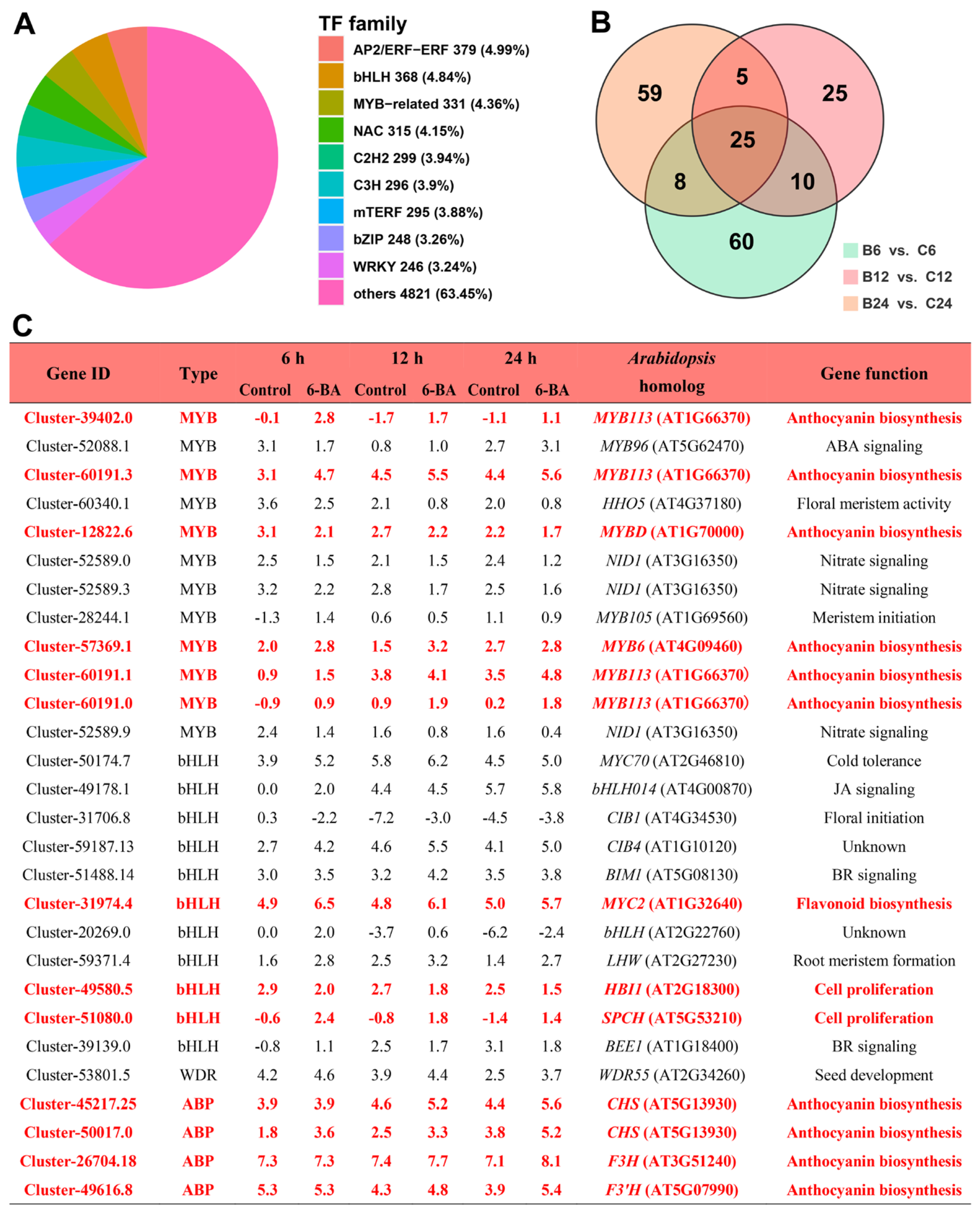
3.6. Identification of Key CK-Responsive Transcription Factors and ABP Genes in H. monogynum

Transcription factors (TFs) play a pivotal role in regulating plant growth and developmental processes. Transcriptome analysis revealed key TF families regulated by cytokinin application. The results showed that the most abundantly regulated TF families were AP2/ERF-ERF, bHLH, and MYB, which constituted 4.99%, 4.84%, and 4.36%, respectively (Figure 7A). A total of 103, 65, and 97 TFs were differentially regulated at 6, 12, and 24 h, respectively, with 25 of them being co-regulated across different time points (Figure 7B). The MBW complex plays a crucial role in regulating flavonoid biosynthesis across various plant species. Consequently, key MYB, bHLH, and WDR genes were identified at different time points following 6-BA treatment (Figure 7C). Based on the functions of their Arabidopsis homologs, five MYBs and one bHLH were predicted to be involved in the regulation of anthocyanin biosynthesis. Among these, the expression of four MYB113 genes (Cluster-39402.0, Cluster-60191.3, Cluster-60191.1, and Cluster-60191.0) and MYC2 (Cluster-31974.4) was increased following 6-BA treatment (Figure 7C). Additionally, two bHLH genes (Cluster-49580.5 and Cluster-51080.0), predicted to be involved in the regulation of cell proliferation, were also differentially regulated by 6-BA treatment (Figure 7C). Furthermore, key structural genes involved in flavonoid biosynthesis were identified. The results showed that two CHS, one F3H, and one F3′H were upregulated by 6-BA treatment (Figure 7C and Figure S4). These findings suggest that 6-BA treatment activates the expression of key transcription factors and ABP genes, thereby inducing flavonoid biosynthesis in H. monogynum leaves.

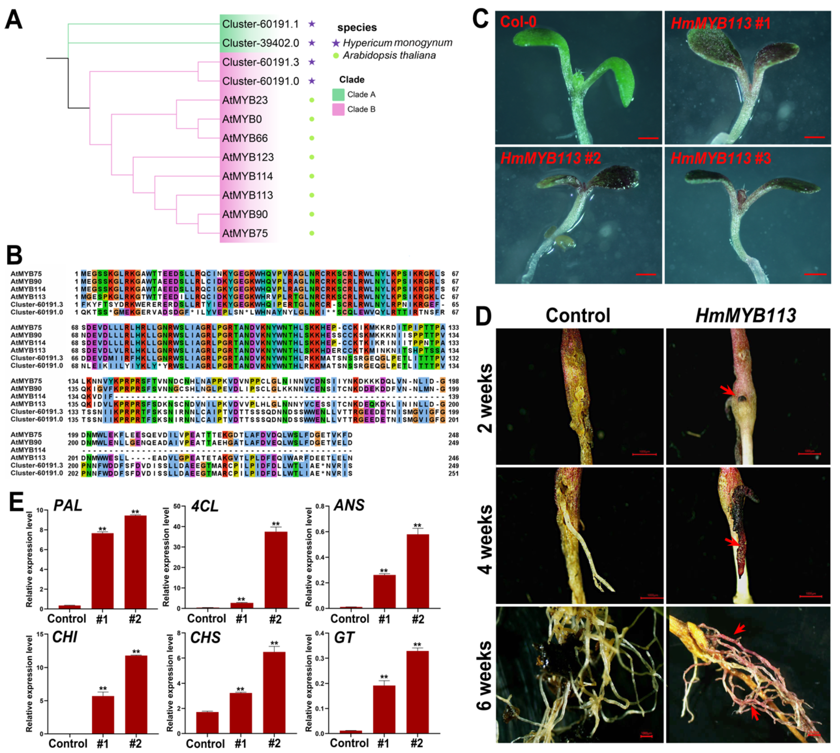
3.7. Functional Characterization of HmMYB113 in the Regulation of Anthocyanin Biosynthesis in the Model Plants Arabidopsis and Eucalyptus

MYB transcription factors, such as MYB75, MYB90, MYB113, and MYB114, have been identified as key regulators of anthocyanin biosynthesis in response to various hormonal and environmental signals in plants. However, the hormonal regulation of these MYB genes in tree species remains poorly understood. Our results showed a significant increase in the expression of several MYBs homologous to AtMYB113, which were upregulated by 6-BA treatment. Phylogenetic analysis revealed that only two HmMYB113 members clustered into the MYB113 clade (Figure 8A). Amino acid sequence alignment indicated that these two HmMYB113 members exhibited high similarity to AtMYB113 (Figure 8B). Furthermore, the full-length cDNA of one HmMYB113 (Cluster-60191.3) was cloned and functionally analyzed in Arabidopsis and Eucalyptus. Overexpression of HmMYB113 in Arabidopsis significantly induced anthocyanin accumulation in the leaves (Figure 8C). In Eucalyptus, we established a highly efficient A. rhizogenes-mediated transformation system, which enables the rapid functional validation of genes involved in anthocyanin biosynthesis in woody plants within one month [27]. Using this system, the function of HmMYB113 in the regulation of anthocyanin biosynthesis was further validated. Our results demonstrated that overexpression of HmMYB113 in Eucalyptus induced the formation of callus with overproduction of anthocyanins within two weeks after inoculation in the seedling hypocotyl (Figure 8D). Additionally, transgenic hairy roots with anthocyanin overproduction were obtained four weeks after inoculation (Figure 8D). The expression of several key ABP genes, including PAL, 4CL, ANS, CHI, CHS, and a glycosyltransferase (GT), was also activated in the transgenic hairy roots (Figure 8E). These findings demonstrate the role of cytokinin-responsive HmMYB113 in the regulation of anthocyanin biosynthesis and further highlight the efficiency of the A. rhizogenes-mediated transformation system in Eucalyptus for gene function analysis in trees.

4. Discussion

The genus Hypericum comprises over 400 species found in temperate regions worldwide, many of which are recognized for their medicinal properties. H. monogynum is a fast-growing shrub that holds both medicinal and ornamental values [1]. In this study, we demonstrate for the first time that anthocyanin biosynthesis can be effectively induced by the exogenous application of cytokinin. Additionally, cytokinin treatment increased leaf size without affecting leaf shape. As leaf color and size are important traits for landscape plants, manipulating these characteristics using a simple and efficient treatment could have significant potential in horticulture. This study not only presented a method for regulating leaf color and size in H. monogynum using a phytohormone but also explored the molecular mechanisms by which cytokinin regulates anthocyanin biosynthesis and leaf expansion through a combination of transcriptomic, metabolomic, and physiological analyses.
In H. monogynum, leaf coloration appears to be determined by both increased anthocyanin biosynthesis and reduced chlorophyll content (Figure 1). Unlike Eucalyptus, where exogenous 6-BA application did not alter chlorophyll levels in the leaves [18], cytokinin treatment drastically increased anthocyanin levels in both species. However, leaf size was unaffected in Eucalyptus, while it was significantly enlarged at the third day post-treatment in H. monogynum (Figure 4). We hypothesize that the reduced chlorophyll content in H. monogynum may result from rapid leaf expansion and energy-consuming anthocyanin biosynthesis. Previous studies have shown that cytokinin-induced anthocyanin biosynthesis in Eucalyptus relies on sugar transport, rather than other nutrients, to the 6-BA-treated leaves [18]. In Arabidopsis, the induction of anthocyanin biosynthesis by multiple phytohormones is thought to depend on the sugar status [19,21], further suggesting that sugar availability is a prerequisite for anthocyanin biosynthesis triggered by other signals. In H. monogynum, cytokinin treatment not only promoted soluble sugar accumulation but also increased levels of amino acids, total proteins, lipids, and starch (Figure 1), indicating that cytokinin regulates leaf growth and cellular metabolism differently across plant species. Notably, metabolomic analysis revealed that 3-indolepropionic acid (IPA) levels in H. monogynum leaves tripled after 6-BA treatment (Figure 6). IPA, a tryptophan-derived indole compound with structural similarities to indole-3-acetic acid (IAA) [38], is widely produced by gut and soil microorganisms. IPA modulates lateral root development via auxin signaling pathways [39]. In contrast to Eucalyptus, where IAA is the primary auxin detected through targeted metabolomic analysis [18,26,27], whereas IAA is absent in H. monogynum, or at least non-detectable in the leaves used in this study (Table S5). IAA represents the main auxin type in so far studied plant species [40]. These results suggested that in H. monogynum, a novel auxin biosynthesis pathway might occur to produce abundant IPA. However, the precise types and concentrations of the auxins in different tissues of H. monogynum need further investigation. As cytokinin and auxin coordinately regulate the plant leaf development and morphogenesis [41], the significantly enhanced leaf expansion observed in H. monogynum might be a combined effect of 6-BA and increased IPA. Further investigations are needed to explore the presence of other auxin types and the biosynthetic pathway of IPA in H. monogynum.
In contrast to herbaceous plants, identifying key genes responsible for the formation of important traits in tree species faces several challenges. One significant limitation is the difficulty in generating sufficient tree mutants, which are crucial for studying the precise interactions between genes and phenotypes. However, with rapid advancements in sequencing technologies, plant transformation, and gene editing techniques, it has become considerably easier to investigate the molecular mechanisms underlying the development of key economic traits. Leaf color is an important trait not only for resistance breeding in trees but also for its horticultural value. In Acer rubrum, the seasonal transition of leaf color has been well-documented, and key transcriptional regulators have been identified through multi-omics analyses [42,43,44]. In the leaves of H. monogynum, a range of secondary metabolites with multiple medicinal uses have been identified through LC-MS/MS, with their levels further enhanced by exogenous cytokinin application (Table S5). Xanthones, which have a wide array of pharmacological applications, including antitumor activity, were identified by metabolomic analysis, revealing over 50 different xanthones (Table S5, Figure 6E). This suggests that the leaves of H. monogynum could serve as a valuable resource for xanthone isolation. Importantly, the content of 24 xanthones was significantly increased by exogenous 6-BA treatment (Figure 6E). This finding holds significant agricultural potential, highlighting the use of cytokinin as a key regulator of medicinal compound content. Meanwhile, the gene regulatory networks involved in anthocyanin biosynthesis in response to various hormonal and environmental signals have been preliminarily studied in Eucalyptus, with key transcription factors such as MYB113 identified [26,27]. In Arabidopsis, MYB75 and MYB90 are known to play crucial roles in regulating anthocyanin biosynthesis in response to hormonal and environmental signals [45,46]. Our recent studies also showed that MYB113 regulates anthocyanin biosynthesis in response to nitrogen deficiency, with its expression strongly induced by nitrogen deficiency in both Arabidopsis and Eucalyptus [27]. In Eucalyptus, cytokinin effectively induces anthocyanin biosynthesis by activating the expression of EgrMYB113, although this process differs from that in Arabidopsis. However, the direct and promotive role of cytokinin in inducing anthocyanin biosynthesis in tree species and its regulatory mechanisms remain to be fully elucidated. Consistent with our previous findings, cytokinin not only directly activates anthocyanin biosynthesis in H. monogynum but also stimulates the expression of several MYB113 homologs (Figure 7C). More importantly, overexpression of HmMYB113 increased anthocyanin biosynthesis in both Arabidopsis and Eucalyptus (Figure 8). These results further emphasize that MYB113 is a direct cytokinin-responsive regulator of anthocyanin biosynthesis in tree species.
In trees, an efficient system for characterizing genes related to anthocyanin biosynthesis has still not been established. Most studies have opted to validate gene functions through transient transformation in tobacco leaves. However, our previous results demonstrated the diverse roles of MYBs in regulating anthocyanin biosynthesis in tobacco and Eucalyptus [18]. Specifically, overexpression of MYB113 did not promote anthocyanin biosynthesis in the tobacco system via transient expression in leaves but significantly induced anthocyanin biosynthesis in callus and hairy roots in Eucalyptus [18]. These findings suggest that tobacco leaves may not be a fully ideal system for functional characterization of anthocyanin biosynthesis. In apple, a highly efficient callus transformation system has been used to study anthocyanin biosynthesis [47]. However, it relies on a mature tissue culture system, which may limit its application in other tree species. In this study, we functionally characterized HmMYB113 in the Eucalyptus system, resulting in the generation of transgenic callus with increased anthocyanin production within two weeks (Figure 8). In comparison, we also overexpressed HmMYB113 in Arabidopsis, which exhibited a similar anthocyanin overproduction phenotype (Figure 8). This study not only revealed the key role of HmMYB113 in mediating cytokinin-induced anthocyanin biosynthesis but also confirmed that the Eucalyptus hairy root transformation system is a valuable tool for rapid screening of anthocyanin biosynthesis genes in tree species. H. monogynum is an important ornamental shrub, and the discovery of methods to alter leaf color in this shrub could have significant practical applications. Through comprehensive transcriptome and metabolome analysis, we not only uncovered the regulatory pattern of cytokinin in controlling both anthocyanin biosynthesis and leaf expansion but also, for the first time, revealed the metabolite profile of H. monogynum, which will facilitate further investigation into the formation of both medicinal and ornamental traits.

5. Conclusions

This study explored the novel role of cytokinin in regulating anthocyanin biosynthesis in the important ornamental tree species H. monogynum through a comprehensive physiological, transcriptomic, and widely targeted metabolomic analysis. The findings revealed that exogenous cytokinin application effectively induced anthocyanin accumulation and promoted leaf expansion, significantly enhancing ornamental value. Transcriptome analysis identified key cytokinin-responsive genes, as well as novel members of the MYB–bHLH–WDR complex. Specifically, cytokinin treatment upregulated the expression of four MYB113 genes, two CHS genes, one F3H gene, and one F3′H gene. Metabolomic analysis revealed that key flavonoids, including anthocyanins, were differentially regulated by cytokinin. Notably, the results also identified key metabolites related to auxins, sugars, amino acids, and other compounds, which were significantly upregulated by cytokinin treatment. These differentially accumulated metabolites likely contribute to the enhanced anthocyanin biosynthesis and leaf expansion observed following cytokinin application. This study not only proposes a novel strategy to enhance the ornamental value of H. monogynum but also provides new insights into the regulatory mechanisms of phytohormones in anthocyanin biosynthesis in perennial tree species.

Supplementary Materials

The following supporting information can be downloaded at https://www.mdpi.com/article/10.3390/f16030465/s1, Figure S1: Expression of HmMYB113 in different transgenic Arabidopsis lines. Expression of HmMYB113 is represented as relative expression level to internal reference AtACT (n = 3).; Figure S2: Expression of HmMYB113 in the hairy roots of different Eucalyptus lines. Expression of HmMYB113 is represented as relative expression level to internal reference EgrACT (n = 3); Figure S3: Expression of nine DEGs was validated by qRT-PCR. Expression of each gene is represented as relative expression level to internal reference ACT2 (n = 3); Figure S4: 6-BA treatment induced the expression of key genes in flavonoid biosynthesis KEGG pathway (24 h). Genes in red box exhibited upregulated expression. Table S1: Information on all the primers used in this study; Table S2: Differentially regulated genes at 6 h after 6-BA treatment; Table S3: Differentially regulated genes at 12 h after 6-BA treatment; Table S4: Differentially regulated genes at 24 h after 6-BA treatment; Table S5: Information of all the metabolites detected by GC-MS/MS in the H. monogynum leaves; Table S6: Identification of the DAMs by 6-BA treatment in the H. monogynum leaves.

Author Contributions

Conceptualization, J.N.; investigation, L.D., Z.Z., J.N., Y.L., K.L., H.H., Y.T. and J.N.; data curation, L.D., Z.Z., K.L., H.H., Y.L., Y.T. and J.N.; formal analysis, L.D., Z.Z., Y.L., Y.T. and J.N.; funding acquisition, J.N. and Z.-F.X.; writing—original draft, L.D. and J.N.; writing—review and editing, L.D., Z.Z., Y.T., Z.-F.X. and J.N. All authors have read and agreed to the published version of the manuscript.

Funding

This work was funded by the National Natural Science Foundation of China (32460399 and 32001345), Natural Science Foundation of Guangxi (2021GXNSFBA220062 and AD22035175), and Guangxi Key Research and Development Program (JB22035001).

Data Availability Statement

RNA-seq data for this article can be downloaded from the GenBank with the BioProject accession number PRJNA1166705. All the data and materials mentioned in this article will be made available upon request.

Conflicts of Interest

The authors declare no conflicts of interest.

References

  1. Caldeira, G.I.; Gouveia, L.P.; Serrano, R.; Silva, O.D. Hypericum genus as a natural source for biologically active compounds. Plants 2022, 11, 2509. [Google Scholar] [CrossRef] [PubMed]
  2. Yang, X.-W.; Grossman, R.B.; Xu, G. Research progress of polycyclic polyprenylated acylphloroglucinols. Chem. Rev. 2018, 118, 3508–3558. [Google Scholar] [CrossRef] [PubMed]
  3. Zhang, S.; Wang, Y.; Cui, Z.; Li, Q.; Kong, L.; Luo, J. Functional characterization of a Flavonol 3-O-rhamnosyltransferase and two UDP-rhamnose synthases from Hypericum monogynum. Plant Physiol. Biochem. 2023, 197, 107643. [Google Scholar] [CrossRef] [PubMed]
  4. Dongfeng, J.; Shuyao, Y.; Qingquan, H.; Qing, L.; Yanqun, H.; Guanglian, L.; Chunhui, H.; Xiaobiao, X. Comprehensive analysis of a red-peel kiwi berry mutant reveals key genes are responsible for anthocyanin biosynthesis in fruit. Sci. Hortic. 2023, 309, 111682. [Google Scholar]
  5. Glover, B.J.; Martin, C. Anthocyanins. Curr. Biol. 2012, 22, R147–R150. [Google Scholar] [CrossRef]
  6. Tanaka, Y.; Sasaki, N.; Ohmiya, A. Biosynthesis of plant pigments: Anthocyanins, betalains and carotenoids. Plant J. 2008, 54, 733–749. [Google Scholar] [CrossRef]
  7. Nakabayashi, R.; Yonekura-Sakakibara, K.; Urano, K.; Suzuki, M.; Yamada, Y.; Nishizawa, T.; Matsuda, F.; Kojima, M.; Sakakibara, H.; Shinozaki, K. Enhancement of oxidative and drought tolerance in Arabidopsis by overaccumulation of antioxidant flavonoids. Plant J. 2014, 77, 367–379. [Google Scholar] [CrossRef]
  8. Kroon, J.; Souer, E.; Graaff, A.D.; Xue, Y.; Koes, R. Cloning and structural analysis of the anthocyanin pigmentation locus Rt of Petunia hybrida: Characterization of insertion sequences in two mutant alleles. Plant J. 2010, 5, 69–80. [Google Scholar] [CrossRef]
  9. Holton, T.A.; Cornish, E.C. Genetics and Biochemistry of Anthocyanin Biosynthesis. Plant Cell 1995, 7, 1071–1083. [Google Scholar] [CrossRef]
  10. Davies, K.M. Modifying Anthocyanin Production in Flowers. In Anthocyanins; Springer: New York, NY, USA, 2008. [Google Scholar]
  11. Mehdy, M.C.; Lamb, C.J. Chalcone isomerase cDNA cloning and mRNA induction by fungal elicitor, wounding and infection. EMBO J. 1987, 6, 1527–1533. [Google Scholar] [CrossRef]
  12. Dongmei, G.; Hanyan, W.; Shumin, Z.; Ting, L. The Type III polyketide synthase supergene family in plants: Complex evolutionary history and functional divergence. Plant J. Cell Mol. Biol. 2022, 112, 414–428. [Google Scholar]
  13. Wenbo, J.; Yaying, X.; Xiaojia, S.; Yongzhen, P. ARF2 positively regulates flavonols and proanthocyanidins biosynthesis in Arabidopsis thaliana. Planta 2022, 256, 44. [Google Scholar]
  14. Jaakola, L. Phenylpropanoid Metabolism and Biosynthesis of Anthocyanins. In The Molecular Biology and Biochemistry of Fruit Ripening; Wiley: Hoboken, NJ, USA, 2013. [Google Scholar]
  15. Grotewold, E. The genetics and biochemistry of floral pigments. Annu. Rev. Plant Biol. 2006, 57, 761–780. [Google Scholar] [CrossRef] [PubMed]
  16. Lafountain, A.M.; Yuan, Y.W. Repressors of anthocyanin biosynthesis. New Phytol. 2021, 231, 933–949. [Google Scholar] [CrossRef]
  17. Li, S.M.; Zheng, H.X.; Zhang, X.S.; Sui, N. Cytokinins as central regulators during plant growth and stress response. Plant Cell Rep. 2021, 40, 271–282. [Google Scholar] [CrossRef]
  18. Zhu, L.; Liao, Y.; Lin, K.; Wu, W.; Duan, L.; Wang, P.; Xiao, X.; Zhang, T.; Chen, X.; Wang, J.; et al. Cytokinin promotes anthocyanin biosynthesis via regulating sugar accumulation and MYB113 expression in Eucalyptus. Tree Physiol. 2024, 44, tpad154. [Google Scholar] [CrossRef]
  19. Das, P.K.; Shin, D.H.; Choi, S.-B.; Yoo, S.-D.; Choi, G.; Park, Y.-I. Cytokinins enhance sugar-induced anthocyanin biosynthesis in Arabidopsis. Mol. Cells 2012, 34, 93–101. [Google Scholar] [CrossRef]
  20. Das, P.K.; Shin, D.H.; Choi, S.-B.; Park, Y.-I. Sugar-hormone cross-talk in anthocyanin biosynthesis. Mol. Cells 2012, 34, 501–507. [Google Scholar] [CrossRef]
  21. Loreti, E.; Povero, G.; Novi, G.; Solfanelli, C.; Alpi, A.; Perata, P. Gibberellins, jasmonate and abscisic acid modulate the sucrose-induced expression of anthocyanin biosynthetic genes in Arabidopsis. New Phytol. 2008, 179, 1004–1016. [Google Scholar] [CrossRef]
  22. Sakamoto, M.; Suzuki, T. Methyl Jasmonate and Salinity Increase Anthocyanin Accumulation in Radish Sprouts. Horticulturae 2019, 5, 62. [Google Scholar] [CrossRef]
  23. Albert, N.W.; Lewis, D.H.; Zhang, H.; Irving, L.J.; Jameson, P.E.; Davies, K.M. Light-induced vegetative anthocyanin pigmentation in Petunia. J. Exp. Bot. 2009, 60, 2191–2202. [Google Scholar] [CrossRef] [PubMed]
  24. Mengqi, X.; WenSha, D.; Chu, W.; Wenjia, W.; Shanwen, Y.; Changyang, C.; Xin, H.; Nannan, W.; Weiyuan, B.; Xiaoshan, T.; et al. Production of purple Ma bamboo (Dendrocalamus latiflorus Munro) with enhanced drought and cold stress tolerance by engineering anthocyanin biosynthesis. Planta 2021, 254, 50. [Google Scholar]
  25. Ma, D.; Sun, D.; Wang, C.; Li, Y.; Guo, T. Expression of flavonoid biosynthesis genes and accumulation of flavonoid in wheat leaves in response to drought stress. Plant Physiol. Biochem. 2014, 80, 60–66. [Google Scholar] [CrossRef] [PubMed]
  26. Zhu, L.; Liao, Y.; Zhang, T.; Zeng, Z.; Wang, J.; Duan, L.; Chen, X.; Lin, K.; Liang, X.; Han, Z.; et al. Reactive oxygen species act as the key signaling molecules mediating light-induced anthocyanin biosynthesis in Eucalyptus. Plant Physiol. Biochem. 2024, 212, 108715. [Google Scholar] [CrossRef]
  27. Zeng, Z.; Liao, Y.; Wang, J.; Liang, X.; Duan, L.; Huang, Y.; Han, Z.; Lin, K.; Hu, H.; Ye, K.; et al. Combined transcriptomic, metabolomic and physiological analysis reveals the key role of nitrogen, but not phosphate and potassium in regulating anthocyanin biosynthesis induced by nutrient deficiency in Eucalyptus. Int. J. Biol. Macromol. 2024, 283, 137564. [Google Scholar] [CrossRef]
  28. Tonelli, P.C. Recent advances on the regulation of anthocyanin synthesis in reproductive organs. Plant Sci. 2011, 181, 219–229. [Google Scholar]
  29. Wu, Y.; Wen, J.; Xia, Y.; Zhang, L.; Du, H. Evolution and functional diversification of R2R3-MYB transcription factors in plants. Hortic. Res. 2022, 9, uhac058. [Google Scholar] [CrossRef]
  30. Liao, Y.; Zeng, Z.; Lin, K.; Jiang, W.; Wang, J.; Duan, L.; Liang, X.; Huang, Y.; Han, Z.; Hu, H.; et al. Gibberellin promotes xylem expansion and cell lignification by regulating sugar accumulation and the expression of JcMYB43 and JcMYB63 in the woody plant Jatropha curcas. Int. J. Biol. Macromol. 2025, 294, 139434. [Google Scholar] [CrossRef]
  31. Pratyusha, D.S.; Sarada, D.V.L. MYB transcription factors-master regulators of phenylpropanoid biosynthesis and diverse developmental and stress responses. Plant Cell Rep. 2022, 41, 2245–2260. [Google Scholar] [CrossRef]
  32. Chen, Z.; Lu, X.; Li, Q.; Li, T.; Zhu, L.; Ma, Q.; Wang, J.; Lan, W.; Ren, J. Systematic analysis of MYB gene family in Acer rubrum and functional characterization of ArMYB89 in regulating anthocyanin biosynthesis. J. Exp. Bot. 2021, 72, 6319–6335. [Google Scholar] [CrossRef]
  33. Gonzalez, A.; Zhao, M.; Leavitt, J.M.; Lloyd, A.M. Regulation of the anthocyanin biosynthetic pathway by the TTG1/bHLH/MYB transcriptional complex in Arabidopsis seedlings. Plant J. 2010, 53, 814–827. [Google Scholar] [CrossRef] [PubMed]
  34. Li, W.; Wen, L.; Chen, Z.; Zhang, Z.; Pang, X.; Deng, Z.; Liu, T.; Guo, Y. Study on metabolic variation in whole grains of four proso millet varieties reveals metabolites important for antioxidant properties and quality traits. Food Chem. 2021, 357, 129791. [Google Scholar] [CrossRef] [PubMed]
  35. Thévenot, E.A.; Roux, A.; Xu, Y.; Ezan, E.; Junot, C. Analysis of the human adult urinary metabolome variations with age, body mass index, and gender by implementing a comprehensive workflow for univariate and OPLS statistical analyses. J. Proteome Res. 2015, 14, 3322–3335. [Google Scholar] [CrossRef] [PubMed]
  36. Chong, J.; Xia, J. MetaboAnalystR: An R package for flexible and reproducible analysis of metabolomics data. Bioinformatics 2018, 34, 4313–4314. [Google Scholar] [CrossRef]
  37. Remali, J.; Sahidin, I.; Aizat, W.M. Xanthone biosynthetic pathway in plants: A review. Front. Plant Sci. 2022, 13, 809497. [Google Scholar] [CrossRef]
  38. Dodd, D.; Spitzer, M.H.; Van Treuren, W.; Merrill, B.D.; Hryckowian, A.J.; Higginbottom, S.K.; Le, A.; Cowan, T.M.; Nolan, G.P.; Fischbach, M.A.; et al. A gut bacterial pathway metabolizes aromatic amino acids into nine circulating metabolites. Nature 2017, 551, 648–652. [Google Scholar] [CrossRef]
  39. Sun, Y.; Yang, Z.; Zhang, C.; Xia, J.; Li, Y.; Liu, X.; Sun, L.; Tan, S. Indole-3-propionic acid regulates lateral root development by targeting auxin signaling in Arabidopsis. iScience 2024, 27, 110363. [Google Scholar] [CrossRef]
  40. Zhao, Y.D. Auxin biosynthesis and its role in plant development. Annu. Rev. Plant Biol. 2010, 61, 49–64. [Google Scholar] [CrossRef]
  41. Hussain, S.; Nanda, S.; Zhang, J.; Rehmani, M.I.A.; Suleman, M.; Li, G.; Hou, H. Auxin and cytokinin interplay during leaf morphogenesis and phyllotaxy. Plants 2021, 10, 1732. [Google Scholar] [CrossRef]
  42. Chen, Z.; Lu, X.Y.; Zhu, L.; Afzal, S.F.; Zhou, J.B.; Ma, Q.Y.; Li, Q.Z.; Chen, J.H.; Ren, J. Chromosomal-level genome and multi-omics dataset provides new insights into leaf pigmentation in Acer palmatum. Int. J. Biol. Macromol. 2023, 227, 93–104. [Google Scholar] [CrossRef]
  43. Chen, Z.; Lu, X.; Xuan, Y.; Tang, F.; Wang, J.; Shi, D.; Fu, S.; Ren, J. Transcriptome analysis based on a combination of sequencing platforms provides insights into leaf pigmentation in Acer rubrum. BMC Plant Biol. 2019, 19, 240. [Google Scholar] [CrossRef] [PubMed]
  44. Chen, Z.; Lu, X.; Gao, J.; Xuan, Y.; Ren, J. Integrating transcriptomic and metabolomic analysis of hormone pathways in Acer rubrum during developmental leaf senescence. BMC Plant Biol. 2020, 20, 410. [Google Scholar] [CrossRef]
  45. Zheng, T.; Tan, W.; Yang, H.; Zhang, L.E.; Li, T.; Liu, B.; Zhang, D.; Lin, H. Regulation of anthocyanin accumulation via MYB75/HAT1/TPL-mediated transcriptional repression. PLoS Genet. 2019, 15, e1007993. [Google Scholar] [CrossRef] [PubMed]
  46. Maier, A.; Schrader, A.; Kokkelink, L.; Falke, C.; Welter, B.; Iniesto, E.; Rubio, V.; Uhrig, J.F.; Hülskamp, M.; Hoecker, U. Light and the E3 ubiquitin ligase COP1/SPA control the protein stability of the MYB transcription factors PAP1 and PAP2 involved in anthocyanin accumulation in Arabidopsis. Plant J. 2013, 74, 638–651. [Google Scholar] [CrossRef]
  47. Wang, Y.; Sun, J.; Wang, N.; Xu, H.; Qu, C.; Jiang, S.; Fang, H.; Su, M.; Zhang, Z.; Chen, X. MdMYBL2 helps regulate cytokinin-induced anthocyanin biosynthesis in red-fleshed apple (Malus sieversii f. niedzwetzkyana) callus. Funct. Plant Biol. 2019, 46, 187–196. [Google Scholar] [CrossRef]
Figure 1. Cytokinin treatment effectively induces anthocyanin accumulation in the leaves of H. monogynum. (A) Photograph of the H. monogynum leaves at 7 and 14 d after control and 6-BA (200 µM) treatment. (B) Effects of different concentrations of 6-BA on anthocyanin biosynthesis at 3 d after treatment. (C,D) Anthocyanin and total chlorophyll content at 7 and 14 d. Total flavonoid (E), soluble sugars (F), starch (G), and amino acids (H) were determined at 3 and 7 d after control and 6-BA (200 µM) treatment (n = 5). Significances between indicated groups and control were marked. Data are represented as means ± standard error. **, p < 0.01; ns, not significant.
Forests 16 00465 g001
Figure 2. Transcriptomic analysis of the H. monogynum leaves in response to cytokinin treatment. (A) Anthocyanin content in the H. monogynum leaves at 1, 2, and 3 d after control and 6-BA (200 µM) treatment (n = 5). (B) Venn diagram of the DEGs of different comparison groups. (CE) Statistics of the DEGs at 6, 12, and 24 h, respectively. (F) Gene functional annotation of different comparison groups by KEGG analysis. (G) Expression pattern of DEGs of different treatments. Significances between indicated groups and control were marked. C, control; B, 6-BA treatment. Data are represented as means ± standard error. **, p < 0.01.
Forests 16 00465 g002
Figure 3. Identification of cytokinin metabolism and signaling DEGs in response to 6-BA treatment. (A) Heatmap of the DEGs of different samples. Red frames indicate specifically expressed group of DEGs by 6-BA treatment. C, control; B, 6-BA treatment. (B) Pathways involved in CK biosynthesis, deactivation, and signaling. (C) Expression level of DEGs related to CK biosynthesis (LOG), deactivation (CKX and APT), and signaling (AHK and RR).
Forests 16 00465 g003
Figure 4. Cytokinin induces leaf expansion in H. monogynum. (A,B) Leaf length and width at 1–7 d after mock and 6-BA (200 µM) treatment (n = 20). (C,D) 6-BA treatment increased the leaf weight (n = 12). FW, fresh weight; DW, dry weight. (E) Identification of key DEGs related to nutrient transport, metabolism, cell cycle, and cell expansion. Significances between indicated groups and control were marked. Data are represented as means ± standard error. *, p < 0.05; **, p < 0.01; ns, not significant.
Forests 16 00465 g004
Figure 5. Widely targeted metabolomic analysis of the H. monogynum leaves in response to cytokinin treatment. (A) PCA analysis of the replicates of different groups. (B) Up- and downregulated metabolites (indicated by red and green dots, respectively) by 6-BA treatment. (C) Classification of the DAMs. (D) Heatmap of the relative abundance of the DAMs in different samples. (E) Anthocyanin identification in the H. monogynum leaves by LC-MS/MS (n = 3).
Forests 16 00465 g005
Figure 6. Cytokinin modulates the metabolism of key metabolites related to amino acids, auxin, and xanthones. (A) KEGG enrichment of the DAMs regulated by 6-BA treatment. (B) Top-regulated DAMs by 6-BA. (C) Amino acids regulated by 6-BA. (D) 6-BA treatment promoted the Trp metabolism (n = 3). (E) 6-BA increased the xanthone content. Detailed information on all DAMs is provided in Table S6.
Forests 16 00465 g006
Figure 7. Identification of the CK-responsive transcription factors in the leaf of H. monogynum. (A) Classification of the CK-responsive TFs. (B) Venn diagram of the TFs of different comparison groups. (C) List of the MBW and ABP genes regulated by CK in H. monogynum leaves. Genes in red font are predicted to be associated with anthocyanin biosynthesis and cell proliferation. Data are presented as Log2FPKM. C, control; B, 6-BA treatment.
Forests 16 00465 g007
Figure 8. Functional characterization of the CK-responsive HmMYB113 in the regulation of anthocyanin biosynthesis. (A) Phylogenetic analysis of two HmMYB113 members with their homologous proteins in Arabidopsis. (B) Amino acid alignment of two HmMYB113 members with AtMYB75, AtMYB90, AtMYB113, and AtMYB114. (C) Overexpression of HmMYB113 promoted anthocyanin biosynthesis in the leaves of Arabidopsis seedlings. (D) Overexpression of HmMYB113 promoted anthocyanin biosynthesis in the callus and hairy root of E. grandis seedlings. (E) Expression of the ABP genes in the hairy roots overexpressing HmMYB113 (6 weeks) (n = 3). Red arrow indicates the transgenic callus or hairy roots with overproduced anthocyanin. Data are represented as means ± standard error. **, p < 0.01.
Forests 16 00465 g008
Disclaimer/Publisher’s Note: The statements, opinions and data contained in all publications are solely those of the individual author(s) and contributor(s) and not of MDPI and/or the editor(s). MDPI and/or the editor(s) disclaim responsibility for any injury to people or property resulting from any ideas, methods, instructions or products referred to in the content.

