Article

Morphological and Molecular Characterization of Botryosphaeria wangensis Causing Branch Blight of Acer saccharum in China

1
Guangzhou Customs Technology Center, Guangzhou 510623, China
2
College of Agricultural Science and Technology, Shandong Agriculture and Engineering University, Jinan 250100, China
*
Author to whom correspondence should be addressed.
Forests 2025, 16(12), 1786; https://doi.org/10.3390/f16121786
Submission received: 8 September 2025 / Revised: 14 November 2025 / Accepted: 23 November 2025 / Published: 28 November 2025
(This article belongs to the Special Issue Forest Fungal Diseases Detection, Diagnosis and Control)

Abstract

Acer saccharum Marsh., valued for its ornamental, material, and edible uses, is an important temperate tree species in the Northern Hemisphere. A blight disease affecting branches of A. saccharum was first identified in 2023 in Shandong, China. The causal agent was identified as Botryosphaeria wangensis G.Q. Li & S.F. Chen based on cultural and morphometric characteristics. Phylogenetic analysis was performed by amplifying and sequencing the internal transcribed spacer (ITS) region of rDNA, the translation elongation factor 1α (tef1) partial gene, the β-tubulin (tub2) partial gene, and the second largest subunit of RNA polymerase II (rpb2), in combination with morphological data. Symptoms observed in the field were replicated in a pathogenicity test through inoculation of A. saccharum branches, thus satisfying Koch’s postulates. To our knowledge, this is the first report worldwide of B. wangensis infecting A. saccharum.

1. Introduction

Sugar maple (Acer saccharum Marsh.) is one of the most important landscape tree species that are widely planted in the Northern Hemisphere [1]. Furthermore, it is recognized as one of the three principal woody plants globally responsible for maple syrup production and serves as a significant species within the forestry sector due to its economic value [2,3]. It is usually used as a traditional medicine in the treatment of various ailments such as back pain and as a diuretic by Native Americans [4,5]. Previous studies demonstrated that phenolic-rich bark extracts from sugar maple have anticancer activities, glucosidase inhibitory activities, and real potential as food additives [3,6]. Because of its combined value for ornamental, timber, and edible purposes, sugar maple was initially introduced to China in 1957 and then widely planted in the northeastern and northern regions [7].
For the past few years, increased diseases caused by fungi and pests such as Phyllosticta negundinis Sacc. & Speg., Verticillium dahilae Klebahn, Neonectria spp., and Anoplophora glabripennis Motschulsky have been reported on sugar maple [8,9,10,11,12]. These pathogens are capable of causing significant symptoms such as cankers and blight in various plant parts, including branches, twigs, shoots, and leaves of sugar maple. At present, the information on fungal diseases affecting sugar maple is still relatively scarce.
Botryosphaeriaceae fungi have been reported on numerous woody plants as endophytes, latent, opportunistic, and primary pathogens worldwide [13,14,15]. In Uruguay, Neofusicoccum cryptoaustrale Pavlic, Maleme, Slippers & M.J. Wingf., and N. occulatum Sakalidis & T. Burgess were identified as pathogenic to olive (Olea europaea subsp. europaea L.) and were found to cause dieback and fruit rot [16]. In Japan, Botryosphaeria tenuispora Y. Hattori & C. Nakash. isolated from Leucothoe fontanesiana (Steud.) Sleumer and insect galls on fruits of Aucuba japonica Thunb. was identified as a new species [17]. It is worth noting that B. wangensis G.Q. Li & S.F. Chen was identified on various economic trees and was found to have caused stem canker and fruit rot in several countries over recent years [15,16,18,19]. Recently, the dieback and branch and twig blight diseases of Eucalyptus caused by Botryosphaeriaceae in China were reported, which confirms the wide host range of Botryosphaeriaceae species on different plants [14]. Among them, the typical symptoms caused by Botryosphaeria spp. usually show brown, dented necrotic lesions with black small dots on the surface of the spots, which are the conidiomata of the pathogen [14,15,16,17,18,19,20], while abundant white mycelia are often observed on the surface of rotten fruits [15,16].
During our investigations in 2023, sugar maple with branch blight was observed in the natural field in Shandong Province, China. Since the causal agent of this disease has not been previously reported, the objective of this study was to identify the causal pathogen by morphological and molecular methods, and a pathogenicity test.

2. Materials and Methods

2.1. Pathogen Isolation and Morphological Identification

Investigations were conducted in “Strawberry Town”, an agriculture park in Heze City, Shandong Province, China (116°2′41″ E, 34°52′26″ N), in 2023. Blight branches from the lower canopy of Acer saccharum (15 cm in diameter breast high) were collected and packed in paper envelopes for further study. Two isolates obtained from infected branches were cultured on potato dextrose agar (PDA) using both tissue transplantation and single-spore methods [21]. Surface-sterilized branch segments were plated on PDA and incubated at 25 °C in the dark for 7 days. The isolates were hyphal tip purified and subcultured for further analysis. Fungal cultures were kept at Shandong Agriculture and Engineering University.
The isolates were cultured on PDA at 25 °C after 7 days for morphological characterization. Mycelial and conidial features were observed using a microscope. The width and length of conidia were measured in this study using 50 measurements per trait.

2.2. Phylogenetic Analyses

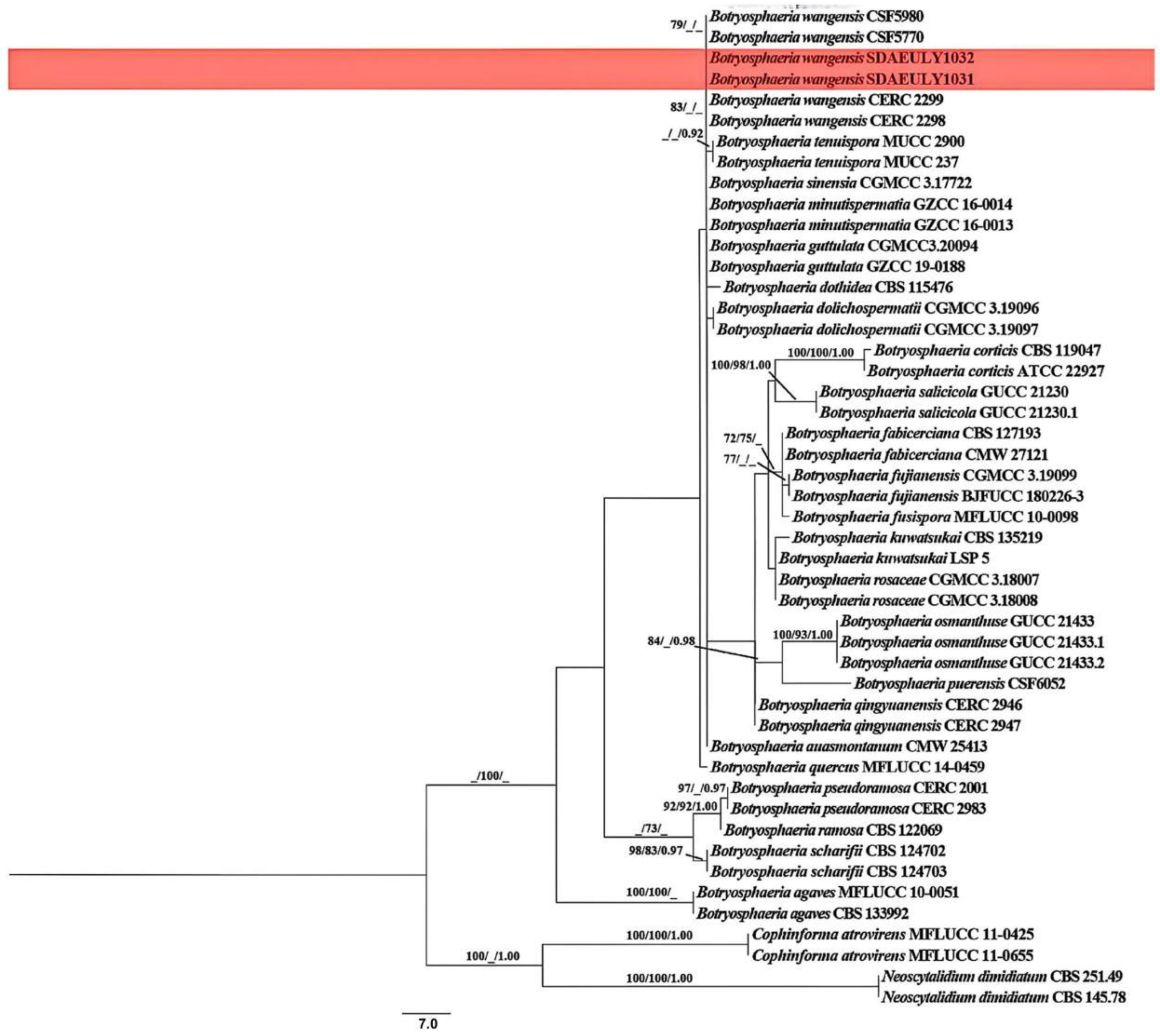
Genomic DNA was extracted using the Ezup Column Fungal Genomic DNA Extraction Kit (Sangon Biotech Co., Ltd., Shanghai, China) following the manufacturer’s instructions. The ITS, tef1, tub2, and rpb2 regions were amplified with primers: ITS1/ITS4 [22], EF1-688/EF1-1251 [23], BT-2a/BT-2b [24], and fRPB2-5f2/fRPB2-7cR [25]. PCR cycling conditions were followed according to a previous study [26]. The PCR products were sequenced by Qingdao Personalbio Gene Technology (Qingdao, China). Sequence alignments were performed by MAFFT v.7 and manually refined in MEGA v.5.0 [21]. Maximum likelihood (ML) analysis was performed by PhyML v.3.0 with 1000 bootstrap replications [27]. Bayesian Inference (BI) analysis was conducted based on the DNA dataset using a Markov Chain Monte Carlo (MCMC) algorithm in MrBayes v. 3.1.2 [28]. For node support, bootstrap values ≥ 70% and Bayesian posterior probabilities ≥ 0.90 were considered significant and are displayed on the respective nodes, separated by a “/”. Lower support values are denoted by “_”. The ITS, tef1, tub2, and rpb2 sequences were combined and aligned with the most related sequences retrieved from NCBI GenBank (Table S1).

2.3. Pathogenicity Test

Fungal isolates (SDAEULY1031 and SDAEULY1032) were used as the inoculum. To assess the pathogenicity, healthy sugar maple branches (two-year-old, average 1.0 cm in diameter) were used for inoculation. The isolates SDAEULY1031 and SDAEULY1032 were cultured on PDA at 25 °C for 7 days. Using the agar plug method, 5 mm diameter mycelial plugs were excised with a sterile cork borer. The inoculation experiments were conducted outdoors by making a partial cut on the branches of A. saccharum, ensuring the plug could be held securely. Inoculation sites were wrapped with sterile water-soaked absorbent cotton and then covered with Parafilm® (Amcor Co., Ltd., Zurich, Switzerland) to preserve moisture. The experiment used an agar plug of PDA alone as the negative control. The pathogenicity experiments were performed in 3 replicates. The lesion area data were measured using the grid method. Fungal isolates were re-isolated and re-identified using morphological and molecular characteristics for confirmation of Koch’s postulates.

3. Results

3.1. Symptom Description and Morphological Characteristics

In 2023, the symptoms of blight disease were observed on Acer saccharum branches in Heze City of Shandong Province. There were 4 out of 38 trees inspected in the area showing symptoms; the incidence of disease was 10.53%. The symptoms of disease on the twig initially appeared as brown striped necrosis. Symptomatic twig tissues gradually became necrotic and began to wilt, while the leaves did not fall from the plants. Eventually, the blight branches became covered with numerous small black dots. The bark of the necrotic branches was arched and showed a fish scale-like appearance (Figure 1A). Five fungal isolates were obtained from the symptomatic sugar maple branches via the tissue transplantation method. Then four pure cultures with a similar colony morphology were obtained via the single-spore method. Among the isolates obtained, isolates SDAEULY1031 and SDAEULY1032 were selected as the representative fungi for further studies. These two isolates were initially white and had achieved 85 mm within 7 days on PDA. The center of the colonies gradually turns gray-black, and multiple rings are formed, while the edges of the colonies remain white (Figure 1B,C). The conidiomatal wall was composed of thick-walled, dark brown textura angularis cells, which were thin-walled and hyaline towards the interior (Figure 1D,E). Conidia were hyaline, elliptical, and thin-walled with granular contents, base subtruncate truncated to rounded, and measured 21.3 ± 1.4 μm × 5.3 ± 0.7 μm (length/width ratio = 4) (Figure 1F). Based on these morphological characteristics, the isolates were identified as belonging to the genus Botryosphaeria.

3.2. Molecular Characterization and Phylogenetic Analyses

The two isolates SDAEULY1031 and SDAEULY1032 were identified by molecular analysis. The ITS, tef1, and tub2 regions of the isolates were cloned and sequenced (Table S1). The ITS, tef1, and tub2 sequences of the isolates revealed 100%, 100%, and 99.29% identity to Botryosphaeria wangensis, respectively. The phylogenetic tree constructed from the ITS, tef1, tub2, and rpb2 sequences showed that both SDAEULY1031 and SDAEULY1032 isolates clustered within B. wangensis (Figure 2).

3.3. B. wangensis Pathogenicity

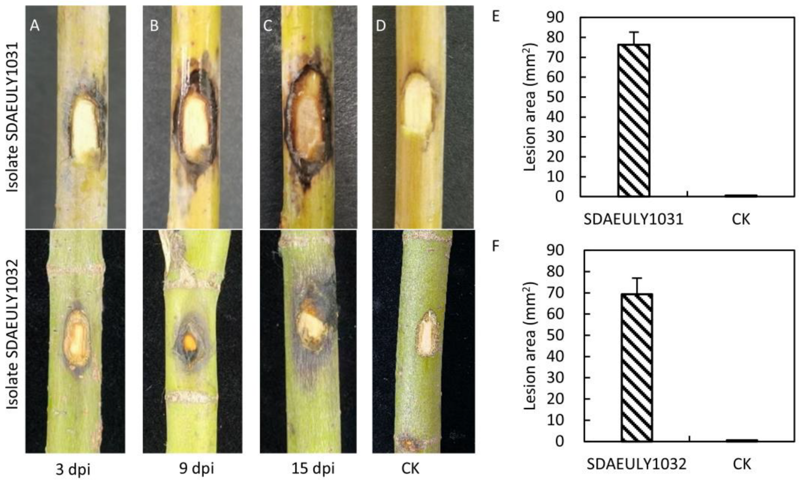
The experiments designed to fulfil Koch’s postulates showed that the symptoms initially appeared 3 days post inoculation (Figure 3A) and attained prominence approximately 15 days after inoculation (Figure 3B,C). All inoculated branches exhibited consistent symptoms, while mock-inoculated controls remained asymptomatic (Figure 3D). The lesion areas caused by isolates SDAEULY1031 and SDAEULY1032 on the inoculated branches were 76.4 mm2 and 69.3 mm2 at 15 days post inoculation, respectively (Figure 3E,F). The pathogen was successfully re-isolated from the inoculated branches, and the fungi were confirmed to be B. wangensis based on consistent colony and conidia morphology, as well as identical ITS, tef1, and tub2 sequences, fulfilling Koch’s postulates.

4. Discussion

The genus Botryosphaeria is commonly associated with dieback, stem canker, shoot blight, and fruit rot in various host plants including both economically important crops and native trees [17,19,20]. The wide host range determination might be related to the secreted hydrolytic enzymes and secondary metabolites of Botryosphaeria [29,30]. In China, there are about 15 Botryosphaeria species reported as endophytes, saprobes, and pathogens on woody plants such as Eucalyptus sp., Malus sp., and Populus sp. [10,14,19,24]. Botryosphaeria wangensis was first identified on Cedrus deodara in South China and then was found in Guizhou Province [18,19]. Recently, B. wangensis was identified as a pathogen on olive trees and plums (Prunus salicina L.) that was associated with stem canker and fruit rot [15,16]. In this study, we report two B. wangensis strains (SDAEULY1031 and SDAEULY1032) isolated from blighted branches of sugar maple in Shandong Province, China. As far as we know, this is the first report in the world of a branch blight disease on a sugar maple tree caused by B. wangensis. The present study suggests that B. wangensis can survive at both a wide host range and a broad range of temperatures. Further studies need to investigate the molecular mechanisms, such as secreted hydrolytic enzymes and secondary metabolites of B. wangensis, which will help us to better understand the infection biology of B. wangensis and the interaction with its plant hosts.
Species identification of Botryosphaeria based on host affiliations and morphology was limited due to the uninformative illustrations and descriptions, weak host specificity, and overlapping morphological characteristics [19,31]. Multi-locus sequence data based on DNA sequence identification represented a more advanced approach for nomenclature and identification of Botryosphaeria [16]. In this study, the ITS, tef1, tub2, and rpb2 regions were used for multi-locus molecular and phylogenetic analyses [19,26]. However, only approximately 7% of Botryosphaeria species had associated DNA sequence data [26]. Consequently, some of the sequence data of Botryosphaeria species used in our study were absent. Moreover, the rpb2 sequences of isolates SDAEULY1031 and SDAEULY1032 were not obtained by PCR. Although B. wangensis is phylogenetically closely related to B. sinensia Y.P. Zhou & Y. Zhang ter, B. auasmontanum F.J.J. van der Walt, Slippers & G.J. Marais, B. dothidea (Moug.) Ces. & De Not., and B. minutispermatia Ariyaw., K.D. Hyde & Z.Y. Liu, it can be distinguished from these species by the size of its conidia [18].
Botryosphaeria species are well known as latent pathogens or endophytes that cause diseases when the host plant is under stress conditions [32]. In our study, B. wangensis produced small lesions in the pathogenicity test, which is similar to previous studies [14,18]. Moreover, Botryosphaeria canker and blight diseases were often associated with insects [33]. In Uruguay, the wounds generated by insects provide additional entryways for Botryosphaeria in olive [16]. In China, the branches of sugar maple are usually attacked by the wood-boring insect A. glabripennis [11]. Despite the fact that the wounds generated by the insect were not detected in our surveys, combining our research and previous studies, the association between Botryosphaeria fungi and branch diseases may potentially present a significant threat to sugar maple. Thus, further studies should be taken into consideration in epidemiology and management strategies of this novel branch blight disease on sugar maple caused by B. wangensis.

5. Conclusions

In this study, Botryosphaeria wangensis was identified as the causal agent of a novel branch blight disease affecting Acer saccharum in Shandong Province, China, based on morphological characteristics, phylogenetic analysis, and a pathogenicity test. This finding represents the first report of this fungal species infecting sugar maple worldwide. The results provide a scientific foundation for future research on disease management and control strategies.

Supplementary Materials

The following supporting information can be downloaded at https://www.mdpi.com/article/10.3390/f16121786/s1, Table S1. Details of fungal isolates used for phylogenetic analysis in this study.

Author Contributions

Conceptualization, C.S.; methodology, W.C. and X.L.; software, M.W.; validation, C.S. and Y.L.; formal analysis, W.C., X.L. and M.W.; writing—original draft preparation, C.S. and Y.L.; writing—review and editing, C.S.; visualization, C.S.; supervision, Y.L.; project administration, Y.L.; funding acquisition, Y.L. All authors have read and agreed to the published version of the manuscript.

Funding

This research was funded by the Research Start-up Foundation for High-level Talents of Shandong Agriculture and Engineering University (30620120).

Data Availability Statement

Accession numbers for the DNA sequence data are available in NCBI GenBank and are listed in Table S1.

Conflicts of Interest

The authors declare no conflicts of interest.

References

  1. van Gelderen, D.M.; Jong, P.C.; Oterdoom, H.J. Maples of the World, 1st ed.; Timber Press: Portland, OR, USA, 1994; pp. 15–20. [Google Scholar]
  2. Dey, D.C.; Dwyer, J.; Wiedenbeck, J. Relationship between tree value, diameter, and age in high-quality sugar maple (Acer saccharum) on the Menominee reservation. Wis. J. For. 2017, 115, 397–405. [Google Scholar] [CrossRef]
  3. Bhatta, S.; Ratti, C.; Poubelle, P.E.; Stevanovic, T. Nutrients, antioxidant capacity and safety of hot water extract from sugar maple (Acer saccharum M.) and red maple (Acer rubrum L.) Bark. Plant Food Hum. Nutr. 2018, 73, 25–33. [Google Scholar] [CrossRef] [PubMed]
  4. Arnason, T.; Hebda, R.; Johns, T. Use of plants for food and medicine by native peoples of eastern Canada. Can. J. Bot. 1981, 59, 2189–2325. [Google Scholar] [CrossRef]
  5. Bi, W.; Gao, Y.; Shen, J.; He, C.; Liu, H.; Peng, Y.; Zhang, C.; Xiao, P. Traditional uses, phytochemistry, and pharmacology of the genus Acer (maple): A review. J. Ethnopharmacol. 2016, 189, 31–60. [Google Scholar] [CrossRef] [PubMed]
  6. Yuan, T.; Wan, C.; González-Sarrías, A.; Kandhi, V.; Cech, N.B.; Seeram, N.P. Phenolic glycosides from sugar maple (Acer saccharum) Bark. J. Nat. Prod. 2011, 74, 2472–2476. [Google Scholar] [CrossRef] [PubMed]
  7. Jiang, Z.; Wang, H.; Wu, Z. North American trees grown in China. Acta Geogr. Sin. 1997, 52, 169–176. [Google Scholar]
  8. Zhang, Y.; Chen, X.; Liu, H.; Zuo, H.; Zhang, H. Efficacy tests on fungicides of Phyllosticta negundinis in laboratory. J. Northeast Agric. Univ. 2011, 42, 76–80. [Google Scholar]
  9. Dodds, K.J.; Hull-Sanders, H.M.; Siegert, N.W.; Bohne, M.J. Colonization of three maple species by Asian longhorned beetle, Anoplophora glabripennis, in two mixed-hardwood forest stands. Insects 2013, 5, 105–119. [Google Scholar] [CrossRef]
  10. Aljawasim, B.; Vincelli, P. Evaluation of polymerase chain reaction (PCR)-based methods for rapid, accurate detection and monitoring of Verticillium dahliae in woody hosts by real-time PCR. Plant Dis. 2015, 99, 866–873. [Google Scholar] [CrossRef]
  11. Ma, X.; Ge, J.; Wang, Q.; Zhao, H.; Sun, Y.; Gao, Y.; Shen, G.; Yu, W. EAG and olfactory behavioral responses of Anoplophora glabripennis (Coleoptera: Cerambycidae) to volatiles of Acer saccharum. J. Nanjing For. Univ. (Nat. Sci. Ed.) 2021, 45, 123–130. [Google Scholar]
  12. Petronek, H.M.; Kasson, M.T.; Metheny, A.M.; Stauder, C.M.; Lovett, B.; Lynch, S.C.; Garnas, J.R.; Kasson, L.R.; Stajich, J.E. Draft genome sequences for Neonectria magnoliae and Neonectria punicea, canker pathogens of Liriodendron tulipifera and Acer saccharum in West Virginia. Microbiol. Resour. Announ. 2025, 14, e0104224. [Google Scholar] [CrossRef]
  13. Osorio, J.A.; Crous, C.J.; De Beer, Z.W.; Wingfield, M.J.; Roux, J. Endophytic Botryosphaeriaceae, including five new species, associated with mangrove trees in South Africa. Fungal Biol. 2017, 121, 361–393. [Google Scholar] [CrossRef] [PubMed]
  14. Li, G.; Slippers, B.; Wingfield, M.J.; Chen, S. Variation in Botryosphaeriaceae from Eucalyptus plantations in YunNan Province in southwestern China across a climatic gradient. IMA Fungus 2020, 11, 22. [Google Scholar] [CrossRef]
  15. Wang, Y.; Huang, B.; Usman, H.M.; Javed, K.; Al-Otibi, F.; Hyde, K.D.; Wang, Y. First report of Botryosphaeria wangensis, Colletotrichum nymphaeae, Diaporthe eres, and Geotrichum candidum causing postharvest fruit rot of plums (Prunus salicina) in China. Crop Prot. 2025, 190, 107089. [Google Scholar] [CrossRef]
  16. Hernández-Rodríguez, L.; Mondino-Hintz, P.; Alaniz-Ferro, S. Diversity of Botryosphaeriaceae species causing stem canker and fruit rot in olive trees in Uruguay. J. Phytopathol. 2022, 170, 264–277. [Google Scholar] [CrossRef]
  17. Hattori, Y.; Ando, Y.; Sasaki, A.; Uechi, N.; Nakashima, C. Taxonomical study of noteworthy species of Botryosphaeria in Japan. Mycobiology 2021, 49, 122–132. [Google Scholar] [CrossRef]
  18. Li, G.; Liu, F.; Li, J.; Liu, Q.; Chen, S. Botryosphaeriaceae from Eucalyptus plantations and adjacent plants in China. Persoonia 2018, 40, 63–95. [Google Scholar] [CrossRef]
  19. Chen, Y.; Dissanayake, A.J.; Liu, Z.; Liu, J. Additions to Karst Fungi 4: Botryosphaeria spp. associated with woody hosts in Guizhou Province, China including B. guttulata sp. nov. Phytotaxa 2020, 454, 186–202. [Google Scholar] [CrossRef]
  20. Wang, C.; Liu, L.; Liu, Y.; Wu, Z.; Li, C.; Pan, H. First report of Botryosphaeria dothidea causing leaf blight on Aesculus chinensis. J. Phytopathol. 2025, 173, e70085. [Google Scholar] [CrossRef]
  21. Lin, L.; Bai, Y.; Pan, M.; Tian, C.; Fan, X. Morphology and molecular analyses reveal three new species of Botryosphaeriales isolated from diseased plant branches in China. MycoKeys 2023, 97, 1–19. [Google Scholar] [CrossRef]
  22. White, T.J.; Bruns, T.; Lee, S.; Taylor, J. Amplification and direct sequencing of fungal ribosomal RNA genes for phylogenetics. PCR Protoc. Guide Methods Appl. 1990, 18, 315–322. [Google Scholar]
  23. Alves, A.; Crous, P.W.; Correia, A.; Phillips, A.J.L. Morphological and molecular data reveal cryptic speciation in Lasiodiplodia theobromae. Fungal Divers. 2008, 28, 1–13. [Google Scholar]
  24. Glass, N.L.; Donaldson, G.C. Development of primer sets designed for use with the PCR to amplify conserved genes from filamentous ascomycetes. Appl. Environ. Microb. 1995, 61, 1323–1330. [Google Scholar] [CrossRef] [PubMed]
  25. Liu, Y.; Whelen, S.; Hall, B.D. Phylogenetic relationships among ascomycetes: Evidence from an RNA polymerse II subunit. Mol. Biol. Evol. 1999, 12, 1799. [Google Scholar] [CrossRef] [PubMed]
  26. Sun, J.; Meng, C.; Phillips, A.J.L.; Wang, Y. Two new Botryosphaeria (Botryosphaeriales, Botryosphaeriaceae) species in China. MycoKeys 2022, 94, 1–16. [Google Scholar] [CrossRef] [PubMed]
  27. Guindon, S.; Dufayard, J.F.; Lefort, V.; Anisimova, M.; Hordijk, W.; Gascuel, O. New algorithms and methods to estimate maximum-likelihood phylogenies: Assessing the performance of PhyML 3.0. Syst. Biol. 2010, 59, 307–321. [Google Scholar] [CrossRef] [PubMed]
  28. Li, Y.; Li, A.; Jiang, N. Identification of the nut rot pathogen affecting Castanopsis carlesii based on morphological and phylogenetic analyses. Forests 2025, 16, 627. [Google Scholar] [CrossRef]
  29. Garcia, J.F.; Laurence, D.P.; Morales-Cruz, A.; Travadon, R.; Minio, A.; Hernandez-Mertinez, R.; Rolshausen, P.E.; Baumgartner, K.; Cantu, D. Phylogenomics of Plant-Associated Botryosphaeriaceae Species. Front. Microbiol. 2021, 12, 652802. [Google Scholar] [CrossRef]
  30. Nagel, J.H.; Wingfield, M.J.; Slippers, B. Increased abundance of secreted hydrolytic enzymes and secondary metabolite gene clusters define the genomes of latent plant pathogens in the Botryosphaeriaceae. BMC Genom. 2021, 22, 589. [Google Scholar] [CrossRef]
  31. Phillips, A.J.; Alves, A.; Abdollahzadeh, J.; Slippers, B.; Wingfield, M.J.; Groenewald, J.Z.; Crous, P.W. The Botryosphaeriaceae: Genera and species known from culture. Stud. Mycol. 2013, 76, 51–167. [Google Scholar] [CrossRef]
  32. Slippers, B.; Wingfield, M.J. Botryosphaeriaceae as endophytes and latent pathogens of woody plants: Diversity, ecology and impact. Fungal Biol. Rev. 2007, 21, 90–106. [Google Scholar] [CrossRef]
  33. Moral, J.; Morgan, D.; Trapero, A.; Michailides, T.J. Ecology and epidemiology of diseases of nut crops and olives caused by Botryosphaeriaceae fungi in California and Spain. Plant Dis. 2019, 103, 1809–1827. [Google Scholar] [CrossRef] [PubMed]
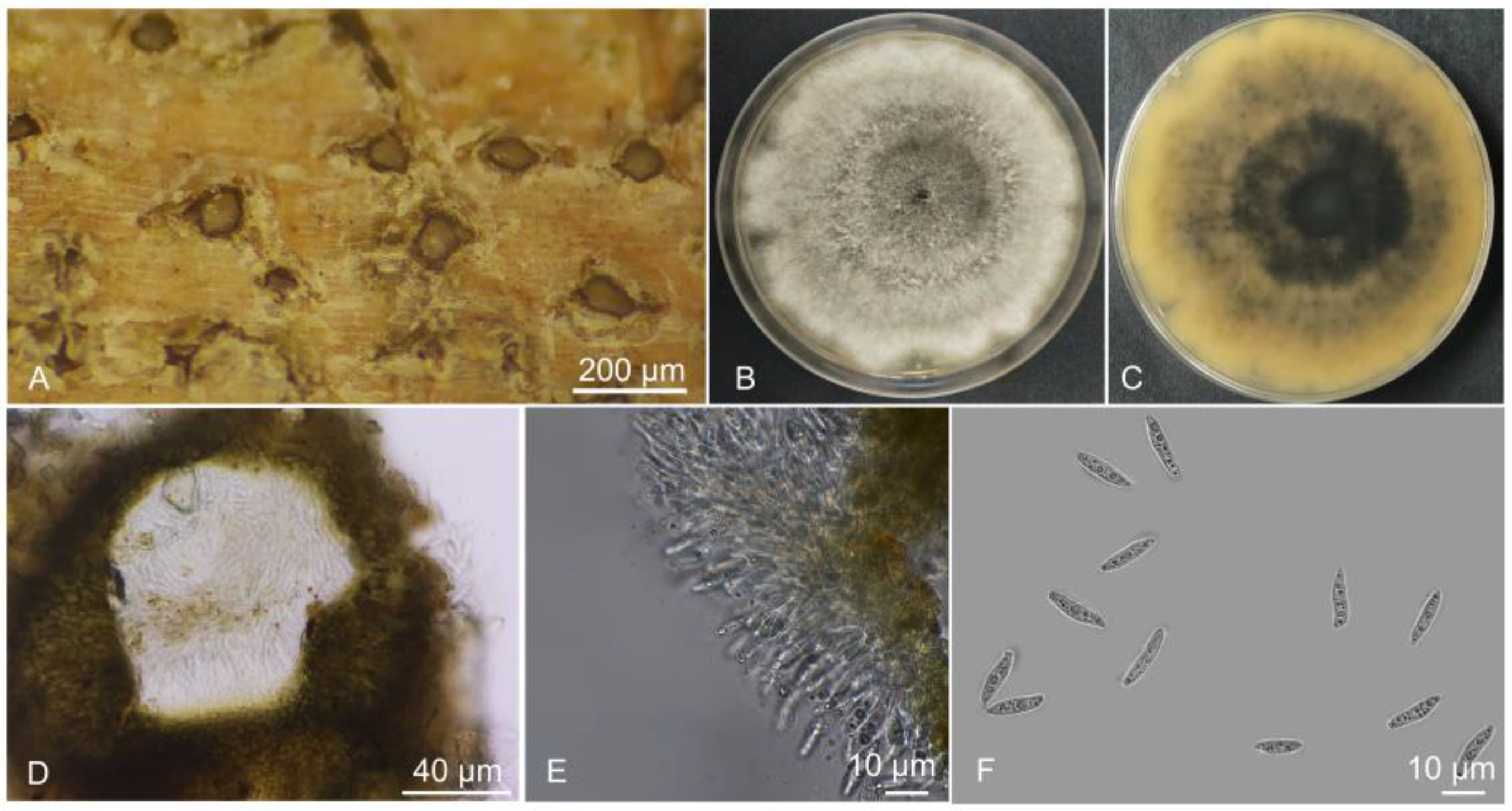
Figure 1. Symptoms of branch blight on Acer saccharum and morphological characteristics of isolate SDAEULY1031. (A) Habit of conidiomata on branches. (B,C) Seven-day-old culture on PDA from top view and from bottom view. (D) Conidia wall. (E) Conidiomatal cells. (F) Conidia.
Forests 16 01786 g001
Figure 2. Phylogenetic tree reconstructed based on the combined alignment of ITS, tef1, tub2, and rpb2 partial sequences of 42 isolates of the Botryosphaeria species. Cophinforma atrovirens (Mehl & Slippers) A. Alves & A.J.L. Phillips (MFLUCC 11-0-0425 and MFLUCC 11-0-0655) and Neoscytalidium dimidiatum (Penz.) Crous & Slippers (CBS 251.49 and CBS 145.78) were used as the outgroup. The scale bar indicates 7.0 expected changes per site. The isolates obtained in this study are highlighted in pale red.
Forests 16 01786 g002
Figure 3. The symptoms of Botryosphaeria wangensis isolates SDAEULY1031 and SDAEULY1032 inoculated on A. saccharum branches. (AC) Symptoms caused by B. wangensis isolates SDAEULY1031 and SDAEULY1032 at 3 dpi (days post inoculation), 9 dpi, and 15 dpi, respectively. (D) CK, inoculated with pure PDA plugs as control at 15 dpi. Scale bar = 1 cm. (E,F) Statistics of the lesion areas caused by isolates SDAEULY1031 and SDAEULY1032, respectively. The lesion area data from three biological replicates at 15 days after inoculation.
Forests 16 01786 g003
Disclaimer/Publisher’s Note: The statements, opinions and data contained in all publications are solely those of the individual author(s) and contributor(s) and not of MDPI and/or the editor(s). MDPI and/or the editor(s) disclaim responsibility for any injury to people or property resulting from any ideas, methods, instructions or products referred to in the content.