Communication

Urban Pathways of Oomycete Dissemination: A Case Study from Warsaw Parks

Diagnosticlab IBL, Department of Forest Protection, Forest Research Institute, Braci leśnej 3, 05-090 Sękocin Stary, Poland
*
Author to whom correspondence should be addressed.
Forests 2025, 16(11), 1736; https://doi.org/10.3390/f16111736
Submission received: 15 October 2025 / Revised: 4 November 2025 / Accepted: 14 November 2025 / Published: 17 November 2025
(This article belongs to the Special Issue Health and Disease Management of Urban Forest Trees)

Abstract

Urban green spaces are essential components of city ecosystems, providing environmental and social benefits while simultaneously serving as potential entry points for invasive plant pathogens. In recent years, increasing attention has been directed toward the role of urban environments as reservoirs and transmission corridors for oomycetes, a group of highly destructive microorganisms affecting trees and shrubs. This study aimed to investigate the diversity and potential introduction pathways of oomycetes in three Warsaw parks representing distinct ecological settings: a historical city park, a large landscape park with aquatic features, and a newly constructed linear park. Samples of soil, and surface water were collected and analysed using standard isolation and molecular identification methods. Four species were identified: Phytophthora cactorum, P. cambivora, Phytopythium vexans, and Ph. montanum—the latter two representing first records for urban parks in Poland. The results indicate that nursery plant material, surface water systems, and wildlife activity, particularly birds, are likely contributors to the introduction and spread of these pathogens in city landscapes. The findings underscore the growing phytosanitary risk associated with urban greenery, where the interplay of anthropogenic disturbance, high plant turnover, and complex hydrological networks facilitates pathogen establishment. This research highlights the urgent need to integrate urban biosecurity strategies with routine molecular monitoring, nursery inspections, and wildlife surveillance to limit further dissemination of invasive oomycetes and enhance the resilience of urban tree populations.

1. Introduction

Urban greenery is a key component of the ecological structure of cities, fulfilling environmental, health, and social functions. Urban parks influence microclimate, reduce air and noise pollution, and improve the well-being of residents [1,2]. At the same time, as ecosystems that are intensively used and subject to constant human interference, they are highly susceptible to phytosanitary disturbances [3]. Under abiotic stress conditions typical of urban habitats, plants are particularly vulnerable to infections caused by soil- and water-borne pathogens, including oomycetes [4,5].
Oomycetes (Oomycota) are among the most destructive pathogens of trees and shrubs, responsible for numerous epizootics and plant epidemics in Europe and worldwide [6]. Species such as Phytophthora cambivora, P. cactorum, and P. plurivora are well-documented causal agents of root rot and dieback in deciduous trees, while representatives of the genus Phytopythium are increasingly reported as opportunistic plant pathogens in aquatic and soil environments [7,8]. The presence of these organisms in urban green areas poses a significant threat to the stability of park ecosystems and increases the risk of pathogen transmission into natural habitats.
A key aspect of the discussion on the occurrence and spread of oomycetes is the role of ornamental and forest plant nurseries. For several decades, research has indicated that nurseries serve as major reservoirs and sources of Phytophthora introduction into new environments [9,10]. Studies conducted in various countries in Europe and North America have shown that a substantial proportion of plant material leaving nurseries was permanently infected, and numerous Phytophthora species were regularly detected in irrigation water [11,12]. In some facilities, more than 30 species of these pathogens were isolated simultaneously [10].
As a result, new plantings in urban parks—often carried out in stages using nursery material of diverse origins—represent particularly vulnerable points in the urban landscape. Pathogen introduction at this stage promotes their establishment in soils and water bodies, followed by further spread through rainwater, puddles, and gardening equipment [4,13]. The presence of oomycetes in parks not only threatens the durability of tree stands and the ecosystem functions of urban greenery but also creates a risk of local epidemic foci that may spread into natural habitats and managed forests [14].
Comparable studies in other European cities have demonstrated high oomycete diversity in managed urban green spaces, with Phytophthora plurivora, P. cinnamomi, and Phytopythium vexans commonly detected in soil and irrigation water [10,13]. Research by Green et al. [15] and Barwell et al. [16] further emphasized the increasing role of trade-related introductions and local hydrological pathways in shaping pathogen assemblages. These studies collectively suggest that urban environments act as epidemiological bridges between nurseries, water systems, and natural forests. The present research builds upon these findings by exploring analogous dynamics in Warsaw’s park network.
In light of the above, it is essential to recognise the diversity and scale of oomycete occurrence in Poland’s urban ecosystems, including parks in Warsaw. Such knowledge can form the basis for preventive actions grounded in biosecurity principles and phytosanitary monitoring, aimed at protecting tree health under urban conditions.
The aim of this study was to determine the species composition of oomycetes inhabiting selected Warsaw parks and to identify potential sources of their introduction into the urban environment. In particular, the research focused on comparing the taxonomic diversity of oomycetes isolated from soil, surface water, and tree rhizospheres across three distinct types of park habitats. The analysis sought to assess the phytosanitary risk associated with the presence of pathogens from the genera Phytophthora and Phytopythium and to provide a foundation for developing recommendations concerning the monitoring and health protection of urban tree stands.

2. Materials and Methods

2.1. Description of the Study Sites

The research was conducted in three distinct urban green areas in Warsaw, representing a historical inner-city park, a large landscape park with an aquatic ecosystem, and a newly developed linear park built atop a road tunnel. Conditions favouring oomycete infections—such as stagnation or periodic waterlogging of the root zone, limited surface water circulation, and anthropogenically modified substrates—are typical of both historical parks with old tree stands and ponds, and new green spaces established on tunnel decks (characterised by heterogeneous soil profiles and episodic flooding). The assessment of potential oomycete occurrence was based on the presence of visible decline symptoms typical of Phytophthora infections (e.g., bark exudates, necrotic lesions, root collar cankers, and crown thinning), following the diagnostic guidelines of Jung et al. [4]. These field symptoms served as indicators for selecting sampling points rather than as quantitative measures of disease incidence. This selection allows for a comparative analysis of the occurrence and potential habitat niches of oomycetes under different habitat and management conditions. Each park represented a distinct environmental setting (soil, water, or plant-associated niche) to capture different potential pathways of oomycete introduction rather than to provide exhaustive sampling within a single location. Figure 1 shows the relative positions of the three sampling sites within Warsaw.

2.1.1. Ujazdowski Park (Śródmieście District, Warsaw)

A historical park established at the end of the 19th century, designed by Franciszek Szanior, located in the central part of the city. It features a classical layout of alleys, old tree stands, and water bodies typical of representative urban parks. Historical and municipal sources emphasise its establishment in the 1890s, sharing its origins with Skaryszewski Park (an initiative of the Municipal Afforestation Committee), which is reflected in their similar compositional design and choice of ornamental tree species.

2.1.2. Skaryszewski Park Named After I.J. Paderewski—Duck Pond (Praga-Południe District, Warsaw)

One of the largest parks in Warsaw (approximately 58 ha), with a landscape character and a system of artificial ponds and canals (total water surface area of about 11.3 ha). The park was established in the early 20th century by F. Szanior. The “Duck Pond” is one of the park’s two main water bodies, characterised by shallow depth and limited water circulation; nearby lies the semi-natural Kamionkowskie Lake.

2.1.3. Linear Park over the Southern Bypass of Warsaw (Ursynów District; Section Along F. Płaskowickiej Street, near Stryjeńskich Street)

A newly designed green space approximately 2 km long, developed in stages on the deck of the S2 expressway tunnel (Southern Bypass of Warsaw). This design creates a sequence of plantings, meadows, and rain gardens on an anthropogenic substrate, featuring local stormwater retention zones and diverse hydrological conditions.

2.2. Fieldwork

Fieldwork was conducted in 2025. In total, eighteen samples were collected across the three sites: eight soil samples in Ujazdowski Park, four water samples in Skaryszewski Park, and six soil samples in the Linear Park. At each selected site, plant, soil, or water samples were collected to isolate organisms belonging to oomycetes (Oomycota).
In Ujazdowski Park, field surveys took place in June 2025. A total of 51 horse chestnut trees (Aesculus hippocastanum L.) were inventoried, of which four showed symptoms of bleeding canker, such as exudates from the trunk, necrosis of the bark, and crown loss (Figure 2). To assess the presence of Phytophthora pathogens, soil samples were collected from the root zones of from the root zones of four symptomatic and four visually asymptomatic (i.e., trees without external signs of bleeding canker or crown decline; no root pruning was performed due to the protected park setting) trees (approximately 250 g each, at a depth of 10–30 cm).
In Skaryszewski Park, the study material comprised water samples collected from the “Duck Pond”. In the immediate vicinity of the pond, dieback symptoms were observed in several tree species (Picea abies (L.) H. Karst and Pseudotsuga menziesii (Mirb.) Franco). In June 2025, samples were collected from four points (Figure 3) corresponding to the cardinal directions, as far from the shore as possible, in sterile containers with a volume of 1 L.
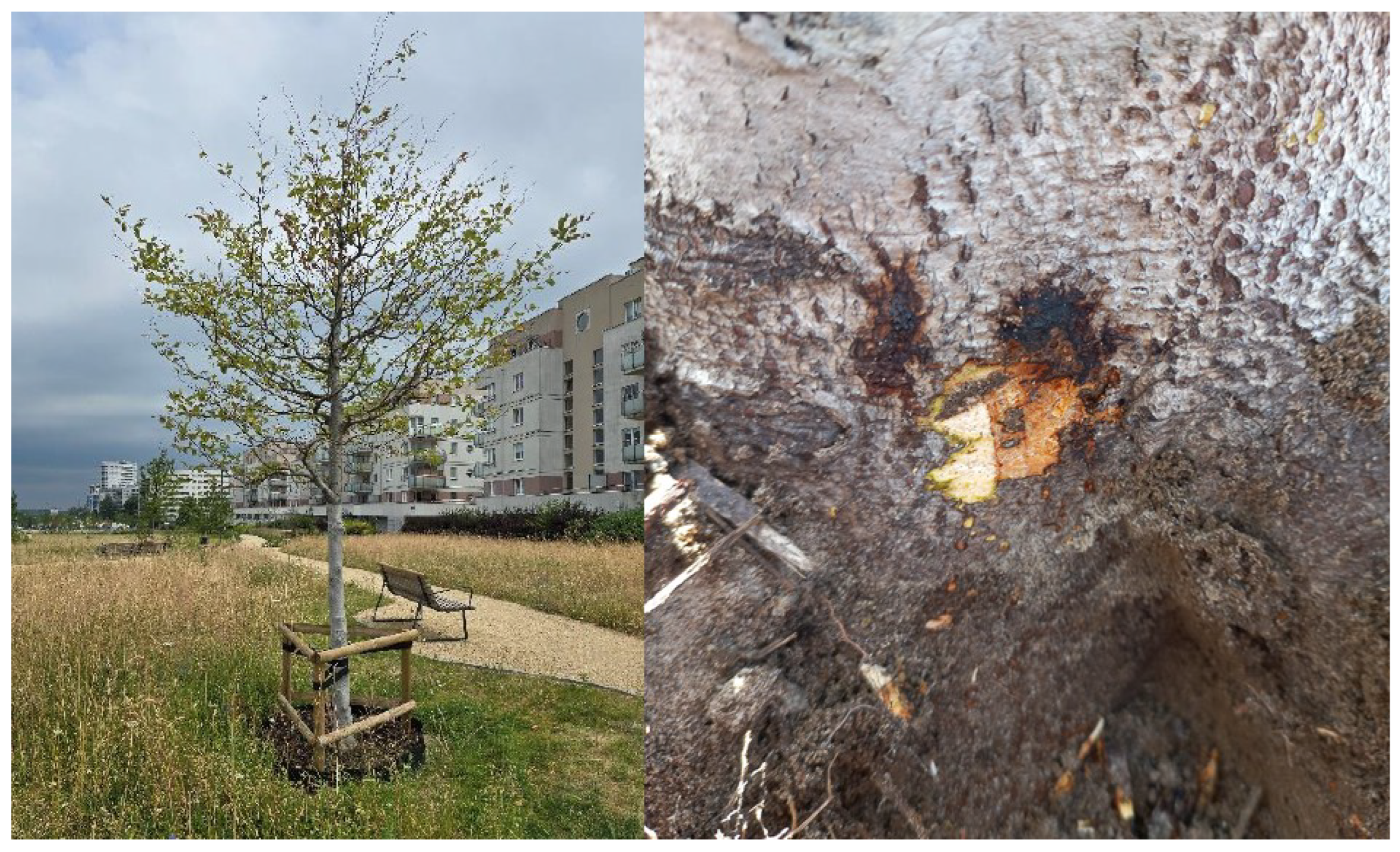
In the linear park above the Southern Bypass of Warsaw (S2), the research focused on European beech (Fagus sylvatica L.), where exudates and necroses were found at the root collar. In July 2025, soil samples (approximately 250 g each, depth 10–30 cm) were taken from the rhizosphere zones of three symptomatic and three asymptomatic trees, as close to the trunk as possible (Figure 4).

2.3. Laboratory Analyses

Soil and water samples were processed to isolate organisms of the Oomycota lineage, using baiting and vacuum filtration methods combined with culturing on selective media.
For soil samples collected beneath trees in Ujazdowski Park and in the linear park above the Southern Bypass of Warsaw, the classical biological baiting method was used, as is common in phytopathological diagnostics of Phytophthora spp. [17,18]. Soil subsamples were placed in sterile containers and flooded with distilled water so that the water level was approximately 4–5 cm above the soil surface. Young leaves of pedunculate oak (Quercus robur L.) and rhododendron (Rhododendron sp.) were placed on the water surface. The leaves were incubated for 3–5 days at room temperature and examined daily for the formation of dark necrotic spots indicative of infection. Fragments of infected tissue (approx. 5 × 5 mm) were then transferred to selective V8–PARPNH medium (16 g/L agar, 2 g/L CaCO3, 100 mL/L vegetable juice, supplemented with inhibitors: pimaricin 10 μg/ml, ampicillin 200 μ/mL, rifampicin 10 μg/mL, PCNB 25 μg/ml, nystatin 50 μg/mL, and hymexazol 50 μg/ml), prepared according to the methodology of Jung and Blaschke [19]. Colonies developing on the medium were subsequently transferred to non-selective V8 medium for further culturing and morphological observation. The non-selective V8 medium consisted of 16 g agar, 100 mL vegetable juice (V8, Campbell Soup Co., Camden, NJ, USA), 2 g CaCO3, and distilled water to a final volume of 1 L (pH 6.0–6.2).
For water samples collected from the “Duck Pond” in Skaryszewski Park, the vacuum filtration method was used [20]. One-litre samples were filtered through 5.0 μm pore-size membranes (MF-Millipore™, Merck Millipore, Burlington, MA, USA) using a MityVac II pump (Mityvac Selectline Superpump, Manufacturer: Lincoln Industrial Corporation/Mityvac, St. Louis, MO, USA). The filters were placed directly onto selective V8–PARPNH medium and incubated at room temperature for 24 h, after which the filters were removed and the plates left for further incubation. As colonies with features typical of oomycetes appeared, they were transferred to fresh V8 medium to obtain pure cultures.

2.4. Preparation of Isolates for Microscopic Observation

Selected isolates grown on non-selective media were examined microscopically to confirm their morphological features. Fragments of actively growing mycelium (approximately 5 × 5 mm) were taken from the colony margins after 3–5 days of incubation and transferred onto microscope slides. Preparations were made in a drop of distilled water, covered with a cover slip, and observed under transmitted light using a ZEISS Axioskop 2 (Carl Zeiss AG, Oberkochen, Germany) or ZEISS Axiolab microscope equipped with a digital camera (magnifications 20×–400×).
To induce the formation of reproductive structures, the soil extract method was used [6]. Fragments of colonies were transferred into Petri dishes flooded with fresh, non-sterile soil extract and incubated at room temperature. After several hours, the extract was replaced with distilled water, and further observations were made at intervals of several hours. This procedure allowed for documentation of characteristic diagnostic structures, such as sporangia, chlamydospores, and sexual organs.

2.5. Molecular Analyses

To confirm the species identification of selected oomycete isolates, sequencing of the ITS rDNA region was performed. This marker is commonly used in the diagnosis of Phytophthora and Phytopythium species [21,22]. Fragments from actively growing colonies (3–7 days old) were taken from the margins of cultures grown on V8 media and suspended in reaction buffer according to the Phire™ Plant Direct PCR Kit protocol (Thermo Fisher Scientific, Waltham, MA, USA). Amplification was carried out using the universal primers ITS4 and ITS6, which cover the ITS1, 5.8S rDNA, and ITS2 regions.
PCR reactions were conducted in a final volume of 20 μL containing 10 μL of Phire Plant PCR buffer, 0.5 μM of each primer, and 0.5 L of the mycelial suspension. The thermal cycling profile consisted of an initial denaturation at 98 °C for 5 min, followed by 40 cycles of denaturation at 98 °C for 5 s, primer annealing at 55 °C for 5 s, and elongation at 72 °C for 50 s, with a final extension at 72 °C for 7 min.
The amplification products were visualized on gels of agarose 1% stained with GelRedTM (Biotium, Inc., Fremont, CA, USA) and subsequently purified using an anti-inhibitor purification kit (A&A Biotechnology Sp. z o.o., Warsaw, Poland). Purified amplicons were sequenced bidirectionally using capillary electrophoresis on an ABI 3730xl DNA Analyzer (Applied Biosystems, Inc., Foster City, CA, USA). The resulting chromatograms were edited in FinchTV (Geospiza Inc., Seattle, WA, USA), and consensus sequences were compared against the GenBank (NCBI) database using the BLAST (version 2.14.1) algorithm. Species identification was considered reliable when sequence similarity with reference entries in the database was ≥99%.
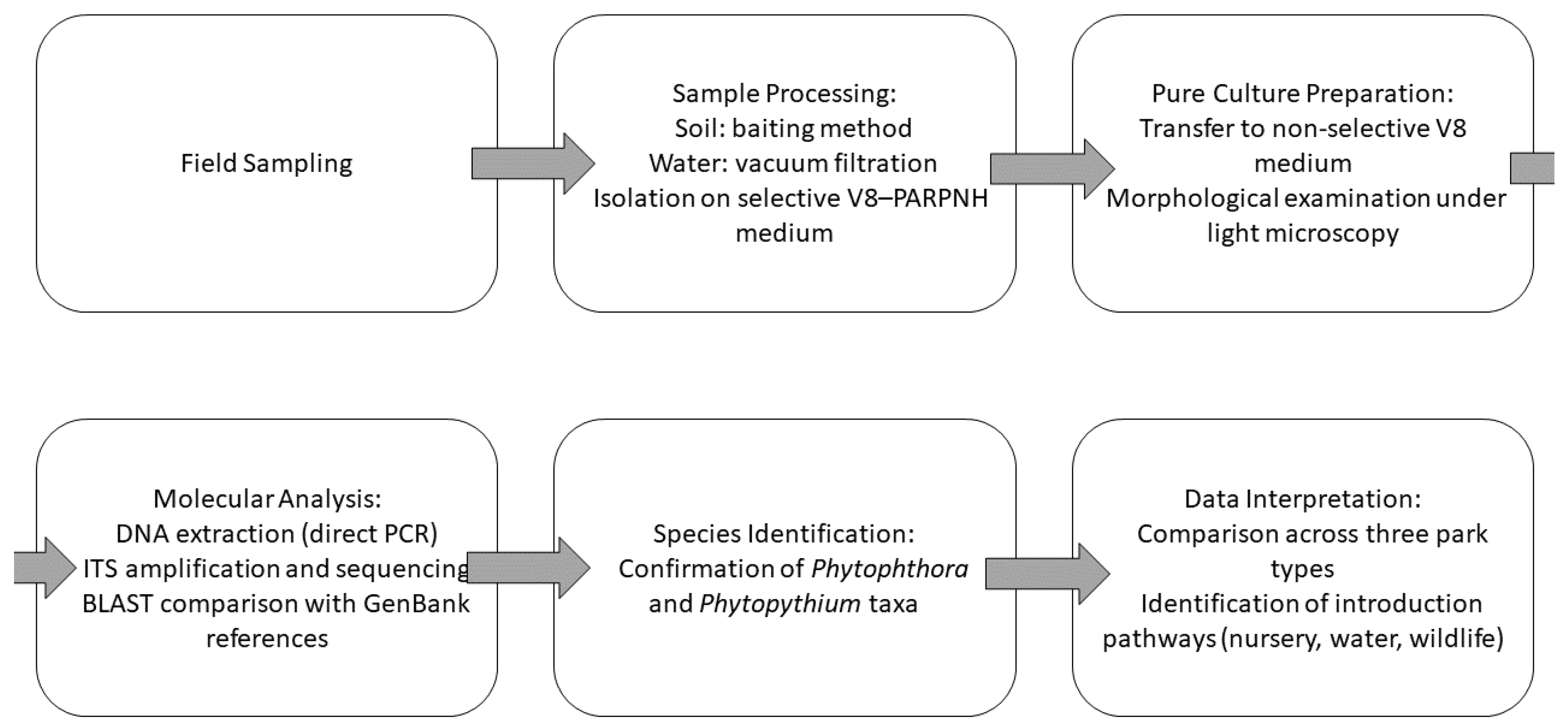
A schematic overview of the experimental workflow is presented in Figure 5.

3. Results

3.1. Ujazdowski Park

Among the 51 inventoried horse chestnut trees (Aesculus hippocastanum L.), four individuals (7.8%) exhibited symptoms typical of bleeding canker, including bark necrosis, dark brown exudates, and progressive crown dieback. Isolates obtained from soil samples collected in their root zones grew on selective V8–PARPNH medium. The colonies were characterized by rapid growth, a distinct radiating hyphal pattern, and the formation of numerous sporangia of spherical to oval shape. Microscopic observations revealed the presence of papillae and occasional internal proliferation. Sexual structures included smooth-walled, terminal oogonia and thick-walled oospores. Species identification was based on the ITS region, which remains the standard and most widely used barcode for oomycetes, including Phytophthora and Phytopythium, due to its high interspecific resolution and extensive reference coverage in GenBank. Identification was additionally supported by detailed morphological observations of colony and reproductive structures. Given that the primary objective of this study was to identify key pathogen taxa and assess potential introduction pathways rather than to perform multilocus phylogenetic analyses, ITS-based identification was considered adequate and reliable for the ecological scope of the work [23]. Nevertheless, we acknowledge the value of additional loci such as COI (cox1) as complementary markers, which may be incorporated in future analyses for enhanced taxonomic verification. ITS sequence analysis showed ≥99.7% similarity with reference sequences of Phytophthora cactorum available in GenBank, allowing unambiguous species identification. No isolates were obtained from soil samples collected under asymptomatic trees.

3.2. Skaryszewski Park (Duck Pond)

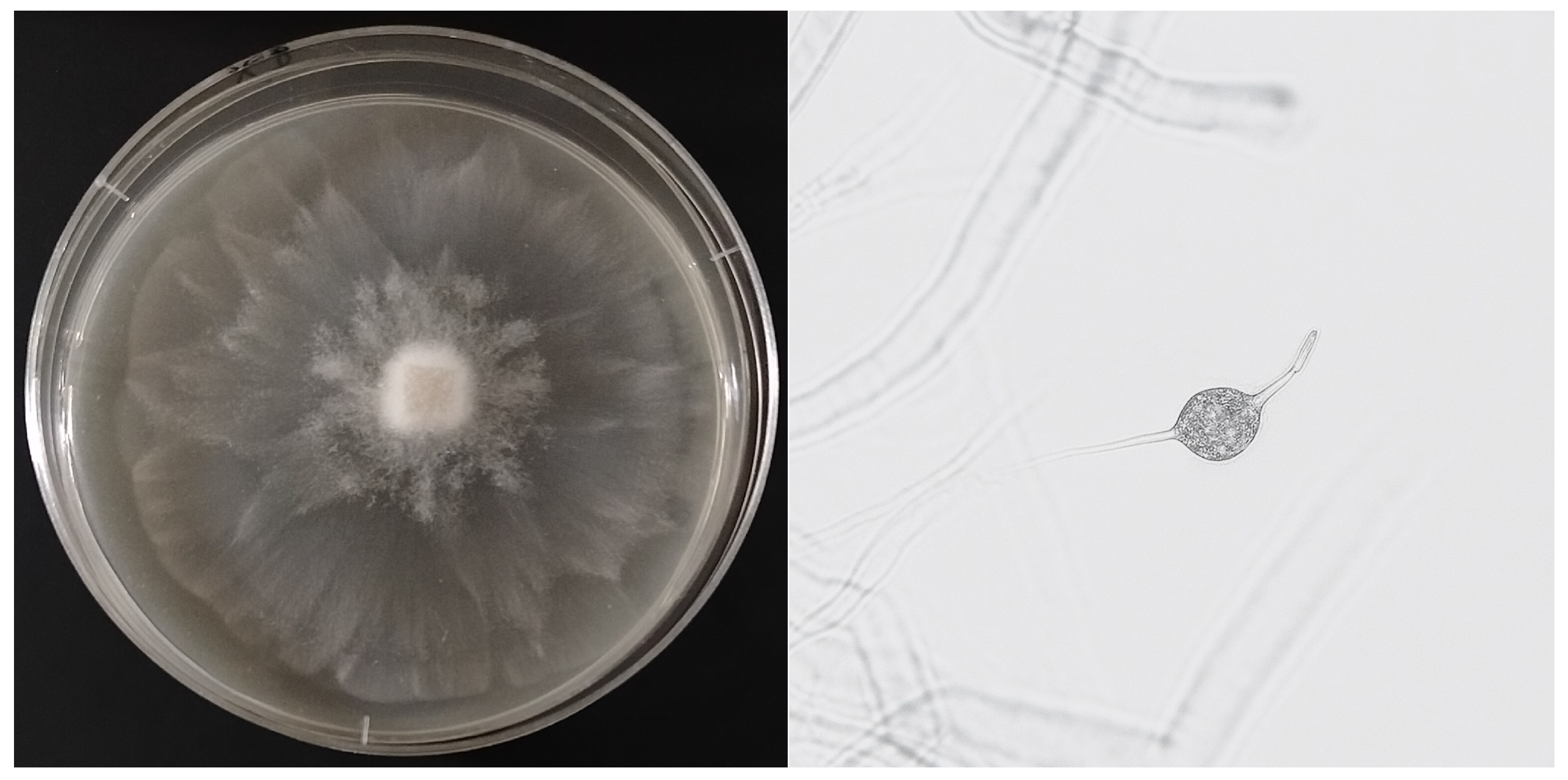
All water samples taken from Duck Pond in June 2025, after vacuum filtration and incubation on V8–PARPNH medium, yielded isolates with slow growth and a characteristic radiating mycelial pattern. Microscopic observations revealed predominantly spherical sporangia with a single, well-developed papilla and numerous cases of internal proliferation. After about three weeks of cultivation, sexual structures were observed: spherical oogonia (21–27 μm), thick-walled, aplerotic oospores, and irregularly shaped antheridia attached laterally to the oogonia (Figure 6). The obtained ITS sequences showed 99%–100% similarity with the reference sequence of Phytopythium montanum (AY162278), confirming the first record of this species in Poland.

3.3. Linear Park Along the Southern Bypass of Warsaw

Soil samples collected from the root zones of European beech (Fagus sylvatica L.) trees showing visible necroses and exudates revealed the presence of three oomycete isolates. Two of them, growing on V8–PARPNH medium, displayed morphological features typical of the genus Phytopythium: oval sporangia and characteristic sexual structures. ITS sequence analysis confirmed their identity as Phytopythium vexans. The third isolate exhibited a different colony morphology and numerous thick-walled oospores. Molecular analysis showed 99% similarity with Phytophthora cambivora sequences, a species known to cause collar rot of beech. As in Ujazdowski Park, no oomycetes were detected in samples collected from asymptomatic trees.

4. Discussion

4.1. Spread of Oomycetes in Urban Areas

The spread of oomycetes, particularly Phytophthora and related taxa, in urban areas presents a significant challenge for the protection of urban greenery and limits the ability to manage plant diseases under anthropogenic conditions. In the urban sites analysed in Warsaw, several likely mechanisms of pathogen introduction were observed: plant material (seedlings), animal vectors (birds), water transport, and human-mediated transmission (shoes or mud). Each of these mechanisms is discussed below with reference to the literature, along with implications associated with the detected species—Phytophthora cactorum and Phytopythium montanum.

4.2. Introduction via Plant Material

The trade in plants and planting material is a well-established and well-documented pathway for the spread of Phytophthora. Numerous studies in different countries have demonstrated that nurseries and the ornamental plant trade are sources of new pathogen introductions and genetic variation [10,15]. Research in British nurseries revealed that approximately half of the samples analysed (water and roots) tested positive for Phytophthora DNA, indicating a high risk of pathogen import through plant material [15]. In Poland, molecular analyses have shown that many Phytophthora species spread through contaminated plant material and soil substrate [24,25].
In the case of beeches growing in a linear park, P. cambivora and Phytopythium vexans were confirmed. P. cambivora is a widely distributed species often responsible for beech decline [10,26]. Ph. vexans has previously been reported from forest nurseries, including in Poland [27,28]. The new beech plantings in the linear park, established on land following tunnel construction, represent a typical example of pathogen spread via planting material. If the root ball or attached soil of the seedlings contained the pathogen, it could have been transferred to the site together with the plants. Even visually healthy seedlings may harbour latent infections, a scenario frequently described in the literature for outbreaks of tree diseases following new plantings [29].
Additionally, the import of plant material from multiple sources (different countries, differing phytosanitary regimes) increases the risk of introducing new isolates and hybrid pathogens [29]. Therefore, it is important to trace the origin of the seedlings, examine nursery production methods (disinfection, clean irrigation water, molecular testing), and, where possible, implement pass–fail testing of plant material before planting.

4.3. Birds as Vectors

The role of birds as potential Phytophthora vectors has been investigated in several studies, though it is often considered a low-probability but realistic pathway. Malewski et al. [30] conducted research in Poland demonstrating that birds can carry Phytophthora propagules on their feathers or feet, and that swab analysis from these surfaces can serve as an environmental detection method [30,31,32]. Although detection rates were generally low, they were significant, suggesting that birds may transfer the pathogen between water bodies, trees, and wooded areas.
Although earlier studies proposed that birds may serve as occasional carriers of Phytophthora propagules via contact with contaminated water or soil [15,30], this hypothesis remains speculative and lacks strong experimental confirmation. Considering the intensity of human activity in urban parks, anthropogenic movement of contaminated soil and water is likely a far more significant factor in pathogen dissemination than potential avian transport. Moreover, many Phytophthora and Phytopythium species naturally inhabit aquatic environments, making direct water-mediated transmission a more plausible mechanism than animal carriage.
In the case of Skaryszewski Park, near the “Duck Pond”, this infection route may be particularly relevant. The pond hosts numerous water birds such as mallards (Anas platyrhynchos) and coots (Fulica atra). These birds may potentially act as mechanical carriers of oomycete propagules; however, this pathway was not directly verified in the present study. Although Ph. montanum is not considered a highly aggressive pathogen [33], continued monitoring is warranted to observe whether nearby trees showing decline could be weakened by the presence of this organism in the water.

4.4. Waterborne Transport—Runoff and Stream Spread

Water transport is a recognised mechanism for Phytophthora dispersal over distances—zoospores and sporangia move in water and can colonise stream banks, wet areas, and irrigation systems. In Poland, various Phytophthora species have been recorded in rivers, canals, and ponds, dominated by P. plurivora, with occurrences of P. cambivora, P. cinnamomi, and others [34]. Polish molecular studies indicate that plant material and water are the main routes of Phytophthora spread [24].
A particularly interesting case was described by Tkaczyk and Sikora [20], where a new Phytophthora species for Poland was detected in an urban watercourse. The authors hypothesise that the pathogen may have been washed off from the surface of Okęcie airport or adjacent areas—for instance, runoff water from aircraft washing or transport surfaces—and subsequently carried further by the water system. This suggests that hydrological corridors in cities constitute real epidemiological threats.
Similar mechanisms have been documented internationally—for instance, in P. ramorum, where the pathogen penetrates streams and rivers and subsequently colonises new areas [35]. In urban conditions, discharge points, drainage systems, and stormwater infrastructure are especially critical as they may connect industrial, airport, or storage zones with green spaces.

4.5. Human Transmission via Footwear

Human activity—such as tourism, gardening, and walking—represents an important but difficult-to-quantify route for pathogen transmission. Under wet conditions, pathogens may adhere to footwear, gardening tools, or equipment and be transported between green zones. Literature on Phytophthora highlights the need for boot and equipment hygiene to prevent pathogen spread in the field [35]. For P. kernoviae and P. ramorum, it has been documented that humans can transport spores on footwear soles, tools, and even through recreational movement [15].
In Ujazdowski Park, the observation that all dead horse chestnut trees were located along footpaths may indicate a pattern consistent with human-mediated transmission—people walking along paths may carry mud containing spores deeper into vegetated zones. Similarly, park maintenance workers might unintentionally spread P. cactorum (confirmed on chestnuts) through contaminated tools or machinery. Although direct studies are lacking for this specific case, analogies from the literature suggest that this mechanism cannot be ignored in urban environments.

4.6. Taxonomic and Epidemiological Significance: Phytophthora cactorum and Phytopythium mntanum

An important finding of the study is that Phytophthora cactorum, although widely distributed in Poland and found in various habitats, was for the first time confirmed on horse chestnut trees. This is a noteworthy observation, as P. cactorum has a broad host range and high adaptive potential, making it an important species from a plant health risk perspective [36,37]. Confirmation of a new host expands its known host spectrum and necessitates the inclusion of this species in future monitoring programmes.
Phytopythium montanum, on the other hand, is a taxon not previously reported from Poland. The inclusion of this species in the local oomycete assemblage indicates a dynamic shift in urban pathogenic communities, likely driven by global movement of biological material and aquatic corridors. The genus Phytopythium (taxonomically separated from Pythium/Phytophthora; [28]) includes species adapted to both aquatic and soil environments, increasing their competitiveness in variable urban conditions. Review literature emphasises the need to monitor newly emerging taxa and hybrids under anthropogenic settings [16].

4.7. Integration of Dispersal Mechanisms and Risk Management Implications

In real urban systems, all the mechanisms mentioned above may act simultaneously—reinforcing each other and enabling “leapfrog” pathogen dispersal between otherwise isolated green patches. For example, a pathogen may be introduced with planting stock, then spread through water to a nearby stream, carried by birds to another pond, and subsequently transferred on human footwear into a neighbouring park section. Such a dispersal network requires a holistic approach consistent with landscape-scale plant disease spread models (e.g., kernel-dispersion or spatially explicit disease models; [38]).
In the context of urban green space management, preventive strategies must address risks from multiple sources simultaneously: quality control of plant material (testing, certification, nursery policy), monitoring of water corridors and drainage infrastructure, research on animal vectors (birds, small mammals) and genotypic mapping of isolates, hygiene measures (footwear, tools, physical barriers) and user education, coordinated preventive actions combining field management, regulation, and monitoring [15].

5. Conclusions

The comparative analysis confirmed the complementary nature of four main dissemination mechanisms: plant material, birds, water, and human-mediated transfer. The integration of these findings emphasizes the need for multi-pathway monitoring in urban green management. In particular, the detection of Phytophthora cactorum on horse chestnut trees, the first confirmed case in Poland, and the presence of Phytopythium montanum, previously unrecorded in the country, emphasise that the monitoring system must be flexible, methodologically robust, and inclusive of newly emerging taxa. Future research will aim to expand the sampling scheme to include multiple sources (soil, water, and plant tissues) from each park, apply multilocus sequencing for improved taxonomic resolution, and establish a long-term monitoring framework assessing the seasonal dynamics of oomycete populations in urban habitats.

Author Contributions

Conceptualization, M.T. and K.S.; methodology, M.T. and K.S.; validation, M.T. and K.S.; formal analysis, M.T. and K.S.; investigation, M.T. and K.S.; writing—original draft preparation, M.T.; writing—review and editing, M.T.; visualization, M.T. All authors have read and agreed to the published version of the manuscript.

Funding

This research received no external funding.

Data Availability Statement

The original contributions presented in the study are included in the article, further inquiries can be directed to the authors. The raw data supporting the conclusions of this article will be made available by the authors on request.

Conflicts of Interest

The authors declare no conflicts of interest.

References

  1. Dale, A.; Frank, S. Warming and drought combine to increase pest insect fitness on urban trees. PLoS ONE 2017, 12, e0173844. [Google Scholar] [CrossRef] [PubMed]
  2. Kabisch, N.; van den Bosch, M.; Lafortezza, R. The health benefits of nature-based solutions to urbanization challenges for children and the elderly—A systematic review. Environ. Res. 2017, 159, 362–373. [Google Scholar] [CrossRef]
  3. Konijnendijk, C.C.; Ricard, R.M.; Kenney, A.; Randrup, T.B. Defining urban forestry–A comparative perspective of North America and Europe. Urban For. Urban Green. 2006, 4, 93–103. [Google Scholar] [CrossRef]
  4. Jung, T.; Pérez-Sierra, A.; Durán, A.; Jung, M.; Balci, Y.; Scanu, B. Canker and decline diseases caused by soil-and airborne Phytophthora species in forests and woodlands. Persoonia-Mol. Phylogeny Evol. Fungi 2018, 40, 182–220. [Google Scholar] [CrossRef]
  5. Hansen, M.; Olivieri, I.; Waller, D.; Nielsen, E.; Group, G.W. Monitoring adaptive genetic responses to environmental change. Mol. Ecol. 2012, 21, 1311–1329. [Google Scholar] [CrossRef]
  6. Erwin, D.; Ribeiro, O.K. Phytophthora Diseases Worldwide; American Phytopathological Society: St Paul, MN, USA, 1996. [Google Scholar]
  7. Abad, Z.G.; Burgess, T.; Bourret, T.; Bensch, K.; Cacciola, S.; Scanu, B.; Mathew, R.; Kasiborski, B.; Srivastava, S.; Kageyama, K.; et al. Phytophthora: Taxonomic and phylogenetic revision of the genus. Stud. Mycol. 2023, 160, 259–348. [Google Scholar] [CrossRef] [PubMed]
  8. Safaiefarahani, B.; Mostowfizadeh-Ghalamfarsa, R.; Hardy, G.; Burgess, T. Re-evaluation of the Phytophthora cryptogea species complex and the description of a new species, Phytophthora pseudocryptogea sp. nov. Mycol. Prog. 2015, 14, 108. [Google Scholar] [CrossRef]
  9. Brasier, C. The biosecurity threat to the UK and global environment from international trade in plants. Plant Pathol. 2008, 57, 792–808. [Google Scholar] [CrossRef]
  10. Jung, T.; Orlikowski, L.; Henricot, B.; Abad-Campos, P.; Aday, A.; Aguín Casal, O.; Bakonyi, J.; Cacciola, S.O.; Cech, T.; Chavarriaga, D.; et al. Widespread Phytophthora infestations in European nurseries put forest, semi-natural and horticultural ecosystems at high risk of Phytophthora diseases. For. Pathol. 2016, 46, 134–163. [Google Scholar] [CrossRef]
  11. Hong, C.; Moorman, G. Plant pathogens in irrigation water: Challenges and opportunities. Crit. Rev. Plant Sci. 2025, 24, 189–208. [Google Scholar] [CrossRef]
  12. Grünwald, B.; Schoeps, B.; Krüger, A. Recognizing the molecular multifunctionality and interactome of TIMP-1. Trends Cell Biol. 2019, 29, 6–19. [Google Scholar] [CrossRef]
  13. Santini, A.; Ghelardini, L.; De Pace, C.; Desprez-Loustau, M.; Capretti, P.; Chandelier, A.; Stenlid, J. Biogeographical patterns and determinants of invasion by forest pathogens in Europe. New Phytol. 2013, 197, 238–250. [Google Scholar] [CrossRef] [PubMed]
  14. Eschen, R.; Britton, K.; Brockerhoff, E.; Burgess, T.; Dalley, V.; Epanchin-Niell, R.; Gupta, K.; Hardy, G.E.S.J.; Huang, Y.; Kenis, M.; et al. International variation in phytosanitary legislation and regulations governing importation of plants for planting. Environ. Sci. Policy 2015, 51, 228–237. [Google Scholar] [CrossRef]
  15. Green, S.; Cooke, D.; Dunn, M.; Barwell, L.; Purse, B.; Chapman, D.; Valatin, G.; Schlenzig, A.; Barbrook, J.; Pettitt, T.; et al. PHYTO-THREATS: Addressing threats to UK forests and woodlands from Phytophthora; identifying risks of spread in trade and methods for mitigation. Forests 2021, 12, 1617. [Google Scholar] [CrossRef]
  16. Barwell, L.; Purse, B.; Green, S.; Hardy, G.; Scott, P.; Williams, N.; Cooke, D.E.; Perez-Sierra, A.; Burgess, T.I.; Chapman, D. Trait-mediated filtering of Phytophthora pathogen invasions through global horticultural trade networks. New Phytol. 2025. [Google Scholar] [CrossRef]
  17. Jung, T.; Blaschke, H.; Neumann, P. Isolation, identification and pathogenicity of Phytophthora Species from Declining Oak Stands. Eur. J. For. Pathol. 1996, 26, 253–272. [Google Scholar] [CrossRef]
  18. Jung, T. Beech decline in Central Europe driven by the interaction between Phytophthora infections and climatic extremes. For. Pathol. 2009, 39, 73–94. [Google Scholar] [CrossRef]
  19. Jung, T.; Blaschke, M. Phytophthora root and collar rot of alders in Bavaria: Distribution, modes of spread and possible management strategies. Plant Pathol. 2004, 53, 197–208. [Google Scholar] [CrossRef]
  20. Tkaczyk, M.; Sikora, K. First report of the occurrence of Phytophthora honggalleglyana in an urban green space in Poland. Urban For. Urban Green. 2025, 108, 128834. [Google Scholar] [CrossRef]
  21. White, T.; Bruns, S.; Taylor, J.W. Amplification and direct sequencing of fungal ribosomal RNA genes for phylogenetics. In Proceedings of the PCR Protocols: A Guide to Methods and Applications; Academic Press Inc.: Cambridge, MA, USA, 1990. [Google Scholar]
  22. Cooke, D.; Drenth, A.; Duncan, J.; Wagels, G.; Brasier, C. A molecular phylogeny of Phytophthora and related oomycetes. Fungal Genet. Biol. 2000, 30, 17–32. [Google Scholar] [CrossRef]
  23. Robideau, G.; De Cock, A.; Coffey, M.; Voglmayr, H.; Brouwer, H.; Bala, K.; Chitty, D.W.; Désaulniers, N.; Eggertson, Q.A.; Gachon, C.M.; et al. DNA barcoding of oomycetes with cytochrome c oxidase subunit I and internal transcribed spacer. Mol. Ecol. Resour. 2011, 11, 1002–1011. [Google Scholar] [CrossRef]
  24. Oszako, T.; Sikora, K.; Belbahri, L.; Nowakowska, J. Molecular analysis of Phytophthora species found in Poland. Folia For. Polonica. Ser. A. For. 2017, 59, 321–328. [Google Scholar] [CrossRef]
  25. Oszako, T.; Żółciak, A.; Tulik, M.; Tkaczyk, M.; Stocki, M.; Nowakowska, J. Influence of Bacillus subtilis and trichoderma asperellum on the development of birch seedlings infected with fine root pathogen phytophthora plurivora. Sylwan 2019, 163, 1006–1015. [Google Scholar]
  26. Tkaczyk, M.; Sikora, K.; Galko, J.; Kunca, A. Incidence and pathogenicity of Phytophthora species in beech (Fagus sylvatica L.) stands in Slovakia. J. Plant Dis. Prot. 2023, 130, 1091–1099. [Google Scholar] [CrossRef]
  27. Bełka, M.; Baranowska, M.; Breza, K. Patogeniczne lęgniowce w szkółce leśnej Trawice w Nadleśnictwie Lipusz. ACTA Sci. Pol. Silv. Colendar. Ratio Ind. Lignar. 2019, 18, 135–140. [Google Scholar]
  28. Tkaczyk, M.; Sikora, K.; Galko, J.; Kunca, A.; Milenković, I. Isolation and pathogenicity of Phytophthora Species from Sessile Oak (Quercus petraea (Matt.) Liebl.) Stands in Slovakia. For. Pathol. 2020, 50, e12632. [Google Scholar] [CrossRef]
  29. Donald, F.; Purse, B.; Green, S. Investigating the role of restoration plantings in introducing disease—A case study using Phytophthora. Forests 2021, 12, 764. [Google Scholar] [CrossRef]
  30. Malewski, T.; Brzezińska, B.; Belbahri, L.; Oszako, T. Role of avian vectors in the spread of Phytophthora species in Poland. Eur. J. Plant Pathol. 2019, 155, 1363–1366. [Google Scholar] [CrossRef]
  31. Dadam, D.; Siasou, E.; Woodward, S.; Clark, J. Migratory passerine birds in Britain carry Phytophthora ramorum inoculum on their feathers and “feet” at low frequency. For. Pathol. 2020, 50, e12569. [Google Scholar] [CrossRef]
  32. Okorski, A.; Paczkowska, G.; Dąbrowska, J.; Rutkiewicz, A.; Borowik, P.; Pszczółkowska, A.; Oszako, T. Characterization of the mycobiome of Pinus sylvestris L. seedlings damaged by forest animals, with an emphasis on pathogenic species. Eur. J. Plant Pathol. 2025, 171, 201–222. [Google Scholar] [CrossRef]
  33. Nechwatal, J.; Oßwald, W. Pythium montanum sp. nov., a new species from a spruce stand in the Bavarian Alps. Mycol. Prog. 2003, 2, 73–80. [Google Scholar] [CrossRef]
  34. Ptaszek, M.; Orlikowski, L. Occurrence of Phytophthora species in watercourses and reservoirs in Poland and the threat to cultivated plants by this genera. Prog. Plant Prot. 2015, 55, 64–70. [Google Scholar] [CrossRef]
  35. Kliejunas, J. Sudden Oak Death and Phytophthora Ramorum: A Summary of the Literature; Gen. Tech. Rep. PSW-GTR-234; US Department of Agriculture, Forest Service, Pacific Southwest Research Station: Albany, CA, USA, 2010. [Google Scholar]
  36. Tkaczyk, M.; Sikora, K.; Nowakowska, J.; Anisko, E.; Oszako, T.; Belbahri, L.; Milenkovic, I. Four different Phytophthora species that are able to infect Scots pine seedlings in laboratory conditions. Folia For. Polonica. Ser. A For. 2016, 58, 123–130. [Google Scholar] [CrossRef]
  37. Chen, X.; Wen, K.; Zhou, X.; Zhu, M.; Liu, Y.; Jin, J.; Nellist, C. The devastating oomycete phytopathogen Phytophthora cactorum: Insights into its biology and molecular features. Mol. Plant Pathol. 2023, 24, 1017–1032. [Google Scholar] [CrossRef] [PubMed]
  38. Fabre, F.; Coville, J.; Cunniffe, N. Optimising reactive disease management using spatially explicit models at the landscape scale. In Plant Diseases and Food Security in the 21st Century; Springer International Publishing: Cham, Switzerland, 2021; pp. 47–72. [Google Scholar]
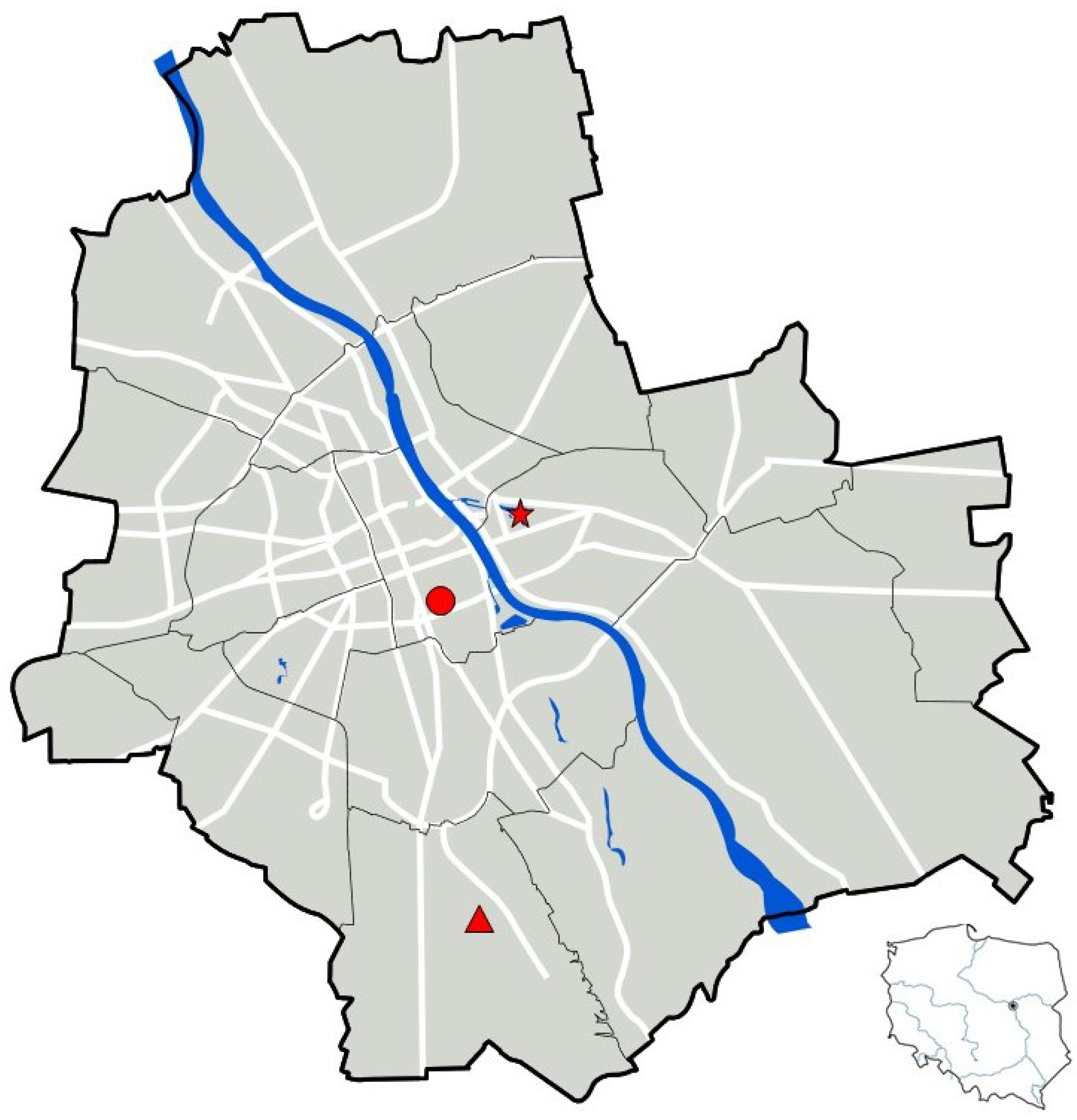
Figure 1. Location of the three study sites within the city of Warsaw (Ujazdowski Park—mark as a circle, Skaryszewski Park—mark as a star, and the Linear Park above the Southern Bypass—mark as a triangle).
Forests 16 01736 g001
Figure 2. Map of the distribution of horse chestnut trees in Ujazdowski Park in Warsaw. Trees on which the leaks shown in the photo on the right were observed are marked in green.
Forests 16 01736 g002
Figure 3. Situational map from Skaryszewski Park. Red stars indicate the water sampling points from Kaczy Pond, and the circle marks a group of dying trees. The photo on the right shows an example of one of the trees (Pseudotsuga menziesii).
Forests 16 01736 g003
Figure 4. An example of a dying European beech in the Linear Park. In the photo on the (left), the tree is shown in its full form; on the (right), necroses are visible in the root collar.
Forests 16 01736 g004
Figure 5. Workflow of the study illustrating key stages: (1) field sampling from three urban parks, (2) isolation and culturing of oomycetes on selective media, (3) microscopic and molecular identification, and (4) ecological interpretation of introduction pathways.
Forests 16 01736 g005
Figure 6. Phytopythium montanum: (left)—sporangium with a distinct single papilla and a short discharge tube (observed under transmitted light microscopy); (right)—colony grown on V8A medium after 10 days of incubation at room temperature, showing characteristic radiating mycelial growth.
Forests 16 01736 g006
Disclaimer/Publisher’s Note: The statements, opinions and data contained in all publications are solely those of the individual author(s) and contributor(s) and not of MDPI and/or the editor(s). MDPI and/or the editor(s) disclaim responsibility for any injury to people or property resulting from any ideas, methods, instructions or products referred to in the content.