Article

Virulence and Pathological Characteristics of a New Metarhizium anisopliae Strain against Asian Long-Horn Beetle Anoplophora glabripennis Larvae

1
Key Laboratory of Plant Protection Resources and Pest Management of Ministry of Education, College of Plant Protection, Northwest Agriculture and Forestry University, Yangling 712100, China
2
Key Laboratory of Integrated Pest Management on the Loess Plateau of Ministry of Agriculture and Rural Affairs, Northwest Agriculture and Forestry University, Yangling 712100, China
*
Author to whom correspondence should be addressed.
Forests 2024, 15(6), 1045; https://doi.org/10.3390/f15061045
Submission received: 16 May 2024 / Revised: 13 June 2024 / Accepted: 13 June 2024 / Published: 17 June 2024
(This article belongs to the Section Forest Health)

Abstract

:
The Asian long-horn beetle (ALB) is a serious wood-boring insect. Continuous isolation of different fungal strains is vital for using fungi for the control of ALB. The virulence and pathological characteristics of a new Metarhizium anisopliae strain DES3 isolated from the desert afforestation stands against the larvae of ALB were assessed in this study. The corrected mortality reached 100% at the conidial concentration of 109 and 108 conidia/mL, and 91.11 ± 4.44% at 107 conidia/mL. Similarly, the LC/LT showed high virulence as well. Meanwhile, the virulence of a commercial M. anisopliae strain against the ALB larvae was evaluated. The corrected mortality was only 33.33% at 109 conidia/mL, and less than 10% at 108 conidia/mL. The pathological characteristics after infection by the M. anisopliae strain DES3 were evident, mainly embodied in the rupture of the adipose tissue, muscle tissue, and midgut. But there was no obvious change after infection by the commercial M. anisopliae strain. In conclusion, these results establish that the M. anisopliae strain DES3 has high virulence in a dosage-dependent manner against ALB larvae, indicating the potential of fungal strain DES3 to be developed as biopesticide for biocontrol of A. glabripennis.

1. Introduction

The Asian long-horn beetle (ALB), Anoplophora glabripennis Motschulsky (Coleoptera: Cerambycidae) is a serious invasive wood-boring pest in forests [1]. Outbreaks of ALB cause huge economic losses. For example, the economic losses in Europe reached approximately EUR 550,000 from 2001 to 2008 [2]. Between 2008 and 2013, the costs of managing ALB totaled EUR 20 million in Lombardy, Italy, alone [3]. Thus, the ALB is taken seriously by many countries [4,5]. Numerous studies researching and using insecticides with high virulence against ALB at different stages have been conducted [1,6,7]. However, excessive and consecutive use of chemical insecticides can have a harmful effect on the environment and cause resistance of pests to pesticides, which is why alternatives to chemical insecticides are needed [8]. As almost no reports on the resistance caused by entomopathogenic fungi can be found, they are a good candidate for insect control.
Entomopathogenic fungi, as an alternative to chemical pesticides, plays an important and necessary role in IPM (integrated pest management) [9]. They receive more and more attention because many strains are highly virulent against many pests, and they are friendly to the environment [10]. Modalities of entomopathogenic fungal infection (Metarhizium and Beauveria) comprise six stages: adhesion, germination, appressorium formation, penetration, colonization of hemolymph, and extrusion and sporulation [11]. During these processes, entomopathogenic fungi also secrete associated enzymes and toxins [12,13]. The entomopathogenic fungi strains finally kill the host through toxins [12,13] and mycelium extrusion [14]. After infection by entomopathogenic fungi, a series of pathological characteristics will occur within the host body [15]. This is not only due to the pathological characteristics of entomopathogenic fungi, but also proves the virulence of entomopathogenic fungi. At present, the typical entomopathogenic fungi, Metarhizium spp., is widely used in biological control of pests [16,17]. In particular, for the management of species such as A. glabripennis, Monochamus alternatus Hope, Aedes aegypti Linnaeus, Nilaparvata lugens Stal, and Melanoplus bivittatus Say, Metarhizium spp. is widely studied and used [15,18,19,20,21]. The IPM requires continuous fungal strains isolation from nature [22]. In order to manage and control the ALB, the aim of our study was to isolate and test a new M. anisopliae strain with high virulence against the ALB.
There are many commercial entomopathogenic fungi strains used for insect control all over the world [23]. Whether commercial strains have high virulence against pests like the ALB is still a question. In this study, the virulence of a commercial strain against the ALB is tested as a comparison.

2. Materials and Methods

2.1. Fungal Isolation and Identification

2.1.1. Fungal Isolation

In June 2023, the M. anisopliae strain DES3 was isolated from soil samples collected at the Hongshixia Afforestation of Sands Experiment Station (HASES) (109°42.757′ E, 38°20.08′ N) in the northern Shaanxi province, China. The HASES is located in the east of Mu Us Desert. The soil samples were collected in desert afforestation stands mainly planted with Pinus sylvestris. The fungal isolation was carried out in the laboratory using the insect bait method with healthy seventh and eighth instar larvae of Tenebrio molitor [24] raised in an artificial incubator. After isolation, the M. anisopliae strain DES3 was stored in the refrigerator at 4 and −80 °C in LIRR (Lab of Insect Relative Resource, College of Plant Protection, Northwest A & F University, Yangling, Shaanxi Province, China).

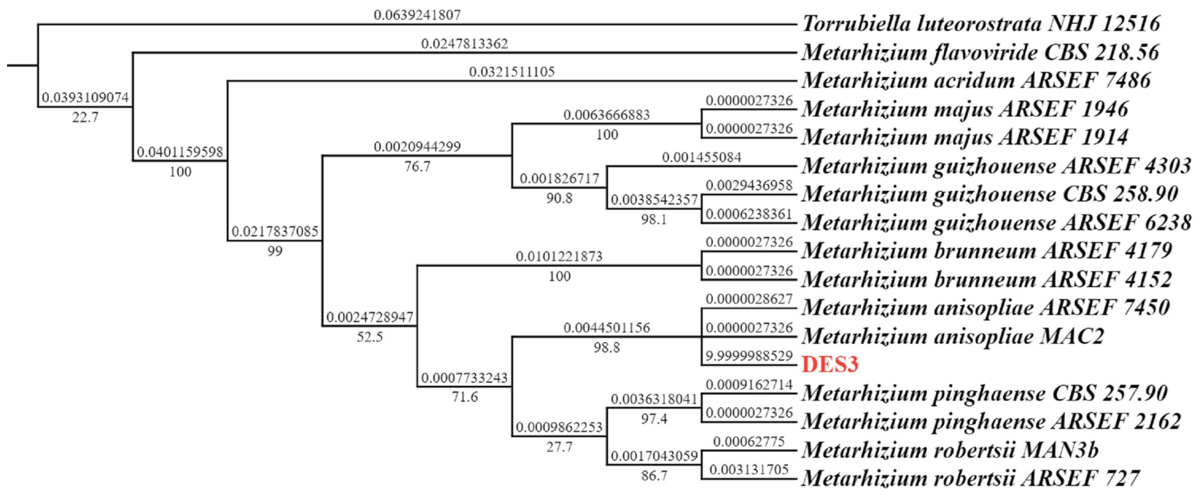
2.1.2. Molecular Identification

After culture on the SDAY medium (10 g/L peptone, 10 g/L yeast extract, 20 g/L agar, 40 g/L D-glucose anhydrous, 1 L sterilized water) for two weeks using the method described by Aljanabi [25] with some modifications, the genomic DNA extraction of the M. anisopliae strain DES3 was carried out. To identify the M. anisopliae strain DES3 molecularly, the internal transcribed spacer (ITS) and αelongation factor 1-alpha (EF1α) regions were amplified via PCR (polymerase chain reactions) using primers (Table S1) [26,27]. The PCR amplicons were sent to a sequencing company (Beijing Tsingke Biotech Co., Ltd., Beijing, China) for Sanger sequencing. All sequences were identified via homology using BLAST from the NCBI GenBank database [28]. After that, a phylogenetic tree was constructed using PhyloSuite v1.2.1 [29,30,31]. After the identification of the M. anisopliae Strain DES3, the obtained sequences were submitted into the NCBI GenBank database (https://www.ncbi.nlm.nih.gov/genbank/}, accessed on 16 April 2024).

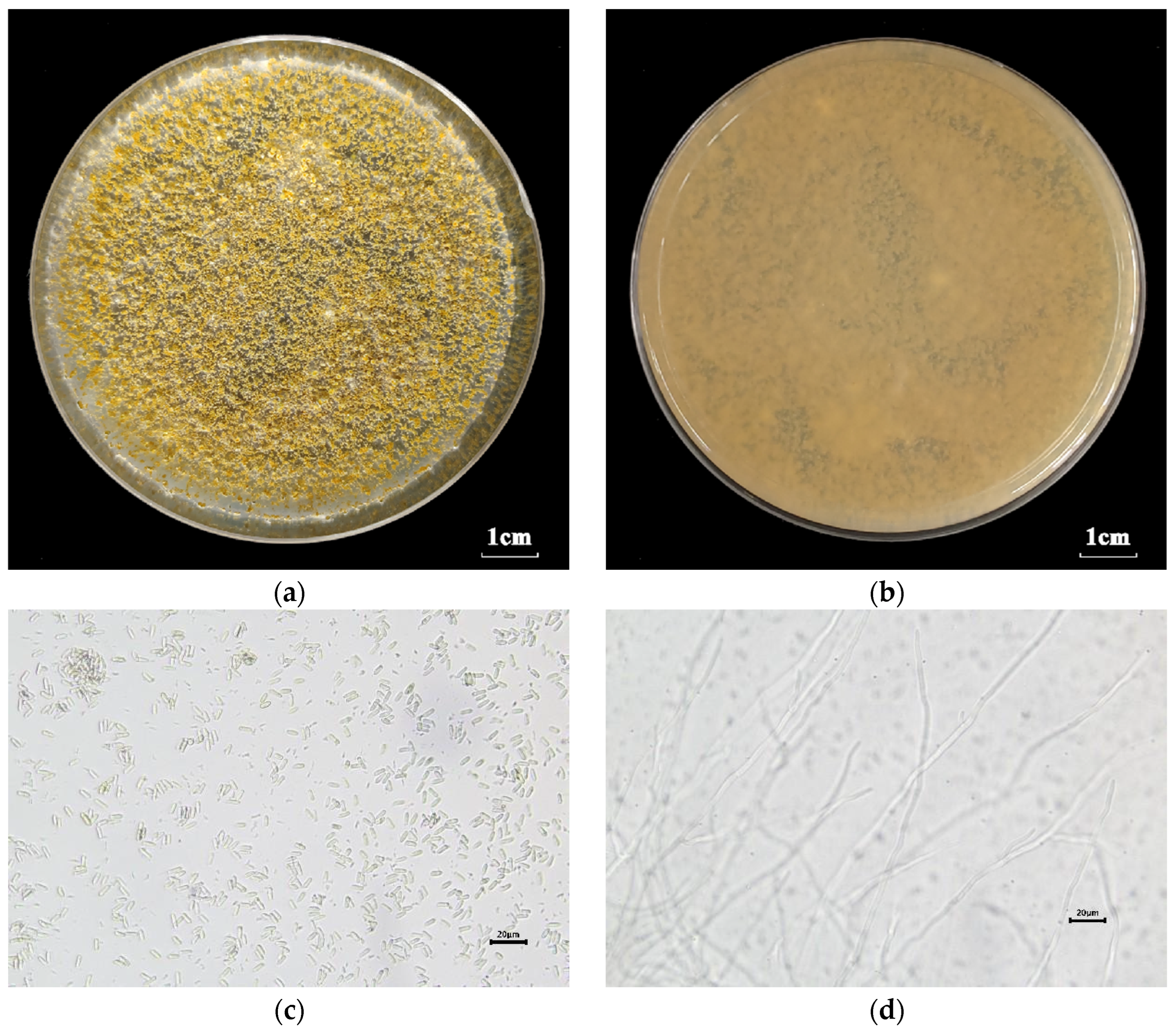
2.1.3. Morphological Identification

On the 1/4 SDAY medium (2.5 g/L peptone, 2.5 g/L yeast extract, 10 g/L D-glucose anhydrous, 20 g/L agar, 1 L sterile water), M. anisopliae strain DES3 was cultured for morphological observation for two weeks to four weeks in an artificial incubator. then, the colony’s morphology, including the characteristics of obverse and reverse of the M. anisopliae strain DES3, was observed. Furthermore, on an optical microscope (Ningbo Sunny Instruments Co., Ltd., Zhejiang, Ningbo, China) at 10× and 40× magnification, the morphological characteristics of mycelia and conidia were observed as well.

2.2. Bioassays of the Metarhizium anisopliae Strain DES3 and Commercial Metarhizium anisopliae Strain against the ALB

2.2.1. Experimental Insects

The ALB larvae used for bioassays and histopathological observations were collected by cutting trees and branches on the Acer negundo and Salix matsudana in November and December 2023 in Yangling, Shaanxi province, China. During the larvae collection, the species identification of larvae referred to the description of Haack [2], and the determination of the instar of larvae referred to the description of He [32]. All larvae used for the bioassays were third instar. The second-instar larvae were also collected, and they were raised to the third instar for experiments. A total of 500 larvae were collected. After the collection of the larvae, they were identified morphologically and molecularly as the Asian long-horn beetle (ALB), Anoplophora glabripennis. And in order to prevent cannibalism, they were reared on the sawdust of Acer negundo separately in 50-well plastic boxes with several small pores in an artificial incubator in complete darkness before bioassays.

2.2.2. Experimental Fungi

To produce conidia, the M. anisopliae strain DES3 was cultured on sterilized 1/4 SDAY medium for two weeks to four weeks in the artificial incubator. Before the bioassay, the conidia were scraped. After that, the scraped conidia were immediately collected into a sterilized 50 mL tube containing 20 mL sterilized water, 0.05% Tween-80, and 1% glycerin. In order to obtain a homogeneous suspension, the conidia suspension was vortexed immediately for 2 min using a vortex oscillator. Then, 10 μL of the conidia suspension was placed into a new 1.5 mL tube and diluted to 100× with 990 μL sterilized water. Under the optical microscope at 10× and 40× magnification, the concentration of the diluted suspension was confirmed using a hemocytometer. After that, the conidia suspension was calculated and diluted with sterilized water containing 0.05% Tween-80 and 1% glycerin to the concentrations of 109, 108, 107, 106, and 105 conidia/mL. For each concentration, a 20 mL suspension was enough to make sure the larvae were completely immersed. For the control group, the sterilized water covered 0.05% Tween-80 and 1% glycerin was used.
The commercial Metarhizium anisopliae strain (Producer: Guangxi NongBao Protection biotechnology Co., Ltd.; date of manufacture: 20 December 2023; quality guarantee period: 12 months) was selected from one of the best-selling commercial products. It was powder, and the number of viable conidia was 1010 g−1. A 5 g sample of the commercial strain was placed into a new 50 mL tube, and 45 g sterilized water containing 0.05% Tween-80 and 1% glycerin was added. Then, the suspension was vortexed immediately for 2 min using a vortex oscillator to make the suspension homogeneous. After this, the concentration of the conidia suspension was 109 conidia/mL. The conidia suspension was then diluted with sterilized water containing 0.05% Tween-80 and 1% glycerin to the concentrations of 109, 108, 107, 106, and 105 conidia/mL. For each concentration, a 20 mL suspension was enough to make sure the larvae were completely immersed. For the control group, sterilized water covered with 0.05% Tween-80 and 1% glycerin was used.

2.2.3. Bioassays

The M. anisopliae strain DES3 and commercial M. anisopliae strain were used for the bioassays. For each concentration (109, 108, 107, 106, and 105 conidia/mL) of different strains, three biological replicates were set. Specifically, in the 109 conidia/mL group, four biological replicates were set for sample collection of the histopathological observations. For one biological replicate, fifteen healthy larvae of the third instar were randomly selected. In this study, the infection method was impregnation. For the experimented larvae, they were submerged in the prepared fungal suspension for 3–5 s in order to ensure they were successfully exposed to the conidia suspension. A sterilized paper was used to absorb excess suspension. After that, fifteen infected larvae per biological replicate were individually transferred to 24-well plastic boxes. The treated larvae were incubated in the artificial incubator in complete darkness. After fungal infection, the larvae were not fed until the end of the experiment. The daily mortality of the ALB larvae was recorded for 21 days.

2.3. Histopathological Observation

After fungal infection by the M. anisopliae strain DES3 and commercial M. anisopliae strain, insect sample collection for histopathological observation was carried out. After 0, 2, 4, 6, and 8 days, three infected larvae were randomly chosen from the 109 conidia/mL group. Then, they were individually placed in 5 mL tubes and 4% paraformaldehyde solution was added until the tube was full. They were all stored at 4 °C. Eventually, after the collection of all the insect samples, the samples were all sent to a biotech company (Shaanxi Y&KBio Bio-Technology Service Co., Ltd., Xi’an, China) for the fabrication of tissue sections (the experimental process was paraffin embedment, paraffin section, and HE staining). Under the microscope at 4× and 10× magnification, the tissue sections were observed in order to describe the pathological characteristics.

2.4. Statistical Analysis

In this study, all figures were produced by GraphPad Prism (version 8.0.2). The calculation of LC50 and LC90 (LC, lethal concentrations) was performed using GraphPad Prism (version 8.0.2) as well. The significant differences were calculated by IBM SPSS Statistics (version 27). And the calculation of LT50 and LT90 (LT, median lethal time) was performed by probit analysis in IBM SPSS Statistics (version 27). Because no larvae died in the control group, the corrected mortality (%) was numerically equivalent to the mortality (%).

3. Results

3.1. Fungal Identification

After sequencing, the accession numbers were submitted to the NCBI database. The phylogenetic tree (Figure 1) suggested that the strain DES3 was Metarhizium anisopliae because the strain DES3 and Metarhizium anisopliae were clustered in the same branch. For morphological identification, the color of the colony was found to be light green (Figure 2a). This was the color of the conidia. On the other hand, the color was found to be light yellow (Figure 2b). Under the microscope, the conidia were clearly elliptic (Figure 2c). The hyphae were filiform (Figure 2d). These were the typical characteristics of the Metarhizium. In conclusion, combining molecular identification and morphological identification, the isolated strain DES3 was identified as Metarhizium anisopliae.

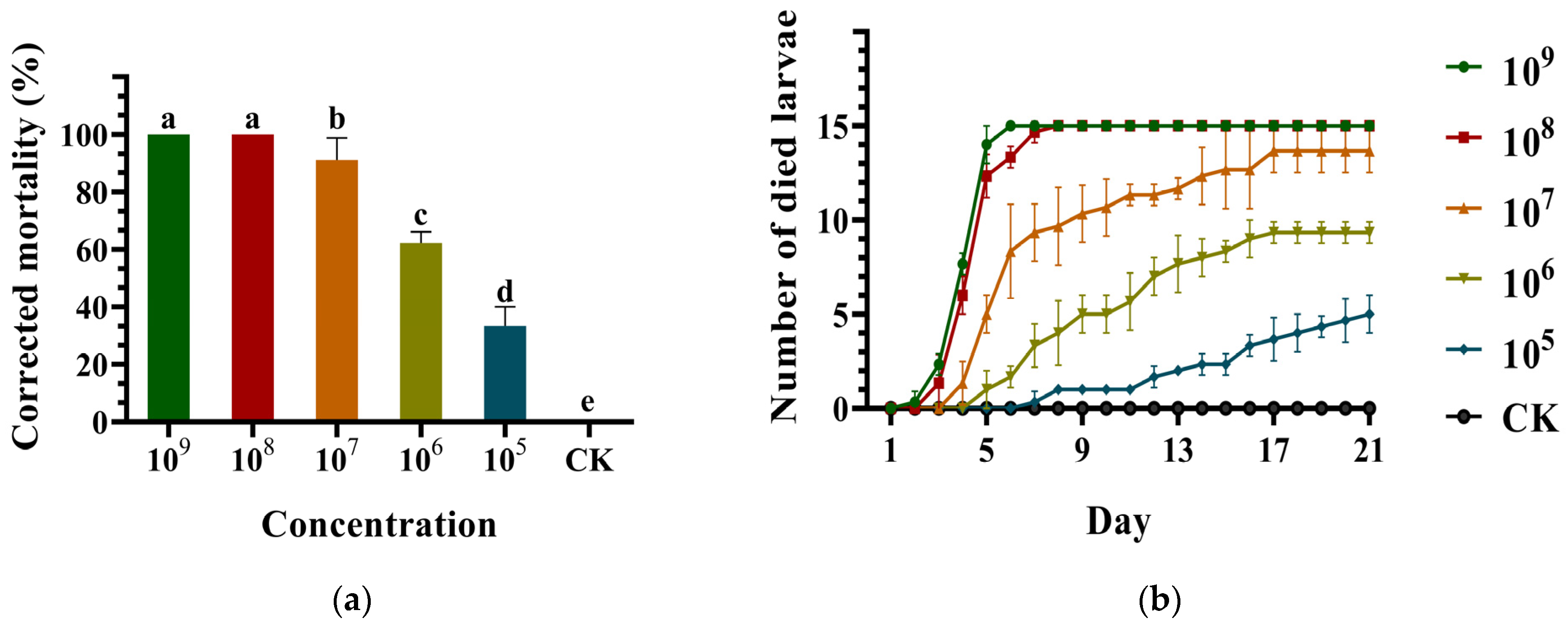
3.2. Virulence of the Metarhizium anisopliae Strain DES3 against the ALB

Overall, the virulence of the M. anisopliae strain DES3 against the ALB larvae was high (Figure 3). The corrected mortality (%) of different conidial suspension concentrations is shown in Figure 3a. Visually, the higher the concentration, the greater the virulence appeared to be. The corrected mortality could reach 100% at the concentration of 109 and 108 conidia/mL. The corrected mortality was 91.11 ± 4.44% at 107 conidia/mL, close to 100%. It showed high virulence. In addition, the corrected mortality was 62.22 ± 2.22% at 106 conidia/mL. The corrected mortality was found to be 33.33 ± 3.85% at 105 conidia/mL, 0% in the CK group. The number of larvae which died per day at different concentrations is shown in Figure 3b. At 109 conidia/mL, after 2 days of infection, the larvae began to die, but only one larva died. Then, the number of larvae which died increased rapidly. All the larvae of 109 conidia/mL died after only 6 days. At 108 conidia/mL, after 3 days of infection, the dead larvae appeared. The larvae all died after 8 days. The dead larvae appeared later at lower concentrations (107, 106, and 105 conidia/mL). As an important indicator of virulence, the LC50 was 1.39×10⁶ conidia/mL and the LC90 was 1.31 × 10⁷ conidia/mL (Table 1), which also showed high virulence. While high virulence was demonstrated, its lethal velocity was also rapid. The LT50 was only 3.90 days and the LT90 was only 5.43 days (Table 2) at 109 conidia/mL.

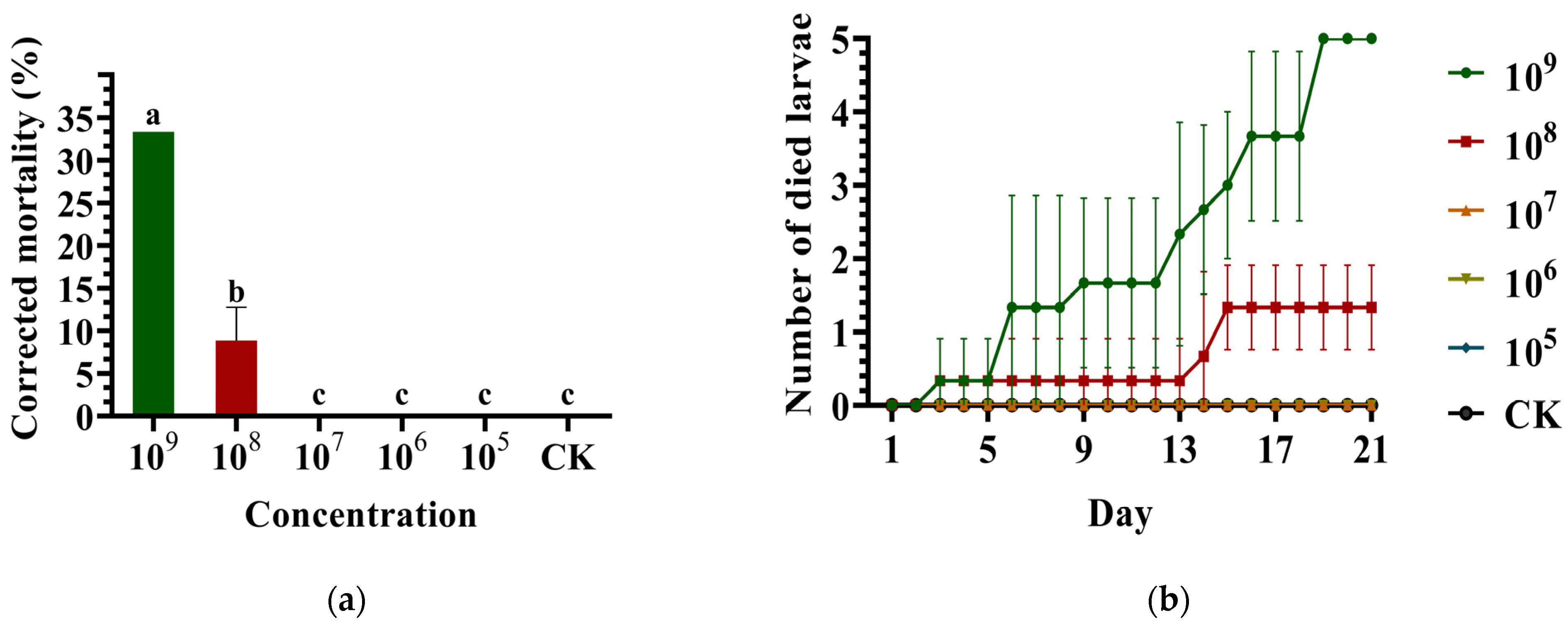
3.3. Virulence of the Commercial Metarhizium anisopliae Strain against the ALB

Overall, the virulence of the commercial M. anisopliae strain against the ALB larvae was low (Figure 4). At 109 conidia/mL, the corrected mortality was only 33.33%. The 109 conidia/mL concentration was the highest in this study. At 108 conidia/mL, the corrected mortality was even less than 10%. However, the corrected mortality was 0% at the other concentration (107, 106, and 105 conidia/mL) (Figure 4a). The number of dead larvae per day at different concentrations of the commercial M. anisopliae strain is shown in Figure 4b. Two days following infection, the larvae began to die (109 and 108 conidia/mL), the same result as the M. anisopliae strain DES3. The number of died larvae was 0 at 107, 106, and 105 conidia/mL. Because the mortality was too low, the LC and LT were not calculated. In conclusion, the commercial M. anisopliae strain showed low virulence in a dosage-dependent manner against the third-instar larvae of the ALB.

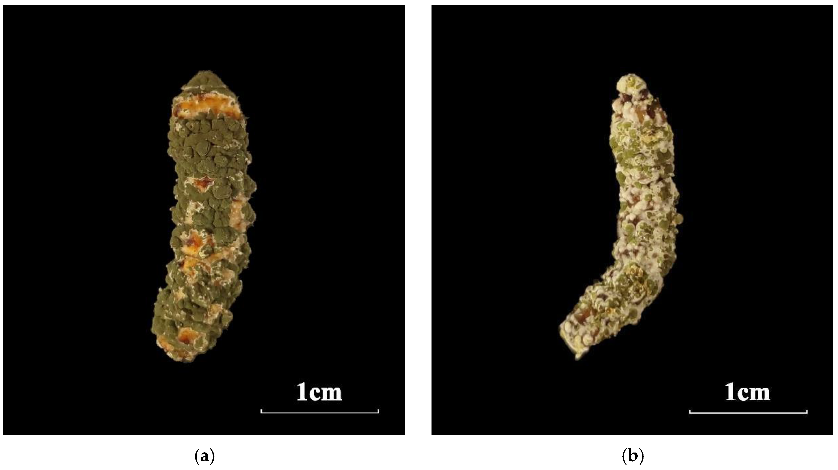
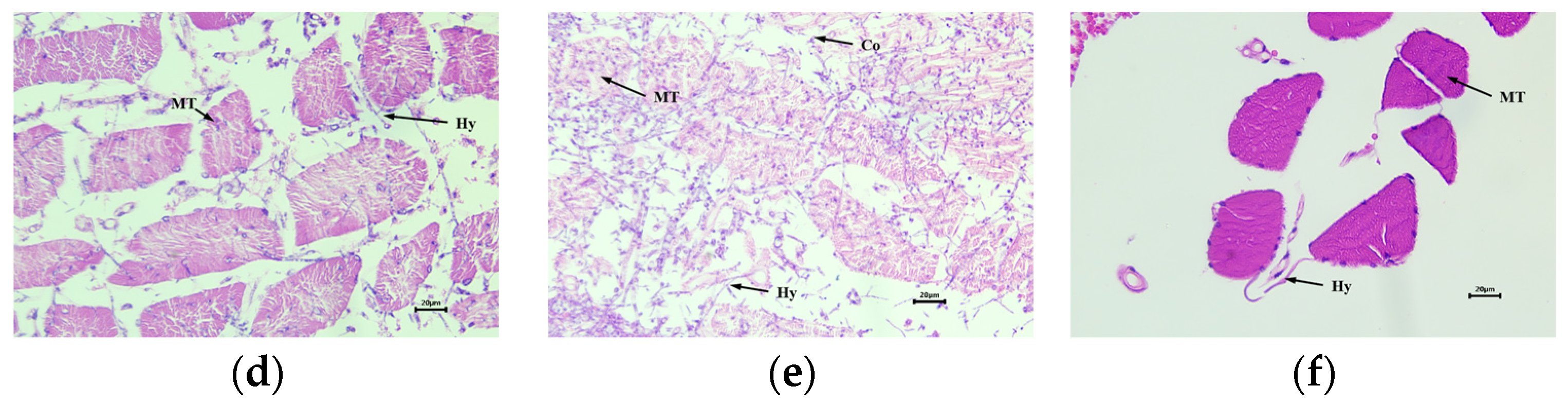
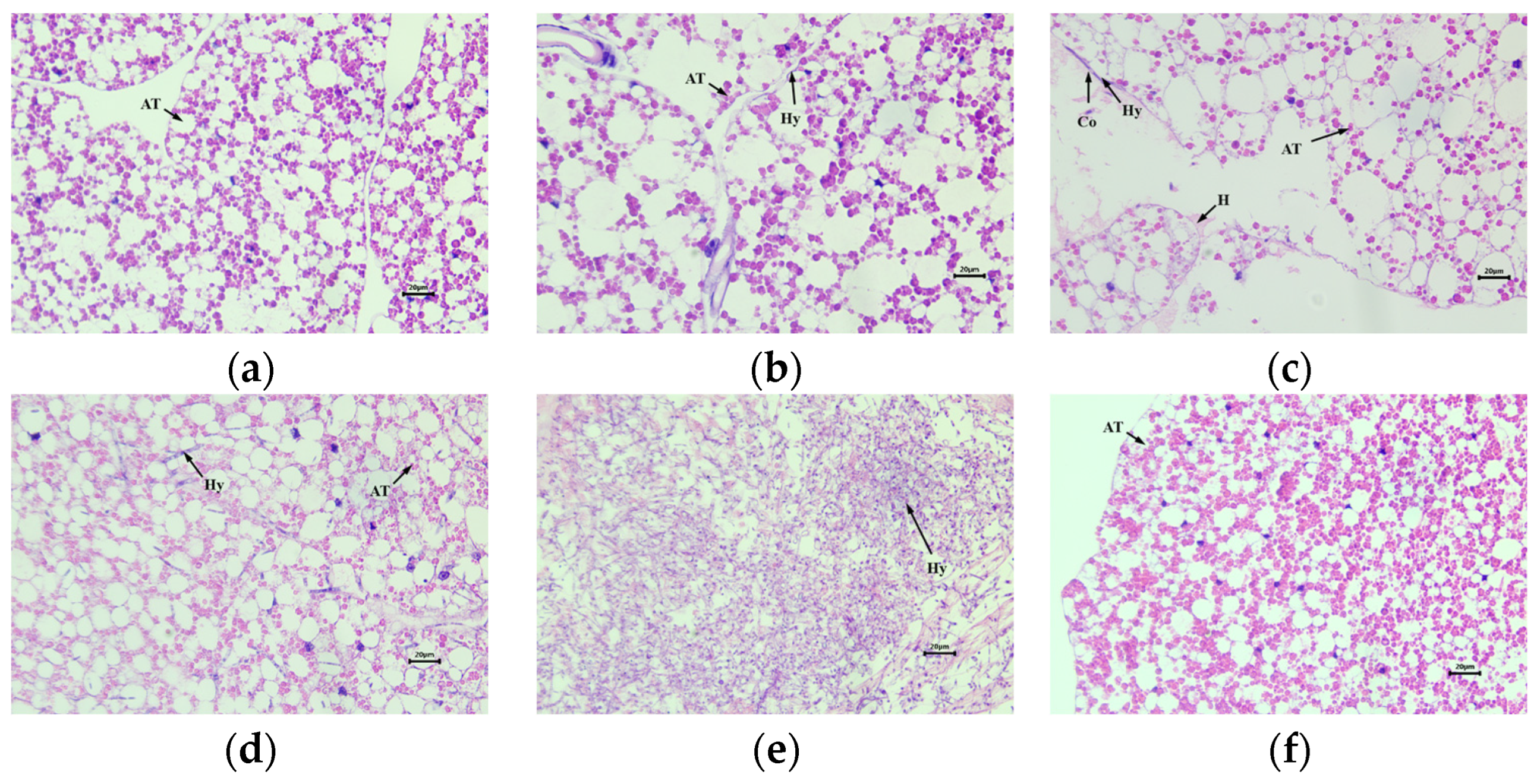
3.4. Histopathological Observation

After infection by the M. anisopliae strain DES3 and the commercial M. anisopliae strain at the same concentration (109 conidia/mL) for 8 days, the symptoms were different (Figure 5). The larvae infected by the M. anisopliae strain DES3 became green (covered by conidia) and stiff (Figure 5a,b). Conversely, the larvae infected by the commercial M. anisopliae strain did not change much compared to the normal dead ALB larvae.
Through observation of the tissue slices using the microscope, histopathological observations of the third-instar larvae of ALB infected by the M. anisopliae strain DES3 and the commercial M. anisopliae strain were accomplished (Figure 6, Figure 7 and Figure 8). The histopathological changes after infection by the M. anisopliae strain DES3 were obvious. Overall, the changes were conspicuous. The noticeable changes were mainly in the big tissues, such as muscle tissue, adipose tissue, and midgut. But there was no obvious change in the larvae infected by the commercial M. anisopliae strain.
Firstly, the muscle tissue was observed (Figure 6). After the larvae were infected by the M. anisopliae strain DES3, this moment, the conidia and hyphae were not found (Figure 6a). Two days after infection, it was found that the muscle tissue was still complete. However, around the muscle tissue, hyphae and conidia appeared (Figure 6b). After 4 days of infection, the muscle tissue became more fragmented. At the same time, the hyphae increased in number, and it was found that a congregation of hematocytes appeared (Figure 6c). After 6 days of infection, the muscle tissue was broken. Around the muscle tissue, large numbers of hyphae appeared (Figure 6d). Eight days after infection, the muscle tissue was almost broken down. It is obvious that the insects’ bodies were filled with conidia and hyphae (Figure 6e). But 8 days after infection, the muscle tissue of the larvae infected by the commercial M. anisopliae strain (Figure 6f) did not change much compared with the normal muscle tissue (Figure 6a). Only a few hyphae appeared (Figure 6f).
After the larvae were infected by the M. anisopliae strain DES3, the adipose tissue changed significantly; the pathological characteristics were similar to those of muscle tissue (Figure 7). In the first 2 days, the adipose tissue did not change obviously (Figure 7a,b), but 2 days after infection, the hyphae appeared (Figure 7b). After 4 days of infection, the hyphae and conidia increased, and a portion of the adipose tissue membrane was destroyed. A congregation of hematocytes appeared (Figure 7c). After 6 days, because of the extrusion of the hyphae, the adipose tissue was broken (Figure 7d). Finally, after 8 days, the adipose tissue was almost blurred (Figure 7e). However, 8 days after infection, the adipose tissue of the larvae infected by the commercial M. anisopliae strain (Figure 7f) did not change much compared with the normal muscle tissue (Figure 7a).
Far away from the body wall, the midgut did not change obviously from 0 to 4 days (Figure 8a–c); only few hyphae appeared (Figure 8c). After 6 days of infection, there were many hyphae around the midgut. Part of the midgut was ruptured, and a few hyphae appeared clearly inside the midgut (Figure 8d). After 8 days, the midgut’s structure became vague, and it was filled with hyphae (Figure 8e). However, 8 days after infection, the midgut structure of the larvae infected by the commercial M. anisopliae strain (Figure 8f) did not change much compared with normal muscle tissue (Figure 8a). After fungal infection, the sampled larvae were not fed until the eighth day. Thus, the shape of the midgut became irregular (Figure 8f).

4. Discussion

The isolated entomopathogenic fungal strain DES3 was morphologically and molecularly identified as Metarhizium anisopliae. In other studies, the M. anisopliae strain F52 has demonstrated high virulence against adult ALB [18]. And the M. anisopliae strains ARSEF 8417, ASSEF 8419, etc., have been shown to have high virulence against ALB larvae; the LC50 was less than one week in 107 conidia/mL [33]. For Beauveria and Metarhizium, it has been defined that the LT50 <5 days indicates high virulence [34]. In our study, for the M. anisopliae strain DES3, the LT50s were 3.90 days at 109 conidia/mL and 4.19 days at 108 conidia/mL. Obviously, high virulence was demonstrated. The LC50s were 1.39 × 10⁶ and 1.31 × 10⁷. High virulence was demonstrated as well. Many M. anisopliae strains, such as JEF-197 and JEF-279, have been found to have high virulence against adult M. alternatus under laboratory conditions [35]. In addition, they have also been found to have high virulence against adult M. alternatus in forests [35]. Remadevi [36] also made a successful attempt at controlling teak defoliators with Metarhizium anisopliae in the laboratory, nursery, and forests [36].
In this study, one of our purposes is to evaluate the virulence of the M. anisopliae strain DES3 against the ALB larvae and compare it with the commercial strain. For the bioassay, the selected commercial strain was M. anisopliae, with a high sales volume and recent production date (date of manufacture was 20 December 2023 and quality guarantee period is 12 months; the experiment began on 4 January 2024). But the virulence of the commercial M. anisopliae strain against the ALB larvae was low, far lower than the virulence of the M. anisopliae strain DES3. As shown in Figure 4a, the corrected mortality was only 33.33% at 109 conidia/mL. Similarly, some M. anisopliae strains isolated from cornfields were found to have higher virulence against the D.v. virgifera larvae than commercial strains [37]. In addition, there was no obvious change after the larvae were infected by the commercial M. anisopliae strain. This is one of the direct reasons for its low virulence. Actually, the hyphae and conidia appeared inside the bodies of the larvae (Figure 6, Figure 7 and Figure 8). But the hyphae and conidia did not proliferate in large numbers. The reason may be that the commercial M. anisopliae strain is inactive during production and storage, because the damage caused by long-term adversity (inappropriate temperature, humidity, etc.) is irreversible and the strain may lose viability permanently [38]. It is worth noting that different species of entomopathogenic fungal strains have different optimal conditions under which they show high virulence [39,40]. In many studies, the commercialized fungal entomopathogens have also shown low virulence against the ALB [41]. In addition, another reason may be that the commercial M. anisopliae strain is not specific to the ALB. This is because different enzymes and virulence genes exist in different strains [42,43]. It also indicates the importance of continuous isolation of entomopathogenic fungi against different pests in different environments.
At the same time, through observation of the tissue slices using a microscope, the pathological characteristics of the ALB larvae infected by the M. anisopliae strain DES3 and the commercial M. anisopliae strain were assessed (Figure 6, Figure 7 and Figure 8). The histopathological changes after infection by the M. anisopliae strain DES3 were obvious. They were mainly embodied in big tissues such as the muscle tissue, adipose tissue, and midgut. The pathological characteristics were roughly the same as the description of Monochamus alternatus larvae infected by the M. robertsii strain GQH6 [15]. Overall, tissue destruction was mainly attributed to the constant proliferation of hyphae and conidia. The hyphae and conidia squeezed the tissue and eventually caused tissue destruction. The M. anisopliae strain DES3 may secrete toxins to affect host tissues [44], but this requires further research.
The soil samples from which the strain DES3 was isolated were collected in desert afforestation stands planted with Populus spp. and Pinus sylvestris at the HASES. The HASES is famous for its great success in the afforestation of deserts [45]. The soil nutrients and soil properties were greatly improved by the afforestation, and the soil microbial communities also underwent great changes [45]. At HASES, the Populus spp. is one of the most common tree species used for afforestation of deserts. The ALB is a potential threat to the Populus spp. [46]. In the future, once the ALB breaks out, the “native” entomopathogenic fungi will be more appropriate to use [47]. However, “non-native” entomopathogenic fungal strains may not adapt to the environment and may even bring many uncertain ecological risks [48]. The “native” entomopathogenic fungi, M. anisopliae strain DES3 isolated from the HASES, was highly virulent against the ALB larvae. Thus, once the ALB breaks out in the future, the M. anisopliae strain DES3 will be suitable for use in the pest management. The ALB larvae are trunk-boring pests in forests. Thus, some methods are being developed to help entomopathogenic fungal strains infect the larvae of ALB. Our previous study also found a creative method to propagate the conidia of Metarhizium and Beauveria through a vector mite into the larvae holes of ALB in the forests of Inner Mongolia, China [49]. This method greatly increases the potential for entomopathogenic fungi strains to control ALB in forests.

5. Conclusions

In this study, the M. anisopliae strain DES3 was found to be highly virulent against ALB larvae of the third instar. The corrected mortality reached 100% at the high conidial concentrations (109 and 108 conidia/mL), and 91.11 ± 4.44% at 107 conidia/mL. Similarly, the LC/LT also showed high virulence. As a comparison, the virulence of a commercial M. anisopliae strain against the ALB larvae was low. The corrected mortality was only 33.33% at 109 conidia/mL, and less than 10% at 108 conidia/mL. Moreover, after the ALB larvae were infected by the M. anisopliae strain DES3 at 109 conidia/mL, the pathological characteristics were evident, mainly embodied in the rupture of the adipose tissue, muscle tissue, and midgut. The hyphae and conidia gradually increased, and they squeezed and destroyed the tissue. But there was no obvious change after infection by the commercial M. anisopliae strain. Our study highlights the potential of the M. anisopliae strain DES3 as a biopesticide against ALB larvae.

Supplementary Materials

The following supporting information can be downloaded at: https://www.mdpi.com/article/10.3390/f15061045/s1, Table S1: The primers used in this study.

Author Contributions

Conceptualization, D.W.; methodology, D.W. and J.-Y.Z.; software, J.-Y.Z.; validation, D.W., J.-Y.Z. and C.-C.J.; formal analysis, J.-Y.Z.; investigation, J.-Y.Z. and C.-C.J.; resources, D.W.; data curation, J.-Y.Z. and C.-C.J.; writing—original draft preparation, J.-Y.Z.; writing—review and editing, D.W. and J.-Y.Z.; visualization, D.W. and J.-Y.Z.; supervision, D.W.; project administration, D.W.; funding acquisition, D.W. All authors have read and agreed to the published version of the manuscript.

Funding

This work was supported by the National Key R&D Program of China (2022YFD1401001) and Science and Technology Innovation Project of Qinling Institute in NWAFU (2452023301).

Data Availability Statement

The sequences of the Metarhizium anisopliae strain DES3 are available in the GenBank database.

Acknowledgments

The authors thank all members from Lab of Insect Related Resources (LIRR) in Northwest A&F University.

Conflicts of Interest

The authors declare no conflicts of interest.

References

  1. Wang, L.X.; Li, C.C.; Luo, Y.Q.; Wang, G.J.; Dou, Z.P.; Haq, I.U.; Shang, S.Q.; Cui, M.M. Current and future control of the wood-boring pest Anoplophora glabripennis. Insect Sci. 2023, 30, 1534–1551. [Google Scholar] [CrossRef] [PubMed]
  2. Haack, R.A.; Hérard, F.; Sun, J.H.; Turgeon, J.J. Managing invasive populations of Asian longhorned beetle and citrus longhorned beetle: A worldwide perspective. Annu. Rev. Entomol. 2010, 55, 521–546. [Google Scholar] [CrossRef] [PubMed]
  3. Cavagna, B.; Ciampitti, M.; Bianchi, A. Lombardy region experience to support the prediction and detection strategies. JJ. Entomol. Acarol. Res. 2013, 45, 1–6. [Google Scholar]
  4. Brabbs, T.; Collins, D.; Hérard, F.; Maspero, M.; Eyre, D. Prospects for the use of biological control agents against Anoplophora in Europe. Pest Manag. Sci. 2015, 71, 7–14. [Google Scholar] [CrossRef] [PubMed]
  5. Cavey, J.F.; Hoebeke, E.R.; Passoa, S.; Lingafelter, S.W. A new exotic threat to North American hardwood forests: An Asian longhorned beetle, Anoplophora glabripennis (motschulsky) (Coleoptera: Cerambycidae): Larval description and diagnosis. Proc. Entomol. Soc. Wash. 1998, 100, 373–381. [Google Scholar]
  6. Wang, J.H.; Che, S.C.; Qiu, L.F.; Li, G.; Shao, J.L.; Zhong, L.; Zhang, G.F.; Xu, H. Efficacy of emamectin benzoate trunk injection against the Asian long-horned beetle [Anoplophora glabripennis (Coleoptera: Cerambycidae)]. J. Econ. Entomol. 2020, 113, 340–347. [Google Scholar] [CrossRef] [PubMed]
  7. Barak, A.V.; Wang, Y.; Xu, L.; Rong, Z.; Hang, X.; Zhan, G. Methyl bromide as a quarantine treatment for Anoplophora glabripennis (Coleoptera: Cerambycidae) in regulated wood packing material. J. Econ. Entomol. 2005, 98, 1911–1916. [Google Scholar] [CrossRef] [PubMed]
  8. Barathi, S.; Sabapathi, N.; Kandasamy, S.; Lee, J.T. Present status of insecticide impacts and eco-friendly approaches for remediation—A review. Environ. Res. 2024, 240, 117432. [Google Scholar] [CrossRef] [PubMed]
  9. van Lenteren, J.C.; Bolckmans, K.; Köhl, J.; Ravensberg, W.J.; Urbaneja, A. Biological control using invertebrates and microorganisms: Plenty of new opportunities. Biocontrol 2018, 63, 39–59. [Google Scholar] [CrossRef]
  10. Perumal, V.; Kannan, S.; Alford, L.; Pittarate, S.; Mekchay, S.; Reddy, G.V.P.; Elangovan, D.; Marimuthu, R.; Krutmuang, P. Biocontrol effect of entomopathogenic fungi Metarhizium anisopliae ethyl acetate-derived chemical molecules: An eco-friendly anti-malarial drug and insecticide. Arch. Insect Biochem. Physiol. 2023, 114, e22037. [Google Scholar] [CrossRef]
  11. Aw, K.M.S.; Hue, S.M. Mode of infection of Metarhizium spp. fungus and their potential as biological control agents. J. Fungi 2017, 3, 3020030. [Google Scholar] [CrossRef] [PubMed]
  12. Olombrada, M.; Medina, P.; Budia, F.; Gavilanes, J.G.; Martínez-del-Pozo, A.; García-Ortega, L. Characterization of a new toxin from the entomopathogenic fungus Metarhizium anisopliae: The ribotoxin anisoplin. Biol. Chem. 2017, 398, 135–142. [Google Scholar] [CrossRef] [PubMed]
  13. Wang, J.B.; Lu, H.L.; Sheng, H.Y.; St Leger, R.J. A Drosophila melanogaster model shows that fast growing Metarhizium species are the deadliest despite eliciting a strong immune response. Virulence 2023, 14, 2275493. [Google Scholar] [CrossRef] [PubMed]
  14. Kim, H.M.; Jeong, S.G.; Choi, I.S.; Yang, J.E.; Lee, K.H.; Kim, J.; Kim, J.C.; Kim, J.S.; Park, H.W. Mechanisms of insecticidal action of Metarhizium anisopliae on adult Japanese pine sawyer beetles (Monochamus alternatus). ACS Omega 2020, 5, 25312–25318. [Google Scholar] [CrossRef] [PubMed]
  15. Zheng, J.Y.; Shi, H.L.; Wang, D. A new strain of Metarhizium robertsii isolated from Loess plateau and its virulence and pathological characteristics against Monochamus alternatus. Microorganisms 2024, 12, 514. [Google Scholar] [CrossRef] [PubMed]
  16. Jeon, I.; Kim, J.S. Soil treatment with Beauveria and Metarhizium to control fall armyworm, Spodoptera frugiperda, during the soil-dwelling stage. J. Asia-Pac. Entomol. 2024, 27, 102193. [Google Scholar] [CrossRef]
  17. Yang, X.M.; Zhang, Y.L.; Zhou, J.C.; Dong, H.; Bai, X.J.; Liu, W.; Gu, Z.M. Pathogenicity, infection process, physiological and biochemical effects of Metarhizium rileyi against Spodoptera frugiperda (JE Smith) (Lepidoptera: Noctuidae) larvae. Egypt. J. Biol. Pest Control 2024, 34, 19. [Google Scholar] [CrossRef]
  18. Shanley, R.P.; Hajek, A.E. Environmental contamination with Metarhizium anisopliae from fungal bands for control of the Asian longhorned beetle, Anoplophora glabripennis (Coleoptera: Cerambycidae). Biocontrol Sci. Technol. 2008, 18, 109–120. [Google Scholar] [CrossRef]
  19. Gomes, S.A.; Carolino, A.T.; Teodoro, T.B.P.; Silva, G.A.; Bitencourt, R.D.B.; Silva, C.P.; Alkhaibari, A.M.; Butt, T.M.; Samuels, R.I. The potential of Metarhizium anisopliae blastospores to control Aedes aegypti larvae in the field. J. Fungi 2023, 9, 759. [Google Scholar] [CrossRef]
  20. Jin, S.F.; Feng, M.G.; Chen, J.Q. Selection of global Metarhizium isolates for the control of the rice pest Nilaparvata lugens (Homoptera: Delphacidae). Pest Manag. Sci. 2008, 64, 1008–1014. [Google Scholar] [CrossRef]
  21. Adatia, A.; Johnson, D.; Entz, S. Pathogenicity of two new isolates of Metarhizium anisopliae from Canadian soil to Melanoplus bivittatus (Orthoptera: Acrididae) and Tenebrio molitor (Coleoptera: Tenebrionidae). Can. Entomol. 2010, 142, 128–134. [Google Scholar] [CrossRef]
  22. Mantzoukas, S.; Lagogiannis, I.; Ntoukas, A.; Eliopoulos, P.A.; Kouretas, D.; Karpouzas, D.G.; Poulas, K. Trapping entomopathogenic fungi from vine terroir soil samples with insect baits for controlling serious pests. Appl. Sci. 2020, 10, 3539. [Google Scholar] [CrossRef]
  23. Wendel, J.; Cisneros, J.; Jaronski, S.; Vitek, C.; Ciomperlik, M.; Flores, D. Screening commercial entomopathogenic fungi for the management of Diaphorina citri populations in the Lower Rio Grande Valley, Texas, USA. Biocontrol 2022, 67, 225–235. [Google Scholar] [CrossRef]
  24. Zimmermann, G. The galleria bait method for detection of entomopathogenic fungi in soil. J. Appl. Entomol. 1986, 102, 213–215. [Google Scholar] [CrossRef]
  25. Aljanabi, S.M.; Martinez, I. Universal and rapid salt-extraction of high quality genomic DNA for PCR-based techniques. Nucleic Acids Res. 1997, 25, 4692–4693. [Google Scholar] [CrossRef]
  26. White, T.J.; Bruns, T.; Lee, S.J.; Taylor, J.W. Amplification and direct sequencing of fungal ribosomal RNA genes for phylogenetics. PCR Protoc. 1990, 18, 315–322. [Google Scholar]
  27. Bischoff, J.F.; Chaverri, P.; White, J.F. Clarification of the host substrate of Ascopolyporus and description of Ascopolyporus philodendrus sp. nov. Mycologia 2005, 97, 710–717. [Google Scholar] [CrossRef]
  28. Zhou, Y.; Zou, X.; Zhi, J.; Xie, J.; Jiang, T. Fast recognition of Lecanicillium spp. and its virulence against Frankliniella occidentalis. Front. Microbiol. 2020, 11, 561381. [Google Scholar] [CrossRef]
  29. Zhang, D.F.; Gao, I.; Jakovlic, H.; Zou, J.; Zhang, W.; Li, X.; Wang, G.T. PhyloSuite: An integrated and scalable desktop platform for streamlined molecular sequence data management and evolutionary phylogenetics studies. Mol. Ecol. Resour. 2020, 20, 348–355. [Google Scholar] [CrossRef] [PubMed]
  30. Nguyen, L.T.; Schmidt, H.A.; Haeseler, A.; Minh, B.Q. IQ-TREE: A fast and effective stochastic algorithm for estimating maximum-likelihood phylogenies. Mol. Biol. Evol. 2015, 32, 268–274. [Google Scholar] [CrossRef]
  31. Lanfear, R.; Frandsen, P.B.; Wright, A.M.; Senfeld, T.; Calcott, B. PartitionFinder 2: New methods for selecting partitioned models of evolution for molecular and morphological phylogenetic analyses. Mol. Biol. Evol. 2017, 34, 772–773. [Google Scholar] [CrossRef] [PubMed]
  32. He, P.; Huang, J.F. Laboratory rearing of Anoplophora glabripennis. J. Beijing For. Univ. 1992, 14, 61–67. [Google Scholar]
  33. Dubois, T.; Lund, J.; Bauer, L.S.; Hajek, A.E. Virulence of entomopathogenic hypocrealean fungi infecting Anoplophora glabripennis. Biocontrol 2008, 53, 517–528. [Google Scholar] [CrossRef]
  34. Gebremariam, A.; Chekol, Y.; Assefa, F. Phenotypic, molecular, and virulence characterization of entomopathogenic fungi, Beauveria bassiana (Balsam) Vuillemin, and Metarhizium anisopliae (Metschn.) Sorokin from soil samples of Ethiopia for the development of mycoinsecticide. Heliyon 2021, 7, e07091. [Google Scholar] [CrossRef]
  35. Kim, J.C.; Lee, S.J.; Kim, S.; Lee, M.R.; Baek, S.; Park, S.E.; Kim, J.; Shin, T.Y.; Kim, J.S. Management of pine wilt disease vectoring Monochamus alternatus adults using spray and soil application of Metarhizium anisopliae JEF isolates. J. Asia-Pac. Entomol. 2020, 23, 224–233. [Google Scholar] [CrossRef]
  36. Remadevi, O.K.; Bai, N.S.; Sasidharan, T.O.; Balachander, M.; Dharmarajan, P. Attempts at controlling Teak Defoliator (Hyblaea puera Cramer, Lepidoptera, Hyblaeidae) with the entomopathogenic fungus, Metarhizium anisopliae (Metsch.): Laboratory, nursery and field trials. Int. J. Pest Manag. 2013, 59, 236–242. [Google Scholar] [CrossRef]
  37. Rudeen, M.L.; Jaronski, S.T.; Petzold-Maxwell, J.L.; Gassmann, A.J. Entomopathogenic fungi in cornfields and their potential to manage larval western corn rootworm Diabrotica virgifera virgifera. J. Invertebr. Pathol. 2013, 114, 329–332. [Google Scholar] [CrossRef]
  38. Wu, S.H.; Toews, M.D.; Oliveira-Hofman, C.; Behle, R.W.; Simmons, A.M.; Shapiro-Ilan, D.I. Environmental tolerance of entomopathogenic fungi: A new strain of Cordyceps javanica isolated from a whitefly epizootic versus commercial fungal strains. Insects 2020, 11, 711. [Google Scholar] [CrossRef]
  39. Meng, Y.; Don, P.I.D.W.H.; Wang, D. A new strain of Lecanicillium uredinophilum isolated from Tibetan plateau and its insecticidal activity. Microorganisms 2000, 10, 1832. [Google Scholar] [CrossRef]
  40. Tomer, H.; Blum, T.; Arye, I.; Faigenboim, A.; Gottlieb, Y.; Ment, D. Activity of native and commercial strains of Metarhizium spp. against the poultry red mite Dermanyssus gallinae under different environmental conditions. Vet. Parasitol. 2018, 262, 20–25. [Google Scholar] [CrossRef]
  41. Clifton, E.H.; Jaronski, S.T.; Hajek, A.E. Virulence of commercialized fungal entomopathogens against asian longhorned beetle (Coleoptera: Cerambycidae). J. Invertebr. Pathol. 2020, 20, 1. [Google Scholar] [CrossRef]
  42. Peng, G.X.; Jin, K.; Liu, Y.C.; Xia, Y.X. Enhancing the utilization of host trehalose by fungal trehalase improves the virulence of fungal insecticide. Appl. Microbiol. Biotechnol. 2015, 99, 8611–8618. [Google Scholar] [CrossRef]
  43. Cao, Y.Q.; Li, M.; Xia, Y.X. Mapmi gene contributes to stress tolerance and virulence of the entomopathogenic fungus, Metarhizium acridum. J. Invertebr. Pathol. 2011, 108, 7–12. [Google Scholar] [CrossRef]
  44. Bawin, T.; Seye, F.; Boukraa, S.; Zimmer, J.Y.; Raharimalala, F.N.; Ndiaye, M.; Compere, P.; Delvigne, F.; Francis, F. Histopathological effects of Aspergillus clavatus (Ascomycota: Trichocomaceae) on larvae of the southern house mosquito, Culex quinquefasciatus (Diptera: Culicidae). Fungal Biol. 2016, 120, 489–499. [Google Scholar] [CrossRef] [PubMed]
  45. Li, J.; Tong, X.G.; Awasthi, M.K.; Wu, F.Y.; Ha, S.; Ma, J.Y.; Sun, X.H.; He, C. Dynamics of soil microbial biomass and enzyme activities along a chronosequence of desertified land revegetation. Ecol. Eng. 2018, 111, 22–30. [Google Scholar] [CrossRef]
  46. Hu, J.F.; Angeli, S.; Schuetz, S.; Luo, Y.Q.; Hajek, A.E. Ecology and management of exotic and endemic Asian longhorned beetle Anoplophora glabripennis. Agric. For. Entomol. 2009, 11, 359–375. [Google Scholar] [CrossRef]
  47. Qayyum, M.A.; Saeed, S.; Wakil, W.; Nawaz, A.; Iqbal, N.; Yasin, M.; Chaurdhry, M.A.; Bashir, M.A.; Ahmed, N.; Riaz, H.; et al. Diversity and correlation of entomopathogenic and associated fungi with soil factors. J. King Saud Univ. Sci. 2021, 33, 101520. [Google Scholar] [CrossRef]
  48. Goble, T.A.; Dames, J.F.; Hill, M.P.; Moore, S.D. Investigation of native isolates of entomopathogenic fungi for the biological control of three citrus pests. Biocontrol Sci. Technol. 2011, 21, 1193–1211. [Google Scholar] [CrossRef]
  49. Tang, H.Y.; Zheng, J.Y.; Wang, D. An effective method for biological control of Anoplophora glabripennis (Coleoptera: Cerambycidae) by using a vector mite, Pyemotes moseri (Acarina: Pyemotidae), carrying insecticidal fungi. Sci. Silvae Sin. 2022, 58, 157–164. [Google Scholar]
Figure 1. Phylogenetic tree constructed by PhyloSuite using the ITS and EF1α regions. DES3: The number of isolated M. anisopliae strain used in this study.
Forests 15 01045 g001
Figure 2. Morphological identification. (a) The obverse of the M. anisopliae strain DES3 (bars = 1 cm); (b) the reverse of the M. anisopliae strain DES3 (bars = 1 cm); (c) the conidia of M. anisopliae strain DES3 under a microscope (bars = 20 µm); (d) the hyphae of M. anisopliae strain DES3 under a microscope (bars = 20 µm).
Forests 15 01045 g002
Figure 3. The corrected mortality (%) histogram and the line graph of the number of dead larvae. (a) The corrected mortality (%) histogram at different concentrations after bioassay; (b) line chart of the number of dead larvae every day at different concentrations after bioassay. Different small letters (a, b, c, d, and e) in Figure 3 (a) indicated significant differences (p < 0.05).
Forests 15 01045 g003
Figure 4. The corrected mortality (%) histogram and line graph of the number of dead larvae. (a) Corrected mortality (%) histogram at different concentrations after bioassay; (b) line chart of the number of dead larvae every day at different concentrations after bioassay. Different small letters (a, b, c) in Figure 4 (a) indicated significant differences (p < 0.05).
Forests 15 01045 g004
Figure 5. Symptoms of the ALB larvae infected by the M. anisopliae strain DES3. (a,b) Symptoms of the ALB larvae infected by the M. anisopliae strain DES3 at 109 conidia/mL for 8 days (bars = 1 cm).
Forests 15 01045 g005
Figure 6. Histopathological observation of muscle tissue (MT). Histopathological observation of muscle tissue of the ALB larvae infected by the M. anisopliae strain DES3 after 0 days (a), 2 days (b), 4 days (c), 6 days (d), and 8 days (e) (bars = 20 µm). (f) Histopathological observation of muscle tissue of ALB larvae infected by the commercial M. anisopliae strain after 8 days (bars = 20 µm). MT: muscle tissue; AT: adipose tissue; Mg: midgut; Hy: hyphae; Co: conidia; H: congregation of hematocytes. Explanations of abbreviations are the same from Figure 6 to Figure 8.
Forests 15 01045 g006aForests 15 01045 g006b
Figure 7. Histopathological observation of adipose tissue (AT). Histopathological observation of muscle tissue of the ALB larvae infected by the M. anisopliae strain DES3 after 0 day (a), 2 days (b), 4 days (c), 6 days (d), and 8 days (e) (bars = 20 µm). (f) Histopathological observation of adipose tissue of the ALB larvae infected by the commercial M. anisopliae strain after 8 days (bars = 20 µm).
Forests 15 01045 g007
Figure 8. Histopathological observation of midgut (Mg). Histopathological observation of muscle tissue of the ALB larvae infected by the M. anisopliae strain DES3 after 0 day (a), 2 days (b), 4 days (c), 6 days (d), and 8 days (e) (bars = 50 µm). (f) Histopathological observation of midgut of the ALB larvae infected by the commercial M. anisopliae strain after 8 days (bars = 50 µm).
Forests 15 01045 g008
Table 1. The LC of the M. anisopliae strain DES3 against the ALB larvae.
Fungal NumberLC50 (Spore/mL)95% Confidence IntervalLC90 (Spore/mL)95% Confidence Interval
DES31.39 × 1061.02 × 106–1.94 × 1061.31 × 1074.04 × 106–4.55 × 10⁷
Table 2. The LT of the M. anisopliae strain DES3 against the ALB larvae.
Fungal NumberConidial
Concentration
LT50 (Days)95% Confidence IntervalLT90 (Days)95% Confidence Interval
DES31093.903.78–4.015.434.95–6.18
1084.194.07–4.305.565.09–6.20
1076.105.72–6.4811.399.15–15.41
1069.518.70–10.70--
Disclaimer/Publisher’s Note: The statements, opinions and data contained in all publications are solely those of the individual author(s) and contributor(s) and not of MDPI and/or the editor(s). MDPI and/or the editor(s) disclaim responsibility for any injury to people or property resulting from any ideas, methods, instructions or products referred to in the content.

