Article

Assessment of Various Iron Extraction Treatments on Waterlogged Archaeological Oak

1
Laboratoire de Conservation-Restauration Arc’Antique, 44300 Nantes, France
2
CNR-IBE, National Research Council of Italy, Institute of Bioeconomy, 50019 Florence, Italy
3
CNRS, Institut des Matériaux de Nantes Jean Rouxel, Nantes Université, IMN, F-44000 Nantes, France
4
Laboratory of Technologies for Heritage Materials, Institute of Chemistry, University of Neuchâtel, 2000 Neuchâtel, Switzerland
*
Author to whom correspondence should be addressed.
Forests 2023, 14(9), 1834; https://doi.org/10.3390/f14091834
Submission received: 1 August 2023 / Revised: 4 September 2023 / Accepted: 6 September 2023 / Published: 8 September 2023
(This article belongs to the Special Issue Wood as Cultural Heritage Material)

Abstract

:
Treatments of organic objects to extract ferrous compounds from waterlogged archaeological wood are well documented. For several years, numerous laboratories have been seeking to determine suitable conservation treatments for such organic objects. For chemical treatments, complexing agents such as EDTA and DTPA, along with acids such as citric and oxalic acids, were selected. In addition, oxidants and reducing agents were tested as pre-treatments to improve extraction rates. In fact, chemicals produce a selective reaction on ferrous compounds, which may improve or be inhibited by complexation or dissolution reactions. Their action depends on the type of compound to be extracted and those present inside the wood. The objective of this study was to make a comparative assessment of the various chemical conservation treatments identified and complement the evaluation of their extraction efficiency with a study of their impact on organic matter by adding criteria such as their visual aspect (using a spectrocolorimeter) and physicochemical actions by means of infrared spectroscopy and micromorphological observations. The effectiveness of EDTA was confirmed, as was that of citric acid, despite some questions arising concerning the presence of wood fibers in the treatment solution, even after rinsing. On the other hand, the extraction rate of oxalic acid, which has a very acidic pH, was unsurprisingly lower, but its visual and anatomical results raised the possibility of using it over a short period of time in view of the effectiveness observed on the wood surface. Pre-treatments improved extraction rates, except in the case of EDTA, which independently had a high extraction rate. It was observed that pre-treatments did not appear to cause any significant chemical degradation of the organic matter. This study provides a tool to assist conservators in selecting a chemical treatment that is in line with the state of decay of the wood, the characteristics of the ferrous compounds to be extracted, and the conservation objectives.

1. Introduction

The experiments described in this study follow on from those that have been carried out in the Arc’Antique laboratory since 2012 as part of a series of research projects on the chemical extraction of ferrous compounds impregnating waterlogged archaeological wood.
Archaeological wood conservation treatments do not adequately ensure wood stability when pollution by ferrous compounds is present. Acidic hydrolysis and the oxidation process of organic matter are known to be caused by the presence of iron salts. The irreversible consequences of this are cracks that open up in the wood, which becomes brittle and fragile, and its color changes [1,2,3,4].
Therefore, for many years, laboratories have been seeking to understand the impact of these compounds, as well as to determine potential curative treatment(s) and define their chemical impact on wood materials [5,6,7]. Since 2012, a selection of chemical treatments have been studied on various artifacts. Beyond effectiveness criteria, their overall impact on the wood is a constant question. Extracting iron from waterlogged archaeological wood is a complex process that requires careful consideration of the impact on organic materials. Despite the increasing interest in the extraction of ferrous compounds from wrecks and other archaeological artifacts, the potential effects of the chemicals used in this process on the characteristics of wood have only recently been investigated [8,9]. These studies examined the effects of extraction procedures with chelating agents of different pHs on the composition and mechanical/viscoelastic properties of waterlogged archaeological wood, including with short immersion times. Pecoraro et al. [7,8] observed that the alkaline pH level resulting from the use of tetrasodium EDTA (ethylenediaminetetraacetate), which is normally used for iron extraction, may significantly affect the chemical and viscoelastic properties of wood. This agent caused changes to the composition of wood hemicelluloses, resulting in a partial loss of wood mass and an increase in the viscous behavior of the treated material. Moreover, Pecoraro et al. [9] showed how disodium EDTA and DTPA extraction treatments slightly modified the physical and chemical characteristics of degraded wood; however, a significant difference exists between the two agents in terms of the viscoelastic modifications of the wood, which means that disodium EDTA is preferable to DTPA (diethylenetriaminepentaacetic acid).
In this article, we set out to test a series of sacrificial archaeological wood samples, on which extraction procedures were assessed by means of several analyses. Anatomical and chemical studies were carried out to evaluate the evolution of the chemical impacts on the structural molecules of cell walls and, in particular, on lignin and cellulose.

2. Materials and Methods

2.1. Waterlogged Archaeological Wood Samples

Tests were performed on a wooden artifact from an oak (Quercus sp. subspecies Quercus) plank found in the archaeological site of “Saint-Lupien” (Rezé, France) [10,11]. The plank was of an orange color on the surface, while the core was black in appearance. The state of decay was initially evaluated using the pin test, a method cited in Bingham J. [12] but currently used by conservators. This non-destructive method was used to estimate the surface hardness of the wood and determine the type and duration of consolidation treatment with polyethylene glycol. The test was performed by manually inserting a needle into the wood to determine its sponginess. Depending on the depth of penetration, the samples can be classified into four grades (very spongy, spongy, dense, and very dense). This method is based on the Pilodyn principle, but here, it is applied with a simple needle [13]. The plank was sawn into 90 cubic samples. Each sample was classified according to its level of decay and the depth of pin penetration. A total of 45 samples were selected with a needle penetration value within the average range (between 2 and 4 mm on a maximum scale of 6 mm; the spongiest samples were discarded). Each sample weighed approximately 20–40 g and was measured to be 3.0 × 3.0 × 3.0 cm (Figure 1). The samples were prepared and treated according to various procedures. The most thorough procedure (on a total of 18 samples) consisted of impregnating samples in a 2-step process using iron(II) chloride and sodium sulfide (see Section 2.2) and then pre-treating (see Section 2.3) with either an oxidizing agent (sodium persulfate) or a reducing agent (sodium dithionite) before complexing or acidic extraction. A further 12 samples were impregnated with iron compounds before undergoing iron extraction treatment, without pre-treatment. A total of 6 samples were first pre-treated and then treated without any preliminary impregnation, and 3 samples were treated without any preliminary impregnation or pre-treatment. The reference samples consisted of the following 2 groups: 3 samples were impregnated only (without being pre-treated or treated), and another 3 were neither impregnated nor treated. Table 1 shows details of the various procedures and the distribution of samples.

2.2. Impregnation Process

The impregnation process followed previous studies carried out to produce archaeological wood samples containing iron compounds [6,14,15]. A total of 30 wood samples (the first two groups were previously described) were first immersed in an iron(II) chloride (0.5 M) solution under partial vacuum (0.6 bars) for 4 h; they were left to dry overnight at room temperature. The same samples were then immersed in sodium sulfide (0.5 M) under partial vacuum (0.6 bars) for 4 h and finally stored in osmosis water.
A structural analysis by X-ray diffraction was conducted to characterize the iron compounds in a reference impregnated wood sample (#43). The measurements were carried out at the Institut des Matériaux Jean Rouxel in Nantes (FRANCE) on a Bruker (Champs-sur-Marne, France) D8 ADVANCE using the 1.5406 Å wavelength of the Kα line of Cu. The measurements lasted for 2 h between 5 and 60° with a resolution of 0.016°. A data analysis was carried out using EVA 9.0 software with the PDF-2004 database and based on results from the literature [16]. To complete the characterization, Raman analyses were carried out on one reference sample after impregnation (#45). Raman spectra were acquired with a Renishaw InVia with the following parameters: 632.8 nm laser (HeNe); 100–2000 cm−1 range. The spectral resolution was 3 cm−1, and the power on the sample was 1 mW, focused with a 100× objective. The resulting compounds were iron(II) and iron(III) hydroxides and sulfides. Pyrite and ferrous oxides such as hematite, lepidocrocite, and goethite were analyzed. A cross section showed that these compounds seemed to be mainly distributed on and just under the surface of the samples (between 1 and 2 mm) (Figure 2).

2.3. Chemical Extraction Methods

Samples were immersed in a chemical solution, whose volume was controlled to maintain a constant 1:10 ratio between the weight of the sample and the volume of the solution. All samples were kept at room temperature in the solutions. Extraction took place in covered plastic boxes to prevent evaporation. The following chemicals were used: two acidic chemicals (1.0%w/w citric acid and 1.0%w/w oxalic acid), a slightly acidic chelating agent (3.7%w/w disodium EDTA), and a neutral complexing agent (0.010 mol/L DTPA) [6,9]. In each treatment, the samples were immersed for 5 days. For samples pre-treated with either an oxidizing agent (0.37%w/w sodium persulfate) or a reducing agent (2.0%w/w sodium dithionite), the treatment lasted 4 h, followed by 24 h of rinsing in osmosis water at room temperature. Three samples were treated by each process. Treatments with oxalic and citric acids were not combined with sodium dithionite because of the risk of the emanation of toxic vapors, such as sulfur dioxide (INRS source https://www.inrs.fr/dms/ficheTox/FicheFicheTox/FICHETOX_228-3/FicheTox_228.pdf accessed on 10 July 2023). Table 1 reports the extraction parameters.

2.4. Analytical Protocol

2.4.1. Atomic Absorption Spectroscopy

The references and one sample of each extraction procedure were digested in 65% nitric acid at room temperature to assess the respective pre- and post-treatment levels of iron. The extracted quantity of iron was measured in all chemical solutions after treatment by atomic absorption spectroscopy (AAS) using an atomic absorption spectrometer (model 3300, Perkin Elmer, Villebon-sur-Yvette, France) equipped with a conventional slit burner head for an air–acetylene flame (flow: 10 L min−1 for air and 2 L min−1 for acetylene) and a hollow cathode lamp. Absorbance values were taken based on an average investigation time of a few seconds. These values were entered on a calibration curve based on a commercial standard solution of 1000 ppm of iron (Plasmanorm, Prolabo, VWR International, Rosny-sous-Bois, France). The standard range of 0–10 mg L−1 resulted in a linear function. The relative error for this analysis, which was estimated at 5%, was due to the disparity between sample preparation and standard results. All results are presented in milligrams of iron extracted per gram of wood (mg g−1) to enable the samples to be compared.

2.4.2. Attenuated Total Reflectance Fourier Transform Infrared Spectroscopy

Infrared analytical analyses were carried out using attenuated total reflectance (ATR) mounted on a Bruker Alpha Platinum-ATR spectrometer equipped with a diamond crystal. The advantage of this technique is that it does not require any pre-treatment of the wood prior to analysis. Additionally, analyses are carried out on moist samples to improve contact and, thus, increase the signal [17,18,19,20]. Spectra were recorded with the following settings: 100 scans per measurement with a spectral resolution of 4 cm−1 and a wavenumber range of between 400 and 4000 cm−1. The samples were then pressed against the crystal with a built-in pressure applicator. Spectra were acquired directly from a small piece of one of the three samples for each series (0.5 cm2) after rinsing with osmosis water and, hence, still in waterlogged conditions, as suggested by Pizzo et al. [17,18]. According to the consistency of the samples, at least three spectra in both the internal and external layers were carried out. Two sessions of FTIR analyses took place 6 months apart. Spectra were managed using OPUS 7.5 software (Bruker Optics, Billerica, MA, USA). For each spectrum, the atmospheric compensation was taken into account. Spectra were normalized in the spectral region of 800–1800 cm−1 by means of min–max normalization. To determine the rate of lignin and carbohydrate decay, areas of the C-H deformation (at 1371 cm−1), C-O-C vibration (1155 cm−1), and C-O vibration (1030 cm−1) bands corresponding to cellulose and hemicellulose were compared to the area of the lignin reference band at 1506 cm−1. Ratios between lignin and each carbohydrate band were calculated.

2.4.3. Colorimetric Assessments

Wood color was measured using a CM-2600d spectrocolorimeter (Konica Minolta, VWR, International, Rosny-sous-Bois, France) under ambient conditions of temperature and relative humidity. The light source D65 (corresponding to daylight at 6500 K) was used as a color parameter to measure and compute the color space. The measurements were carried out using both specular component included (SCI) and excluded (SCE) modes, but no difference was observed. We used SCI values. The CIE HUNTERLAB 1995 color system evaluates wood color based on the following three criteria: black and white axis or lightness L* (L = 0 for black, L = 100 for white), red and green axis a* (+100 values for red, −100 values for green), and yellow and blue axis b* (+100 values for yellow, −100 values for blues). The wood color difference magnitude, referred to as ΔE* and described by Keey [21], was used to compare the samples before and after treatment as follows:
Δ E * = ( Δ L * ) 2 + ( Δ a * ) 2 + ( Δ b * ) 2 ;
with: Δ L * = L f * L i * ; Δ a * = a f * a i *   ;   Δ b * = b f * b i *
i: initial color before treatment; f: final color after treatment.
To cover a maximum surface area, an 8 mm window was used for each measurement. ΔE* was calculated using the window’s average color values. We calculated the average of six values (corresponding to each side of the wood sample) to obtain an overall evaluation of the color of each cube and then an average of 3 cubes treated by the same procedure. Through the calculation of ΔE* before and after treatment, we measured the impact of each extraction procedure on the color.

2.4.4. Anatomical and Micromorphological Observations

A micromorphological analysis was performed on small samples to evaluate the state of preservation at the anatomical level. The results yield a degradation grading score according to a specific classification [22]. In hardwood xylems, only the fibers may have thick cell walls, which makes it easier to describe micromorphological decay. Thus, observations were made on the cell walls of the fibers and on the vessel walls. Micromorphological investigations were carried out using a transmission light microscope (DM LB 2, Leica, Nanterre, France) equipped with polarized light.
The wood samples were hand-sliced with a chisel following the natural grain of the wood to obtain three cubes of roughly 3–4 mm on each side, oriented along the three anatomical wood directions. Each cube was held in place on a freezing plate and hand-cut into thin slices transversally, radially, and tangentially using a razor blade. The thin, oriented slices were placed on an object slide, embedded in drops of glycerol, and covered with a coverslip. Micromorphological analyses allowed the state of wood conservation to be determined by observing the state of the cell walls and how intact the physical structure remained at the microscopic level.

3. Results

3.1. Efficiency of Iron Removal Treatments: Iron Impregnation and Titration

The measurements revealed that the initial iron concentration in the reference samples that had not undergone an impregnation process (#40–#42; Table 2) was 1.2 mg of iron per gram of wood compared to 9.4 mg of iron per gram of wood in the reference samples that had undergone the process (#43–#45; Table 2).
The extraction treatments were assessed by measuring the amount of iron in the solution together with that remaining in the wood samples. All results are presented in Table 3. To determine the overall amount of iron present in the samples before impregnation, the quantity of iron in the solution was added to that still present in the wood samples after they had been completely dissolved. An average of 8.5 mg of iron per gram of wood was obtained; this was similar to the value measured after the references were dissolved (i.e., 9.4 mg). The stability of the iron content in the samples confirmed the efficacy and reproducibility of the impregnation process. The iron extraction rates for the samples treated by the same procedure were similar, demonstrating, again, a high level of homogeneity. The coefficient of variation was also under 15% for the majority of treatments, with the exception of the sodium dithionite and DTPA treatments, where it reached 21%. Among the chelating–complexing treatments, EDTA yielded the best results for iron extracted from the samples (up to 80%; Table 3). Citric acid extracted 57% of the iron, whereas lower values were obtained with oxalic acid and DTPA (they extracted 22% and 13%, respectively). Except for the EDTA treatments, which always have very high extraction efficiency rates, the use of a sodium dithionite pre-treatment improved iron extraction. The positive effect was even more marked when sodium persulfate was used. With regard to DTPA, sodium dithionite improved iron extraction by 8% and sodium persulfate improved it by 24% compared to samples treated with DTPA alone. For citric and oxalic acids, sodium persulfate improved iron extraction rates by 22% and 14%, respectively. These results confirmed previous experiments carried out on an archaeological wood beam [6] and marine hemp objects [5].

3.2. Impacts of the Chemical Extraction Treatments

3.2.1. Color Evolution of Samples before and after Treatment

The average colorimetric values from the 39 impregnated samples prior to the extraction treatment were as follows: 18.95 ± 0.86 for L* (5%); 1.28 ± 0.80 for a* (64%); and 1.90 ± 1.29 for b* (69%). The post-treatment results showed higher variations in the colorimetric measurements than before treatment. The L* axis values were multiplied by 20, and those of the a* and b* axes were multiplied by 5. This suggests that samples reacted differently according to the extraction procedures. Values were between 3 and 6 times wider than before. It can also be observed that the main heterogeneity was in the values of the L* axis for all treatments. Delta values allowed evolutions of color to be determined. Positive ΔL* values mean lightening. This evolution could point to efficient extraction with black iron compounds on the surface being dissolved. Δa* values move toward red when positive or green when negative. Δb* values tend toward yellow for positive values and blue for negative values.
The samples treated by DTPA (with and without pre-treatment) presented lower values of ΔE* (Table 4), indicating that the color of the samples had undergone only slight variations: the ΔE* values were between 5 and 9. In the other treatments, the ΔE* levels were higher: 15 for citric acid, 21 for EDTA with impregnation, and 31 for oxalic acid. The main difference was an increase in the L axis, indicating a significant increase in the degree of lightening.
The pre-treatments did not seem to have a significant influence on the color of the samples. Only a slight increase in ΔE* for samples pre-treated with persulfate can be observed. For the above treatments, a positive difference in the ΔL* value suggests that the sample surfaces have lightened. Concerning Δa*, the differences between pre- and post-treatment color were less significant, except, to a lesser extent, in the samples treated with oxalic acid and EDTA. These values increased, suggesting an evolution toward the red axis. On the contrary, the Δa* values of the citric acid treatments decreased, suggesting an evolution toward green. Similarly, after treatment, the solutions also turned greenish. A positive Δb* value indicates yellowing of the sample. This was the case when oxalic acid and EDTA were used. The citric acid treatments led to negative values, suggesting a slight evolution toward the blue axis.

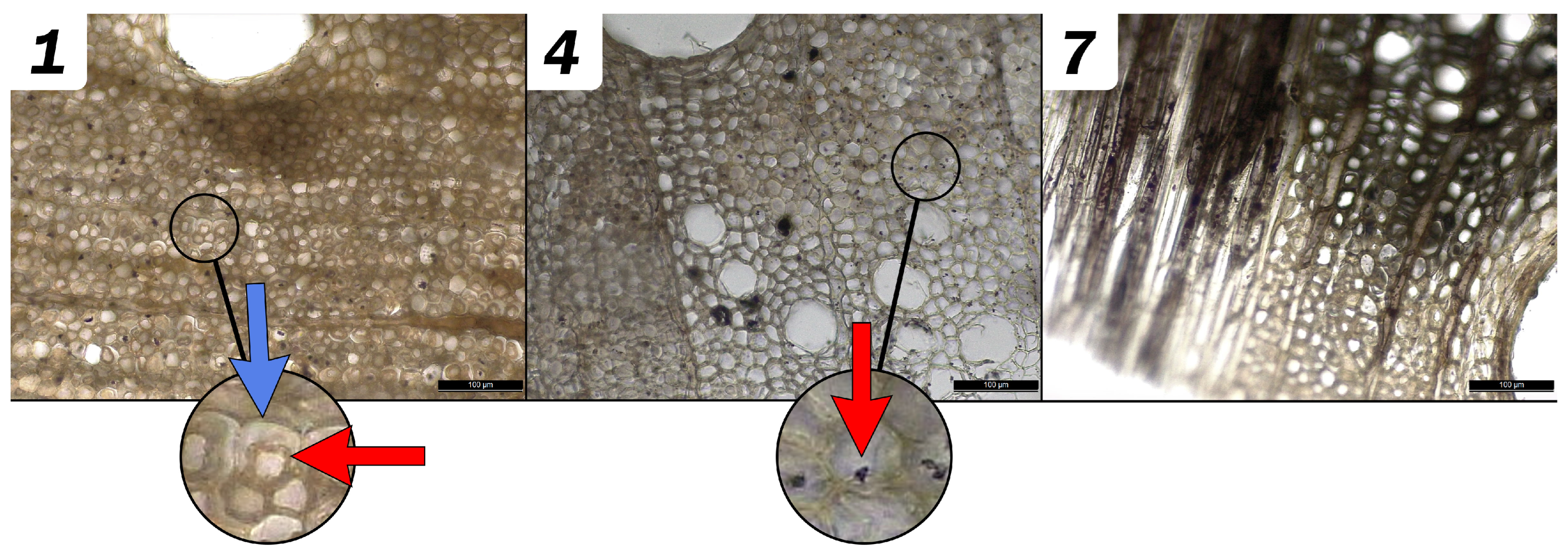
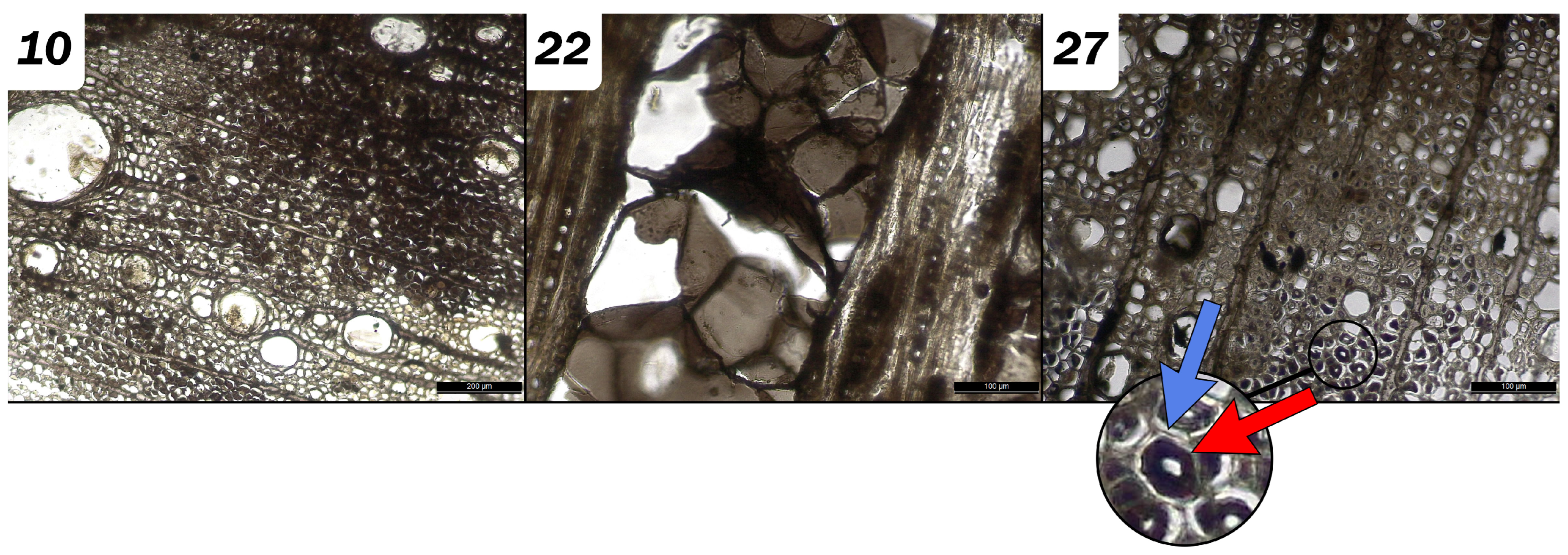
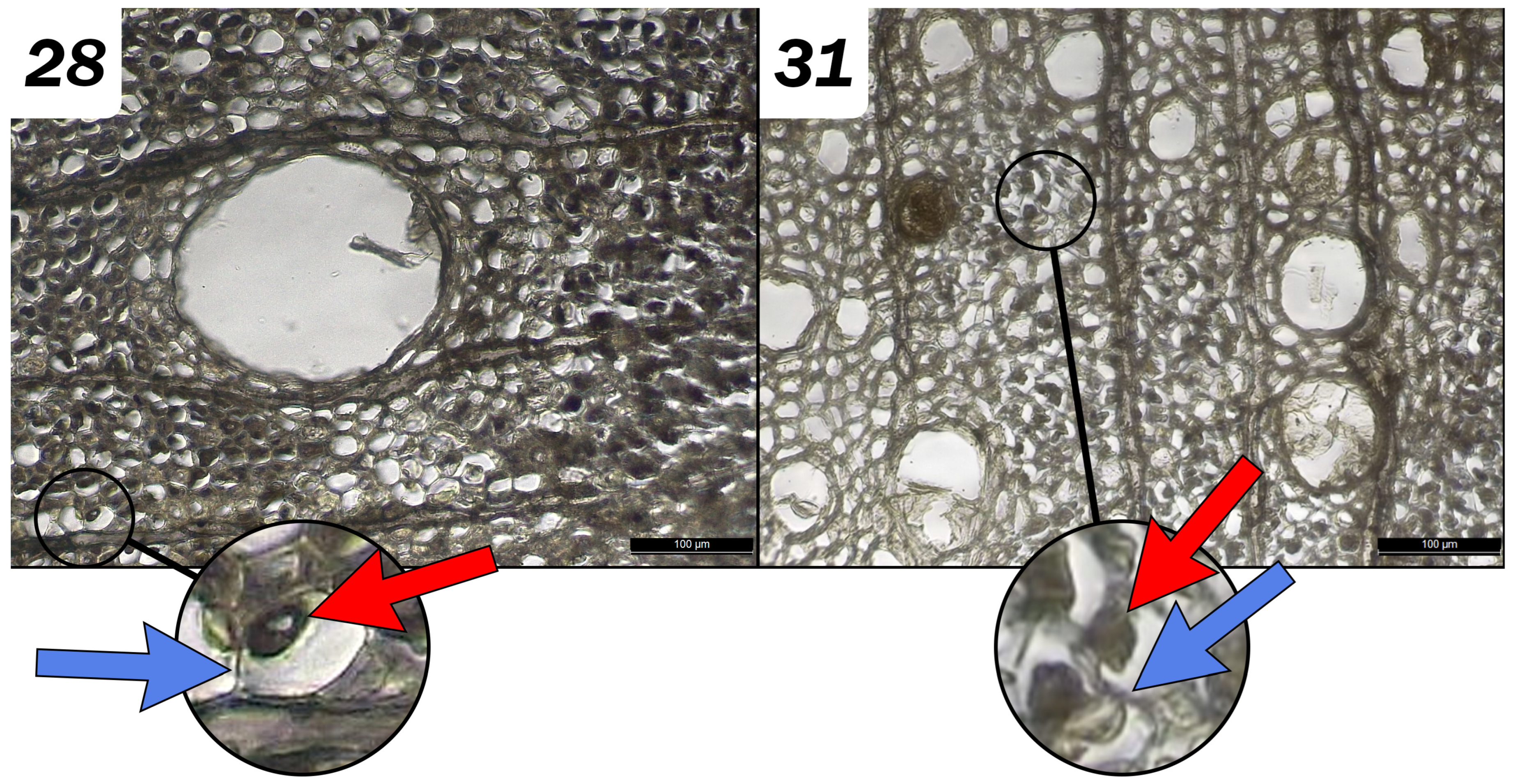
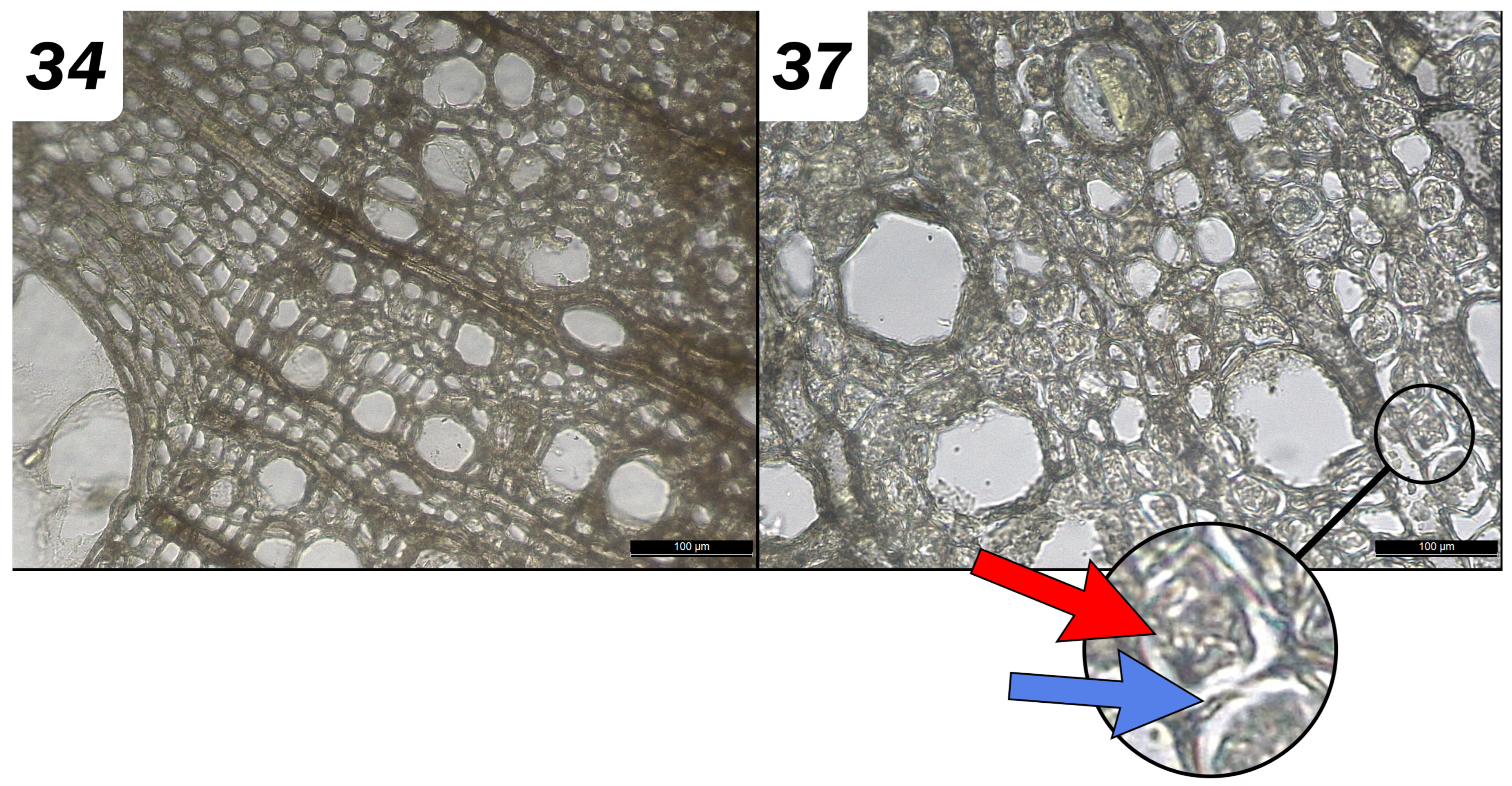
3.2.2. Effects of the Chemical Extractions on Micromorphology: Diagnostic Evaluation

Concerning the archaeological references, their state of decay was assessed as slightly spongy by the pin test. Most of the fibers’ cell walls were decayed. The S2 secondary wall layer was often detached from the compound middle lamella. Longitudinal sections revealed that cells were extremely distorted. The lumen of the cells was very dark, as were the vessel walls. Cell lumens could not be distinguished, as they were generally filled with material digested by bacteria. In the longitudinal sections, signs of fungal and bacterial attacks were evident and widespread. The lumens of parenchymatic cells and vessel walls contained a large quantity of black material, most probably ferrous compounds distributed heterogeneously.
EDTA on impregnated samples. The samples treated with EDTA (with or without pre-treatments) showed a darker inner central halo. In the samples treated with EDTA with or without sodium dithionite, the lumens of the parenchymatic cells were lighter than the references. The disintegrated material in the fibers’ lumina had probably been removed by the treatment. In some cases, only the compound middle lamella and the S3 secondary wall layer could be observed due to the removal of the degraded secondary wall by the treatment. Blackish inclusions were visible inside some cells (fibers and axial parenchyma) that could be due to ferrous compound impregnation. In the tangential longitudinal section, there were also dark streaks, but the distribution of ferrous compounds seemed to be less clear within the cell lumens in the samples treated with EDTA. In those treated with sodium dithionite and EDTA, observations revealed that a large quantity of iron remained in the tangential longitudinal section, where extraction probably did not occur. Small fungal hyphae were noted, probably due to an attack after excavation. These fungal hyphae resemble those typical of imperfect fungi (ascomycetes), visible in the axial parenchyma in the longitudinal section. The so-called soft rot is caused by imperfect fungi.
Samples treated with EDTA and sodium persulfate were non-homogeneous. Some cellular lumens in the fibers sometimes appeared empty while the secondary walls were partially degraded. The samples presented some traces of iron, i.e., black-colored areas and lighter areas alternating with darker areas, especially in the lumens of the parenchyma. Some black spots, probably due to iron impregnation, were observed, as was the presence of ascomycetes and basidiomycetes, such as spores and hyphae (Figure 3).
EDTA on non-impregnated samples. The samples treated with EDTA without impregnation appeared to be free from ferrous compounds, except for a few small spots, especially in the lumina of small latewood vessels. The contents of some cells yellowed, probably due to the complexation of EDTA. The samples appeared very “clean”, and the walls were similar to those of the references. It should be noted that some fibers’ cell walls were decomposed. Hyphae were also present in sample #16.
DTPA. The samples treated with DTPA with or without pre-treatment were very dark. Compared to the reference, areas of the fibers’ walls had collapsed to a greater extent, and the samples were darker. Some similarities with the reference samples were reported as follows: some black remained in the parenchymatic ray cells, although some of it seemed to have been partially washed away. In the DTPA samples with pre-treatment, some observations seemed to show that the fibers’ walls had collapsed and darkened, but this was not homogeneous. In the longitudinal section, collapsed areas appeared as very dark-colored streaks. In the same sections, hyphae inside the vessels were observed to be located in the middle of the tyloses (Figure 4). The parenchymatic cells of the rays seemed to have been impregnated with iron, as the cellular lumina were very dark.
Citric acid. Samples treated with citric acid produced a strong sulfur odor. Macroscopically, their inner color was darker than the outside. In the cross sections, the overall color was dark but less marked than those treated with DTPA. It was noted that some portions of the walls had collapsed but not systematically, with some maintaining the same dimensions. In the radial longitudinal section, dark streaks with marked tissue destruction were observed. The longitudinal sections featured dark streaks with widespread hyphae and ascomycetes fungi spores. In the samples treated with citric acid and sodium persulfate, the tissue appeared to be very degraded in cross sections and much lighter than the sample treated with citric acid alone. The fiber walls had widely deteriorated and collapsed. Dark streaks were observed, but these were generally lighter than when citric acid alone was used. In the longitudinal tangential section, the larger streaks were still quite dark, while the smaller ones had faded more. We also noticed the presence of hyphae in the longitudinal sections in this sample, but to a lesser extent than in the sample treated with citric acid only (Figure 5). Even after rinsing, significant amounts of wood fibers were visible.
Oxalic acid. The cross sections of the samples treated only with oxalic acid were generally very light and yellowed—more so than those treated with sodium persulfate. Very few black spots in some axial parenchyma cells were observed in the samples treated with oxalic acid alone, with a higher frequency in the samples treated with persulfate. Yellow spots were also visible in the tangential longitudinal sections. A very small number of radial parenchyma cells with dark residues were also observed. Even the widest streaks were considerably lightened by the treatment, and this was more apparent in the samples where only oxalic acid was used. Some degradation of the fiber walls led to limited cell collapse, but, in general, the cells retained their shape. Some black concretions were observed inside the vessels in the tangential–longitudinal and cross sections (Figure 6).

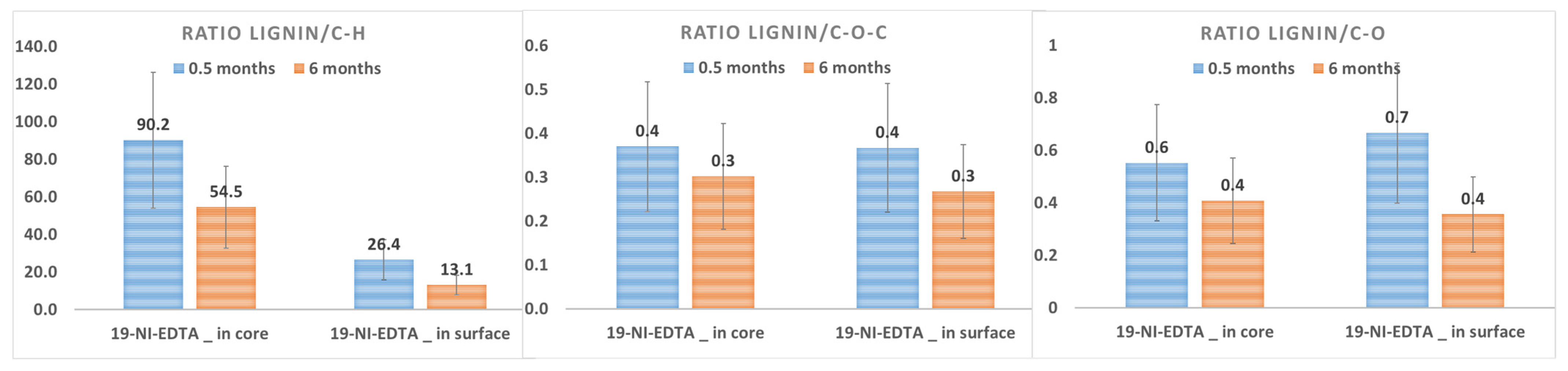
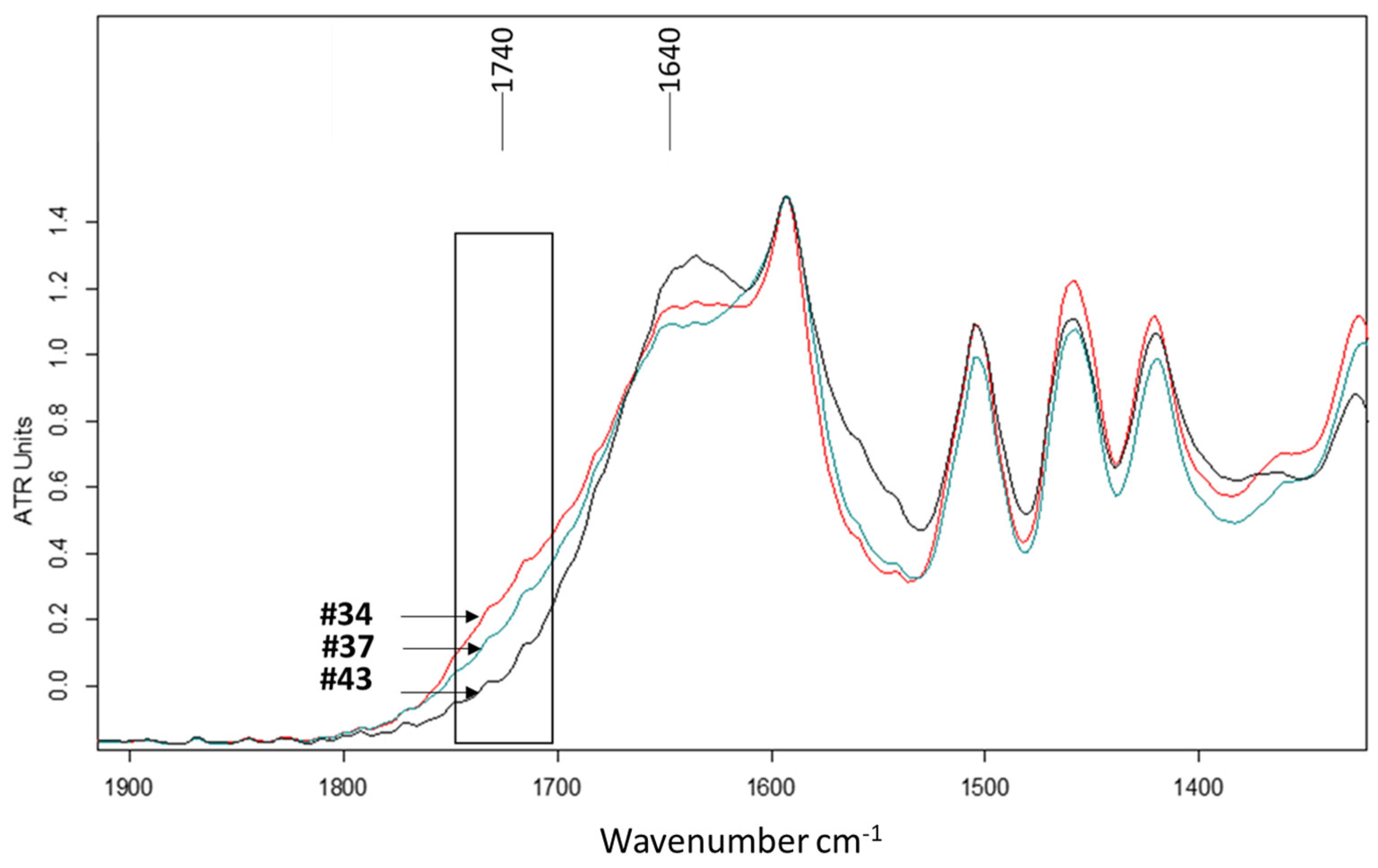
3.2.3. Chemical Impacts on Lignin and Carbohydrates

In most samples, there was no significant difference between surface and core analyses. An exception was sample #19 (non-impregnated_EDTA), in which the ratio of lignin/C-H was four times higher in the core than in the surface analyses (Figure 7). This was due to its surface, which was spongier than the other samples. In fact, samples were chosen because of their homogeneity, but it is widely acknowledged that fully homogeneous samples in archeology are the exception rather than the rule.
Compared to the untreated materials (#41), the process of ferrous compound impregnation seemed to slightly degrade the C-O bonds and, to a lesser extent, the C-H bonds (#43); the ratio was multiplied by 6 after impregnation for the C-O bonds and by 4 for the C-H bonds, both in the core and surface areas (Figure 8). This is consistent with the natural degradation of archaeological wood impregnated with iron compounds [1,2,23,24]. It underlines the need to extract iron compounds and define extraction procedures.
The variability coefficients in the treated samples were high, considerably limiting the interpretation of the infrared results. However, a decrease in the lignin/carbohydrate ratio can be noted for some samples, suggesting a trend toward lignin degradation, especially for sample #22 (sodium dithionite + DTPA) and sample #34 (oxalic acid). Concerning #22, it is difficult to specify the reasons for this degradation because the samples that were treated separately with sodium dithionite or DTPA did not show the same evolution. On the other hand, DTPA was used at a neutral pH and seemed to have had a chelating effect on the iron compounds without any obvious interaction with the organic matrix. Moreover, oxalic acid is a powerful acid and oxidant whose extraction efficiency has been determined in previous studies [6]. Its acidic pH and bi-carboxylic function seem to favor complexation with iron compounds [25], but its highly acidic pH can interact with organic materials. In fact, oxalic acid hydrolyzes hemicellulose, making cellulose fibers more accessible to cellulases. It is possible that oxalic acid also directly initiated the depolymerization of amorphous cellulose. When Fourier transform infrared spectra were overlapped, a difference appeared in the signal at 1740 cm−1, which, for samples #37 and #34, became similar to a shoulder of the broad band at 1640 cm−1 (however, this is associated with absorbed moisture) (Figure 9). The difference in signal with the reference sample is very weak, but since the 1740 cm−1 band is normally assigned to C=O stretching of the carbonyl group, it could suggest a possible esterification of cellulose by oxalic acid [26,27,28,29]. An acidic solution could degrade cellulose fibers by acid hydrolysis and reduce the mechanical stability of the wood [23].

4. Discussion

This study focused on archaeological oak wood impregnated with iron oxides and sulfides. The state of degradation was classified as slightly spongy. The analysis techniques used were the most accessible ones, i.e., infrared spectroscopy and microscopic observations for anatomical and chemical evaluation, atomic absorption spectroscopy to evaluate iron extraction, and spectrocolorimetry to assess changes in surface color. The treatments and parameters are among the most widely used in the field, i.e., EDTA, DTPA, citric acid, and oxalic acid. The two complexing agents were tested alone and with an additional pre-treatment with either sodium dithionite or sodium persulfate. Acids were also tested both alone and with a single pre-treatment, such as sodium persulfate, due to the risk of toxic fumes being released when an acid and a reducing agent, such as sodium dithionite, were mixed.
Table 5 provides an overall summary of our results. It should be noted that all observations refer to a defined corpus under specific conditions. Chemical agents selected for a treatment must be defined according to the ferrous compounds that are to be extracted. In this case, the samples were artificially impregnated, and goethite, lepidocrocite, and pyrite were identified.
The performance of chelating agents with slightly acidic or acidic pHs, such as EDTA and citric acid, in dissolving iron compounds is well established. The different chemical solutions proposed and tested in these studies, alone or in combination with a pre-treatment, yielded effective results in the extraction of iron compounds. However, each of these solutions presents chemical advantages and disadvantages that we need to evaluate to determine the best compromise in each case.
The main evolution in color was the lightening of surfaces, and this was intensified when oxalic acid and EDTA, followed by citric acid, were used. Pre-treatment with sodium persulfate or sodium dithionite did not significantly improve the results, except in the case of oxalic acid. DTPA treatments did not have a significant effect on the surface color of the samples. Colorimetric measurements assessed the visual impact of the various extraction treatments on the wood samples by measuring changes in color and, thus, enabling us to assess the efficiency of extraction treatments by surface evaluation. In this study, despite the typical heterogeneity of archaeological wood, when the samples were treated with EDTA and citric and oxalic acids, the results of each set of samples were largely homogeneous. However, when DTPA was used, there was considerable heterogeneity in the results within each sample set. Samples treated with oxalic acid, EDTA, and, to a lesser extent, citric acid showed the most significant changes. Increased values on the L* axis indicated a constant lightening of the samples, which can be attributed to the removal of corrosion products, such as iron and sulfide compounds. The pre-treatments did not seem to impact the visual aspect of the samples. It can be observed that pre-treatments are commonly used to focus their chemical impact on iron compounds in order to destabilize them and improve the complexing process.
The efficiency of the chemical product EDTA in terms of its extraction rate is confirmed in this study. The lightening of the sample color after treatment also suggested a leaching of iron compounds, at least on the surface (orange and/or black). Micromorphological observations revealed a lighter-colored sample with cells appearing similar to the reference. However, the infrared results revealed changes in bands linked to lignin and carbohydrates, which are heterogeneous. It should be noted that the use of a pre-treatment did not improve extraction rates compared to EDTA alone (in both cases, the efficiency was 80%). As observed in previous studies, EDTA can form a hexadentate complex, which assumes a strong iron complex [30,31]. Some authors have determined that the dissolution rate in EDTA is higher at a low pH for magnetite and lepidocrocite (the observed values were pH 4.7 for magnetite and pH 3.0 for lepidocrocite) and, conversely, at a high pH for goethite (pH 8) [30,32,33].
Citric acid yielded effective results in terms of iron extraction; the color was homogeneous, although slightly greenish. On the other hand, the chemical impact seemed low or undetectable, whereas solutions showed significant amounts of wood fibers, even after rinsing. Citric acid is an α-hydroxyl tricarboxylic acid that can form tridentate mononuclear complexes with hydroxyl and carboxyl groups [34,35,36]. Under oxidized conditions, FeCit species dominate at pH 5.4 to the detriment of Fe(OH)3 [37]. Authors have previously reported the formation of FeCit species for values below pH 3 and, to a lesser extent, the formation of FeCitOH for values from pH 4.0 to 6.5 [38]. The formation constant of the citrate complex is higher for ferric iron than for ferrous ions under the same conditions [38]. Citric acid, therefore, has a good capacity to extract iron compounds but is not without risk for the wood.
Extraction with oxalic acid offers certain advantages. Although the extraction rate is lower than with other solutions, the chemical impact on wood tissue is low, and the evolution of sample color is satisfactory overall. Oxalic acid is not specific to iron and can rapidly inhibit reactions if other environmental factors are present. It is a bi-carboxylic acid that can form mononuclear or binuclear bidentate complexes [25]. Each carboxylic function is associated with a pH value that corresponds to the property change between the acidic and basic functions [35]. The maximum dissolution was identified at pH 2.6; the rates decreased sharply at higher and lower pHs. The highest adsorption occurs at pH 2.5–3 for synthesized goethite [31,39,40] and at pH 2.5 for hematite [41,42,43]. Some authors have found that oxalic acid is more efficient at dissolving hematite [31,39]. Oxalic acid can be very effective in dissolving ferric compounds but is much less so in the presence of ferrous compounds. It has a strong, rapid effect on the surface, which may be an advantage or a disadvantage depending on the surface of the wood to be treated.
Despite the widely acknowledged heterogeneity of archaeological wood, when the samples were treated with EDTA and citric and oxalic acids, the results of each set of samples were homogeneous. However, when DTPA was used, there was considerable heterogeneity in the results within each sample set. The iron–DTPA complex causes the solution to turn reddish-brown, which could partly explain the very dark appearance of the samples, which were rinsed before the observations. The extraction rate was relatively low compared to other solutions. However, the maximum range was about 7–9 to form the complex [2]. Anatomical observations indicated collapses and a blackened surface, but the heterogeneity of the results of these observations made interpretation difficult. DTPA proved to be inefficient in these tests. It may be added that polyphenols (such as tannin) present in the parenchyma and vessels of oak wood would react closely with iron compounds, which would certainly influence extractions. It has previously been shown that iron–tannin precipitates are difficult to remove with DTPA, probably because the stability constants of iron–tannin and iron–DTPA are of the same order of magnitude [44].
The pre-treatments (sodium dithionite and sodium persulfate) did not significantly improve the efficiency of the complexing agents; there was no difference for EDTA and a limited effect for citric and oxalic acids. It should be recalled that the role of pre-treatments is to help dissolve iron sulfides by oxidizing with sodium persulfate or by reducing iron III oxides/hydroxides to iron II to facilitate complexation reactions. In the absence or reduced presence of these compounds, pre-treatments are not necessary. Although pyrite was detected by XRD and Raman analysis, the amount of each iron compound was not calculated. We do not, therefore, really know how much of these compounds to extract. This could influence the interest in using pre-treatments where pyrite is in the minority. Moreover, sodium dithionite acts on iron(III) oxide compounds when present. However, dithionite was rapidly consumed due to decomposition; its concentration decreased considerably after five minutes [45]. Thus, identification and quantification of the various iron compounds could help in the choice of extraction methods by tailoring each solution to the compounds to be extracted.
In 1996, Schwertmann and Cornell [39] recognized the difficulty in establishing a dissolution model that takes all of these factors into account. They considered a tendency in how the ion behaves in solution [39]. The chemical’s efficiency depends on pH and ligand speciation. Thus, it remains an important endeavor to define an optimal domain to dissolve iron oxides (specifically for goethite and lepidocrocite) based on each complexing agent according to certain pH values and environmental conditions. This may also explain the differences in the extraction effectiveness of different chemicals, each of which has a different influence on the type of ferrous compound to be extracted. The particular archaeological context may interfere with the dissolution process; this is especially true for the presence of iron sulfide.
One additional point arising from the tests is the importance of rinsing and the risks if it is not carried out properly. For all treatments (except for EDTA with and without pre-treatment), after 6 months, a decrease in the signals attributable to the lignin/carbohydrate ratio was observed due to the action of chemicals. It was shown that 2 weeks after treatment, the samples treated with EDTA and oxalic acid (without persulfate) showed similar results to the reference samples. This suggests a non-significant decay of organic compounds. Six months later, analyses highlighted significant rinsing issues, mainly for DTPA and oxalic acid. If rinsing is not thorough, it can lead to the degradation of organic matter such as lignin. In view of the variability coefficients, it is difficult to distinguish the potential impact of chemical solutions on these bonds. The impact of the impregnation of ferrous compounds on lignin and, specifically, on the progress of cellulose degradation (and lignin to a certain extent) on archaeological objects upstream of extraction treatments, which may subsequently influence or bias the results, raises a number of questions [15,46,47].

5. Conclusions

The results fall into the following three main categories: conservation–restoration objectives to be reached (aesthetics, stability, short/medium term, etc.); a preliminary diagnosis of the object with a characterization of the ferrous compounds to be removed and their influence on the selection of chemical products and state of degradation; and, finally, the chemical properties of the solutions tested and available to conservator–restorers. These data can form the basis of a tool capable of broadening and optimizing the range of suitable treatment proposals for each object studied.
In this study, EDTA confirmed its effectiveness in relation to these chemical parameters, with the highest extraction efficiency rate, the lowest chemical impact, and an aesthetically acceptable visual result. Its action as a complexing agent on ferrous compounds, especially iron(II) oxyhydroxides, is wide-ranging. However, the oxalic acid results are interesting because they may provide a potent solution that can rapidly yield a satisfactory aesthetic result (color), with a relatively limited impact on organic matter.
Adding a pre-treatment improves the extraction rate of all treatments, but it should be used with caution. Sodium dithionite can produce toxic vapors (sulfide dioxide) in acidic conditions. Sodium persulfate is a powerful oxidant, but it can be harmful to a person’s health if inhaled. In fact, the addition of pre-treatments is not necessarily systematic because their influence depends on which iron compounds are to be extracted. In relation to this, it would be interesting to supplement the diagnostic stage with an identification of the ferrous compounds and their volume distribution in order to determine the objective (surface or deep extraction, which may, in turn, influence immersion time, for example, or the power of a complexing agent) by CT scans or with synchrotron analyses.
In defining certain criteria, such as wood species, degradation state, ferrous compounds, and the final objective of the conservation treatment (stabilization, aesthetics, etc.), a flowchart could help conservators choose from a range of treatments for which relevant chemical and physical studies have been carried out. We hope that this study makes a positive contribution to the numerous experiments in this field and to fostering the pooling of results, enabling colleagues to create a sharing tool to improve and optimize extraction treatments—a tool that would also embrace new research, especially related to the use of live microorganisms for curative/preventive treatments.
It is certain that these tests need to be multiplied and applied to complementary corpora with varying levels of degradation and species, but hopefully this will give rise to a decision support system that can be widely applied in the context of curative treatments.

Author Contributions

Conceptualization, E.G. and C.P.-M.; methodology, C.P.-M. and E.G.; validation, M.M., C.P.-M., E.G., N.M. and B.P. formal analysis, C.P.-M., N.M., L.S. and J.Y.M.; investigation, C.P.-M., N.M., L.S. and J.Y.M.; writing—original draft preparation, C.P.-M.; writing—review and editing, G.L., B.P. and E.P.; supervision, E.G.; project administration, E.G.; funding acquisition, E.G. All authors have read and agreed to the published version of the manuscript.

Funding

The «Direction Régionale des Affaires Culturelles des pays de la Loire» (DRAC).

Institutional Review Board Statement

Not applicable.

Informed Consent Statement

Not applicable.

Data Availability Statement

Not applicable.

Acknowledgments

The authors express their sincere gratitude to Nathalie Guichard (CEISAM), Pierre-Emmanuel Petit (IMN), Mark Irle (ESB), and Julia Büchner (ESB) for their valuable assistance. We would also like to thank Raphaël Moreau, who participated in this study as a student.

Conflicts of Interest

The authors declare no conflict of interest.

References

  1. Sandström, M.; Fors, Y.; Jalilehvand, F.; Damian, E.; Gelius, U. Analyses of Sulfur and Iron in Marine Archaeological Wood. In Proceedings of the 9th ICOM Conservation Group on Wet Organic Archaeological Naterials, Copenhagen, Denmark, 4 July 2004; Hoffmann, P., Spriggs, J.A., Straetkvern, K., Gregory, D., Eds.; The International Council of Museums, Committee for Conservation Working Group on Wet Organic Archaeological Materials: Copenhagen, Denmark, 2004; pp. 181–202. [Google Scholar]
  2. Almkvist, G.; Persson, I. Extraction of iron compounds from wood from the Vasa. Holzforschung 2006, 60, 678–684. [Google Scholar] [CrossRef]
  3. Fors, Y.; Jalilehvand, F.; Risberg, E.D.; Björdal, C.; Phillips, E.; Sandström, M. Sulfur and iron analysis of marine archaeological wood in shipwrecks from the Baltic Sea and Scandinavian waters. J. Archaeol. Sci. 2012, 39, 2521–2532. [Google Scholar] [CrossRef]
  4. Zhang, H.; Shen, D.; Zhang, Z.; Ma, Q. Characterization of degradation and iron deposit of the wood of Nanhai I shipwreck. Herit. Sci. 2022, 10, 202. [Google Scholar] [CrossRef]
  5. Pelé, C.; Moulhérat, C.; Lemoine, G.; Labroche, S.; Guilminot, E.; Baron, G. Extraction of iron compounds from marine organic objects. In Proceedings of the 12th ICOM Wet Organic Archaeological Materials Conference (WOAM), Istanbul, Turkey, 13–17 May 2013. [Google Scholar]
  6. Pelé, C.; Lemoine, G.; Labroche, S.; Guilminot, E.; Baron, G. Iron removal from waterlogged wood: Extraction by electrophoresis and chemical treatments. Stud. Conserv. 2015, 60, 155–171. [Google Scholar] [CrossRef]
  7. Pecoraro, E. New Studies about the Effects of Selected Treatments on the Chemical and Dynamic Mechanical Properties of Waterlogged Archaeological Wood. Ph.D. Thesis, Università degli Studi Firenze, Florence, Italy, 2016. [Google Scholar]
  8. Pecoraro, E.; Pelé-Meziani, C.; Macchioni, N.; Lemoine, G.; Guilminot, E.; Pizzo, B. Effects of iron removal treatments on the chemical and viscoelastic properties of waterlogged wood. J. Cult. Herit. 2022, 56, 149–158. [Google Scholar] [CrossRef]
  9. Pecoraro, E.; Pelé-Meziani, C.; Macchioni, N.; Lemoine, G.; Guilminot, E.; Shen, D.; Pizzo, B. The removal of iron from waterlogged archaeological wood: Efficacy and effects on the room temperature wood properties. Wood Mater. Sci. Eng. 2023, 18, 672–689. [Google Scholar] [CrossRef]
  10. Mouchard, J.; Guitton, D.; Monteil, M.; Favreau, X.; Ménez, N.; Yacger, M. Le Port Romain du Quartier de Saint-Lupien à Rezé/Ratiatum (Loire-Atlantique): Origine et Evolution/The Roman Port Neighborhood of Saint-Lupien in Rezé/Ratiatum (Loire-Atlantique): Origin and Evolution. Gallia; Archéologie de la Gaulle soutenu par le Ministère de la Culture: Paris, France, 2020; pp. 67–97. [Google Scholar]
  11. Arthuis, R.; Guitton, D.; Monteil, M.; Mouchard, J.; de Peretti, O. Rezé–Saint-Lupien, Fouille Programmée (2009); Edition Electronique; Ministère de la Culture: Paris, France, 2020. Available online: http://journals.openedition.org/adlfi/36713 (accessed on 31 July 2023).
  12. Bingham, J. A Quantitative and Nondestructive Method for Determining the Degradation of Waterlogged Archaeological Wood. Ph.D. Thesis, The Faculty of the Department of Chemistry, East Carolina University, Greenville, NC, USA, 2013. [Google Scholar]
  13. Gregory, D.; Jensen, P.; Matthiesen, H.; Straetkvern, K. The correlation between bulk density and shock resistance of waterlogged archaeological wood using the Pilodyn. Stud. Conserv. 2007, 52, 289–298. [Google Scholar] [CrossRef]
  14. Tran, Q.K.; Remazeilles, C.; Guilminot, É. Prévention de l’Acidification des Objets Archéologiques Humides Issus de Fouilles sous-Marines par Extraction des Composés Soufrés; Programme National de Recherche sur la Connaissance et la Conservation des Matériaux du Patrimoine Culturel: Paris, France, 2008.
  15. Monachon, M.; Albelda-Berenguer, M.; Pelé, C.; Cornet, E.; Guilminot, E.; Rémazeilles, C.; Joseph, E. Characterization of model samples simulating degradation processes induced by iron and sulfur species on waterlogged wood. Microchem. J. 2020, 155, 104756. [Google Scholar] [CrossRef]
  16. Bourdoiseau, J.-A. Rôle des Espèces Sulfures sur le Comportement d’un Acier non Allié en Milieu de Stockage des Déchets Radioactifs de Type C: Interaction Sulfures/Produits de Corrosion. Ph.D. Thesis, Université de la Rochelle, La Rochelle, France, 2011. [Google Scholar]
  17. Pizzo, B.; Pecoraro, E.; Macchioni, N. A new method to quantitatively evaluate the chemical composition of waterlogged wood by means of Attenuated Total Reflectance Fourier Transform Infrared (ATR-FT-IR) measurements carried out on wet material. Appl. Spectrosc. 2013, 67, 553–562. [Google Scholar] [CrossRef]
  18. Pizzo, B.; Pecoraro, E.; Alves, A.; Macchioni, N.; Rodrigues, J.-C. Quantitative evaluation of attenuated total reflectance infrared (ATR-FTIR) spectroscopy to the chemical composition of decayed wood preserved in waterlogged conditions. Talanta 2015, 131, 14–20. [Google Scholar] [CrossRef]
  19. High, K.E.; Penkman, K.E.H. A review of analytical methods for assessing preservation in waterlogged archaeological wood and their application in practice. Herit. Sci. 2020, 8, 83. [Google Scholar] [CrossRef]
  20. Picollo, M.; Cavallo, E.; Macchioni, N.; Pignatelli, O.; Pizzo, B.; Santoni, I. Spectral characterization of ancient wooden artefacts with the use of traditional IR techniques and ATR device: A methodological approach. In Proceedings of the 9th Conference of the Infrared and Raman Users’ Group, Buenos Aires, Argentina, 3–6 March 2010; Morana RTD: Ljubljana, Slovenia, 2010; pp. 23–28. [Google Scholar]
  21. Keey, R. Colour Development on Drying. Maderas Cie. Tecnol. 2005, 7, 3–16. [Google Scholar] [CrossRef]
  22. Macchioni, N.; Capretti, C.; Sozzi, L.; Pizzo, B. Grading the decay of waterlogged archaeological wood according to anatomical characterization. The case of the Fiavé site (N-E Italy). Int. Biodeterior. Biodegrad. 2013, 84, 54–64. [Google Scholar] [CrossRef]
  23. Sandström, M.; Jalilehvand, F.; Damian, E.; Fors, Y.; Gelius, U.; Jones, M.; Salomé, M.V. Sulfur accumulation in the timbers of King Henry VIII’s warship Mary Rose: A pathway in the sulfur cycle of conservation concern. Proc. Natl. Acad. Sci. USA 2005, 102, 14165–14170. [Google Scholar] [CrossRef]
  24. Fords, Y.; Richards, V. The effect of the ammonia neutralizing treatment on marine archaeological Vasa wood. Stud. Conserv. 2010, 55, 41–54. [Google Scholar] [CrossRef]
  25. Panias, D.; Taxiarchiou, M.; Douni, I.; Paspaliaris, I.; Kontopoulos, A. Dissolution of hematite in acidic oxalate solutions: The effect of ferrous ions addition. Hydrometallurgy 1996, 43, 219–230. [Google Scholar] [CrossRef]
  26. Chen, L.; Zhu, J.Y.; Baez, C.; Kitin, P.; Elder, T. Highly thermal-stable and functional cellulose nanocrystals and nanofibrils produced using fully recyclable organic acids. Green Chem. 2016, 16, 3835–3843. [Google Scholar] [CrossRef]
  27. Kaneko, S.; Yoshitake, K.; Itakura, S.; Tanaka, H.; Enoki, A. Relationship between production of hydroxyl radicals and degradation of wood, crystalline cellulose, and a lignin-related compound or accumulation of oxalic acid in cultures of brown-rot fungi. J. Wood Sci. 2005, 51, 262–269. [Google Scholar] [CrossRef]
  28. Beck-Anderson, J. Production, Function, and Neutralization of Oxalic Acid Produced by the Dry Rot Fungus and Other Brown-Rot Fungi; International Research Group on Wood Preservation: Stockholm, Sweden, 1987. [Google Scholar]
  29. Shimada, M.; Akamatsu, Y.; Ohta, A.; Takahashi, M. Biochemical Relationships between Biodegradation of Cellulose and Formation of Oxalic Acid in Brown-Rot Wood Decay; International Research Group on Wood Preservation: Stockholm, Sweden, 1991; Doc No. IRG/WP/1472. [Google Scholar]
  30. Nowack, B.; Sigg, L. Adsorption of EDTA and Metal-EDTA Complexes onto Goethite. J. Colloid Interface Sci. 1995, 177, 106–121. [Google Scholar] [CrossRef]
  31. Belattar, S. Réactivités Thermiques et Photochimiques du Fer en Solution Aqueuse vis-à-vis de Molécules Organiques en Phase Homogène et Hétérogène; Mentouri-Constantine University: Constantine, Algeria, 2009; pp. 20–41. [Google Scholar]
  32. Schwertmann, U. Solubility and dissolution of iron oxides. Plant Soil 1991, 130, 1–25. [Google Scholar] [CrossRef]
  33. Bondietti, G.; Sinniger, J.; Stumm, W. The reactivity of Fe(III) (hydroxides: Effects of ligands in inhibiting the dissolution. Colloids Surf. A Physicochem. Eng. Asp. 1993, 79, 157–167. [Google Scholar] [CrossRef]
  34. Abrahamson, H.B.; Rezvani, A.B.; Brushmiller, J.G. Photochemical and spectroscopic studies of complexes of iron(III) with citric acid and other carboxylic acids. Inorg. Chim. Acta 1994, 226, 117–127. [Google Scholar] [CrossRef]
  35. Abida, O. Impact des Complexes de Fer et de la Lumière Solaire sur le Devenir de Polluants de l’Environnement Aquatique. Ph.D. Thesis, Université Blaise Pascal de Clermont-Ferrand, Clermont-Ferrand, France, 2005. [Google Scholar]
  36. Jean, L. Mobilisation du Chrome et du Nickel à Partir de Sols Contaminés, en Présence de Complexants: Transfert et Accumulation de ces Métaux chez Datura innoxia. Ph.D. Thesis, Université de Limoges, Limoges, France, 2007. [Google Scholar]
  37. Sandström, T.P.A.; Hall-Roth, I.; Karlsson, A. Salt precipitation on Vasa Timbers; an introduction to a problem. In Proceedings of the 8th ICOM Group on Wet Archaeological Materials Conference, Stockholm, Sweden, 11–15 June 2001; Committee for Conservation Working Group on Wet Organic Archaeological Materials: Paris, France, 2001; pp. 54–55. [Google Scholar]
  38. Königsberger, L.C.; Königsberger, E.; May, P.M.; Hefter, G.T. Complexation of iron(III) and iron(II) by citrate. Implications for iron speciation in blood plasma. J. Inorg. Biochem. 2000, 78, 175–184. [Google Scholar] [CrossRef] [PubMed]
  39. Cornell, R.M.; Schwertmann, U. The Iron Oxides: Structure, Properties, Reactions, Occurrence and Uses; VCH Verlagsgesellschaft GmbH: Weinheim, Germany, 1996. [Google Scholar]
  40. Stumm, W. Reactivity at the mineral-water interface: Dissolution and inhibition. Colloids Surf. A Physicochem. Eng. Asp. 1997, 120, 143–166. [Google Scholar] [CrossRef]
  41. Taxiarchou, M.; Panias, D.; Douni, I.; Paspaliaris, I.; Kontopoulos, A. Removal of iron from silica by leaching with oxalic acid. Hydrometallurgy 1997, 46, 215–227. [Google Scholar] [CrossRef]
  42. Sung, O.L.; Tran, T.; Jung, B.H.; Kim, S.J.; Kim, M.J. Dissolution of iron oxide using oxalic acid. Hydrometallurgy 2007, 87, 91–99. [Google Scholar]
  43. Cornell, R.M.; Schindler, P.W. Photochemical dissolution of goethite in acid/oxalate solution. Clays Clay Miner. 1987, 35, 347–352. [Google Scholar] [CrossRef]
  44. Henrik-Klemens, A.; Bengtsson, F.; Björdal, C.G. Raman spectroscopic investigation of iron-tannin precipitates in waterlogged archaeological oak. Stud. Conserv. 2021, 67, 237–247. [Google Scholar] [CrossRef]
  45. Blesa, M.A.; Rueda, E.H.; Ballesteros, M.C.; Grassi, R.L. Dithionite as a dissolving reagent for goethite in the presence of EDTA and citrate. Application to soils analysis. Clays Clay Miner. 1992, 40, 575–585. [Google Scholar]
  46. Gelbrich, J.; Mai, C.; Militz, H. Chemical changes in wood degradred by bacteria. Int. Biodeterior. Biodegrad. 2008, 61, 24–32. [Google Scholar] [CrossRef]
  47. Broda, M.; Hill, C.A.S. Conservation of waterlogged wood-past, present and future perspectives. Forests 2021, 12, 1193. [Google Scholar] [CrossRef]
Figure 1. Waterlogged archaeological wood from the archaeological site “Saint-Lupien”, Rezé, Nantes (FRANCE), and below, samples after cutting (3 × 3 × 3 cm) used for extraction treatments.
Forests 14 01834 g001
Figure 2. Raman spectra carried out on a reference sample (#45), pyrite (with bands at 472, 435, 243, 220, and 151 cm−1), and hematite (with bands at 617, 410, 295, and 227 cm−1).
Forests 14 01834 g002
Figure 3. Cross sections of archaeological samples after treatment. #1—EDTA, the red arrow shows the decayed material inside the lumen, and the blue arrow shows the compound middle lamella of the same cell; #4—Sodium dithionite, the red arrow points to a black spot of ferrous compounds in cell lumens; #7—Sodium persulfate + EDTA, the picture highlights a section of latewood, while on the right is a large ray well impregnated with black ferrous compounds. Scale bars: 100 µm.
Forests 14 01834 g003
Figure 4. Sections of archaeological samples after treatment. #10—DTPA cross section showing the decayed secondary wall impregnated with black iron compounds. Scale bar: 200 µm; #22—Sodium dithionite + DTPA, longitudinal tangential section showing blackish tyloses in a vessel lumen; #27—Sodium dithionite + DTPA, cross section: the red arrow points to the decayed secondary wall impregnated with black iron compounds, while the blue arrow points to the remaining compound middle lamella. Scale bar: 100 µm.
Forests 14 01834 g004
Figure 5. Cross sections of archaeological samples after treatment. #28—Citric acid, a latewood vessel, surrounded by longitudinal parenchymatic cells and some fibers showing decayed blackish secondary walls detached from the compound middle lamella, which are more visible in #31—Sodium persulfate + citric acid—where the red arrow points to the darker secondary wall, detached from the compound middle lamella (blue arrow). Scale bar: 100 µm.
Forests 14 01834 g005
Figure 6. Cross sections of archaeological samples after treatment. #34—Oxalic acid shows a growth ring limit, with cell walls and cell lumens being quite light colored; #37—Sodium persulfate + Oxalic acid, the red arrow points to the decayed light-colored secondary wall, and the blue arrow points to the compound middle lamella of the same cell. Some of the latewood vessels are losing their rounded/oval shape. Scale bars: 100 µm.
Forests 14 01834 g006
Figure 7. Ratios of lignin and carbohydrates for impregnated and non-impregnated samples subsequently treated with EDTA. FTIR analysis, carried out after an immersion period of 0.5 months and 6 months.
Forests 14 01834 g007
Figure 8. Ratios of lignin and carbohydrates for impregnated and non-impregnated references after an immersion period of 0.5 months and 6 months in water.
Forests 14 01834 g008
Figure 9. FTIR spectra of the reference #43 (in black) and of samples #37 (in green) and #34 (in red) immersed in oxalic acid.
Forests 14 01834 g009
Table 1. Parameters of the chemical products used for extraction treatment.
Chemical SolutionsConcentrationspH
EDTA3.7%w/w5.0
DTPA0.010 mol/L7.0
Citric acid1.0%w/w2.2
Oxalic acid1.0%w/w1.0
Sodium persulfate0.37%w/v6.0
Sodium dithionite2.0%w/w6.5
Table 2. Treatment applications with samples and chemical procedures.
Sample n°ImpregnationPre-TreatmentTreatment
#40-41-42No--
#19-20-21No-EDTA
#13-14-15NoSodium dithionite EDTA
#16-17-18NoSodium persulfateEDTA
#43-44-45Yes--
#1-2-3Yes-EDTA
#4-5-6YesSodium dithionite EDTA
#7-8-9YesSodium persulfateEDTA
#10-11-12Yes-DTPA
#22-23-24YesSodium dithionite DTPA
#25-26-27YesSodium persulfateDTPA
#28-29-30Yes-Citric acid
#31-32-33YesSodium persulfateCitric acid
#34-35-36Yes-Oxalic acid
#36-37-38YesSodium persulfateOxalic acid
Table 3. Average amount of iron extracted in solutions and remaining in wood samples (with a standard deviation assessed at 5%).
Chemical TreatmentsIron Extracted in Solution
(mgFe/g Wood)
Iron Remained in Wood
(mgFe/g Wood)
Total Iron in Wood
before Treatment
(mgFe/g Wood)
% of Iron Extracted
EDTA6.31.57.880%
Sodium dithionite–EDTA6.61.48.082%
Sodium persulfate–EDTA5.91.87.776%
DTPA1.38.59.813%
Sodium dithionite–DTPA2.38.510.821%
Sodium persulfate–DTPA3.25.48.637%
Citric acid5.03.88.857%
Sodium persulfate–citric acid6.01.67.679%
Oxalic acid0.66.87.28%
Sodium persulfate–oxalic acid1.86.58.322%
Table 4. Evolution of colorimetry for each sample, assessed after extraction procedures. The ΔL*, Δa*, Δb*, and ΔE values were calculated from the L*, a*, and b* of the reference.
ΔL* (D65)Δa* (D65)Δb* (D65)ΔE
#1EDTA20.640.902.5921
#4Sodium dithionite–EDTA17.281.994.6018
#7Sodium persulfate–EDTA23.403.125.8424
#19Non-impregnated–EDTA4.713.054.977
#13Non-impregnated–Sodium dithionite–EDTA7.023.447.1711
#16Non-impregnated–Sodium persulfate–EDTA5.084.386.539
#10DTPA4.81−1.25−2.426
#22Sodium dithionite–DTPA4.37−1.25−1.965
#25Sodium persulfate–DTPA8.36−1.67−1.389
#28Citric acid13.24−2.41−3.2314
#31Sodium persulfate–Citric acid17.09−0.341.2017
#34Oxalic acid21.665.3011.5125
#37Sodium persulfate–Oxalic acid30.958.6819.2237
Table 5. Summary of results.
Corpus:
Archaeological Oak Samples
Degradation Level:
Slightly Spongy
Impregnation:
Defined by Previous Studies
(Monachon et al., 2020)
Concentration:
Defined by Main Treatments Used in the Field of Conservation—Restoration
Duration Treatment:
4 h in Pre-Treatment, 5 Days in Chelating or Acidic Solutions
DiagnosticEDTADTPACitric acidOxalic acid
Chemicals aloneLumens seem lighter
Disintegrated material removed
Secondary wall seems washed away
Forests 14 01834 i001Very dark
Fiber collapsed
Forests 14 01834 i002A strong sulfur odor
Core is darker
Some portions of walls have collapsed
Forests 14 01834 i002Very light and yellowing, a few black spots
Some wall areas have collapsed but remain in position
Forests 14 01834 i003
With Na DithioniteIron spots remain
Small fungal hyphae
Forests 14 01834 i001Very dark, Intensification of collapse, Nonhomogeneous
Hyphae
Forests 14 01834 i002X toxic emanation X toxic emanation
With Na PersulfateNon-homogeneous
Secondary walls partially degraded
Cellular lumens partially empty
Black-colored areas
Spores and hyphae
Forests 14 01834 i002Very dark
Intensification of collapse
Nonhomogeneous
Hyphae
Forests 14 01834 i002Tissue appears very degraded
Much lighter
Hyphae
Forests 14 01834 i002Light and yellowed (less than without pre-treatment)
A few black spots
Some wall areas have collapsed but remain in position
Forests 14 01834 i003
ColorimetryEDTADTPACitric acidOxalic acid
Chemicals aloneColor is homogeneous
Lighter color
Forests 14 01834 i003Lower evolution
Forests 14 01834 i001Color is homogeneous
Lighter color
Forests 14 01834 i003Color is homogeneous
Lighter color
Yellowing surface
Forests 14 01834 i003
With Na DithioniteNo significant changeForests 14 01834 i001No significant changeForests 14 01834 i001X toxic emanation X toxic emanation
With Na PersulfateNo significant changeForests 14 01834 i001Slightly increase the changeForests 14 01834 i001Slightly increase the changeForests 14 01834 i003Slightly increase the changeForests 14 01834 i003
Extraction rateEDTADTPACitric acidOxalic acid
Chemicals alone80%Forests 14 01834 i00313%Forests 14 01834 i00157%Forests 14 01834 i0038%Forests 14 01834 i001
With Na Dithionite82%Forests 14 01834 i00121%Forests 14 01834 i003X toxic emanation X toxic emanation
With Na Persulfate76%Forests 14 01834 i00137%Forests 14 01834 i00379%Forests 14 01834 i00322%Forests 14 01834 i003
No interest to use pre-treatment Pre-treatment improves extraction Pre-treatment improves extraction Pre-treatment improves extraction
Chemical impactsEDTADTPACitric acidOxalic acid
Chemicals aloneForests 14 01834 i004lignin/C-HForests 14 01834 i001Forests 14 01834 i004lignin/C-HForests 14 01834 i001Forests 14 01834 i004lignin/C-HForests 14 01834 i001Forests 14 01834 i004lignin/C-HForests 14 01834 i001
Rinsing
With Na DithioniteForests 14 01834 i004lignin/C-HForests 14 01834 i001Forests 14 01834 i004lignin/C-HForests 14 01834 i001X toxic emanation X toxic emanation
Rinsing Forests 14 01834 i003Forests 14 01834 i004lignin/C-O-C and C-OForests 14 01834 i002
With Na PersulfateForests 14 01834 i004lignin/C-HForests 14 01834 i001Forests 14 01834 i005lignin/C-H Forests 14 01834 i005lignin/C-H Forests 14 01834 i005lignin/C-H
Rinsing
Disclaimer/Publisher’s Note: The statements, opinions and data contained in all publications are solely those of the individual author(s) and contributor(s) and not of MDPI and/or the editor(s). MDPI and/or the editor(s) disclaim responsibility for any injury to people or property resulting from any ideas, methods, instructions or products referred to in the content.

