Article

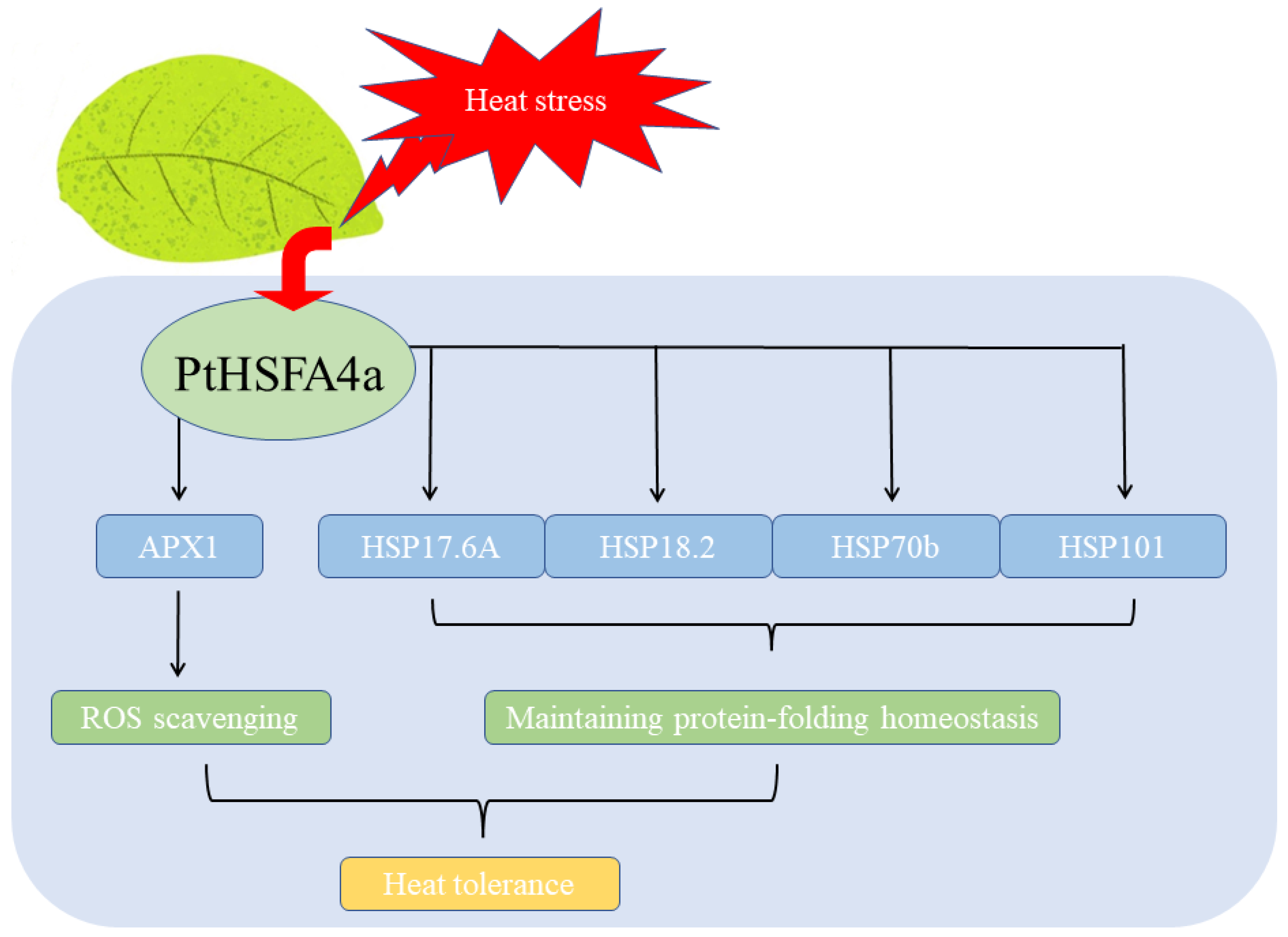
Populus trichocarpa PtHSFA4a Enhances Heat Tolerance by Regulating Expression of APX1 and HSPs

College of Horticulture and Landscape Architecture, Northeast Agricultural University, Harbin 150030, China
*
Author to whom correspondence should be addressed.
These authors contributed equally to this work.
Forests 2023, 14(10), 2028; https://doi.org/10.3390/f14102028
Submission received: 25 August 2023 / Revised: 26 September 2023 / Accepted: 3 October 2023 / Published: 10 October 2023
(This article belongs to the Special Issue Forest Tree Genetics and Breeding in Response to Different Threats)

Abstract

:
Heat stress can severely inhibit plant growth and reproduction, resulting in heavy financial and crop yield losses. Heat shock transcription factors (HSFs) play an important role in regulating plant responses to abiotic stress. However, compared with the in-depth study of HSF gene function in herbaceous species, reports on the regulatory mechanism of the response of HSFs to heat stress in trees are scarce. Here, we demonstrated that PtHSFA4a is induced by high temperatures in Populus trichocarpa leaves. Intense GUS activity was detected in the leaves of PtHSFA4a promoter-GUS reporter transgenic line under heat stress. Ectopic expression of PtHSFA4a in Arabidopsis thaliana enhanced heat stress tolerance, which reduced malondialdehyde and reactive oxygen species levels. RT-qPCR revealed that the expression of key heat stress-related genes (that is, AtMBF1c, AtZAT12, AtAPX1, AtHSA32, and AtHSPs) was upregulated in PtHSFA4a transgenic plants. Additionally, PtHSFA4a directly bind to the promoters of AtAPX1 and AtHSPs under heat stress to enhance heat tolerance by upregulating the antioxidant defense system and maintaining protein folding homeostasis in A. thaliana leaves. These findings provide novel insights into the molecular mechanisms underlying PtHSFA4a-mediated regulation of plant responses to heat stress.

1. Introduction

High ambient temperature is one of the most important abiotic stress factors affecting plant growth and development, causing impaired fertility [1], reduced photosynthetic efficiency, membrane lipid peroxidation, and adverse effects on cell division and growth. Furthermore, following global warming and the resulting ongoing climate change, forest mortality events induced by high-temperature stress have become widespread worldwide [2,3]. Therefore, it is necessary to genetically improve the adaptability of trees to cope with global warming trends in order to ease the heat threat. Also, a thorough understanding the photosynthetic physiology and molecular mechanisms whereby tree species sense and respond to heat stress is necessary to cope with the adverse effects of climate change on forest yields.
Plants have developed several molecular mechanisms of response to heat stress that minimize heat damage and help them to adapt to increased ambient temperatures. Cells can sense heat stress signals at the surface and intracellular sites almost simultaneously, and trigger cellular responses through various mechanisms. Thus, several pathways reportedly respond to high ambient temperatures as follows: first, the CNGC-calcium-calmodulin pathway plays a fundamental role in stress signal transduction [4,5]; second, plant hormone-mediated signaling pathways play a significant role in regulating the heat stress response via biochemical pathways [6]; third, a reactive oxygen species (ROS)-induced pathway transduces a signal that triggers anti-oxidation-related genes such as the cytosolic ascorbate peroxidase gene (APX) and temperature-induced lipocalins (TILs) [7,8]; finally, in the transcriptional regulation pathway, several transcription factors (TFs), such as WRKY, dehydration responsive element binding (DREB), and heat shock transcription factors (HSFs), can regulate heat stress-related genes and play an important role in the plant responses and adaptation to heat stress [9,10,11].
As terminal components of the signal transduction chain, HSFs are key regulators of heat stress (HS)-responsive genes encoding molecular chaperones and other heat shock-induced proteins [12,13]. HSFs form trimers and bind the highly conserved heat shock element (HSE; the core sequence nGAAnnTTCn or nTTCnnGAAn) in the promoters of target genes [14]. HSFs are divided into three classes, namely, A, B, and C, based on their HR-A/B regions [12]. Among the members of the HSF family, HSFA1 has been identified as a master regulator of thermotolerance in plants [13]. In particular, in Arabidopsis thaliana, AtHSFA1 (AtHSFA1a, AtHSFA1b, AtHSFA1d, and AtHSFA1e) is redundant and shares the role of the main regulator in the heat shock response [14]. AtHSFA1a and AtHSFA1b induce HS-responsive genes such as molecular chaperones (AtHSP100, AtHSP90, AtHSP70, and small HSP families) and galactinol synthase (AtGolS1 and AtGolS2) under heat stress [15]. Further, AtHSFA1a induces a set of classical HSP genes, namely, AtHSP17.4, AtHSP18.2, AtHSP21, AtHSP81-1, and AtHSP101 [16]. to respond to heat stress. In tomato, the overexpression of HSFA1 enhances thermotolerance by increasing heat-inducible HSP expression compared to that in wild-type tomatoes [17]. Similarly, the overexpression of GmHSFA1 in transgenic soybean plants leads to the activation of GmHSP genes in thermotolerance reactions [18]. As an induced transcription factor, HSFA2 increases acquired thermotolerance, particularly during repeated cycles of HS and recovery [19,20,21]. In A. thaliana, AtHSFA2 serves as a regulatory amplifier of APX2 and HSPs (HSP26.5-P(r), HSP25.3-P, HSP22.0-ER, HSP18.1-CI, HSP70b, and HSP101-3) in the heat stress response [19], and AtHSFA2 alleviates oxidative stress caused by HS [22]. In addition, AtHSFA2 interacts with AtHSFA1a/b and synergistically activates the expression of HSP genes during thermotolerance reactions [21]. Additionally, AtHSFA3 confers thermotolerance by regulating the expression of galactinol synthase (GolS1 and GolS2) genes that translate into GolS enzymes that function as antioxidants in A. thaliana [23]. Furthermore, under heat stress, AtHSFA3 is transcriptionally induced by dehydration-responsive element binding protein 2A (AtDREB2A) [24], whereas the expression of the AtDREB2A gene is regulated by AtHSFA1 subtribes [25]. Consistently, the suppression of AtHSFA4a prevents the expression of H2O2-scavenging APX1 and zinc-finger protein (ZAT12) induced by light stress [26]. In turn, the overexpression of AtHSFA4a in A. thaliana reportedly improved plant survival and reduced lipid peroxidation in separate salt and combined heat and salt stress by targeting HSP17.6A, ZAT12, and WRKY30 genes [27]. Meanwhile, a heat-stress transcription factor (Spl7) mutant of rice showed more spotted leaves compared to the wild type in a high-temperature growth chamber [28], and the suppression of HSFA9 in tobacco caused a reduction in seed basal thermotolerance and suppressed the expression of sHSP (HSP101, sHSP CI, and sHSP CII) genes [29]. As for the plant Class HSFB, A. thaliana HSFB1 and HSFB2b act as repressors of the expression of HSFA2, HSFA7a, HSFB1, and HSFB2b and some HSPs genes under heat stress conditions in A. thaliana [30]. In summary, thermotolerance mechanisms involving HSFs are well known in A. thaliana, rice, and tomato. However, little information is currently available regarding the transcriptional modulation of HSFs in woody species in response to heat stress.
The heat stress response in Populus species is an important issue; however, its physiological and molecular functions remain unclear. This study aimed to gain a more in-depth and comprehensive understanding of the structure of the HSF family gene PtHSFA4a from P. trichocarpa. Reverse transcription-quantitative PCR and GUS analyses revealed that high-temperature treatment induced the expression of PtHSFA4a. To investigate the biological function of PtHSFA4a, it was ectopically overexpressed in A. thaliana. Heterologous PtHSFA4a lines showed increased resistance to heat stress. Furthermore, chromatin immunoprecipitation (ChIP)-qPCR assay revealed that PtHSFA4a regulated thermotolerance by directly binding to the promoters of HSPs and ROS-scavenging enzyme-encoding genes and activating their expression.

2. Materials and Methods

2.1. Plant Material, Growth Conditions, and Stress Treatments

Seeds of Arabidopsis thaliana ecotype Columbia-0 were obtained from the Arabidopsis Biological Resources Center (ABRC) and Populus trichocarpa seedings clone Donglin plants. A. thaliana seedings were grown at 22 °C under a 16 h light/8 h dark photoperiod and a light intensity of 80−100 µmol photons m−2s−1 for use in transformation experiments. Additionally, 3-week-old P. trichocarpa seedlings were cultured in vitro on 1/2 Murashige and Skoog (MS) medium with 0.6% (w/v) agar, 2% (w/v) sucrose, and, respectively, supplemented with 6% (w/v) PEG6000, 150 mM NaCl, 25 mM ABA, 80 μM CdCl2, 100 μM CuSO4, or 1.0 mM FeSO4, as stress treatments, and incubated for 0, 1, 3, 6, and 12 h. As for heat stress, 3-week-old wild-type P. trichocarpa seedlings were cultured in vitro on 1/2 MS medium with 0.6% (w/v) agar, 2% (w/v) sucrose, and incubated at 45 °C in the dark for 0, 1, 3, 6, and 9 h. The pH of medium set at 5.8–6.0. Roots and leaves of P. trichocarpa exposed to various stress conditions cited above were flash-frozen in liquid nitrogen and stored in a refrigerator at −80 °C for gene expression analysis.

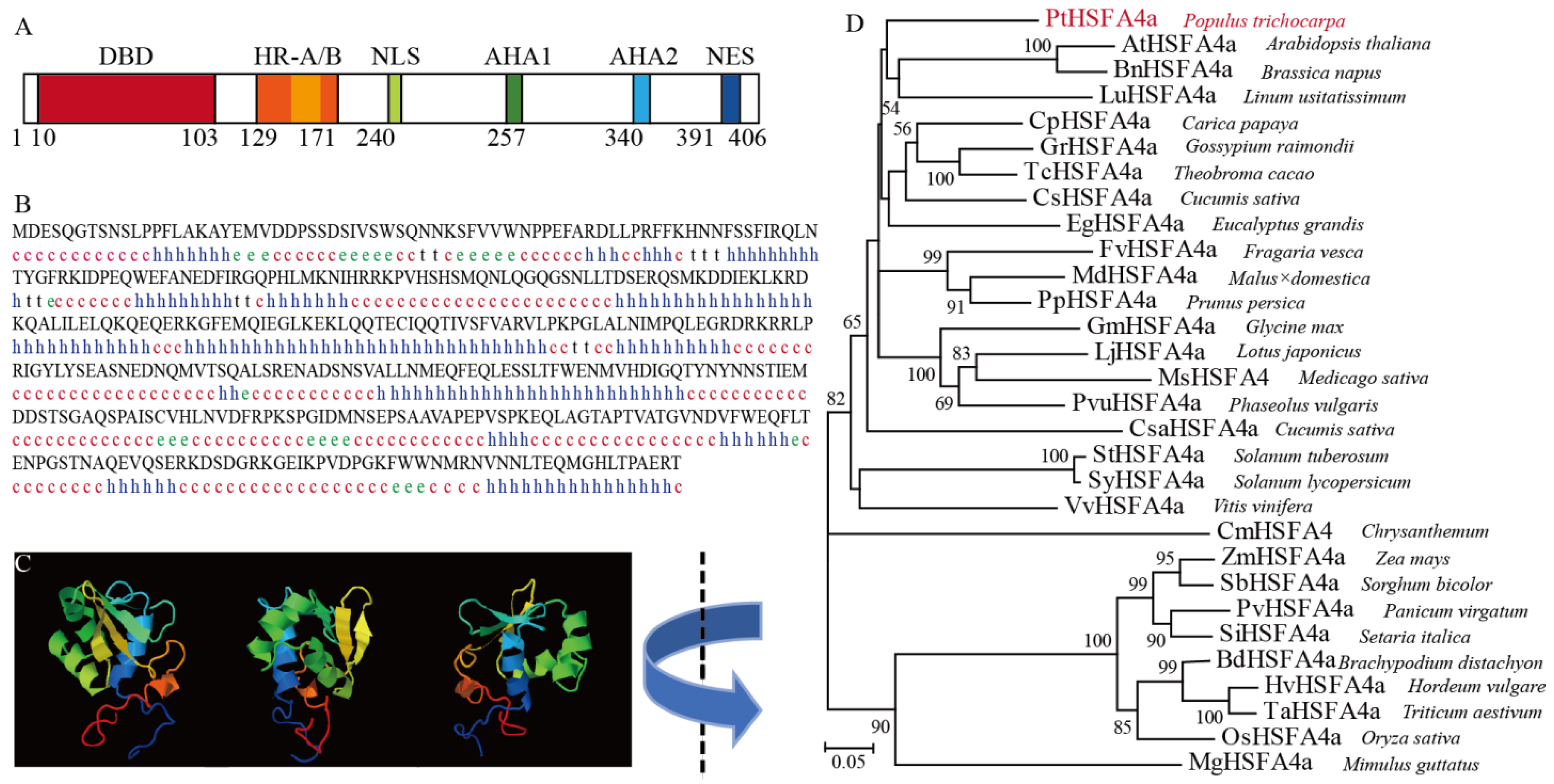
2.2. Secondary and Three-Dimensional (3D) Structural Analysis of the PtHSFA4a Protein

The online SOPMA secondary structure-prediction method (https://npsa-pbil.ibcp.fr/cgi-bin/npsa_automat.pl?page=npsa_sopma.html, accessed on 18 February 2022) was used to analyze the secondary structure of the PtHSFA4a (Potri.011G071700) protein [31]. In turn, the SWISS-MODEL server was used to predict the three-dimensional (3D) structure of the protein homology model using automated protein structure-modeling [32].

2.3. Phylogenetic Analysis of the HSFA4a Family

All HSFA4a protein sequences of P. tricochapa and 29 other species with known developmental functions were downloaded from Heatster (http://www.cibiv.at/services/hsf/info, accessed on 20 February 2022) or Phytozome 13 (https://phytozome-next.jgi.doe.gov/info/Creinhardtii_v5_6, accessed on 20 February 2022) [33]. The HSF protein sequences were aligned using the Biological Sequence Alignment Editor (Bioedit) software version 7.2.5. Subsequently, a phylogenetic tree generated by maximum likelihood method in which distance was estimated by the Jones–Taylor–Thornton (JTT) model implemented in the program MEGA5.2 software (https://www.megasoftware.net, accessed on 25 February 2022). Bootstrap values were estimated using bootstrap analysis of 1000 replicates. Accession numbers and species for all amino acid sequences are listed in Table S1.

2.4. RNA Extraction and Reverse Transcription-Quantitative PCR

Total RNA was extracted from 0.5 g roots and leaves of P. trichocarpa or A. thaliana seedlings using the cetyltrimethylammonium bromide (CTAB) method [34]. Reverse transcription was performed using the PrimeScript RT First-Strand cDNA Synthesis Kit (Takara, Tokyo, Japan) following the manufacturer’s instructions. Reverse transcription-quantitative PCR (RT-qPCR) was performed using the TransStart Top Green qPCR SuperMix (TransGen Biotech, Beijing, China) on a CFX96 Real-Time PCR system (Bio-Rad, Hercules, CA, USA). The relative expression of target genes was calculated using the 2−ΔΔCT method. Specifically, for the relative expression data of PtHSFA4a in P. trichocarpa, the P. trichocarpa actin (Ptactin) gene (Potri.001 G309500) was used as an internal standard control, and the primers are listed in Table S1. Meanwhile, the A. thaliana actin (AtActin) (At3g18780) gene was selected as an internal standard control for the relative expression data of the downstream genes in A. thaliana, and the related primers are listed in Table S1. RT-qPCR was performed with three technical replicates from three independent biological replicates.

2.5. Generation of A. thaliana Transgenic Plants

To construct the ProPtHSFA4a::GUS plasmid, we cloned the regions 1415 bp upstream of the transcription start site of PtHSFA4a, the sequences of which were downloaded from Phytozome 13 (https://phytozome-next.jgi.doe.gov/info/Creinhardtii_v5_6, accessed on 20 February 2022), amplified these regions by PCR, and then inserted them into pBI121 carrying the GUS reporter gene. The vector was introduced into Agrobacterium tumefaciens strain EHA105 and transformed into wild-type A. thaliana using the floral dip method [35]. Seeds from the dipped plants were plated on MS medium supplemented with 50 mg mL−1 kanamycin to select transformants. Effective transformants were confirmed via PCR using the gene- and vector sequence-specific primers listed in Table S1. To generate the overexpression constructs, the PtHSFA4a coding sequence was inserted into the pBI121-GFP vector, where expression was driven by the CaMV 35S promoter and transformed into wild-type A. thaliana plants. Ten PtHSFA4a-OE independent transgenic lines have been developed in previous studies. The T2 or later generations of transgenic A. thaliana lines (PtHSFA4a-OE1, PtHSFA4a-OE2, and PtHSFA4a-OE3) were used in this study.

2.6. Analysis of PtHSFA4a Promoter Activity under Heat Stress

For promoter analysis, 5-day-old ProPtHSFA4a::GUS transgenic A. thaliana seedlings grown in a petri dish were placed in a temperature-controlled chamber at 45 °C for 1 h and then incubated in staining solution for the GUS assay. Five-day-old ProPtHSFA4a:GUS transgenic seedlings treated at 22 °C were used as controls. The protocol for GUS activity was as described by Zhang et al. [36], and the GUS assay was performed using 5–6 seedlings per genotype from three independent replicates.

2.7. Thermotolerance Test

Seeds of PtHSFA4a-overexpressing lines and wild-type A. thaliana (WT) were first stratified for 3 day in the dark at 4 °C and then sterilized in bleach solution for 20–25 min, followed by rinsing five times with sterile water. Sterilized seeds were germinated on 1/2 MS medium. For the basal thermotolerance test, 5-day-old seedlings grown in a petri dish were placed in a temperature-controlled chamber at 45 °C (heat stress) or 22 °C (control) for 1 h. Seedlings were photographed, and the survival rate was determined after recovery at 22 °C for 10 day. For the basal thermotolerance, 4-week-old WT and transgenic lines were germinated on soil and then were kept at 46 °C (heat stress) or 22 °C (control) for 3 h. Photographs were acquired after a 15-day recovery period of incubation under the original growth conditions.

2.8. Measurement of Malondialdehyde, Electroleakage, and Reactive Oxygen Species Accumulation

Four-week-old A. thaliana seedlings were grown at 46 °C for 0, 1, and 3 h. Treated leaves were collected and used for malondialdehyde (MDA), electrolyte leakage (EL), and reactive oxygen species (ROS) analysis at 0 and 3 h after treatment. MDA content was quantified as described by Metwally et al. [37]; in turn, EL measurements were performed as described by Zhang et al. [36], and for histochemical detection of hydrogen peroxide (H2O2) and superoxide radicals (O2) accumulation, leaves were treated with 3,3′-diaminobenzidine (DAB) solution (Sigma–Aldrich (St. Louis, MI, USA), 1.0 mg mL−1 DAB-HCl, pH 3.8) and nitro blue tetrazolium (NBT) solution (Sigma–Aldrich, 1.0 mg NBT in 10 mM sodium azide and 10 mM potassium phosphate buffer (PBS), pH 7.8), respectively [38]. Stained leaves were bleached in acetic acid-ethanol (1:3, v/v) solution at 25 °C for 3 h and then stored in a glycerol-ethanol (1:4, v/v) solution. Images were acquired using an Olympus SZ Stereo microscope. The H2O2 content was measured as described by Zhang et al. [36]. The measurement of O2 content was performed as described by Baliardini et al. [39] and Ramel et al. [40]. Each experiment was repeated at least three times.

2.9. Chromatin Immunoprecipitation-Quantitative PCR (ChIP-qPCR) Assays

In A. thaliana, AtMBF1c, AtZAT12, AtAPX1, AtHSA32, AtHSP17.6A, AtHSP18.2, AtHSP22, AtHSP70b, and AtHSP101 play important roles in heat tolerance. Therefore, ChIP-qPCR analysis was performed to determine whether PtHSFA4 regulates these genes to improve heat tolerance. Thus, to investigate the binding of PtHSFA4a to its DNA-binding site in the promoters of AtMBF1c, AtZAT12, AtAPX1, AtHSA32, AtHSP17.6A, AtHSP18.2, AtHSP22, AtHSP70b, and AtHSP101 in vivo, 1-month-old stable transgenic (PtHSFA4a-OE) and WT A. thaliana plants grown in soil were subjected to heat stress at 46 °C for 3 h, after which approximately 2 g of leaf sample was collected for ChIP assays. Wild-type cells were used as negative control. ChIP assay was performed as previously described by Li et al. [41]. Anti-GFP antibody (Beyotime) bound to Protein-A agarose beads (Sigma-Aldrich) was used for the pull-down assay, and rabbit anti-hemagglutinin antibody was used as the negative control. Previous reports revealed that HSE-binding sites exist in some of these gene promoters [27,42,43,44]. The promoter fragment of the A. thaliana tubulin α-3 (TUA3, AT5G19770) gene lacking the HSE motif was used as a nonspecific target, as described previously [27]. The GFP-specific enrichment of the fragments in these promoters was analyzed using qPCR according to the percent input method [45], and the specific primers are listed in Table S1. The experiments were performed in triplicate.

2.10. Statistical Analysis

The SPSS software v26.0 (SPSS Inc., Chicago, IL, USA) was used to perform statistical analyses. Statistically significant differences are evaluated using Student’s t-test or one-way analysis of variance (ANOVA) with Tukey’s honestly significant difference (HSD) post hoc test. In Student’s t-test, single and double asterisks indicate p < 0.05 and p < 0.01, respectively. In one-way ANOVA, different letters above the data points are used to indicate differences that are statistically significant.

2.11. Accession Numbers

Sequence data can be found in Populus trichocarpa v3.1 (Poplar) and Arabidopsis thaliana TAIR10 of Phytozome database (https://phytozome-next.jgi.doe.gov/, accessed on 25 October 2022) under accession numbers (Table S2). HSFA4a protein sequences of different species were downloaded from Heatster under accession numbers (given in Table S2).

3. Results

3.1. Isolation and Characterization of PtHSFA4a

Full-length PtHSFA4a (Potri.011G071700) was obtained from P. trichocarpa. The identified PtHSFA4a gene encodes a protein containing 406 amino acids, with an isoelectric point of 5.23. Further, PtHSFA4a shares ~99% sequence identity with P. ussuriensis PuHSFA4a [36]. Specifically, the primary structure of PtHSFA4a also includes an N-terminal DNA-binding domain (DBD), a hydrophobic heptad repeat region for oligomerization (HR-A/B), a nuclear import signal (NLS), a leucine-rich nuclear export signal (NES), and the transcriptional activator domains AHA1 and AHA2, as previously described (Figure 1A). The predicted secondary structure of PtHSFA4a is shown in Figure 1B and consists of 44.77% random coil, 42.12% alpha helix, 6.4% extended strand, and 2.71% beta-turn. In turn, the 3D structure of the modeled PtHSFA4a is shown in Figure 1C and was confirmed using the SWISS-MODEL server. A phylogenetic tree constructed based on homologous proteins of HSFA4a in different species, including five that have been functionally characterized, showed that PtHSFA4a is more closely related to the HSFA4a protein from A. thaliana (AtHSFA4a), Brassica napus (BnHSFA4a), and Linum usitatissimum (LuHSFA4a) than to those from other plant species (Figure 1D). Particularly, PtHSFA4a shared 58.67%, 59.15%, and 63.21% amino acid identity with AtHSFA4a, BnHSFA4a, and LuHSFA4a, respectively.

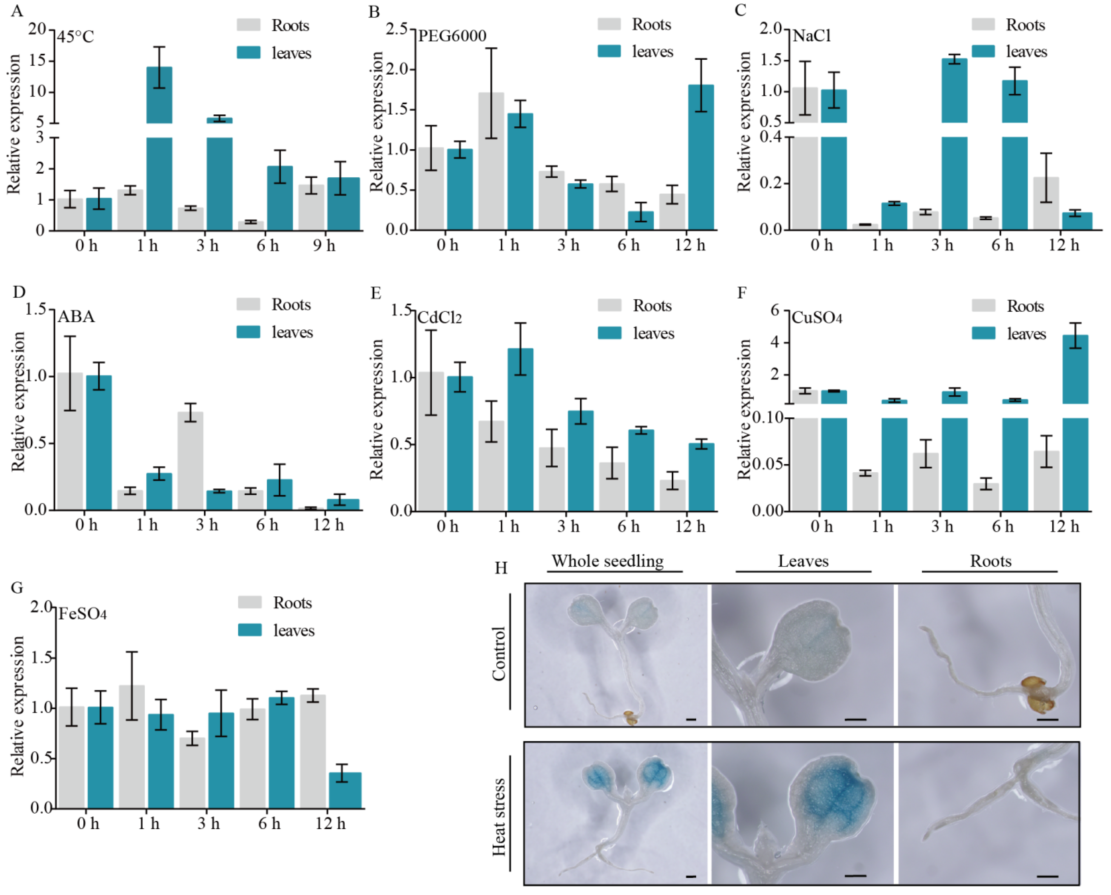
3.2. High Temperature Influences PtHSFA4a Expression in Leaf Tissues

To investigate the expression pattern of PtHSFA4a under various abiotic stress conditions, in vitro cultured P. trichocarpa seedlings were separately exposed to 45 °C, PEG6000, NaCl, ABA, CdCl2, CuSO4, or FeSO4 treatment. RT-qPCR revealed that leaf expression of the PtHSFA4a gene increased markedly and peaked at 1 h at 45 °C, remaining high over the following 9 h (Figure 2A). Meanwhile, in roots, PtHSFA4a transcript levels remained unchanged or were even downregulated under high-temperature stress (Figure 2A). In contrast, the expression of PtHSFA4a did not change significantly at some time points and downregulated at others under any other (PEG6000, NaCl, ABA, CdCl2, CuSO4, or FeSO4) stress condition tested (Figure 2B–G). We generated a PtHSFA4a promoter-glucuronidase (GUS) reporter line in which the 1415-bp PtHSFA4a promoter region was fused to the GUS gene (Figure S1). Histochemical GUS assays revealed that ProPtHSFA4a::GUS showed more vital staining in leaf cells under heat stress (Figure 2H) compared to control conditions. In addition, ProPtHSFA4a::GUS activity did not change in root tissues under heat stress, compared with control conditions. Consistent with RT-qPCR results, a similar histochemical expression pattern was observed in the majority of transgenic lines. These results suggest that the expression of PtHSFA4a was actively regulated in leaves under heat stress.

3.3. PtHSFA4a Regulates Resistance to Heat Stress and High Temperature-Induced ROS Production in A. thaliana

To reveal the function of PtHSFA4a under heat stress in vivo, we used three PtHSFA4a-overexpressing A. thaliana lines, OE1, OE2, and OE3, as described in our previous study [46]. We compared the thermotolerance phenotypes of wild-type (WT) A. thaliana and PtHSFA4a-overexpressing seedlings. First, the survival rates of 5-d old WT and PtHSFA4a-overexpressing OE1, OE2, and OE3 transgenic plants were analyzed under heat (45 °C, 1 h) and normal conditions. There was no significant difference among WT, OE1, OE2, and OE3 at 22 °C, whereas PtHSFA4a-overexpressing lines showed a significantly higher survival rate than WT plants at 45 °C (Figure 3A). Thus, approximately 80% of the PtHSFA4a-overexpressing plants and only 20% of the WT plants recovered completely from exposure to heat stress. (Figure 3B). Additionally, we compared the thermotolerance phenotype, MDA, EL, and ROS contents in adult (4-week old) plants. Under control conditions, WT, OE1, OE2, and OE3 had similar fresh weights (Figure 4A). In contrast, PtHSFA4a-overexpressing transgenic plants exhibited higher fresh weight than WT plants after recovery from heat stress (Figure 4A,B). In turn, MDA content reflected a certain degree of lipid peroxidation induced by ROS, and although increased MDA content was observed in both WT and PtHSFA4a-overexpressing cells after heat stress, it was significantly lower in PtHSFA4a-overexpressing cells than in WT cells (Figure 4C). In addition, we examined EL extent in 4-week-old plants after treatment at 46 °C for 3 h, in which case, PtHSFA4a-overexpressing plants displayed substantially lower EL than that shown by WT plants (Figure 4D). Similarly, our study revealed ROS production in both WT and transgenic A. thaliana plants, regardless of temperature. The accumulation of two major ROS species (H2O2 and O2) in the leaves was analyzed via NBT and DAB staining. We detected no remarkable difference in NBT and DAB staining between the WT and transgenic lines (Figure 4E) under normal temperature conditions. However, after heat treatment, the leaves of WT exhibited darker spots than those of the PtHSFA4a-overexpressing transgenic lines (Figure 4E), indicating that the latter exhibited considerably less accumulation of H2O2 and O2 than WT plants under heat stress. In addition, H2O2 and O2 levels were further confirmed using quantitative measurement (Figure 4F,G), and the leaves of PtHSFA4a-overexpressing plants were found to have significantly less H2O2 content than did WT plants under heat stress (Figure 4F). In addition, the accumulation of O2 had a similar pattern to that of H2O2 (Figure 4G). These results revealed that PtHSFA4a-overexpressing transgenic lines accumulated less ROS in leaves than did the WT under heat stress. Briefly, all results demonstrated that overexpression of PtHSFA4a in A. thaliana positively enhanced thermotolerance and reduced ROS production in leaves after heat stress.

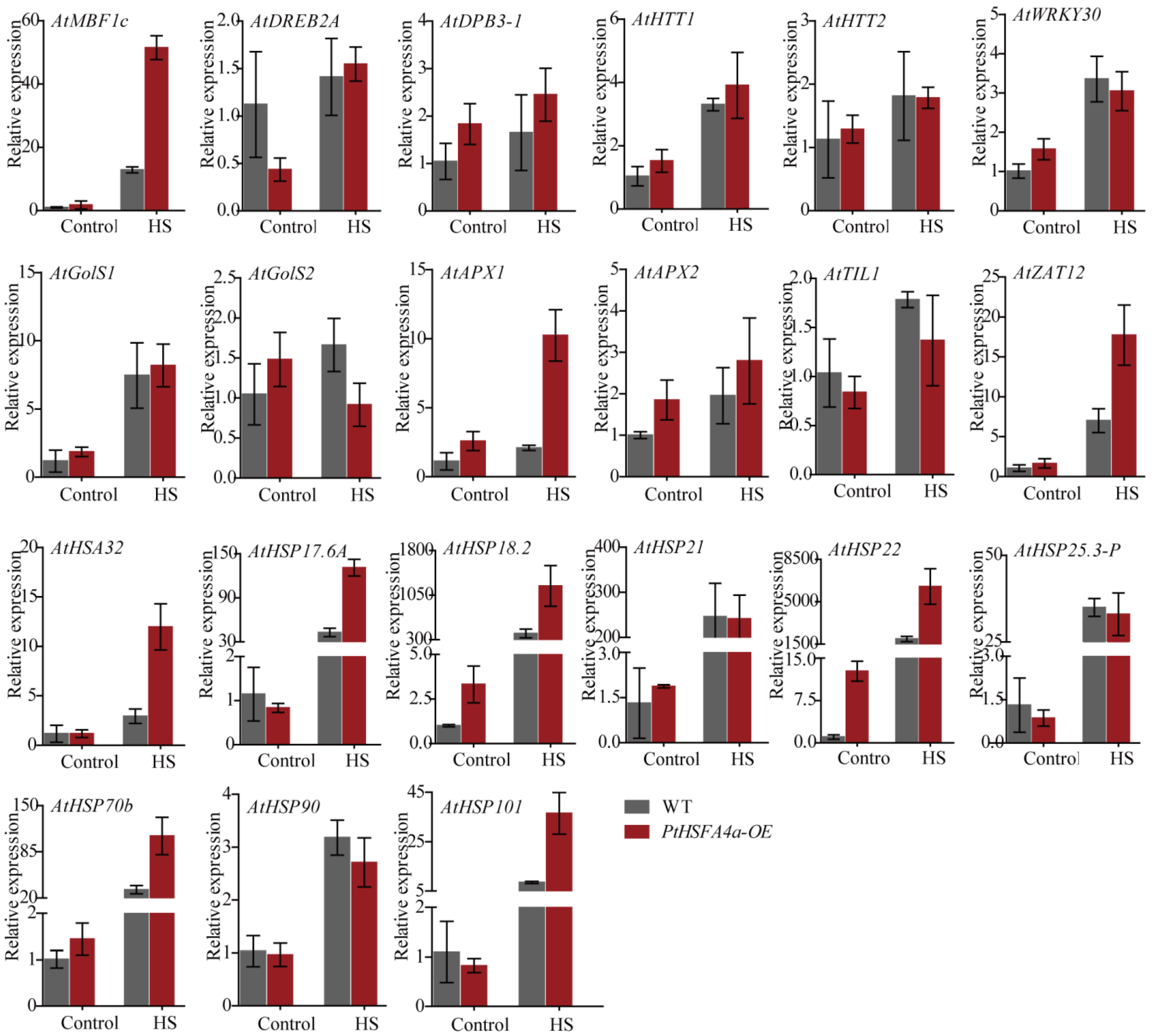
3.4. Overexpression of PtHSFA4a Upregulated Heat Stress-Responsive Genes in Transgenic A. thaliana Plants

As the PtHSFA4a-overexpressing A. thaliana lines exhibited higher thermotolerance and lower ROS content, we analyzed whether heat stress-related genes were regulated in these transgenic plants. A battery of TFs and molecular chaperone genes are known to be essential for thermotolerance in A. thaliana. These include: multiprotein bridging factor 1c (AtMBF1c, At3g24500), AtDREB2A (At5g05410), DNA polymerase II subunit B3-1 (AtDPB3-1, At1g07980), heat-induced tas1 target1 (AtHTT1, At4g29770), heat-induced tas1 target2 (AtHTT2, At5g18040), AtWRKY30 (At5g24110), AtGolS1 (At2g47180), AtGolS2 (At1g56600), AtAPX1 (At1g07890), AtAPX2 (At3g09640), AtTIL1 (At5g58070), AtZAT12 (At5g59820), heat-stress-associated 32-kDa protein (AtHSA32, At4g21320), AtHSP17.6A (At5g12030), AtHSP18.2 (At5g59720), AtHSP21 (At4g27670), AtHSP22 (At4g10250), AtHSP25.3-P (At4g27670), AtHSP70b (At1g16030), AtHSP90 (At5g52640), and AtHSP101 (At1g74310). We investigated the expression of these genes in the leaves of WT and PtHSFA4a-overexpressing transgenic plants under heat stress (Figure 5). The data revealed that, under normal conditions, the transcript levels of these genes did not significantly differ between WT and the transgenic plants (Figure 5). Conversely, the expression of AtMBF1c, AtZAT12, AtAPX1, AtHSA32, AtHSP17.6A, AtHSP18.2, AtHSP22, AtHSP70b, and AtHSP101 was significantly upregulated in the PtHSFA4a-overexpressing lines upon exposure to heat stress, compared with that in WT (Figure 5). Altogether, our data allowed us to conclude that PtHSFA4a-overexpressing transgenic plants regulate the expression of HSPs, TFs, and ROS-scavenging marker genes, thereby enhancing thermotolerance in A. thaliana.

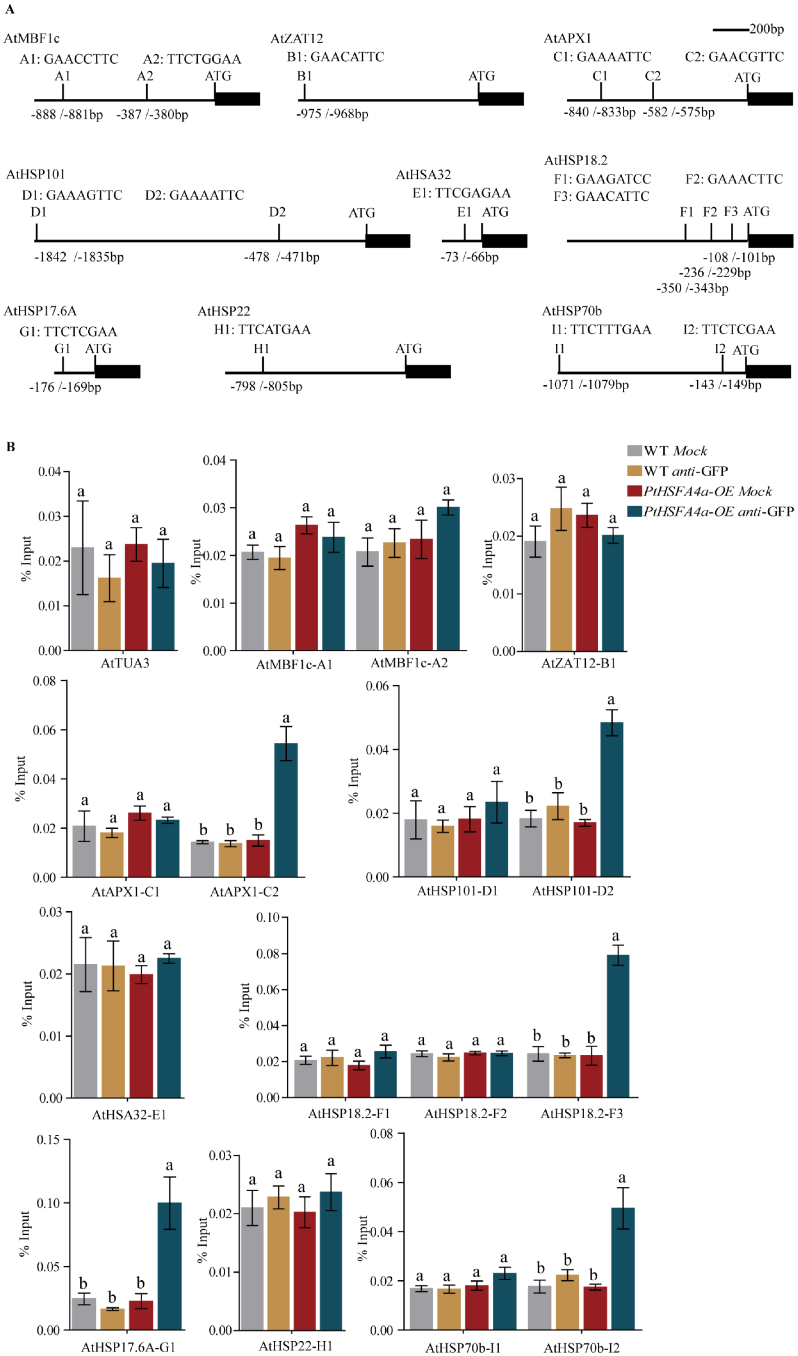
3.5. PtHSFA4a Directly Targets AtAPX1 and AtHSPs Promoters

We selected genes upregulated by PtHSFA4a for ChIP-qPCR assays using an anti-GFP antibody. This set of genes includes AtMBF1c, AtZAT12, AtAPX1, and six HSP-encoding genes, namely, AtHSA32, AtHSP17.6A, AtHSP18.2, AtHSP22, AtHSP70b, and AtHSP101. HSF-related TFs generally regulate the expression of their target genes by binding to the HSE motifs in their promoters. Moreover, we used known HSEs to test whether PtHSFA4a would associate with the nine genes identified as HSE-binding elements. HSE motifs in the promoters of AtZAT12, AtAPX1, AtHSP17.6A, AtHSP18.2, and AtHSP22 (Figure 6A) have been previously reported [27,42,43,44]. We identified the HSE motif once in the AtHSA32 promoter and twice in the AtMBF1c, AtHSP70b, and AtHSP101 promoters (Figure 6A). Further, according to the ChIP-qPCR assay, the HSE motifs in the promoters of AtAPX1, AtHSP17.6A, AtHSP18.2, AtHSP70b, and AtHSP101 were strongly enriched with anti-GFP immunoprecipitated chromatin in PtHSFA4a-overexpressing plants, compared with those in WT plants under heat stress (Figure 6B). In addition, the promoters of these genes were activated more efficiently than the promoter of AtTUA3 (Figure 6B). These results indicate that PtHSFA4a directly activates the expression of AtAPX1, AtHSP17.6A, AtHSP18.2, AtHSP70b, and AtHSP101 by directly binding to their promoters, which underlies the function of PtHSFA4a in thermotolerance.

4. Discussion

High temperature has become a major limiting factor for plant growth and productivity [1]. Therefore, interest in thoroughly understanding the mechanisms underlying plant adaptation to heat stress is on the rise. Several genes are well known to be involved in plant adaptation to heat stress [14]. The response of plants to high temperature is mainly regulated by HSFs. In this study, we identified an HSF-type transcription factor, PtHSFA4a, from P. trichocarpa which plays an important role in heat stress. Heterologous expression of PtHSFA4a in A. thaliana enhanced resistance to high-temperature stress. Furthermore, ectopic expression of PtHSFA4a in A. thaliana resulted in a larger fresh weight than that of non-transgenic plants, and lower MDA, EL, and ROS contents than those in WT plants subjected to heat stress. PtHSFA4a acts as an upstream regulator that directly binds to the HSE of HSPs and APX promoter, thereby activating the expression of these genes to maintain protein-folding homeostasis and enhancing the activation of the antioxidant defense system.
The results of RT-qPCR and GUS activity showed that PtHSFA4a functions specifically in the leaves in response to heat stress. Thus, A. thaliana plants expressing the GFP-tagged PtHSFA4a protein from the CaMV 35S promoter (PtHSFA4a-OE) exhibited enhanced tolerance to heat stress (Figure 3 and Figure 4), as well as reduced ROS contents, compared to WT (Figure 4). Several key genes are reportedly involved in thermotolerance in A. thaliana. For instance, AtMBF1c enhances basal thermotolerance by functioning upstream of the salicylic acid, pathogenesis-related protein 1, ethylene, and trehalose pathways [47]. Further, cooperation between AtDREB2A and AtDPB3-1 enhanced heat tolerance by upregulating the expression of AtHSF and AtHSP genes in A. thaliana [48]. Consistently, the overexpression of AtHTT1 and AtHTT2 in A. thaliana leads to greater thermotolerance, and AtHSFA1a targets AtHTT1 and AtHTT2 at high temperatures [49]. Additionally, in this species, AtTIL1 plays a pivotal role in thermotolerance by acting against heat stress-induced lipid peroxidation [7]. Specifically, AtAPX1 is a central component of ROS-scavenging enzymes, and AtAPX2 is induced by AtHSF3 under heat stress in leaf tissues [50]. As master regulators of the heat shock response in A. thaliana, AtHSFA1s and AtHSFA2 upregulate a set of classical HSPs (AtHSA32, AtHSP17.6A, AtHSP18.2, AtHSP21, AtHSP22, AtHSP25.3-P, AtHSP70b, AtHSP90, and AtHSP101) and AtGolS1 and AtGolS2 genes as part of the plant heat stress-resistance response [16,17,21,22,25,51].
Heat shock proteins function as molecular chaperones to ensure protein functional stability under high temperatures and other stress conditions. Herein, we compared the expression of key HSP-related genes between PtHSFA4a-OE and WT to elucidate the molecular mechanism whereby PtHSFA4a enhances heat tolerance. Our RT-qPCR data showed that PtHSFA4a enhanced heat tolerance and reduced ROS levels by promoting the expression of central thermotolerance genes in A. thaliana, such as molecular chaperones (AtHSA32, AtHSP17.6A, AtHSP18.2, AtHSP22, AtHSP70b, and AtHSP101), ROS-scavenging enzymes (AtZAT12 and AtAPX1), and AtMBF1c.
This study further identified the direct target genes of PtHSFA4a in A. thaliana to elucidate the stress-resistance phenotypes caused by overexpression of PtHSFA4a. First, heat stress causes overproduction of ROS in plant chloroplasts and mitochondria [52]. Then, when the accumulation of ROS reaches harmful levels, it causes membrane lipid peroxidation and cell membrane damage in plant tissues [53]. Some antioxidant defense mechanisms (enzymatic and non-enzymatic antioxidants) protect plants from ROS and repair oxidative damage [54]. Particularly, APX belongs to a small enzymatic gene family that plays a vital role in ROS scavenging in plant cells [55,56,57]. Specifically, AtAPX1 is a cytosolic member of APXs in A. thaliana required to protect the cytoplasm against ROS accumulation under stress. AtAPX1 is induced by heat and oxidative stress [57], and an A. thaliana AtAPX1-deficient mutant (apx1) was significantly more sensitive to the combination of drought and heat stress, such that it accumulated ROS to a larger extent than did the WT [56]. Further, knockout mutant plants deficient in AtAPX1 (KO-Apx1) showed higher H2O2 levels and more protein oxidation during a moderate level of light stress than the WT [55]. Our study showed that AtAPX1 is a direct target gene of PtHSFA4a. In addition, PtHSFA4a-overexpressing A. thaliana plants accumulated less MDA, EL, and endogenous ROS in the leaves under heat stress compared with the WT (Figure 4C–G). These results indicate that the function of PtHSFA4a in heat tolerance may be associated with the regulation of antioxidant ability by directly upregulating the expression of AtAPX1 (Figure 7).
Heat stress induces cellular reactions including misfolding and terminal aggregation of proteins [58]. To maintain protein-folding homeostasis, plants rapidly synthesize and accumulate HSPs at extreme temperatures. As molecular chaperones, HSPs play critical roles in preventing proteins from aggregating and in helping denatured proteins to regain their functional folding. Additionally, they are involved in protein homeostasis (translocation and degradation) under stress [59]. However, each central HSP family member has a unique mechanism of action. For example, the expression of AtHSP17.6A in A. thaliana is induced by heat and drought stress; however, its overexpression increased drought but not heat tolerance in [60]. Consistently, the AtHSP18.2 promoter of A. thaliana exhibited approximately a 1000-fold induction in cultured tobacco cells under heat stress compared with control conditions [61]. Another case yet is A. thaliana AtHSP70b, a cytosolic member of HSP70s whose expression is induced by thermal stress alone [62]. Similarly, AtHSP101 expression is strongly induced by high temperature in A. thaliana [63], and pollen from AtHSP101-overexpressing transgenic cotton and tobacco plants exhibit higher germination rates and much better pollen tube growth under heat stress [64]. Furthermore, overexpression of AtHSP101 in A. thaliana resulted in high heat tolerance [63]. In summary, AtHSP101 plays a crucial role in thermotolerance in A. thaliana. Here, PtHSFA4a directly activated AtHSP17.6A, AtHSP18.2, AtHSP70b, and AtHSP101 by binding to highly conserved heat-shock elements in their promoters. Therefore, our findings strongly indicate that PtHSFA4a enhances heat tolerance, at least partly, by regulating AtHSP17.6A, AtHSP18.2, AtHSP70b, and AtHSP101 to maintain protein folding homeostasis under heat stress conditions (Figure 7).

5. Conclusions

In this study, our data demonstrated that PtHSFA4a responds to heat stress and acts as a positive regulator of heat tolerance by regulating antioxidant defense responses and maintaining protein-folding homeostasis in A. thaliana leaves. In addition, PtHSFA4a seemingly directly targets AtAPX1, AtHSP17.6A, AtHSP18.2, AtHSP70b, and AtHSP101 to enhance heat tolerance by regulating antioxidant defenses and maintaining protein-folding homeostasis in A. thaliana leaves. Overall, these results clearly draw a possible molecular mechanism involving PtHSFA4a in heat stress-responses and provide novel insights into the HSFA4a of woody plant species.

Supplementary Materials

The following supporting information can be downloaded at: https://www.mdpi.com/article/10.3390/f14102028/s1, Figure S1: Promoter sequence of PtHSFA4a; Table S1: All the primers used in this study; Table S2: The accession numbers of the genes mentioned in the paper.

Author Contributions

H.Z., J.W. and X.Z. conceived and designed the experiments, H.Z., X.Z. and M.M. conducted the experiments, X.Z., M.M. and H.D. analyzed the data, H.Z. and J.W. wrote the paper. All authors have read and agreed to the published version of the manuscript.

Funding

This work was funded by the National Natural Science Foundation of China (31972450) and the “Young Talent” Project of Northeast Agricultural University of China (20QC05).

Data Availability Statement

The data that support the findings of this study are available from the corresponding author upon reasonable request.

Conflicts of Interest

The authors declare no conflict of interest.

References

  1. Chand, G.; Sinha, B.K.; Sharma, M.; Dogra, S. High temperature stress: Responses mechanism and management. In Abiotic & Biotic Stress Management in Plants, 1st ed.; Sinha, B.K., Reena, Eds.; E-Publishing Inc.: London, UK, 2022; Volume 1, pp. 51–93. [Google Scholar]
  2. Hartmann, H.; Bastos, A.; Das, A.J.; Esquivel-Muelbert, A.; Hammond, W.M.; Martínez-Vilalta, J.; McDowell, N.G.; Powers, J.S.; Pugh, T.A.M.; Ruthrof, K.X.; et al. Climate change risks to global forest health: Emergence of unexpected events of elevated tree mortality worldwide. Annu. Rev. Plant Biol. 2022, 73, 673–702. [Google Scholar] [CrossRef] [PubMed]
  3. Gong, X.W.; Hao, G.Y. The synergistic effect of hydraulic and thermal impairments accounts for the severe crown damage in Fraxinus mandshurica seedlings following the combined drought-heatwave stress. Sci. Total Environ. 2023, 856, 159017. [Google Scholar] [PubMed]
  4. Elzanati, O.; Mouzeyar, S.; Roche, J. Dynamics of the transcriptome response to heat in the moss, physcomitrella patens. Int. J. Mol. Sci. 2020, 21, 1512. [Google Scholar] [CrossRef] [PubMed]
  5. Kour, J.; Khanna, K.; Singh, A.D.; Dhiman, S.; Bhardwaj, T.; Devi, K.; Sharma, N.; Ohri, P.; Bhardwaj, R. Calcium’s multifaceted functions: From nutrient to secondary messenger during stress. S. Afr. J. Bot. 2023, 152, 247–263. [Google Scholar]
  6. Li, N.; Euring, D.; Cha, J.Y.; Lin, Z.; Lu, M.; Huang, L.J.; Kim, W.Y. Plant hormone-mediated regulation of heat tolerance in response to global climate change. Front. Plant Sci. 2021, 11, 627969. [Google Scholar]
  7. Chi, W.T.; Fung, R.W.; Liu, H.C.; Hsu, C.C.; Charng, Y.Y. Temperature-induced lipocalin is required for basal and acquired thermotolerance in Arabidopsis. Plant Cell Environ. 2009, 32, 917–927. [Google Scholar] [CrossRef]
  8. He, F.; Li, H.G.; Wang, J.J.; Su, Y.; Wang, H.L.; Feng, C.H.; Yang, Y.; Niu, M.X.; Liu, C.; Yin, W.; et al. PeSTZ1, a C2H2-type zinc finger transcription factor from Populus euphratica, enhances freezing tolerance through modulation of ROS scavenging by directly regulating PeAPX2. Plant Biotechnol. J. 2019, 17, 2169–2183. [Google Scholar] [CrossRef]
  9. Ding, L.; Wu, Z.; Teng, R.; Xu, S.; Cao, X.; Yuan, G.; Zhang, D.; Teng, N. LlWRKY39 is involved in thermotolerance by activating LlMBF1c and interacting with LlCaM3 in lily (Lilium longiflorum). Hortic. Res. 2021, 8, 36. [Google Scholar]
  10. Fortunato, S.; Lasorella, C.; Dipierro, N.; Vita, F.; de Pinto, M.C. Redox signaling in plant heat stress response. Antioxidants 2023, 12, 605. [Google Scholar]
  11. Mao, L.; Deng, M.; Jiang, S.; Zhu, H.; Yang, Z.; Yue, Y.; Zhao, K. Characterization of the DREBA4-type transcription factor (SlDREBA4), which contributes to heat tolerance in tomatoes. Front. Plant Sci. 2020, 11, 554520. [Google Scholar] [CrossRef]
  12. Nover, L.; Bharti, K.; Döring, P.; Mishra, S.K.; Ganguli, A.; Scharf, K.D. Arabidopsis and the heat stress transcription factor world: How many heat stress transcription factors do we need? Cell Stress Chaperones 2001, 6, 177–189. [Google Scholar] [CrossRef]
  13. Shi, H.; Tan, D.X.; Reiter, R.J.; Ye, T.; Yang, F.; Chan, Z. Melatonin induces class A1 heat-shock factors (HSFA 1s) and their possible involvement of thermotolerance in Arabidopsis. J. Pineal Res. 2015, 58, 335–342. [Google Scholar] [CrossRef] [PubMed]
  14. Zhang, T.; Zhou, Y. Plant transcription factors and temperature stress. In Plant Transcription Factors, 1st ed.; Srivastava, V., Mishra, S., Eds.; E-Publishing Inc.: London, UK, 2023; pp. 287–300. [Google Scholar]
  15. Busch, W.; Wunderlich, M.; Schöffl, F. Identification of novel heat shock factor-dependent genes and biochemical pathways in Arabidopsis thaliana. Plant J. 2005, 41, 1–14. [Google Scholar] [CrossRef] [PubMed]
  16. Guo, L.; Chen, S.; Liu, K.; Liu, Y.; Ni, L.; Zhang, K.; Zhang, L. Isolation of heat shock factor HSFA1a-binding sites in vivo revealed variations of heat shock elements in Arabidopsis thaliana. Plant Cell Physiol. 2008, 49, 1306–1315. [Google Scholar] [CrossRef] [PubMed]
  17. Fragkostefanakis, S.; Mesihovic, A.; Simm, S.; Paupière, M.J.; Hu, Y.; Paul, P.; Mishra, S.K.; Tschiersch, B.; Theres, K.; Bovy, A.; et al. HSFA2 controls the activity of developmentally and stress-regulated heat stress protection mechanisms in tomato male reproductive tissues. Plant Physiol. 2016, 170, 2461–2477. [Google Scholar] [CrossRef] [PubMed]
  18. Chen, X.J.; Ye, C.J.; Lü, H.Y.; Xu, M.X.; Li, W.; Zhang, L.M.; Wang, C.; Luo, S.P.; Zhu, B.G. Cloning of GmHSFA1 gene and its overexpression leading to enhancement of heat tolerance in transgenic soybean. Yi Chuan Hered. 2006, 28, 1411–1420. [Google Scholar] [CrossRef] [PubMed]
  19. Charng, Y.Y.; Liu, H.C.; Liu, N.Y.; Chi, W.T.; Wang, C.N.; Chang, S.H.; Wang, T.T. A heat-inducible transcription factor, HSFA2, is required for extension of acquired thermotolerance in Arabidopsis. Plant Physiol. 2007, 143, 251–262. [Google Scholar]
  20. Cohen-Peer, R.; Schuster, S.; Meiri, D.; Breiman, A.; Avni, A. Arabidopsis heat shock factor A2 (HSFA2) modifies its activity during acquired thermotholerance. Plant Mol. Biol. 2010, 74, 33–45. [Google Scholar]
  21. Nishizawa-Yokoi, A.; Nosaka, R.; Hayashi, H.; Tainaka, H.; Maruta, T.; Tamoi, M.; Ikeda, M.; Ohme-Takagi, M.; Yoshimura, K.; Yabuta, Y.; et al. HSFA1d and HSFA1e involved in the transcriptional regulation of HSFA2 function as key regulators for the HSF signaling network in response to environmental stress. Plant Cell Physiol. 2011, 52, 933–945. [Google Scholar] [CrossRef]
  22. Zhang, L.; Li, Y.; Xing, D.; Gao, C. Characterization of mitochondrial dynamics and subcellular localization of ROS reveal that HSFA2 alleviates oxidative damage caused by heat stress in Arabidopsis. J. Exp. Bot. 2009, 60, 2073–2091. [Google Scholar]
  23. Song, C.; Chung, W.S.; Lim, C.O. Overexpression of heat shock factor gene HSFA3 increases galactinol levels and oxidative stress tolerance in Arabidopsis. Mol. Cells 2016, 39, 477–483. [Google Scholar] [CrossRef] [PubMed]
  24. Schramm, F.; Larkindale, J.; Kiehlmann, E.; Ganguli, A.; Englich, G.; Vierling, E.; Von Koskull-Döring, P. A cascade of transcription factor DREB2A and heat stress transcription factor HSFA3 regulates the heat stress response of Arabidopsis. Plant J. 2008, 53, 264–274. [Google Scholar] [PubMed]
  25. Yoshida, T.; Ohama, N.; Nakajima, J.; Kidokoro, S.; Mizoi, J.; Nakashima, K.; Maruyama, K.; Kim, J.M.; Seki, M.; Todaka, D.; et al. Arabidopsis HSFA1 transcription factors function as the main positive regulators in heat shock responsive gene expression. Mol. Genet. Genom. 2011, 286, 321–332. [Google Scholar] [CrossRef]
  26. Huang, H.Y.; Chang, K.Y.; Wu, S.J. High irradiance sensitive phenotype of Arabidopsis hit2/xpo1a mutant is caused in part by nuclear confinement of AtHSFA4a. Biol. Plant. 2018, 62, 69–79. [Google Scholar] [CrossRef]
  27. Andrási, N.; Rigó, G.; Zsigmond, L.; Pérez-Salamó, I.; Papdi, C.; Klement, E.; Pettkó-Szandtner, A.; Baba, A.I.; Ayaydin, F.; Dasari, R.; et al. The mitogen-activated protein kinase 4-phosphorylated heat shock factor A4A regulates responses to combined salt and heat stresses. J. Exp. Bot. 2019, 70, 4903–4918. [Google Scholar] [CrossRef] [PubMed]
  28. Yamanouchi, U.; Yano, M.; Lin, H.; Ashikari, M.; Yamada, K. A rice spotted leaf gene, Spl7, encodes a heat stress transcription factor protein. Proc. Natl. Acad. Sci. USA 2002, 99, 7530–7535. [Google Scholar] [PubMed]
  29. Tejedor-Cano, J.; Prieto-Dapena, P.; Almoguera, C.; Carranco, R.; Hiratsu, K.; Ohme-Takagi, M.; Jordano, J. Loss of function of the HSFA9 seed longevity program. Plant Cell Environ. 2010, 33, 1408–1417. [Google Scholar]
  30. Ikeda, M.; Mitsuda, N.; Ohme-Takagi, M. Arabidopsis HSFB1 and HSFB2b act as repressors of the expression of heat-inducible HSFs but positively regulate the acquired thermotolerance. Plant Physiol. 2011, 157, 1243–1254. [Google Scholar] [CrossRef]
  31. AlGhamdi, R.; Aziz, A.; Alshehri, M.; Pardasani, K.R.; Aziz, T. Deep learning model with ensemble techniques to compute the secondary structure of proteins. J. Supercomput. 2021, 77, 5104–5119. [Google Scholar] [CrossRef]
  32. Bienert, S.; Waterhouse, A.; De Beer, T.A.; Tauriello, G.; Studer, G.; Bordoli, L.; Schwede, T. The SWISS-MODEL Repository-new features and functionality. Nucleic Acids Res. 2017, 45, D313–D319. [Google Scholar] [CrossRef]
  33. Berz, J.; Simm, S.; Schuster, S.; Scharf, K.D.; Schleiff, E.; Ebersberger, I. Heatster: A database and web server for identification and classification of heat stress transcription factors in plants. Bioinform. Biol. Insights 2019, 13, 1–5. [Google Scholar] [CrossRef] [PubMed]
  34. Gang, H.; Li, R.; Zhao, Y.; Liu, G.; Chen, S.; Jiang, J. Loss of GLK1 transcription factor function reveals new insights in chlorophyll biosynthesis and chloroplast development. J. Exp. Bot. 2019, 70, 3125–3138. [Google Scholar] [PubMed]
  35. Clough, S.J.; Bent, A.F. Floral dip: A simplified method for Agrobacterium-mediated transformation of Arabidopsis thaliana. Plant J. 1998, 16, 735–743. [Google Scholar] [CrossRef] [PubMed]
  36. Zhang, H.; Yang, J.; Li, W.; Chen, Y.; Lu, H.; Zhao, S.; Li, D.; Wei, M.; Li, C. PuHSFA4a enhances tolerance to excess zinc by regulating reactive oxygen species production and root development in Populus. Plant Physiol. 2019, 180, 2254–2271. [Google Scholar] [CrossRef] [PubMed]
  37. Metwally, A.; Finkemeier, I.; Georgi, M.; Dietz, K.J. Salicylic acid alleviates the cadmium toxicity in barley seedlings. Plant Physiol. 2003, 132, 272–281. [Google Scholar] [CrossRef] [PubMed]
  38. Zhang, X.; Wang, L.; Meng, H.; Wen, H.; Fan, Y.; Zhao, J. Maize ABP9 enhances tolerance to multiple stresses in transgenic Arabidopsis by modulating ABA signaling and cellular levels of reactive oxygen species. Plant Mol. Biol. 2011, 75, 365–378. [Google Scholar]
  39. Baliardini, C.; Meyer, C.L.; Salis, P.; Saumitoulaprade, P.; Verbruggen, N. CATION EXCHANGER1 cosegregates with cadmium tolerance in the metal hyperaccumulator Arabidopsis halleri and plays a role in limiting oxidative stress in Arabidopsis spp. Plant Physiol. 2015, 169, 549–559. [Google Scholar] [CrossRef]
  40. Ramel, F.; Sulmon, C.; Bogard, M.; Couée, I.; Gouesbet, G. Differential patterns of reactive oxygen species and antioxidative mechanisms during atrazine injury and sucrose-induced tolerance in Arabidopsis thaliana plantlets. BMC Plant Biol. 2009, 9, 28. [Google Scholar]
  41. Li, W.; Lin, Y.C.; Li, Q.; Shi, R.; Lin, C.Y.; Chen, H.; Chuang, L.; Qu, G.Z.; Sederoff, R.R.; Chiang, V.L. A robust chromatin immunoprecipitation protocol for studying transcription factor-DNA interactions and histone modifications in wood-forming tissue. Nat. Protoc. 2014, 9, 2180–2193. [Google Scholar] [CrossRef]
  42. Lämke, J.; Brzezinka, K.; Altmann, S.; Bäurle, I. A hit-and-run heat shock factor govern sustained histone methylation and transcriptional stress memory. EMBO J. 2016, 35, 162–175. [Google Scholar] [CrossRef]
  43. Rizhsky, L.; Davletova, S.; Liang, H.; Mittler, R. The zinc finger protein Zat12 is required for cytosolic ascorbate peroxidase 1 expression during oxidative stress in Arabidopsis. J. Biol. Chem. 2004, 279, 11736–11743. [Google Scholar] [CrossRef] [PubMed]
  44. Storozhenko, S.; De Pauw, P.; Van Montagu, M.; Inzé, D.; Kushnir, S. The heat-shock element is a functional component of the Arabidopsis APX1 gene promoter. Plant Physiol. 1998, 118, 1005–1014. [Google Scholar] [PubMed]
  45. Yamaguchi, N.; Winter, C.M.; Wu, M.F.; Kwon, C.S.; William, D.A.; Wagner, D. Protocols: Chromatin immunoprecipitation from Arabidopsis tissues. Arab. Book 2014, 12, e0170. [Google Scholar] [CrossRef] [PubMed]
  46. Zhang, H.; Yang, J.; Li, D.; Wei, M.; Li, C. PtHSFA4a gene play critical roles in the adaptation of Arabidopsis thaliana plants to high-Zinc stress. Plant Signal. Behav. 2019, 14, e1654353. [Google Scholar] [CrossRef] [PubMed]
  47. Suzuki, N.; Sejima, H.; Tam, R.; Schlauch, K.; Mittler, R. Identification of the MBF1 heat-response regulon of Arabidopsis thaliana. Plant J. 2011, 66, 844–851. [Google Scholar]
  48. Sato, H.; Mizoi, J.; Tanaka, H.; Maruyama, K.; Qin, F.; Osakabe, Y.; Morimoto, K.; Ohori, T.; Kusakabe, K.; Nagata, M.; et al. Arabidopsis DPB3-1, a DREB2A interactor, specifically enhances heat stress-induced gene expression by forming a heat stress-specific transcriptional complex with NF-Y subunits. Plant Cell 2014, 26, 4954–4973. [Google Scholar] [CrossRef]
  49. Li, S.; Liu, J.; Liu, Z.; Li, X.; Wu, F.; He, Y. Heat-induced TAS1 TARGET1 mediates thermotolerance via heat stress transcription factor A1a–directed pathways in Arabidopsis. Plant Cell 2014, 26, 1764–1780. [Google Scholar]
  50. Yer, E.N.; Baloglu, M.C.; Ayan, S. Identification and expression profiling of all HSP family member genes under salinity stress in different poplar clones. Gene 2018, 678, 324–336. [Google Scholar]
  51. Nishizawa, A.; Yabuta, Y.; Shigeoka, S. Galactinol and raffinose constitute a novel function to protect plants from oxidative damage. Plant Physiol. 2008, 147, 1251–1263. [Google Scholar]
  52. Medina, E.; Kim, S.H.; Yun, M.; Choi, W.G. Recapitulation of the function and role of ROS generated in response to heat stress in plants. Plants 2021, 10, 371. [Google Scholar] [CrossRef]
  53. Naveen, J.; Hithamani, G.; Pushpalatha, H.G. Stress and Its Influence on the Generation of Reactive Oxygen Species and Oxidative Damage in Plants. In Plant Metabolites under Environmental Stress, 1st ed.; Desai, N.M., Pawar, U.R., Eds.; E-Publishing Inc.: New York, NY, USA, 2023; pp. 219–248. [Google Scholar]
  54. Mansoor, S.; Ali, W.O.; Lone, J.K.; Manhas, S.; Kour, N.; Alam, P.; Ahmad, A.; Ahmad, P. Reactive oxygen species in plants: From source to sink. Antioxidants 2022, 11, 225. [Google Scholar] [CrossRef] [PubMed]
  55. Davletova, S.; Rizhsky, L.; Liang, H.; Shengqiang, Z.; Oliver, D.J.; Coutu, J.; Shulaev, V.; Schlauch, K.; Mittler, R. Cytosolic ascorbate peroxidase 1 is a central component of the reactive oxygen gene network of Arabidopsis. Plant Cell 2005, 17, 268–281. [Google Scholar] [CrossRef] [PubMed]
  56. Koussevitzky, S.; Suzuki, N.; Huntington, S.; Armijo, L.; Sha, W.; Cortes, D.; Shulaev, V.; Mittler, R. Ascorbate peroxidase 1 plays a key role in the response of Arabidopsis thaliana to stress combination. J. Biol. Chem. 2008, 283, 34197–34203. [Google Scholar] [CrossRef] [PubMed]
  57. Panchuk, I.I.; Volkov, R.A.; Schöffl, F. Heat stress-and heat shock transcription factor-dependent expression and activity of ascorbate peroxidase in Arabidopsis. Plant Physiol. 2002, 129, 838–853. [Google Scholar]
  58. Yadav, A.; Singh, J.; Ranjan, K.; Kumar, P.; Khanna, S.; Gupta, M.; Kumar, V.; Wani, S.H.; Sirohi, A. Heat shock proteins: Master players for heat-stress tolerance in plants during climate change. In Heat Stress Tolerance in Plants: Physiological, Molecular and Genetic Perspectives, 1st ed.; Wani, S.H., Kumar, V., Eds.; E-Publishing Inc.: New York, NY, USA, 2020; pp. 189–211. [Google Scholar]
  59. Wu, J.; Gao, T.; Hu, J.; Zhao, L.; Yu, C.; Ma, F. Research advances in function and regulation mechanisms of plant small heat shock proteins (sHSPs) under environmental stresses. Sci. Total Environ. 2022, 825, 154054. [Google Scholar]
  60. Osthoff, A.; Donà Dalle Rose, P.; Baldauf, J.A.; Piepho, H.P.; Hochholdinger, F. Transcriptomic reprogramming of barley seminal roots by combined water deficit and salt stress. BMC Genom. 2019, 20, 325. [Google Scholar]
  61. Yoshida, K.; Kasai, T.; Garcia, M.R.; Sawada, S.; Shoji, T.; Shimizu, S.; Yamazaki, K.; Komeda, Y.; Shinmyo, A. Heat-inducible expression system for a foreign gene in cultured tobacco cells using the HSP18.2 promoter of Arabidopsis thaliana. Appl. Microbiol. Biotechnol. 1995, 44, 466–472. [Google Scholar] [CrossRef]
  62. Sung, D.Y.; Vierling, E.; Guy, C.L. Comprehensive expression profile analysis of the Arabidopsis Hsp70 gene family. Plant Physiol. 2001, 126, 789–800. [Google Scholar]
  63. Babbar, R.; Tiwari, L.D.; Mishra, R.C.; Shimphrui, R.; Singh, A.A.; Goyal, I.; Rana, S.; Kumar, R.; Sharma, V.; Tripathi, G.; et al. Arabidopsis plants overexpressing additional copies of heat shock protein HSP101 showed high heat tolerance and endo-gene silencing. Plant Sci. 2023, 330, 111639. [Google Scholar]
  64. Kumar, R.; Khungar, L.; Shimphrui, R.; Tiwari, L.D.; Tripathi, G.; Sarkar, N.K.; Agarwal, S.K.; Agarwal, M.; Grover, A. AtHSP101 research sets course of action for the genetic improvement of crops against heat stress. J. Plant Biochem. Biotechnol. 2020, 29, 715–732. [Google Scholar]
Figure 1. Structure of PtHSFA4a and phylogenetic analysis of HSFA4a proteins. (A) Bar diagram of the domains and signature sequences of HSFA4a in Populus trichocarpa (PtHSFA4a). Sequence length: 406. (B) Prediction of Secondary structure of protein PtHSFA4a using SOPMA. h: Alpha helix; e: Extended strand; t: Beta turn; c: Random coil. (C) Best predicted three-dimensional (3D) model of PtHSFA4a generated using the SWISS-MODEL server. The N terminus of the PtHSFA4a is shown in red. The C terminus of the PtHSFA4a is shown in blue. Figures are shown from left to right, with the 3D molecular model of PtHSFA4a rotating 180° horizontally. (D) The unrooted phylogenetic tree shows the evolutionary relationship of PtHSFA4a, AtHSFA4a, BnHSFA4a, LuHSFA4a, CpHSFA4a, GrHSFA4a, TcHSFA4a, CsHSFA4a, EgHSFA4a, FvHSFA4a, MdHSFA4a, PpHSFA4a, GmHSFA4a, LjHSFA4a, MsHSFA4a, PvuHSFA4a, CsaHSFA4a, StHSFA4a, SyHSFA4a, VvHSFA4a, CmHSFA4a, ZmHSFA4a, SbHSFA4a, PvHsfA4a, SiHSFA4a, BdHSFA4a, HvHSFA4a, TaHSFA4a, OsHSFA4a, and MgHSFA4a proteins. Scale bar = 0.05 substitutions per site. The scientific names were added after the gene name.
Forests 14 02028 g001
Figure 2. The expression of PtHSFA4a is significantly induced by heat stress. (A) The PtHSFA4a expression level in roots and leaves under heat stress as measured using RT-qPCR. Three-week-old wild-type P. trichocarpa treated at 45 °C in the dark for 0, 1, 3, 6, or 9 h. Data shown are means of three biological replicates, and error bars = SD. (BG) PtHSFA4a expression level in roots and leaves under other abiotic stress factors, as measured using RT-qPCR. Three-week-old wild-type P. trichocarpa were treated separately with 6% (w/v) PEG6000, 150 mM of NaCl, 25 mM of ABA, 80 μM of CdCl2, 100 μM of CuSO4, or 1.0 mM of FeSO4, for 0, 1, 3, 6, or 12 h. Data shown are the mean values of three biological replicates, and error bars = SD. (H) GUS staining in 5-day-old ProPtHSFA4a:GUS transgenic Arabidopsis seedlings treated with or without high temperature (45 °C, 1 h). Scale bar = 200 µm. Three independent experiments were performed using at least 5–6 Arabidopsis seedlings per experiment with similar results. One representative picture is shown.
Forests 14 02028 g002
Figure 3. Analysis of thermotolerance in PtHSFA4a-overexpressing transgenic plants. (A) Thermotolerance test model. Five-day-old wild-type Arabidopsis thaliana (WT) and PtHSFA4a-overexpressing OE1, OE2, and OE3 seedlings grown on MS medium were transferred to 45 °C or 22 °C for 1 h, and then allowed to recover during 10 day before photographing. (B) Survival rate of control (22 °C) and heat-stressed (45 °C) seedlings. Error bars indicate SD for 10 replicates with at least 50 seeds in each. Statistically significant differences between control condition and heat stress condition were analyzed by Student’s t-test (** p < 0.01).
Forests 14 02028 g003
Figure 4. Thermotolerance of PtHSFA4a-overexpressing plants in the soil. Four-week-old WT and PtHSFA4a-overexpressing lines OE1, OE2, and OE3 were grown at 22 °C (left) or 46 °C (right) for 3 h, and then at 22 °C for 15 day (A). (B) Comparison of fresh weight among WT, OE1, OE2, and OE3 after heat-stress recovery. For B, error bars indicate SD for 20 plants. Asterisks indicate significant differences between WT and PtHSFA4a-overexpressing lines: ** p < 0.01, as per Student’s t-test. (C,D) MDA content and electrolyte leakage in leaves of WT, OE1, OE2, and OE3 under control or high temperature conditions, respectively. Leaves were sampled from 4-week-old seedlings grown at 22 °C or incubated at 46 °C for 3 h. For (C,D), error bars indicate SD for at least three replicates. Asterisks indicate significant differences between WT and PtHSFA4a-overexpressing lines: ** p < 0.01, as per Student’s t-test. (E) H2O2 and superoxide radical contents in leaves of WT, OE1, OE2, and OE3 were measured via DAB and NBT staining, respectively. Leaves were sampled from 4-week-old seedlings grown at 22 °C or incubated at 46 °C for 0, 1, and 3 h. Three independent experiments were performed using at least 5–6 seedlings per genotype per experiment with similar results. One representative picture is shown. (F,G) Measurement of the levels of H2O2 and O2 in leaves of WT, OE1, OE2, and OE3 under control or high temperature conditions, respectively. Data are represented as mean ± SD from three biological replicates. Statistically significant differences between WT and PtHSFA4a-overexpressing lines (OE1, OE2 or OE3) were analyzed by Student’s t-test: * p < 0.05 and ** p < 0.01.
Forests 14 02028 g004
Figure 5. RT-qPCR comparative analysis of transcript levels of heat stress-responsive genes between WT and transgenic A. thaliana plants (PtHSFA4a-OE) under normal or heat stress conditions. Data shown are the mean values of three biological replicates; error bars = SD.
Forests 14 02028 g005
Figure 6. ChIP-qPCR analysis of PtHSFA4a-binding to heat stress-responsive gene promoters in A. thaliana using anti-GFP tag antibody. (A) Structural diagrams of the AtMBF1c, AtZAT12, AtAPX1, and six HSP-encoding gene promoters for AtHSP101, AtHSA32, AtHSP18.2, AtHSP17.6A, AtHSP22, and AtHSP70b. Locations and sequences of putative HSEs (A1-2, B1, C1-2, D1-2, E1, F1-3, G1, H1, I1-2) in these gene promoters are shown. (B) ChIP-qPCR assays of selected HSEs from the promoters of AtMBF1c, AtZAT12, AtAPX1, AtHSP101, AtHSA32, AtHSP18.2, AtHSP17.6A, AtHSP22, and AtHSP70b in stable transgenic A. thaliana plants (PtHSFA4a-OE) and WT. Anti-GFP antibody (Beyotime) bound to Protein-A agarose beads was used for the pull-down assay (Anti-GFP). A rabbit anti-hemagglutinin antibody was used as a negative control (Mock). AtTUA3 lacking the HSE region served as a control. Data shown are the mean values of three biological replicates, and error bars = SD. Different letters above the columns indicate significant differences (p < 0.05), as determined by one-way ANOVA.
Forests 14 02028 g006
Figure 7. A proposed model illustrating heat tolerance regulated by the PtHSFA4a.
Forests 14 02028 g007
Disclaimer/Publisher’s Note: The statements, opinions and data contained in all publications are solely those of the individual author(s) and contributor(s) and not of MDPI and/or the editor(s). MDPI and/or the editor(s) disclaim responsibility for any injury to people or property resulting from any ideas, methods, instructions or products referred to in the content.

