Article

Seed Dormancy Characteristics of Kadsura coccinea (Lem.) A. C. Smith, a Unique Medicinal Plant in Southeast Asia

1
Key Laboratory of Cultivation and Protection for Non-Wood Forest Trees of Ministry of Education, College of Forestry, Central South University of Forestry and Technology, Changsha 410004, China
2
The Belt and Road International Joint Research Center for Tropical Arid Non-Wood Forest in Hunan, Changsha 410004, China
3
Experimental Center for Subtropical Forestry, Chinese Academy of Forestry, Xinyu 336600, China
*
Author to whom correspondence should be addressed.
Forests 2023, 14(10), 1928; https://doi.org/10.3390/f14101928
Submission received: 14 August 2023 / Revised: 5 September 2023 / Accepted: 15 September 2023 / Published: 22 September 2023
(This article belongs to the Special Issue Non-timber Forestry Breeding, Cultivation and Processing Technology)

Abstract

:
Kadsura coccinea (Lem.) A. C. Smith is a significant tree species of non-timber forest. However, the low germination percentage and lengthy germination time of its seeds pose obstacles to industry development. Aiming at the problem of seed dormancy, this study first determined the seed dormancy period through seed germination test, and then comprehensively evaluated the dormancy characteristics by observing the seed coat structure, measuring endogenous inhibitors, and in vitro embryo culture. The results indicated the dormancy of K. coccinea seeds, and that the germination period was up to 80 days. Its seed coat is composed of exotesta and endotesta. The dense seed coat structure causes water absorption and air permeability obstacles to the seeds and causes mechanical restraint to the development of the embryo. Meanwhile, its seeds have substances that inhibit seed germination, and there are germination inhibitors in distilled water, methanol, petroleum ether, and ethyl acetate extracts of kernels and seed coats. The inhibitory activity of kernel petroleum ether extract was the highest, and the inhibitory activity of seed coat methanol extract was the highest. In addition, the embryo of K. coccinea developed completely and could germinate normally under in vitro conditions. This study has basically proved that the dormancy of K. coccinea seeds is caused by the seed coat (physical dormancy) and endogenous inhibitors (physiological dormancy), which provides a scientific theoretical basis to further explore the method of seed dormancy release of K. coccinea.

1. Introduction

Kadsura coccinea (Lem.) A. C. Smith is an evergreen woody vine plant belonging to Kadsura genus of the Schisandraceae family [1], naturally distributed in Southeast Asian countries [2,3]. K. coccinea is an important tree species of non-timber forest with a wide range of economic uses [4]. Its roots and rattan stems are traditional Chinese medicinal materials, as they promote blood circulation, reduce swelling, and relieve pain [5]. Its fruit is edible and medicinal, containing essential amino acids, trace elements, vitamins and other nutrients, and medicinal ingredients [6,7]. K. coccinea is rich in lignans and triterpenoids with pharmacological activity [8,9]. Modern medical research has shown that K. coccinea root extracts have anti-inflammatory, anti-tumor, and hepatoprotective effects [10,11,12], stem extracts exhibit anti-HIV activity [13], and fruit extracts have antioxidant, blood lipid-regulating, and skin-whitening effects [14,15,16]. In addition, K. coccinea is also an excellent tree species for landscaping due to its long flowering period and beautiful fruit shape [17].
The main breeding method of K. coccinea is seed breeding, but its seeds exhibit dormancy, resulting in a long germination period and low germination percentage. Seed dormancy is a physiological phenomenon in plants that enables them to resist adverse environmental conditions and ensure successful reproduction, which is the result of natural selection in the long-term evolution of plants to adapt to the environment [18]. The reasons for seed dormancy are complex, including anatomical structure, seed metabolism physiology, heredity, external environment, and so on. Usually, seed dormancy is not caused by one reason but is the result of multiple factors. The relationships between various factors are also complex. When one factor is eliminated and the other factors are not eliminated, the seed is still in a dormant state. Sometimes one factor is eliminated, and other factors are subsequently eliminated, leading to the release of seed dormancy [18,19]. In short, all influencing factors can be eliminated before the seeds can be released from dormancy. Many scholars have defined different types of seed dormancy, but the classification system proposed by Baskin is widely recognized. Based on the Nikolaeva classification system, Baskins categorized seed dormancy into five types: physiological dormancy, morphological dormancy, morphophysiological dormancy, physical dormancy, and combinational dormancy [20]. The main reasons for seed dormancy include embryo dormancy, seed coat obstruction, germination inhibitors, and adverse external conditions. Embryo dormancy is caused by the immature embryo or seed without physiological after-ripening [21,22]. The seed coat leading to seed dormancy is usually dense, hard, or attached with appendages and has cuticle, waxy layer, palisade tissue, and stone cells [23,24,25]. Most seeds contain endogenous inhibitors in some parts (pericarp, seed coat, endosperm, and embryo), including phenols, organic acids, alkaloids, and aldehydes, which may cause seed dormancy [26,27]. Seed germination is closely related to the external environment, such as temperature, moisture, light, oxygen, and soil. When some conditions of the environment fail to meet the requirements of germination, seeds will produce dormancy [28,29,30,31].
The seed dormancy of K. coccinea seeds hinders the development and utilization of germplasm resources. However, there is no report on the dormancy characteristics of K. coccinea seeds at present. Facing this scientific problem, the purpose of this article is to determine the dormancy characteristics of K. coccinea seeds and provide a theoretical basis for releasing dormancy. Specifically, we investigated the dormancy period of seeds, structure and permeability of the seed coat, the endogenous inhibitors of seeds, and the germination of isolated embryos.

2. Materials and Methods

2.1. Source of Materials

The seeds used in the experiment were collected from the experimental base of K. coccinea in Huaihua City, Hunan Province, China (lat. 26°8′35″ N, long. 109°44′53″ E). Healthy, undamaged, and fully mature seeds were selected as the experimental materials.

2.2. Seed Germination Experiment

We took 300 coated seeds and 300 dehulled seeds of K. coccinea and divided them into 3 groups, 100 seeds in each group. The seeds were germinated on the sand bed in an artificial climate chamber (Yanghui RDN-260, Ningbo, China) at 25 °C for 100 days. The sand bed’s moisture content was maintained, and each treatment was repeated 3 times. The germination number of seeds was recorded every 5 days, and the germination percentage was calculated.

2.3. Observation of Seed Coat Structure

Stereomicroscope observation and paraffin section technique were used to observe the seed coat of K. coccinea. The seed coat was cut into the appropriate size and observed under the stereomicroscope (Leica MZ101, Wetzlar, Germany), and then fixed with FAA fixative. After pumping, it was stored in a refrigerator at 4 °C for 24 h, dehydrated with gradient concentration of alcohol, soaked in wax, embedded in paraffin, sliced by slicing machine, stained and observed by microscope (Leica DM4P, Wetzlar, Germany).

2.4. Seed Coat Permeability

Two groups of coated seeds and dehulled seeds of K. coccinea were selected, with a total of 6 samples, each weighing 10 g. One group had the seed coats removed. The weighed seeds were placed in beakers containing 200 mL of water. The seeds were weighed every 2 h from 0 to 12 h, then every 6 h during the daytime, and every 12 h at night. The experiment lasted for 120 h, with a total of 20 measurements. When weighing the seeds, the surface was wiped dry before weighing. The net water absorption of the seeds was calculated, and a water absorption curve was plotted. The respiration rate of K. coccinea seeds was determined using the small basket method [32].

2.5. Endogenous Seed Germination Inhibitor

The effect of endogenous substances in K. coccinea seeds on germination was determined using cabbage as an indicator plant [32,33]. After drying and grinding the seed kernel and seed coat of K. coccinea into powder, they were separately extracted with distilled water, methanol, petroleum ether, and ethyl acetate. The extraction process was repeated three times, and the combined extract was adjusted to a concentration of 0.1 g/mL, which served as the extraction solution for each solvent. Filter paper was placed in a 9 cm Petri dish, and 5 mL of the extraction solution at dilutions of 0%, 25%, 50%, 75%, and 100% were added to the dishes. The solvent was allowed to evaporate, and after the filter paper was completely dry, 5 mL of distilled water was added to moisten the filter paper. Cabbage seeds were then placed on the moistened filter papers to observe germination. Each treatment was replicated three times, with 100 cabbage seeds in each replicate. The germination percentage of cabbage seeds was observed after 24 h, and the root length and seedling height of cabbage seedlings were observed after 72 h.

2.6. Embryo Culture

We took the complete, plump, and swollen seeds of K. coccinea, removed the exotesta, and rinsed them under running water for 5 h. Then, the seeds were placed on a sterile workbench and sterilized by wiping with 70% alcohol for 30 s. They were rinsed with sterile water and then treated with 0.1% mercuric chloride for 10 min. After that, the seeds were thoroughly rinsed with sterile water. Under aseptic conditions, the embryos were carefully dissected and transferred to sterile MS (Murashige and Skoog) culture medium with an agar concentration of 1.5%. The cultures were placed in a growth chamber at 25 °C with a 12 h photoperiod. The experiment was replicated 3 times, with 50 embryos in each replicate.

2.7. Statistical Analysis

The data were analyzed using one-way analysis of variance (ANOVA) and mean comparison, and the differences were statistically compared using the Duncan test with a significance level of p < 0.05. Data are expressed as mean ± standard error. All statistical analyses were carried out using the SPSS software version 18.0. The figures were prepared using Origin 2022 (Origin Laboratory, Northampton, MA, USA).

3. Results

3.1. Effect of K. coccinea Seed Coat on Germination

The germination period of K. coccinea seeds lasted for 80 days, and the final germination percentage was 62.5% at the end of the experiment. K. coccinea seeds exhibited slow germination, with seed germination observed at 35 days. However, once the seeds started to germinate, the germination percentage was relatively fast, with an average daily germination percentage of 1.3%. The germination percentage of 35~80 days was significantly different in every two observation cycles (p < 0.05). On the 30th day, the germination percentage of K. coccinea seeds was 0, which was far from 80% of the non-dormant seeds, indicating that K. coccinea seeds had dormancy (Figure 1).
There are differences in the germination process between coated seeds and dehulled seeds. The dehulled seeds had seed germination at the 20th day, 15 days earlier than the coated seeds, but the germination percentage was slow at the early stage of germination, and there was no significant difference in germination percentage between the 20th day and the 25th day (p > 0.05), and there was no significant difference in germination percentage between the 30th day and the 35th day (p > 0.05). The germination period of the dehulled seeds was 70 days, which was 10 days shorter than that of the coated seeds, and the final germination percentage was 86.5% at the end of the experiment, which was 24.1% higher than that of the coated seeds. The germination percentage of dehulled K. coccinea seeds could reach 86.5%, indicating that the viability of K. coccinea seeds was higher. The seed coat obviously inhibited the germination of K. coccinea seeds, and the inhibition effect was mainly reflected in two aspects: first, it inhibited the germination time of K. coccinea seeds, making the germination start late; second, it inhibited the germination percentage of K. coccinea seeds, making the seed germination percentage decrease.

3.2. The Structure of K. coccinea Seed Coat

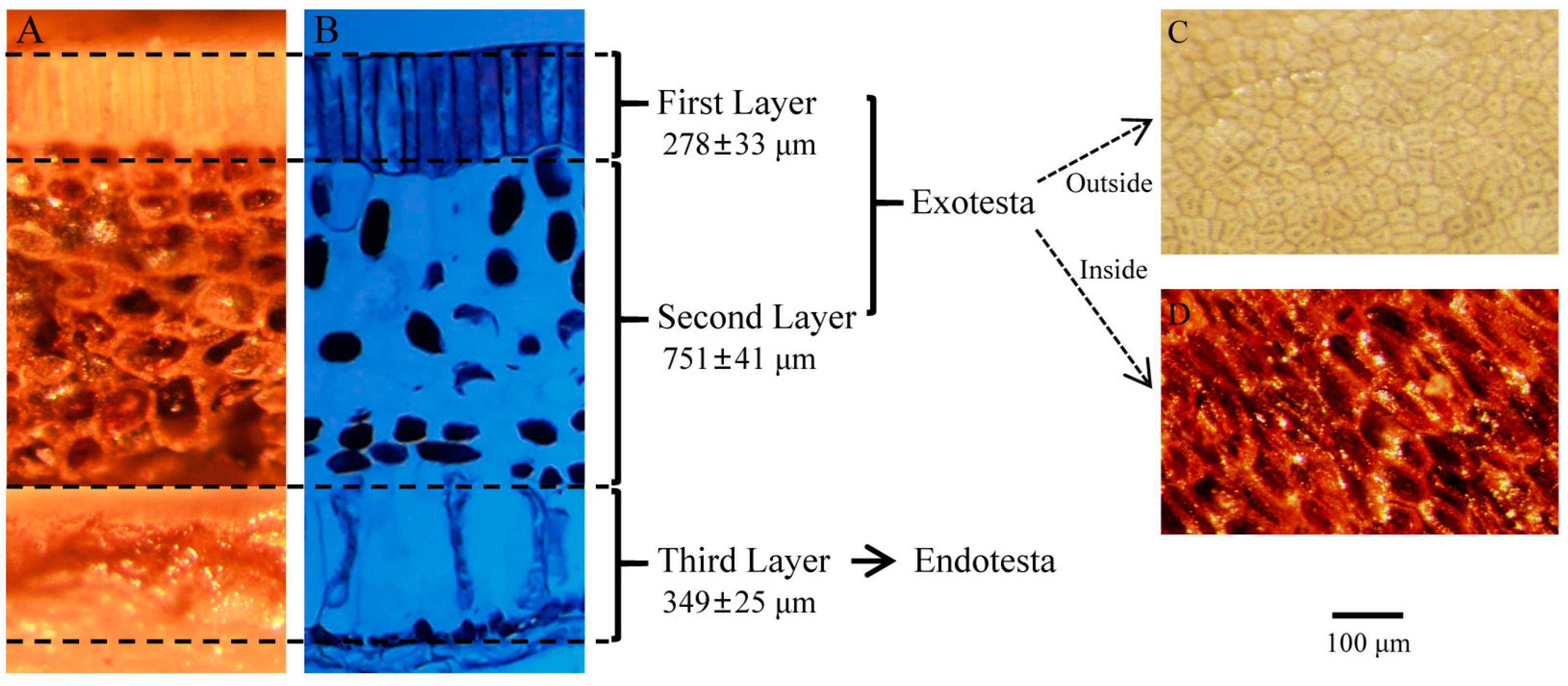
The seed coat is developed from the integument and is an important part of the seed. It can resist and protect the seed from the adverse environment. The seed coat of K. coccinea is composed of the exotesta and the endotesta. The exotesta is light yellow in the dehydrated state, and the inner side of the exotesta is dark brown. The endotesta is the outermost layer of the kernel, which is light brown. When the seed is mature, the hardness of the exotesta is very high, but it can be dehulled. The endotesta is tightly attached to the endosperm to form the kernel, which cannot be peeled off. Only after the cotyledon develops and absorbs the endosperm during seed germination can the endotesta be completely separated (Figure 2).
Under an optical microscope, the thickness of the exotesta was observed to be 1029 ± 101 μm, and the thickness of the endotesta to be 349 ± 25 μm. The thickness of the exotesta was 2.60 times that of the endotesta. The exotesta of K. coccinea can be divided into two layers. The first layer is a dense stratum corneum, which is composed of a layer of slender cells arranged closely and vertically. The thickness is 278 ± 33 μm, accounting for 26.7% of the exotesta. The second layer is the palisade layer, the cell volume is large, and no obvious structure is observed under the optical microscope. However, under the stereomicroscope, it can be clearly seen that the palisade layer is composed of fully lignified cells, and there are cavities in the cells. The thickness is 751 ± 41 μm, accounting for 73.3% of the thickness of the exotesta. The endotesta is composed of mature cells, and the cell structure can be clearly seen under the optical microscope. The cell volume is large, and there are no other organelles in the cell, and the nucleus is not observed.
There are obvious differences between the outer and inner sides of the exotesta of K. coccinea. The outer side of the exotesta is flat, and the surface shape seems to be composed of irregular quadrilaterals, pentagons, and hexagons, and the arrangement is very close, but there is no fixed order or rule, which is messy. The inner side of exotesta is uneven and dense compared to the outer side. The dense structure of K. coccinea exotesta hinders the permeability of the seeds and causes mechanical constraints on the seeds.

3.3. Permeability of K. coccinea Seed Coat

The water absorption rate of coated seeds and dehulled seeds of K. coccinea was measured. It was found that 0~12 h was the rapid water absorption period of seeds. During this period, the net water absorption amount per 10 g of coated seed was 1.43 g, while the water absorption amount of dehulled seed was 3.12 g, which was 2.18 times that of coated seed. Moreover, 12~78 h was the slow water absorption period of seeds, and the water absorption rate decreased significantly. The water absorption rate of dehulled seeds was 0.0549 g/h, and the water absorption rate of dehulled seeds was 0.0490 g/h. After 78 h, the seed was saturated. The net water absorption of the coated seed was 5.06 g, and the net water absorption of the dehulled seed was 7.15 g, which was quite different (Figure 3). In summary, the seed coat of K. coccinea slowed down the water absorption rate of the seed during the rapid water absorption period, and the seed coat had little effect on the water absorption rate of the seed during the slow water absorption period, and the seed coat inhibited the maximum water absorption of the seed. The seed coat of K. coccinea has certain water permeability, but the water absorption rate is slow, and the time required for water absorption to reach saturation is long, which may be caused by the dense structure of the seed coat.
Respiration is the most basic reaction process in plant life activities and intensity is an important indicator to reflect the intensity of seed metabolism. The respiration intensity of K. coccinea seeds was weak, but the seed coat had an effect on the respiration intensity. The respiration intensity of coated seeds was 0.5921 ± 0.0087 mg CO2·g−1·h−1, and the respiration intensity of dehulled seeds was 0.8652 ± 0.0153 mg CO2·g−1·h−1. The respiration intensity of dehulled seed was 1.46 times that of coated seed. The dense seed coat structure of coated seeds seriously hindered the gas exchange inside and outside the seeds, thus affecting the metabolic activity of seeds.

3.4. Effect of K. coccinea Seed Extract on Germination Percentage

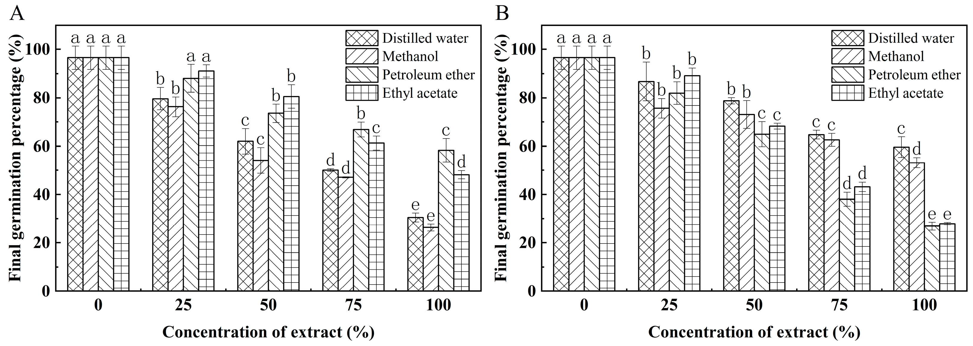
The effect of different solvent extracts of seed coat on the germination percentage of cabbage seeds after dilution with different multiples is shown in Figure 4A. Compared with the germination percentage of 96.5% of the control treated cabbage seeds, the germination percentage of cabbage seeds treated with distilled water 25%, 50%, 75%, and 100% extracts decreased by 16.9%, 34.5%, 46.5%, and 66.1%, respectively, compared with the control. The germination percentage of cabbage seeds treated with methanol 25%, 50%, 75%, and 100% extracts decreased by 20.3%, 42.4%, 49.5%, and 70.2%, respectively, compared with the control. The germination percentage of cabbage seeds treated with 25%, 50%, 75%, and 100% petroleum ether extracts decreased by 8.5%, 22.9%, 29.6%, and 38.2%, respectively, compared with the control. The germination percentage of cabbage seeds treated with 25%, 50%, 75%, and 100% ethyl acetate extracts decreased by 5.4%, 16.0%, 35.2%, and 48.3%, respectively, compared with the control. It shows that the different solvent extracts of K. coccinea seed coat have obvious inhibitory effect on the germination percentage of the cabbage seeds. The inhibitory effect of different solvent extracts of K. coccinea seed coat on the germination percentage of cabbage seeds was in the order of methanol > distilled water > ethyl acetate > petroleum ether.
The effect of different solvent extracts of seed kernel on the germination percentage of cabbage seeds after dilution with different multiples is shown in Figure 4B. The germination percentage of cabbage seeds treated with distilled water 25%, 50%, 75%, and 100% extracts decreased by 9.8%, 17.8%, 31.7%, and 36.9%, respectively, compared with the control. The germination percentage of cabbage seeds treated with methanol 25%, 50%, 75%, and 100% extracts decreased by 20.9%, 23.4%, 33.9%, and 43.4%, respectively, compared with the control. The germination percentage of cabbage seeds treated with 25%, 50%, 75%, and 100% petroleum ether extracts decreased by 14.6%, 31.5%, 58.5%, and 69.6%, respectively, compared with the control. The germination percentage of cabbage seeds treated with 25%, 50%, 75%, and 100% ethyl acetate extracts decreased by 7.3%, 28.3%, 50.4%, and 58.6%, respectively, compared with the control. The results showed that the different solvent extracts of the K. coccinea kernel had obvious inhibitory effects on the germination percentage of cabbage seeds. The inhibitory effect of different solvent extracts of K. coccinea seed kernel on the germination percentage of cabbage seeds was as follows: petroleum ether > ethyl acetate > methanol > distilled water.

3.5. Effect of K. coccinea Seed Extract on Growth

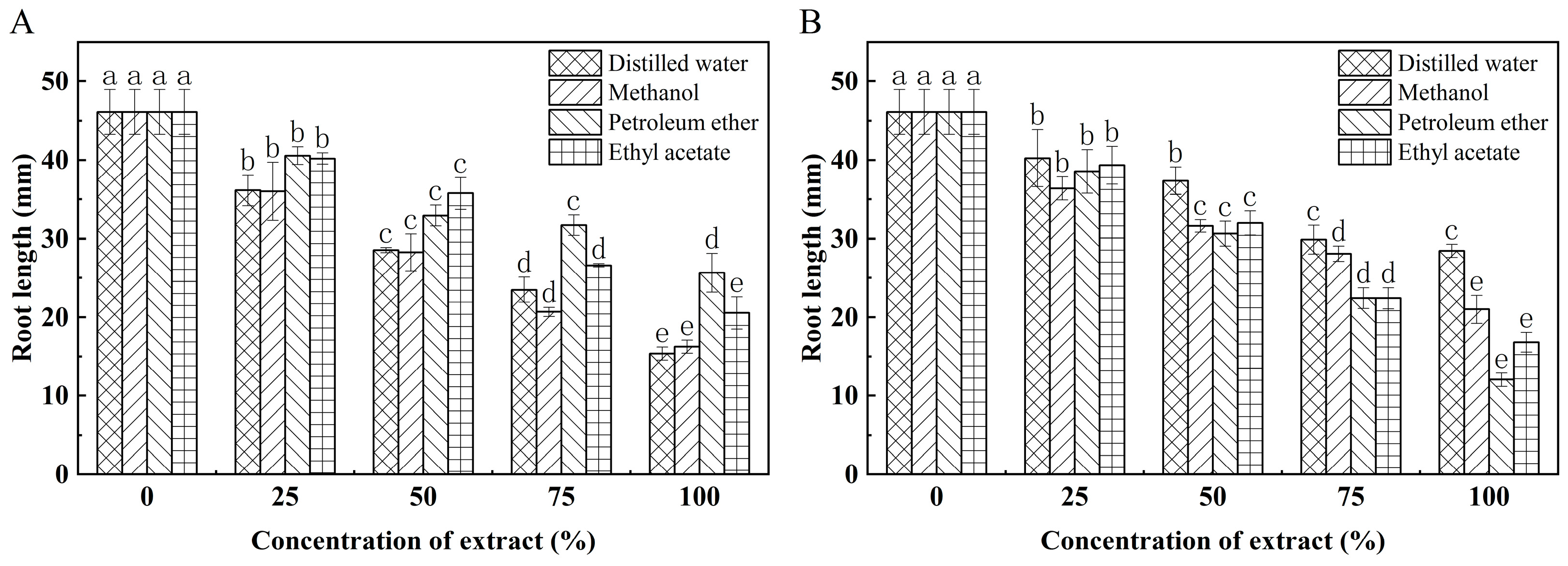
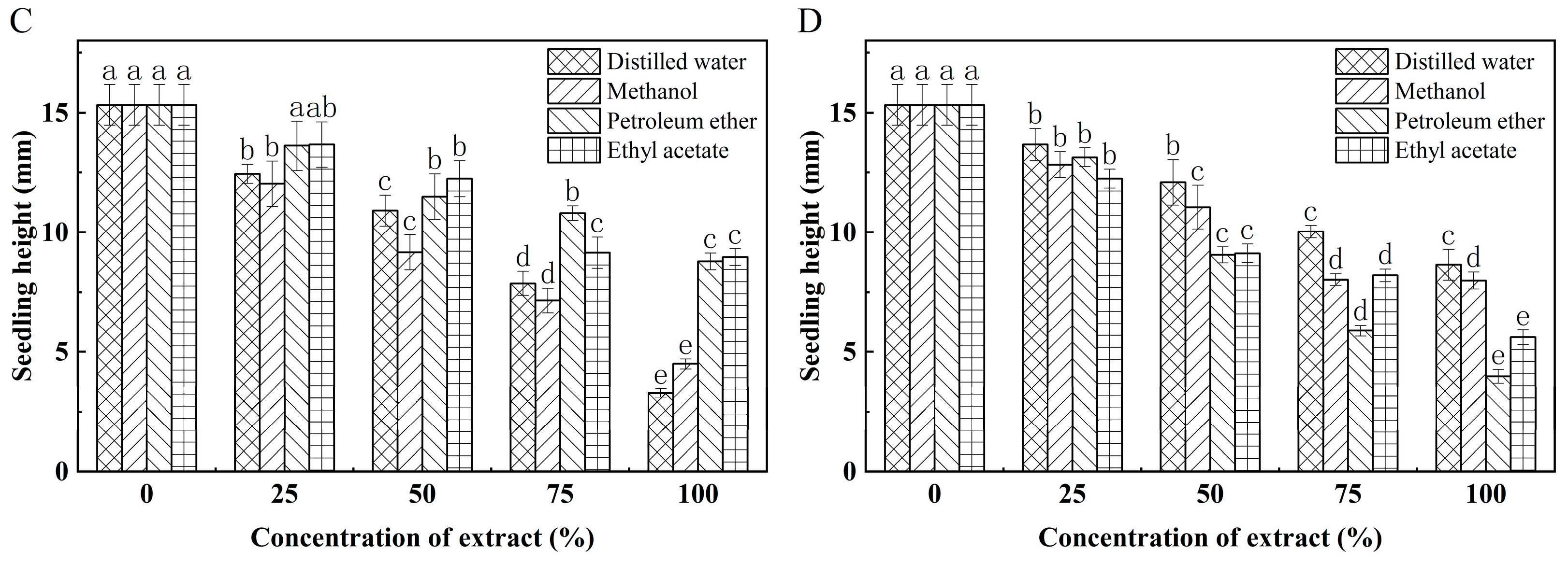
The different solvent extracts of K. coccinea seed coat and kernel had obvious inhibitory effect on the root length of cabbage seeds (Figure 5A,B). The inhibitory effect of different solvent extracts of seed coat on root length was in the order of distilled water > methanol > ethyl acetate > petroleum ether. The inhibitory effect of different solvent extracts on root length was in the order of petroleum ether > ethyl acetate > methanol > distilled water. Meanwhile, this different solvent extract has a significant inhibitory effect on the cabbage seedling’s height (Figure 5C,D). The inhibitory effect of different solvent extracts of seed coat on seedling height was in the order of distilled water > methanol > petroleum ether > ethyl acetate. The inhibitory effect of different solvent extracts on seedling height was in the order of petroleum ether > ethyl acetate > methanol > distilled water.

3.6. Embryo of K. coccinea Culture In Vitro

The embryo volume of K. coccinea is small, and the embryo percentage of mature seeds is only 7.1%. However, the results of the in vitro embryo culture experiment indicate that despite their small size, the embryos have undergone complete differentiation and development and can germinate on a blank MS culture medium (Figure 6). During the first 5 days of K. coccinea embryo in vitro culture, an increase in embryo size can be observed. The growth percentage of the embryo follows a sigmoidal pattern, with the proportion of embryo enlargement reaching 61.2% by day 30 of culture. At day 20, the emergence of embryonic roots and subsequent germination of the embryos can be observed, with a germination percentage of 21.5% at day 30. It should be noted that due to the small size of embryos and the potential for contamination during the isolation process, the contamination percentage in this experiment reached 15.6%.

4. Discussion

Only when the seeds germinate in the appropriate time and space can the growth and development of the seedlings be guaranteed and the successful renewal of the population be realized [34]. In this study, the coated seeds of K. coccinea could not germinate after 30 days of germination, indicating that the seeds had dormancy [35]. The seed coat of K. coccinea has a dense cuticle and poor permeability, which hinders the air permeability and water permeability of seeds, thus slowing down the physiological metabolic activity of kernels. The germination time of coated seeds is longer than that of dehulled seeds, and the germination percentage is also lower, indicating that the seed coat has certain mechanical resistance to the kernel, so it can be inferred that K. coccinea seeds have physical dormancy. The distilled water, methanol, petroleum ether and ethyl acetate extracts of the seed kernel and seed coat of K. coccinea seed have obvious inhibitory effects on the germination percentage, root length and seedling height of cabbage, indicating that there are substances inhibiting germination in the seed of K. coccinea, which can determine the existence of physiological dormancy of the seed of K. coccinea. The embryo of K. coccinea can germinate under in vitro conditions, indicating that the embryo is fully developed and has vitality and does require after-ripening, so its seed does not exist or has a shallow morphological dormancy [31]. In summary, the experimental results show that the seeds of K. coccinea have physical and physiological compound dormancy. The seeds with compound dormancy need to break the physical and physiological dormancy of the seeds, respectively. The physical and physiological dormancy of the seeds of K. coccinea is discussed in detail below.

4.1. Physical Dormancy of K. coccinea Seeds

The seed coat is the main cause of physical dormancy, and the structure of the seed coat is directly related to seed dormancy. Many studies have shown that mechanical resistance or poor permeability of the seed coat is the main reason for plant seed dormancy. The seeds of Schisandra sphenanthera, which belongs to the same genus as K. coccinea, have certain water absorption barriers, and the impermeability of the seed coat is one of the reasons for its dormancy [21]. The coated seeds of Schisandra chinensis, which belong to the same family as K. coccinea, take 7 days to reach water absorption saturation, while the dehulled seeds only takes 3 day; The oxygen consumption of dehulled seeds during seed stratification is also much larger than that of coated seeds. The seed coat of Schisandra chinensis is dense and impermeable, which is the main cause of seed dormancy [36]. In conclusion, the seeds of Schisandraceae family plants generally have dormancy caused by poor seed coat permeability. The seed coat of Cercis canadensis contains dense structures such as palisade layer, cuticle and bright line. Under appropriate germination conditions, its seeds can not germinate via water absorption, which leads to a serious dormancy phenomenon [37]. The outermost layer of the seed coat of Melicope pteleifolia has a thick and dense wax and is rich in hydrophobic oils, which makes it difficult for seeds to absorb water, causing the seeds to be dormant [38]. The exotesta of Cyclocarya paliurus has a cork texture, and the mesotesta cells are closely arranged and highly woody and hard. This dense and hard seed coat structure plays a mechanical constraint on the germination of the embryo and the elongation of the radicle, resulting in difficulty in seed germination [39]. In summary, the palisade layer, cuticle, wax, bright line, and other structures of the seed coat are the main reasons for the poor permeability of the seed. The dense and hard seed coat texture causes mechanical resistance to the seed embryo protruding the seed coat. By observing the anatomical structure of the seeds of K. coccinea, the results showed that the first layer of the exotesta was the stratum corneum, the second layer was the palisade layer, and the endotesta was membranous. The seed coat structure of K. coccinea has a certain degree of protective effect on the kernel, but it also causes water and air permeability obstacles and prevents the leakage of endogenous inhibitory substances. This dense structure also plays a mechanical constraint on seed germination.

4.2. Physiological Dormancy of K. coccinea Seeds

The existence of endogenous germination inhibitors in seeds is the main cause of physiological dormancy. These germination inhibitors affect cell division, differentiation and elongation, thereby inhibiting seed germination. The embryo of Celtis julianae is fully developed and can germinate normally after being cultured in vitro for 1 week. However, its seed cannot germinate under suitable conditions. The presence of germination inhibitors in the seeds is the main reason for dormancy [40]. The germination inhibitors also exist in the seeds of Tulipa thianschanica, Taxus wallichiana, and Pugionium dolabratum [41,42,43]. In this study, four different solvents (distilled water, methanol, petroleum ether, and ethyl acetate) were used to extract the substances in the kernels and seed coats of K. coccinea. The biological identification of inhibitors using extracts revealed that all extracts had significant inhibitory effects on the germination and growth of cabbage seeds. The inhibitory effect of the petroleum ether extract of the kernel was the most obvious, and the inhibitory effect of the methanol extract of the seed coat was the most obvious. The results showed that there were germination inhibitors in K. coccinea seeds, which was the main reason for the dormancy of K. coccinea seeds. However, there were differences in the activity of inhibitors extracted from kernels and seed coats and different solvents. The specific reason was that the number and type of inhibitors were different, which needed further study.

5. Conclusions

The seed coat of fresh K. coccinea had low water permeability, and the seeds had difficulties in absorbing water. There were some germination inhibitors in K. coccinea seeds, which inhibit the germination and growth of indicator plant seeds. The embryo of K. coccinea can germinate normally under in vitro conditions, and the embryo has high vitality and no dormancy. All these findings indicated that the K. coccinea seeds belonged to physiological dormancy and physiological dormancy. The seed of K. coccinea have exotesta and endotesta. The first layer of exotesta is the stratum corneum, and the second layer is palisade layer; The endotesta is membranous. The dense seed coat structure causes water absorption and air permeability obstacles to the seeds. There were germination inhibitors in distilled water, methanol, petroleum ether, and ethyl acetate extracts of K. coccinea kernels and seed coats.

Author Contributions

Conceptualization, S.W. and T.H.; methodology, S.W. and T.H.; validation, W.Z.; formal analysis, T.H., H.G. and C.Y.; investigation, T.H. and X.G.; resources, T.H. and J.W.; data curation, T.H. and F.S.; writing—original draft preparation, T.H.; writing—review and editing, S.W., W.Z. and T.H.; visualization, T.H., F.S. and H.G. All authors have read and agreed to the published version of the manuscript.

Funding

This research was funded by the Research and Utilization of Tropical Arid Non-wood Forest Germplasm Resources in China and Pakistan (Grant No. 2022WK2021).

Data Availability Statement

The datasets generated during the current study are available from the corresponding author upon reasonable request.

Conflicts of Interest

The authors declare no conflict of interest.

References

  1. Sritalahareuthai, V.; Aursalung, A.; On-nom, N.; Temviriyanukul, P.; Charoenkiatkul, S.; Suttisansanee, U. Nutritional composition of conserved Kadsura spp. plants in northern Thailand. Heliyon 2020, 6, e04451. [Google Scholar] [CrossRef]
  2. Deng, B.L.; Xie, B.X.; Zhang, C. Germplasm resources of Kadsura and their utilization in China. J. Central South Univ. For. Technol. 2008, 28, 90–94. [Google Scholar] [CrossRef]
  3. Bi, H.Y.; Lin, Q.; Liu, C.J.; Zhao, J.C. Seed morphology of Kadsura Juss. (Schisandraceae) in relation to its taxonomic significance. Acta Phytotaxon. Sin. 2002, 40, 501–510. [Google Scholar]
  4. Liang, Z.H.; Li, X.; Li, P.; Deng, Y.; Zhong, Y.; Yang, H. Kadsura coccinea lignan metabolism based on metabolome and transcriptome analysis. J. Oncol. 2022, 2022, 3152155. [Google Scholar] [CrossRef] [PubMed]
  5. Sun, Y.H.; Cao, C.Y.; Jin, A.; Wu, W.H. Advances in chemical constituents and pharmacological activities of lignans from Kadsura coccinea. Nat. Prod. Res. Dev. 2022, 34, 491–504. [Google Scholar] [CrossRef]
  6. Zou, J.W.; Rao, H.X.; Luo, X.Q.; Zhang, M.; Liu, W. Characterization of the processing characteristics and nutritional components of Kadsura coccinea fruit. J. Hunan Agric. Univ. 2019, 45, 679–684. [Google Scholar] [CrossRef]
  7. Wang, L.J.; Liao, S.Q.; Long, H.R.; Xia, X.H.; Chen, Q.P.; Liang, J.; Wei, S.G. Analysis and evaluation of nutritional components of Kadsura coccinea fruit peel and pulp. Food Ferm. Ind. 2021, 47, 124–131. [Google Scholar] [CrossRef]
  8. Xu, L.; Su, K.D.; Wei, X.C.; Zhang, J.; Li, H.R. Chemical constituents of Kadsura coccinea. Chem. Nat. Compd. 2018, 54, 242–244. [Google Scholar] [CrossRef]
  9. Liu, Y.B.; Yang, Y.P.; Tasneem, S.; Hussain, N.; Daniyal, M.; Yuan, H.; Xie, Q.L.; Liu, B.; Sun, J.; Jian, Y.Q.; et al. Lignans from Tujia ethnomedicine Heilaohu: Chemical characterization and evaluation of their cytotoxicity and antioxidant activities. Molecules 2018, 23, 2147. [Google Scholar] [CrossRef]
  10. Zhao, Q.J.; Song, Y.; Chen, H.S. Cytotoxic dibenzocyclooctadiene lignans from Kadsura coccinea. Arch. Pharm. Res. 2014, 37, 1375–1379. [Google Scholar] [CrossRef]
  11. Ban, N.K.; Thanh, B.V.; Kiem, P.V.; Minh, C.V.; Cuong, N.X.; Nhiem, N.X.; Huong, H.T.; Anh, H.T.; Park, E.J.; Sohn, D.H.; et al. Dibenzocyclooctadiene lignans and lanostane derivatives from the roots of Kadsura coccinea and their protective effects on primary rat hepatocyte injury induced by t-butyl hydroperoxide. Planta Med. 2009, 75, 1253–1257. [Google Scholar] [CrossRef] [PubMed]
  12. Fang, L.; Xie, C.; Wang, H.; Jin, D.Q.; Xu, J.; Guo, Y.; Ma, Y. Lignans from the roots of Kadsura coccinea and their inhibitory activities on LPS-induced NO production. Phytochem. Lett. 2014, 9, 158–162. [Google Scholar] [CrossRef]
  13. Liang, C.Q.; Shi, Y.M.; Luo, R.H.; Li, X.Y.; Gao, Z.H.; Li, X.N.; Yang, L.M.; Shang, S.Z.; Li, Y.; Zheng, Y.T.; et al. Kadcoccitones A and B, two new 6/6/5/5-fused tetracyclic triterpenoids from Kadsura coccinea. Org. Lett. 2012, 14, 6362–6365. [Google Scholar] [CrossRef] [PubMed]
  14. Li, Z.C.; Sun, J.; Feng, Y.; Zhuo, S.Y.; Le, N.; Yao, J.Y.; Long, X. An experimental animal investigation on toxicity and blood lipid modulating effect of Kadsura coccinea fruit. Food Sci. 2011, 32, 203–205. [Google Scholar]
  15. Sun, J.; Yao, J.Y.; Huang, S.X.; Long, X.; Wang, J.B.; Garcia-Garcia, E. Antioxidant activity of polyphenol and anthocyanin extracts from fruits of Kadsura coccinea (Lem.) AC Smith. Food Chem. 2009, 117, 276–281. [Google Scholar] [CrossRef]
  16. Yan, Z.H.; Gao, Y.Q.; Kong, L.Y.; An, S.S.; Wang, X.; Li, L. Whitening effect of essence product containing Kadsura coccinea extract. Chin. J. Dermatovenereol. 2015, 29, 528–530. [Google Scholar] [CrossRef]
  17. He, T.D.; Shao, F.X.; Zhang, S.Y.; Chen, J.; Yang, C.H.; Wang, S. Fruit development and seed oil accumulation of Kadsura coccinea. Sci. Silv. Sin. 2023, 59, 75–85. [Google Scholar] [CrossRef]
  18. Bewley, J.D.; Black, M. Dormancy and the control of germination. In Seeds; Springer: Boston, MA, USA, 1994; pp. 199–271. [Google Scholar]
  19. Sun, H.F. Some dormancy performance of angiosperm zygotes. Bull. Biol. 1990, 12. [Google Scholar]
  20. Baskin, J.M.; Baskin, C.C. A classification system for seed dormancy. Seed Sci. Res. 2004, 14, 1–16. [Google Scholar] [CrossRef]
  21. Cao, Y.M.; Zhang, J.; Xiang, Y.; Zhao, Z.J.; Fei, Y.J. Study on the dormancy characteristics and dormancy breaking techniques of Paliurus hemsleyanus Rehd. Seed. Seed 2019, 38, 73–76. [Google Scholar] [CrossRef]
  22. Chen, W.; Chen, S.A.; Li, J.; Meng, M.; Feng, X.; Liu, J.M. A preliminary study on dormancy mechanism of Kadsura ananosma seeds. J. West Chin. For. Sci. 2020, 49, 43–47+56. [Google Scholar] [CrossRef]
  23. Ma, L.Q.H. Study on Dormancy Characteristics and Germination Physiology of Four Varieties sabina chinensis Seeds. Master’s Thesis, Inner Mongolia Agricultural University, Hohhot, China, 2019. [Google Scholar]
  24. Ke, B.Y.; Zhou, C.Y.; Li, J.Y.; Pan, J.; Zhou, P. Study on methods of seed dormancy and dormancy-release for rare species Acrocarpus fraxinifolius. Seed 2020, 39, 139–143. [Google Scholar] [CrossRef]
  25. Bhat, H.A.; Asif, M.; Mir, N.; Aijaz-Un-Nabi; Gatto, A.A.; Ahmad, F.; Hussain, N. Maturity indices and dormancy breaking methods of black locust (Rhobinia pseudoacacia) seeds under temperate Kashmir condition. Ecol. Environ. Cons. 2014, 20, 1769–1775. [Google Scholar]
  26. Pipinis, E.; Stampoulidis, A.; Milios, E.; Kitikidou, K.; Akritidou, S.; Theodoridou, S.; Radoglou, K. Effects of seed moisture content, stratification and sowing date on the germination of Corylus avellana seeds. J. For. Res. 2020, 31, 743–749. [Google Scholar] [CrossRef]
  27. Chien, C.T.; Chen, S.Y.; Chang, S.H.; Chung, J.D. Dormancy and germination in seeds of the medicinal Asian tree species Phellodendron amurense var. Wilsonii (Rutaceae). Seed Sci. Technol. 2006, 34, 561–571. [Google Scholar] [CrossRef]
  28. Qi, Y.; Bilan, M.; Chin, K. New method for breaking Korean pine seed dormancy. J. Arbor. 1993, 19, 113–117. [Google Scholar] [CrossRef]
  29. Cao, D.C.; Xu, H.M.; Zhao, Y.Y.; Deng, X.; Liu, Y.X.; Soppe, W.J.J.; Lin, J.X. Transcriptome and degradome sequencing reveals dormancy mechanisms of Cunninghamia Lanceolata seeds. Plant Phys. 2016, 172, 2347–2362. [Google Scholar] [CrossRef]
  30. Li, Z.H.; Xu, R.H.; Ren, M.J.; Li, L.H. Advances in phytochrome regulating seed dormancy and germination by sensing light and temperature signals. Plant Phys. J. 2019, 55, 539–546. [Google Scholar] [CrossRef]
  31. Qin, A.L.; Guo, Q.S.; Ma, F.Q.; Jian, Z.J.; Zheng, X.K. Effects of temperature, light and water conditions on seed germination of Thuja sutchuenensis franch. Seed 2020, 39, 15–20. [Google Scholar] [CrossRef]
  32. Li, T.H.; Min, X.H. Dormancy characteristics and germination requirements of Phoebe bournei seed. Sci. Hortic. 2020, 260, 108903. [Google Scholar] [CrossRef]
  33. Lu, J.J.; Tan, D.Y.; Baskin, J.M.; Baskin, C.C. Trade-offs between seed dispersal and dormancy in an amphi-basicarpic cold desert annual. Ann. Bot. 2013, 112, 1815–1827. [Google Scholar] [CrossRef] [PubMed]
  34. Finch-Savage, W.E.; Leubner-Metzger, G. Seed dormancy and the control of germination. New Phytol. 2006, 171, 501–523. [Google Scholar] [CrossRef] [PubMed]
  35. Zhang, K.L.; Yao, L.J.; Zhang, Y.; Baskin, J.M.; Baskin, C.C.; Xiong, Z.M.; Tao, J. A review of the seed biology of Paeonia species (Paeoniaceae), with particular reference to dormancy and germination. Planta 2019, 249, 291–303. [Google Scholar] [CrossRef] [PubMed]
  36. Zhu, H.; Zhou, D.B.; Wang, W.Z.; Nie, S.Q. Preliminary study on the causes of seed dormancy of Schisandra chinensis (Turcz.) Baill. Terr. Nat. Res. Study 1993, 2, 59–61. [Google Scholar] [CrossRef]
  37. Yang, J.H. Dormancy and Germination Characteristics of Cercis canadensis ‘Forest Pansy’ Seeds. Master’s Thesis, Yangzhou University, Yangzhou, China, 2021. [Google Scholar]
  38. Lu, Q. Study on the Methods and Preliminary Mechanism of Seed Dormancy Release of Melicope pteleifolia. Ph.D. Thesis, Guangzhou University of Chinese Medicine, Guangzhou, China, 2019. [Google Scholar]
  39. Shang, X.L.; Xu, X.Z.; Fang, S.Z. Seed dormancy mechanism of Cyclocarya paliurus. Sci. Silv. Sin. 2011, 47, 68–74. [Google Scholar]
  40. Liu, X. Studies on Seed Dormancy Mechanism of Celtis julianae Schneid. Seeds. Master’s Thesis, Nanjing Forestry University, Nanjing, China, 2019. [Google Scholar]
  41. Zhang, W.; Qu, L.W.; Zhao, J.; Xue, L.; Dai, H.P.; Xing, G.M.; Lei, J.J. Practical methods for breaking seed dormancy in a wild or-namental tulip species Tulipa thianschanica regel. Agronomy 2020, 10, 1765. [Google Scholar] [CrossRef]
  42. Liu, C.G.; Chen, L.; Li, Y.; Dai, S.J.; Chen, W.J.; Pan, L.; Xu, Y.J. Study on dormancy and germination technology of Taxus chinensis var. mairei seeds. J. Southwest For. Coll. 2015, 35, 25–29. [Google Scholar]
  43. Zheng, Q.L.; Hao, L.Z.; Yang, Z.R.; Zhang, F.L. Research progress of Pugionium gaertn plant seeds. Seed 2017, 36, 46–50. [Google Scholar] [CrossRef]
Figure 1. Effect of K. coccinea seed coat on seed germination. Note: Different letters represent significant differences (p < 0.05). The vertical lines indicate the standard error of the mean (±SEM).
Forests 14 01928 g001
Figure 2. Seed coat structure of K. coccinea. (A) Stereomicroscopy of a cross section of the seed coat. (B) Optical microscope image of a cross section of the seed coat. (C) Stereomicroscopy of the outside of the exotesta. (D) Stereomicroscopy of the inside of the exotesta.
Forests 14 01928 g002
Figure 3. Water absorption curve of K. coccinea seed. Note: Different letters represent significant differences (p < 0.05). The vertical lines indicate the standard error of the mean (±SEM).
Forests 14 01928 g003
Figure 4. (A) Effect of K. coccinea seed coat extract on germination. (B) Effect of K. coccinea kernel extract on germination. Note: Different letters represent significant differences (p < 0.05). The vertical lines indicate the standard error of the mean (±SEM).
Forests 14 01928 g004
Figure 5. (A) Effect of K. coccinea seed coat extract on root length. (B) Effect of K. coccinea seed kernel extract on root length. (C) Effect of K. coccinea seed coat extract on seedling height. (D) Effect of K. coccinea seed kernel extract on seedling height. Note: Different letters represent significant differences (p < 0.05). The vertical lines indicate the standard error of the mean (±SEM).
Forests 14 01928 g005aForests 14 01928 g005b
Figure 6. In vitro culture growth of K. coccinea embryo. Note: Different letters represent significant differences (p < 0.05). The vertical lines indicate the standard error of the mean (±SEM).
Forests 14 01928 g006
Disclaimer/Publisher’s Note: The statements, opinions and data contained in all publications are solely those of the individual author(s) and contributor(s) and not of MDPI and/or the editor(s). MDPI and/or the editor(s) disclaim responsibility for any injury to people or property resulting from any ideas, methods, instructions or products referred to in the content.