Communication

Detection and Quantification of Serpula himantioides in the Wood of Chamaecyparis pisifera Butt Rot Trees by Real-Time PCR

The University of Tokyo Chichibu Forest, Graduate School of Agricultural and Life Sciences, The University of Tokyo, 1-1-49 Hinoda-machi, Chichibu, Saitama 368-0034, Japan
*
Author to whom correspondence should be addressed.
Forests 2022, 13(9), 1429; https://doi.org/10.3390/f13091429
Submission received: 11 July 2022 / Revised: 30 August 2022 / Accepted: 1 September 2022 / Published: 6 September 2022
(This article belongs to the Section Forest Health)

Abstract

:
Serpula himantioides is a wood-decaying fungal pathogen which is widespread and causes potentially serious butt rot in living trees of numerous coniferous plantation species. This study aimed to quantify S. himantioides in the wood of Sawara cypress (Chamaecyparis pisifera) with butt rot using real-time polymerase chain reaction (real-time PCR). Species–specific primers were designed for the internal transcribed spacer 2 (ITS2) regions of ribosomal DNA (rDNA) of S. himantioides. The specificity of the designed primer set was tested by end-point PCR and amplicon sequencing. End-point PCR assays were positive for S. himantioides and negative for S. lacrymans, which belongs to the same genus as S. himantioides, Coniophora puteana of the same family as S. himantioides, and other wood decay fungi. Taxonomy assignment based on amplicon sequencing detected only Serpula spp., and most of them were identified as S. himantioides. The linearity of the calibration curve for absolute quantification by real-time PCR assays was confirmed in the range from 101 to 108 copies. This molecular assay method using real-time PCR could detect trace amounts of S. himantioides in decayed wood, showing the applicability for early diagnostics of butt rot emergence in forests.

1. Introduction

Rot disease is one of the most common diseases that degrades timber quality, including timber discoloration and structural weakness, leading to significant economic losses [1]. For instance, root and butt rot caused by Heterobasidion annosum is one of the most destructive diseases affecting conifers in the northern temperate regions of the world, particularly in Europe. The annual economic loss attributable to H. annosum infection in Europe is estimated at 800 million euros [2]. However, detecting this disease is difficult, until the pathogen is widespread and wood decay reaches an advanced stage [3]. Instrumental analyses, such as tomographic technologies [4,5,6], magnetic resonance imaging [7], and ultrasonic measurements [8], have helped in the detection of internal wood decay. The identification of causal agents for such decay is not always feasible without detecting the presence of fungal fruiting bodies, which are only sporadically visible, especially in advanced stages of infection [9]. Traditional methods for the identification of wood decay fungi primarily rely on the visual inspection of fruiting bodies [1]. Pure fungal cultures isolated from mycelia or decayed wood when no fruiting bodies are available may help identify causal agents. However, diagnostic methods based on pure culture tests are time-consuming and sometimes cannot accurately distinguish between closely related species [10]. Furthermore, isolation of wood decay fungi from environmental samples is often difficult despite the use of selective media, due to contamination from other fast-growing fungus [1]. Hence, rapid and accurate detection of fungal pathogens is essential for taking preventive measures to reduce the damage caused by butt rot.
Serpula himantioides is a wood-decay fungus that causes butt rot in living trees and has been found in all continents, except Antarctica. A recent phylogenetic study of S. himantioides reported five major clades as phylogenetic species (PS1–PS5) of which, PS1 is found in South America, PS2 and PS3 include isolates from North America only, PS4 is dominant in North America and Europe, and PS5 is found in most continents [11]. S. himantioides infects a wide range of woody plant species, including Japanese larch (Larix kaempferi); Douglas fir (Pseudotsuga menziesii) in Germany [12]; P. menziesii in Denmark [13]; white spruce (Picea glauca), black spruce (Picea mariana), and balsam fir (Abies balsamea) in Canada [14]; and A. balsamea and red spruce (Picea rubens) in the USA [15]. Recently, butt rot caused by S. himantioides was found in the living trees of Sawara cypress (Chamaecyparis pisifera), a major plantation species in Japan (Figure 1) [16,17]. Therefore, it is important to monitor the status of S. himantioides infection in forests to prevent heart rot damage in Japanese cedar (Cryptomeria japonica) [17], the most dominant plantation species in Japan.
Real-time polymerase chain reaction (PCR) has been widely used as an effective method for the rapid detection and identification of plant pathogens. It combines the sensitivity of end-point PCR with the generation of a specific fluorescent signal, providing real-time analysis of polymerase chain reaction kinetics, and allowing quantification of a specific DNA target [18]. This tool has been successfully used to detect plant pathogens in various environmental samples, such as plant tissue [19,20,21], soil [22,23], and air [24,25]. Wood decay fungi, such as Heterobasidion annosum sensu lato [26], Phaeolus schweinitzii [26], Gloeophyllum trabeum [27], Coniophora puteana [27], Serpula lacrymans [27], and Trametes versicolor [27,28] have also been detected in decaying wood by real-time PCR using species-specific primers, suggesting that this method is a simple and rapid diagnostic tool for their detection. Although there are few reports on the causative agents of butt rot, it is anticipated that real-time PCR assays using a species-specific primer set can also be used to quantitatively detect S. himantioides in environmental samples. Schmidt et al. [29] developed a species-specific primer set for detecting S. himantioides using end-point PCR; however, this was not initially designed for real-time PCR. Therefore, a real-time PCR assay that can quantitatively detect S. himantioides with high specificity and sensitivity is needed. In this study, we aimed to develop a real-time PCR assay for detecting trace amounts of S. himantioides in wood by designing a new primer set with high specificity and sensitivity and to verify its specificity using end-point PCR and amplicon sequencing.

2. Materials and Methods

2.1. Design of Species-Specific Primer Set

Sequences of the internal transcribed spacer (ITS) region of the ribosomal DNA (rDNA) of S. himantioides strains from all continents except Antarctica were collected from the National Center for Biotechnology Information (NCBI) nucleotide database (Table S1). We compared these sequences of the ITS region with those of other fungal groups and found that the ITS2 region was suitable for S. himantioides. Therefore, we designed the following species-specific primer set using OligoEvaluator™, an online oligonucleotide sequence calculator [30]. The designed forward primer was SHF (5′-CTCGCATCGATGAAGAAC-3′), and the reverse primer was SHR (5′-CAAAACATTGTCTTACGACG-3′). This primer set was designed with a GC content ranging from 40% to 50% and similar melting temperatures (Table S2). In this primer set, the reverse primer was specific to S. himantioides and the amplicon size was 286 bp. The specificity of the reverse primer was verified using the Basic Local Alignment Search Tool (BLAST) in silico [31].

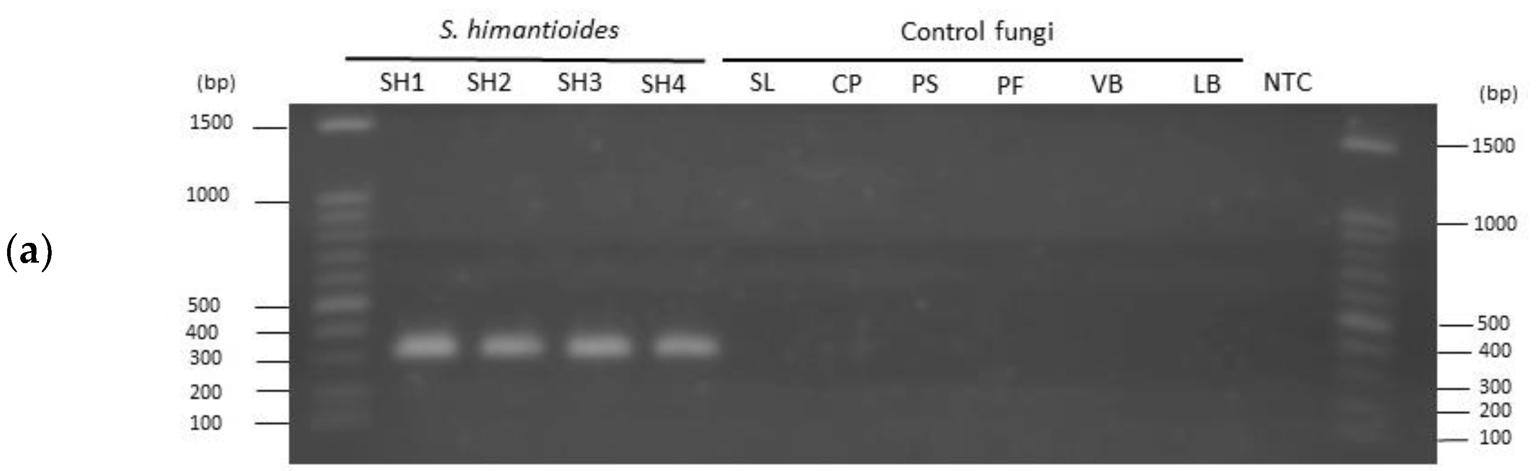
2.2. Verification of Specificity by End-Point PCR

The specificity of the designed primer set was tested using isolates of S. himantioides, S. lacrymans (of the same genus), and Coniophora puteana (of the same family), and other phylogenetically distant wood decay fungi (Phaeolus schweinitzii, Postia floriformis, Lenzites betulinus, and Veluticeps berkeleyi) (Table S3). These isolates were cultured on potato dextrose agar (PDA) in an incubator at 22 °C for 10 days, and genomic DNA was extracted from mycelia collected from the agar surface using a NucleoSpin Plant II kit (Macherey-Nagel GmbH & Co. KG, Düren, Germany) according to the manufacturer’s instructions.
PCR was performed using the genomic DNA template with a species-specific primer set (SHF/SHR) and a universal primer set for fungi, using a thermal cycler (GeneAtlas G, ASTEC, Fukuoka, Japan). ITS5 (5′-GGAAGTAAAAGTCGTAACAAGG-3′; White et al. [32]) was the forward primer, and ITS4 (5′-TCCTCCGCTTATTGATATGC-3′; White et al. [32]) was the reverse primer in the universal primer set for fungi. PCR was performed in a 25 µL reaction mixture containing 5 ng of template DNA, 0.25 µM of each forward and reverse primers, 12.5 µL of 2 × Gflex PCR Buffer, and 0.5 µL of Tks Gflex DNA polymerase (Takara Bio Inc., Shiga, Japan). PCR conditions were as follows: 94 °C for 1 min, followed by 30 cycles of 10 s at 98 °C, 15 s at 60 °C, and 30 s at 68 °C, and a final extension step at 68 °C for 7 min. PCR conditions for the reaction with the universal primer set were 94 °C for 1 min, followed by 30 cycles of 10 s at 98 °C, 15 s at 55 °C, and 30 s at 68 °C, with a final extension at 68 °C for 7 min. Purified water without a genomic DNA template was used as the negative control.
After PCR, agarose gel electrophoresis was performed to verify the presence or absence of bands using a Benchtop 2UV transilluminator (Analytik Jena US, An Endress+Hauser Company, Upland, CA, USA). The results of the species-specific primer set and the universal primer set were compared.

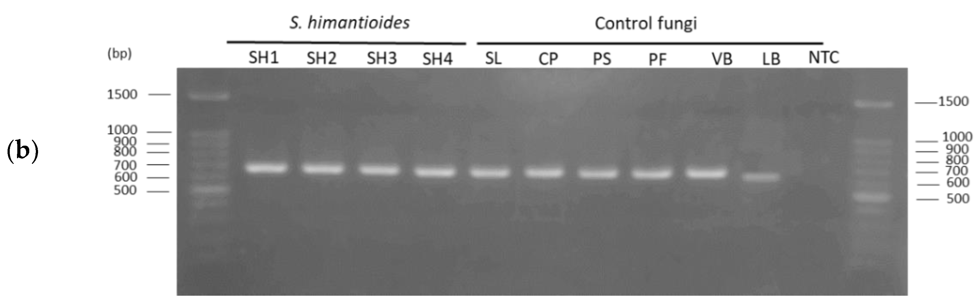
2.3. Verification of Specificity by Amplicon Sequencing

Amplicon sequencing was used to ascertain whether the designed primer set could specifically detect S. himantioides in decayed wood, in which numerous species of fungi were present.
Two decayed C. pisifera trees (CP1 and CP2; Table S4) were cut in the University of Tokyo Chichibu Forest (35°56′ N, 138°52′ E) in Central Japan, where the most recent butt rot damage by S. himantioides was reported [16]. S. himantioides was isolated from these trees. Taking wood discoloration as an indicator, nine samples of decayed wood with varying degrees of brown rot were obtained from the decayed parts of the trees using a flame-sterilized electric drill (9 mm in diameter). The degree of wood discoloration was classified into three types: non-discoloration, moderate discoloration, and severe discoloration. Three samples were collected from each of the above region (Table S5). Decayed wood samples were freeze-dried for 72 h and powdered using a bead-beater homogenizer. Genomic DNA was extracted from 5 mg of powdered wood samples using the NucleoSpin Plant II kit according to the manufacturer’s protocol. DNA yields were determined using a Qubit dsDNA HS Assay Kit (Thermo Fisher Scientific, Waltham, MA, USA).
PCR was performed using the genomic DNA template with a species-specific primer set (SHF/SHR) and fungal community primer set (gITS7/ITS4ngs) for amplicon sequencing. gITS7 (5′-GTGARTCATCGARTCTTTG-3′; Ihrmark et al. [33]) was the forward primer and ITS4ngs (5′-TTCCTSCGCTTATTGATATGC-3′; Tedersoo et al. [34]) was the reverse primer in the fungal community primer set. A two-step tailed PCR method was used for library preparation using both primer sets.
The first PCR was performed in 50 µL reaction mixtures containing 1 ng of template DNA from a wood sample, 10 µM of each forward and reverse primer, 5 µL of 10× PCR Buffer for KOD -Plus- Neo, 5 µL of 2 mM dNTPs, 3 µL of 25 mM MgSO4, and 1 µL of KOD -Plus- Neo (Toyobo Co., Ltd., Osaka, Japan). The initial conditions for PCR with the species-specific primer set were 94 °C for 2 min, followed by 35 cycles of 10 s at 98 °C, 15 s at 60 °C, and 30 s at 68 °C, and a final extension at 68 °C for 7 min. The initial conditions for PCR with the fungal community primer set were 94 °C for 2 min, followed by 35 cycles of 10 s at 98 °C, 15 s at 56 °C, and 30 s at 68 °C, and a final extension at 68 °C for 7 min. AMPure XP beads (Beckman Coulter, Brea, CA, USA) were used to purify the amplicons. The first PCR product was quantified using the Qubit dsDNA HS assay.
The second PCR was performed in 25 µL reaction mixtures, including 10 ng of template DNA amplified in the first PCR, 0.25 µM each forward and reverse primers for adding index sequences, 12.5 µL of 2× Gflex PCR Buffer, and 0.5 µL of Tks Gflex DNA polymerase. The second PCR conditions were 94 °C for 5 min, eight cycles of 98 °C for 10 s, 60 °C for 30 s, and 68 °C for 30 s, with a final extension at 68 °C for 7 min. The second PCR products were purified using AMPure XP beads and measured using the Qubit dsDNA HS assay. Finally, all samples were pooled at equimolar concentrations.
Sequencing of the fungal communities based on the species-specific primer set and the fungal community primer set was carried out on a MiSeq platform (Illumina, San Diego, CA, USA) with 2 × 300 bp paired-end reads (FASMAC Co., Ltd., Kanagawa, Japan).
The amplicon sequencing data were processed using PIPITS version 2.3 pipeline (Gweon et al. [35]). The fungal ITS2 region was extracted using ITSx (Bengtsson-Palme et al. [36]). Subsequently, both short (<100 bp) and singleton sequences were removed, and the operational taxonomic units (OTUs) were defined at a 97% sequence similarity threshold using VSEARCH, a versatile open-source tool for metagenomics (Rognes et al. [37]). Taxonomy was assigned using the Ribosomal Database Project (RDP) classifier trained on the UNITE fungal ITS reference database (Abarenkov et al. [38]). The raw sequence data reported are available in the DNA Data Bank of Japan (DDBJ) Sequenced Read Archive under the accession number DRA013086. The sequences of each sample were rarefied to 18,667 sequences in the fungal community primer set and 43,669 sequences in the species-specific primer set based on the sample with the lowest number of sequencing reads. Finally, the results of amplicon sequencing analyses with the species-specific and fungal community primer sets were compared to verify whether only S. himantioides was detected among the fungal communities with the species-specific primer set.

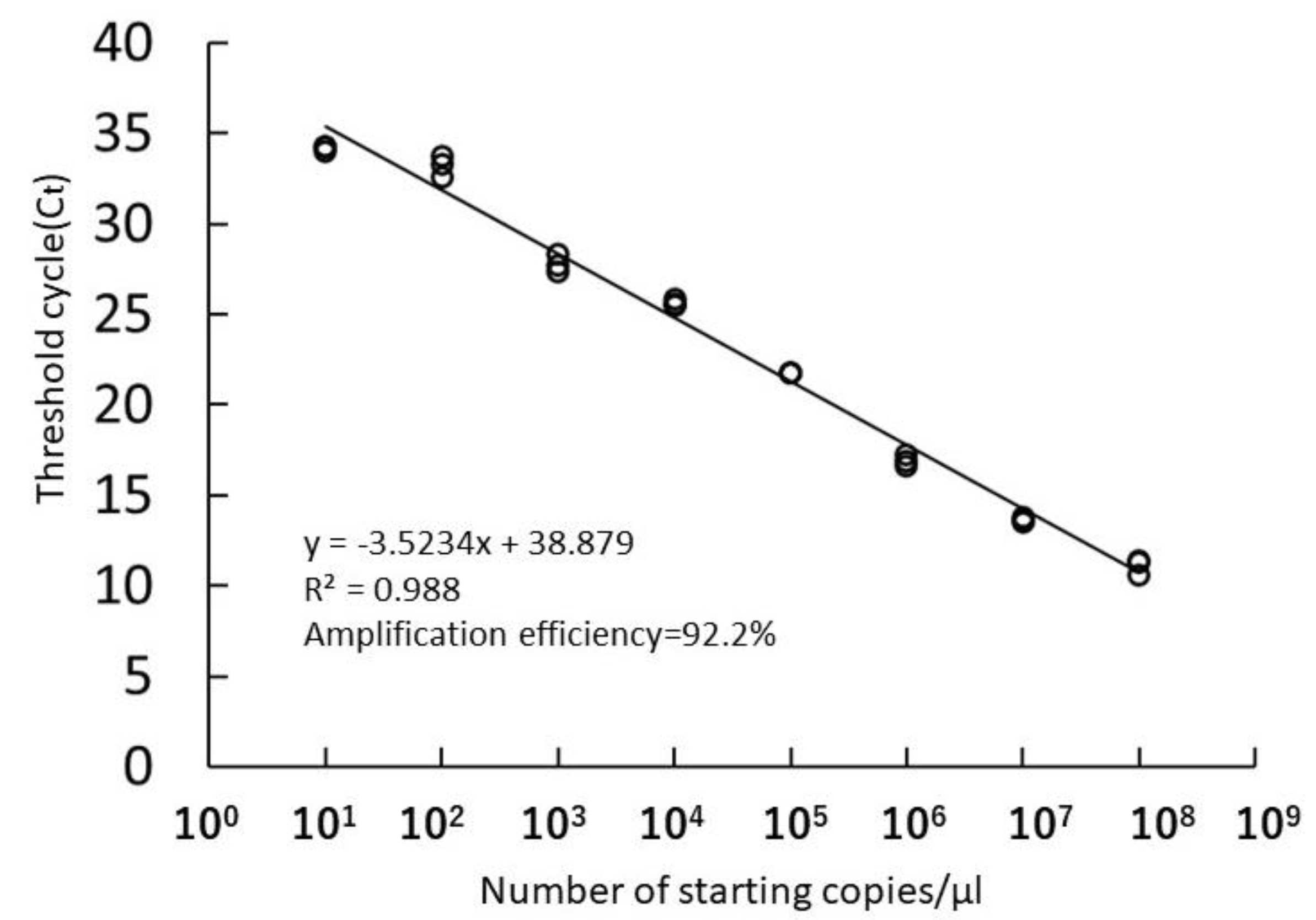
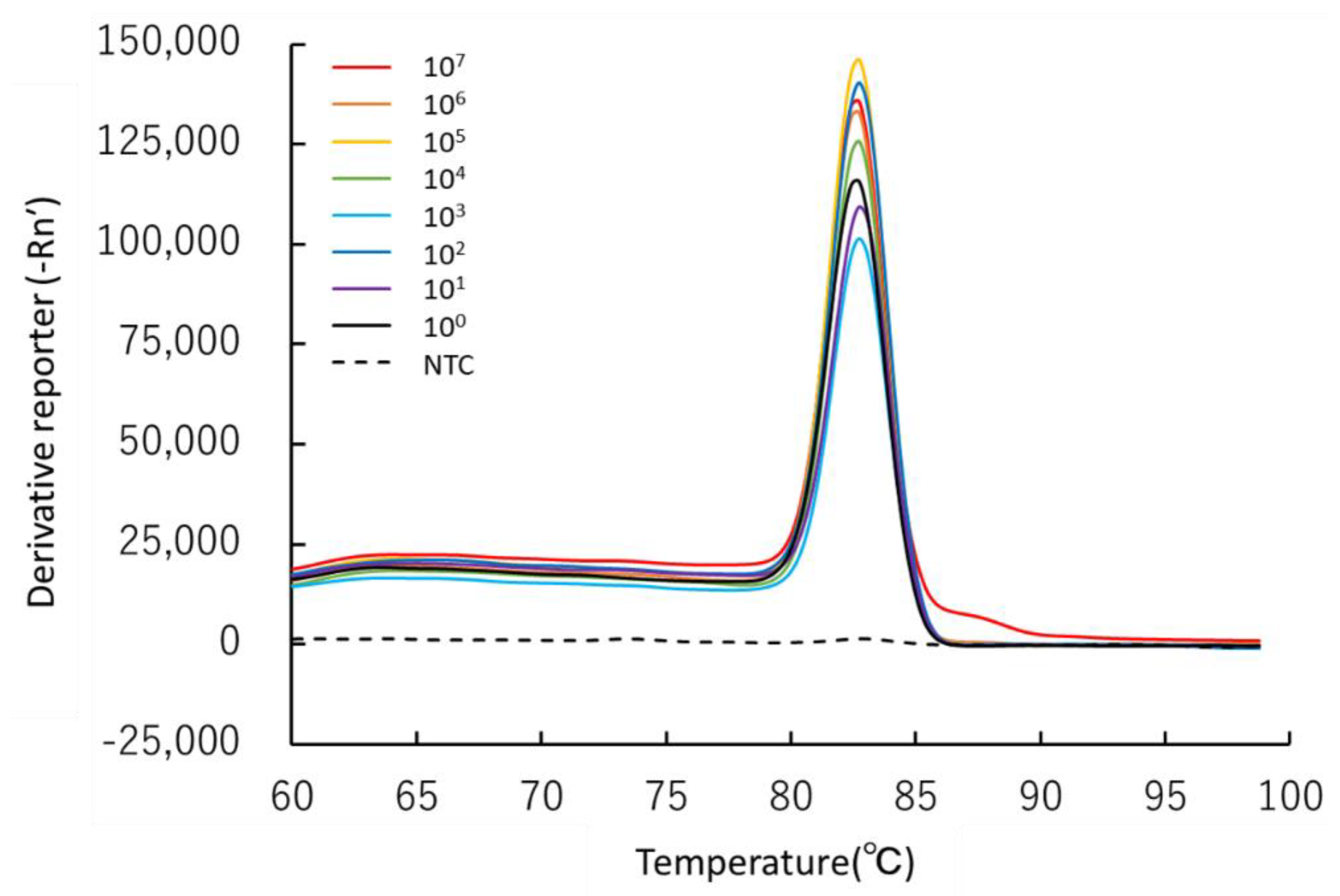
2.4. Real-Time PCR Experiments

The same samples used for amplicon sequencing were assayed using a QuantStudio 3 real-time PCR system (Applied Biosystems, Carlsbad, CA, USA) to detect and quantify S. himantioides in the decayed wood. The reaction was performed in 20 µL reaction mixtures containing 1 µL of template DNA, 10 µL KOD SYBR qPCR Mix, 0.2 µM of each forward and reverse primer, and 0.04 µL 50 × ROX Reference Dye (Toyobo). The real-time PCR conditions were 98 °C for 2 min, followed by 45 cycles at 98 °C for 10 s, 60 °C for 10 s, and 68 °C for 30 s. Melting curve analysis was performed at 95 °C for 15 s, 50 °C for 1 min, and 99 °C for 15 s. The calibration curve for absolute quantification was prepared in eight steps by diluting a plasmid (347 bp, 8.93 ng/uL) containing a fragment of the rDNA ITS region of S. himantioides derived from sawara1 in Table S1.
Real-time PCR was performed thrice for each sample, and the mean value was used as the quantification result. Next, the number of DNA copies per microliter was determined from quantification results.

3. Results

3.1. Specificity of the Newly Developed Primer Set

In the end-point PCR with the species-specific primer set (SHF/SHR), a single band (approximately 300 bp) was amplified for S. himantioides, whereas no amplified product was detected for the other fungi (Figure 2). In contrast, in the end-point PCR with universal primer set (ITS5/ITS4), single bands (approximately 600 bp) were amplified for all the fungi, and no amplified product was found in the negative control. This result suggests that the specific primer set developed in this study was successful in detecting S. himantioides.
Diverse basidiomycetes (including S. himantioides), ascomycetes, and other fungi were identified using a fungal community primer set (Table 1). Basidiomycetes was the most abundant group of which S. himantioides and Serpula spp. had almost the same percentage. A low percentage of S. lacrymans was detected in the samples collected from CP2 (DWS_7–DWS_9). In contrast, only S. himantioides and Serpula spp. were detected by amplicon sequencing using species-specific primer set. No S. lacrymans, Ascomycetes, or other fungi were detected (Table 2).

3.2. Sensitivity of the Real-Time PCR Assay for Environmental Samples

The calibration curve for absolute quantification showed linearity (R2 > 0.988) in the range of 101–108 copies, with a PCR efficiency of 92.2% (Figure 3). Melting curve analysis revealed a single peak at approximately 82.6 ± 0.1 °C, confirming the absence of non-specific products; along with absence of amplified products in the negative control (Figure 4). In addition, there were 1.6 × 101–1.6 × 10¹⁰ copies of S. himantioides per microliter of sample.
Concentration of DNA extracted from decayed wood samples ranged from 0.04 to 0.49 ng/µL, and S. himantioides was detected in all samples by real-time PCR (Table S6). The estimated range of rDNA copies of S. himantioides per microliter of sample was from 7.78 × 10³ copies (DWS_9) to 4.53 × 10⁶ copies (DWS_1). The melting curve analysis showed a single peak at 83.0 ± 0.2 °C in all samples (Figure S1).

4. Discussion

4.1. Specificity of the Designed Primer Set

The results of end-point PCR showed that the newly developed primer set could specifically detect S. himantioides among isolates of closely related fungi. The species-specific primer set for S. himantioides developed in a previous study was also validated for specificity among closely related species using end-point PCR [29]. This validation was performed on DNA extracted from fungal isolates; however, the specificity was not verified using environmental DNA containing DNA from a variety of fungi. In this study, the specificity was verified using both end-point PCR and amplicon sequencing. End-point PCR was performed with the addition of control fungi, which were not used in the previous study. Amplicon sequencing offers more accurate confirmation of the specificity of primer sets for environmental DNA from various fungal species. Bradley et al. [39] performed amplicon sequencing of microalgal mock communities using a primer set specific to the V4 region of the 18S rRNA gene. This set failed to reliably amplify 2 of the 12 mock community members. The findings of amplicon sequencing in the current study showed that the designed primer set could accurately and specifically detect S. himantioides in decayed wood samples among different fungal species. In addition, melting curve analysis demonstrated a single peak, and real-time PCR analysis using decayed wood samples confirmed the absence of non-specific products. These results indicate that S. himantioides can be monitored on-site using this primer set, even if it is not isolated from wood tissue.
Furthermore, amplicon sequencing using a species-specific primer set identified OTU as Serpula spp. Intraspecific diversification leading to varieties and cryptic species within S. himantioides is estimated to have occurred during the Miocene and Pliocene [40]. Kauserud et al. [41] reported the occurrence of at least three clades within the S. himantioides species complex. Carlsen et al. [11] analyzed a wider range of specimens and detected five clades. A new clade was discovered after adding the Japanese S. himantioides strains used in this study for phylogenetic analysis [42]. With the accumulation of more sequence data on Japanese S. himantioides strains, it is possible that the OTUs identified as S. himantioides and Serpula spp. belong to the new clade of S. himantioides. However, amplicon sequencing using a species-specific primer set could not be included in the negative control because it did not yield amplified products and could not read the amplicon. Therefore, at this time, the possibility that the detected Serpula spp. was a false positive cannot be ruled out. However, the amount of Serpula spp. detected was less (less than 12.66% of the samples), and end-point PCR and real-time PCR showed no false positives. Therefore, the detection of S. himantioides in environmental samples using real-time PCR does not appear to be a practical problem. The species-specific primer set developed in this study was designed to include all five clades identified by Carlsen et al. [11], and thus could detect not only Japanese S. himantioides but also other clades of S. himantioides.

4.2. Sensitivity of the Real-Time PCR Assay for Environmental Samples

The detection limit of the calibration curve used for real-time PCR was 160 copies/µL. This limit is comparable to that for fungal communities from soil samples (3 × 10² copies [43]) and arbuscular mycorrhizal fungal isolates (100–1000 copies) [44]. The detection limit of the method developed in this study was comparable to that observed in previous studies. Therefore, this real-time PCR assay can be used to detect trace amounts of S. himantioides in environmental samples.
S. himantioides has been detected in both conifers and decayed Quercus logs and stumps [45]. The S. himantioides strains used by Carlsen et al. [11] were obtained from hardwoods, soil, and indoors. This suggests that S. himantioides has a wide range of habitats which are yet to be documented clearly, such as distribution patterns in forests and soil, and the mode of dispersal by spores.
The real-time PCR method proposed in this study will be a powerful tool for early monitoring and detection of S. himantioides in softwood, hardwood, soil, and air and will be able to reveal its habitat. In addition, it may help forest owners decide when to cut down trees and reduce the economic losses caused by butt rot.

5. Conclusions

In this study, we successfully developed a real-time PCR assay, the species-specific primer set SHF (5′-CTCGCATCGATGAAGAAC-3′) and SHR (5′-CAAAACATTGTCTTACGACG-3′), designed in the ITS2 region to detect and quantify S. himantioides causing serious butt rot in C. pisifera. This method can be applied not only for the early detection of S. himantioides-induced butt rot but also for detecting the presence of S. himantioides in decaying wood, soil, and air. Overall, this method may help to identify the infection pathway of S. himantioides and to provide timely and effective control measures.

Supplementary Materials

The following are available online at https://www.mdpi.com/article/10.3390/f13091429/s1: Table S1: List of collected Serpula himantioides sequences; Table S2: Primer sequences, names, GC%, and melting temperatures; Table S3: Serpula himantioides strains and other fungal species used in end-point PCR; Table S4: Decayed tree size of Chamaecyparis pisifera, Table S5: Detail of decayed wood samples, Table S6: Results of absolute quantification of Serpula himantioides in the decayed wood samples; Figure S1: Melting curves of DNA extracted from decayed wood samples as measured by real-time PCR.

Author Contributions

Conceptualization, R.H., T.H. and T.Y.; methodology, R.H. and T.H.; software, R.H. and T.H.; validation, R.H., T.H. and T.Y.; formal analysis, R.H. and T.H.; investigation, R.H. and T.Y.; resources, R.H. and T.Y.; data curation, R.H.; writing—original draft preparation, R.H. and T.H.; writing—review and editing, R.H., T.H. and T.Y.; visualization, R.H.; supervision, T.Y.; project administration, T.Y.; funding acquisition, T.Y. All authors have read and agreed to the published version of the manuscript.

Funding

This research was funded by a Grant-in-Aid for Scientific Research from the Japan Society for the Promotion of Science (grant numbers: 18H02232 and 18K19870) and a joint project between the University of Tokyo Chichibu Forest and the Suntory Natural Water Sanctuary.

Institutional Review Board Statement

Not applicable.

Informed Consent Statement

Not applicable.

Data Availability Statement

Not applicable.

Acknowledgments

We are grateful to the technical staff of the University of Tokyo Chichibu Forest for helping with fieldwork. We also thank Tsutomu Hattori for providing the strains and Yuko Ota for her advice on the experiments.

Conflicts of Interest

The authors declare no conflict of interest.

References

  1. Nicolotti, G.; Gonthier, P.; Guglielmo, F. Advances in Detection and Identification of Wood Rotting Fungi in Timber and Standing Trees. In Molecular Identification of Fungi; Gherbawy, Y., Voight, K., Eds.; Springer: Berlin/Heidelberg, Germany, 2010; pp. 251–276. [Google Scholar] [CrossRef]
  2. Woodward, S.; Stenlid, J.; Karjalainen, R.; Hüttermann, A. Heterobasidion Annosum, Biology, Ecology, Impact and Control; CAB International: Oxon, UK, 1998; pp. xi–xii. [Google Scholar]
  3. Regue, A.; Bassie, L.; de-Miguel, S.; Colinas, C. Environmental and stand conditions related to Fistulina hepatica heart rot attack on Castanea sativa. For. Pathol. 2019, 49, 7. [Google Scholar] [CrossRef]
  4. Tomikawa, Y.; Iwase, Y.; Arita, K.; Yamada, H. Nondestructive inspection of a wooden pole using ultrasonic computed tomography. IEEE Trans. Ultrason. Ferroelectr. Freq. Control 1986, 33, 354–358. [Google Scholar] [CrossRef] [PubMed]
  5. Habermehl, A.; Ridder, H.W.; Seidl, P. Computerized tomographic systems as tools for diagnosing urban tree health. Int. Symp. Urban Tree Health 1999, 496, 261–268. [Google Scholar] [CrossRef]
  6. Nicolotti, G.; Socco, L.V.; Martinis, R.; Godio, A.; Sambuelli, L. Application and comparison of three tomographic techniques for detection of decay in trees. J. Arbor. 2003, 29, 66–78. [Google Scholar] [CrossRef]
  7. Muller, U.; Bammer, R.; Halmschlager, E.; Stollberger, R.; Wimmer, R. Detection of fungal wood decay using magnetic resonance imaging. Holz. Als Roh-Und Werkst. 2001, 59, 190–194. [Google Scholar] [CrossRef]
  8. Sambuelli, L.; Socco, L.V.; Godio, A.; Nicolotti, G.; Martinis, R. Ultrasonic, electric and radar measurements for living tree assessment. Boll. Di Geofis. Teor. Ed Appl. 2003, 44, 253–279. [Google Scholar]
  9. Guglielmo, F.; Bergemann, S.E.; Gonthier, P.; Nicolotti, G.; Garbelotto, M. A multiplex PCR-based method for the detection and early identification of wood rotting fungi in standing trees. J. Appl. Microbiol. 2007, 103, 1490–1507. [Google Scholar] [CrossRef]
  10. Schena, L.; Nigro, F.; Ippolito, A.; Gallitelli, D. Real-time quantitative PCR: A new technology to detect and study phytopathogenic and antagonistic fungi. Eur. J. Plant. Pathol. 2004, 110, 893–908. [Google Scholar] [CrossRef]
  11. Carlsen, T.; Engh, I.B.; Decock, C.; Rajchenberg, M.; Kauserud, H. Multiple cryptic species with divergent substrate affinities in the Serpula himantioides species complex. Fungal Biol. 2011, 115, 54–61. [Google Scholar] [CrossRef]
  12. Seehann, G. Butt rot in conifers caused by Serpula himantioides. Karst. For. Pathol. 1986, 16, 207–217. [Google Scholar]
  13. Koch, J.; Thomsen, I.M. Serpula himantioides, Heterobasidion annosum and Calocera viscosa as butt rot fungi in a Danish Douglas-fir stand. For. Pathol. 2003, 33, 1–6. [Google Scholar] [CrossRef]
  14. Whitney, R.D. Root-rotting fungi in white spruce, black spruce, and balsam fir in northern Ontario. Can. J. For. Res. 1995, 25, 1209–1230. [Google Scholar] [CrossRef]
  15. Rizzo, D.M.; Harrington, T.C. Root and butt rot fungi on balsam fir and red spruce in the White Mountains, New Hampshire. Plant Dis. 1988, 72, 329–331. [Google Scholar] [CrossRef]
  16. Haraguchi, R.; Hirao, T.; Yamada, T. Butt-rot damage situation of Chamaecyparis pisifera in the University of Tokyo Chichibu Forest. Kanto J. For. Res. 2017, 68, 181–184. (In Japanese) [Google Scholar]
  17. Abe, Y. Some properties of Chamaecyparis pisifera butt rot fungus Serpula himantioides. Trans. JPN For. Soc. 2002, 113, 87. (In Japanese) [Google Scholar]
  18. Schmidt, O. Indoor wood-decay basidiomycetes: Damage, causal fungi, physiology, identification and characterization, prevention and control. Mycol. Prog. 2007, 6, 261–279. [Google Scholar] [CrossRef]
  19. Boehm, J.; Hahn, A.; Schubert, R.; Bahnweg, G.; Adler, N.; Nechwatal, J.; Oehlmann, R.; Osswald, W. Real-time quantitative PCR: DNA determination in isolated spores of the mycorrhizal fungus Glomus mosseae and monitoring of Phytophthora infestans and Phytophthora citricola in their respective host plants. J. Phytopathol. 1999, 147, 409–416. [Google Scholar] [CrossRef]
  20. Luchi, N.; Capretti, P.; Pinzani, P.; Orlando, C.; Pazzagli, M. Real-time PCR detection of Biscogniauxia mediterranea in symptomless oak tissue. Lett. Appl. Microbiol. 2005, 41, 61–68. [Google Scholar] [CrossRef]
  21. Luchi, N.; Capretti, P.; Vettraino, A.M.; Vannini, A.; Pinzani, P.; Pazzagli, M. Early detection of Biscogniauxia nummularia in symptomless European beech (Fagus sylvatica L.) by TaqMan (TM) quantitative real-time PCR. Lett. Appl. Microbiol. 2006, 43, 33–38. [Google Scholar] [CrossRef]
  22. Filion, M.; St-Arnaud, M.; Jabaji-Hare, S.H. Direct quantification of fungal DNA from soil substrate using real-time PCR. J. Microbiol. Methods 2003, 53, 67–76. [Google Scholar] [CrossRef]
  23. Berry, S.D.; Fargette, M.; Spaull, V.W.; Morand, S.; Cadet, P. Detection and quantification of root-knot nematode (Meloidogyne javanica), lesion nematode (Pratylenchus zeae) and dagger nematode (Xiphinema elongatum) parasites of sugarcane using real-time PCR. Mol. Cell. Probes 2008, 22, 168–176. [Google Scholar] [CrossRef] [PubMed]
  24. Schweigkofler, W.; O’Donnell, K.; Garbelotto, M. Detection and quantification of airborne conidia of Fusarium circinatum, the causal agent of pine pitch canker, from two California sites by using a real-time PCR approach combined with a simple spore trapping method. Appl. Environ. Microbiol. 2004, 70, 3512–3520. [Google Scholar] [CrossRef] [PubMed]
  25. Luchi, N.; Ghelardini, L.; Belbahri, L.; Quartier, M.; Santini, A. Rapid Detection of Ceratocystis platani Inoculum by Quantitative Real-Time PCR Assay. Appl. Environ. Microbiol. 2013, 79, 5394–5404. [Google Scholar] [CrossRef] [PubMed] [Green Version]
  26. Gonthier, P.; Guglielmo, F.; Sillo, F.; Giordano, L.; Garbelotto, M. A molecular diagnostic assay for the detection and identification of wood decay fungi of conifers. For. Pathol. 2015, 45, 89–101. [Google Scholar] [CrossRef]
  27. Horisawa, S.; Sakuma, Y.; Doi, S. Qualitative and quantitative PCR methods using species-specific primer for detection and identification of wood rot fungi. J. Wood Sci. 2009, 55, 133–138. [Google Scholar] [CrossRef]
  28. Eikenes, M.; Hietala, A.; Alfredsen, G.; Fossdal, C.G.; Solheim, H. Comparison of chitin, ergosterol and Real-Time PCR based assays for monitoring colonization of Trametes versicolor in birch wood. Holzforschung 2005, 59, 568–573. [Google Scholar] [CrossRef]
  29. Schmidt, O.; Moreth, U. Species-specific PCR primers in the rDNA-ITS region as a diagnostic tool for Serpula lacrymans. Mycol. Res. 2000, 104, 69–72. [Google Scholar] [CrossRef]
  30. OligoEvaluator™. Available online: http://www.oligoevaluator.com/ (accessed on 7 June 2021).
  31. National Center for Biotechnology Information Home Page. Available online: http://www.ncbi.nlm.nih.gov/BLAST/ (accessed on 7 June 2021).
  32. White, T.J.; Bruns, T.; Lee, S.; Taylor, J. Amplification and direct sequencing of fungal ribosomal RNA genes for phylogenetics. In Pcr Protocols: A Guide to Methods and Applications; Innis, M.A., Gelfand, D.H., Sninsky, J.J., White, T.J., Eds.; Academic Press Inc.: San Diego, CA, USA, 1990; pp. 315–322. [Google Scholar]
  33. Ihrmark, K.; Bodeker, I.T.M.; Cruz-Martinez, K.; Friberg, H.; Kubartova, A.; Schenck, J.; Strid, Y.; Stenlid, J.; Brandstrom-Durling, M.; Clemmensen, K.E.; et al. New primers to amplify the fungal ITS2 region—evaluation by 454-sequencing of artificial and natural communities. FEMS Microbiol. Ecol. 2012, 82, 666–677. [Google Scholar] [CrossRef]
  34. Tedersoo, L.; Bahram, M.; Polme, S.; Koljalg, U.; Yorou, N.S.; Wijesundera, R.; Villarreal Ruiz, L.; Vasco-Palacios, A.M.; Pham Quang, T.; Suija, A.; et al. Global diversity and geography of soil fungi. Science 2014, 346, 1256688. [Google Scholar] [CrossRef]
  35. Gweon, H.S.; Oliver, A.; Taylor, J.; Booth, T.; Gibbs, M.; Read, D.S.; Griffiths, R.I.; Schonrogge, K. PIPITS: An automated pipeline for analyses of fungal internal transcribed spacer sequences from the Illumina sequencing platform. Methods Ecol. Evol. 2015, 6, 973–980. [Google Scholar] [CrossRef]
  36. Bengtsson-Palme, J.; Ryberg, M.; Hartmann, M.; Branco, S.; Wang, Z.; Godhe, A.; De Wit, P.; Sanchez-Garcia, M.; Ebersberger, I.; de Sousa, F.; et al. Improved software detection and extraction of ITS1 and ITS2 from ribosomal ITS sequences of fungi and other eukaryotes for analysis of environmental sequencing data. Methods Ecol. Evol. 2013, 4, 914–919. [Google Scholar] [CrossRef]
  37. Rognes, T.; Flouri, T.; Nichols, B.; Quince, C.; Mahe, F. VSEARCH: A versatile open source tool for metagenomics. PeerJ 2016, 4, e2584. [Google Scholar] [CrossRef]
  38. Abarenkov, K.; Nilsson, R.H.; Larsson, K.H.; Alexander, I.J.; Eberhardt, U.; Erland, S.; Hoiland, K.; Kjoller, R.; Larsson, E.; Pennanen, T.; et al. The UNITE database for molecular identification of fungi—Recent updates and future perspectives. New Phytol. 2010, 186, 281–285. [Google Scholar] [CrossRef]
  39. Bradley, I.M.; Pinto, A.J.; Guest, J.S. Design and Evaluation of Illumina MiSeq-Compatible, 18S rRNA Gene-Specific Primers for Improved Characterization of Mixed Phototrophic Communities. Appl. Environ. Microbiol. 2016, 82, 5878–5891. [Google Scholar] [CrossRef]
  40. Skrede, I.; Engh, I.B.; Binder, M.; Carlsen, T.; Kauserud, H.; Bendiksby, M. Evolutionary history of Serpulaceae (Basidiomycota): Molecular phylogeny, historical biogeography and evidence for a single transition of nutritional mode. BMC Evol. Biol. 2011, 11, 230. [Google Scholar] [CrossRef] [PubMed]
  41. Kauserud, H.; Hogberg, N.; Knudsen, H.; Elborne, S.A.; Schumacher, T. Molecular phylogenetics suggest a North American link between the anthropogenic dry rot fungus Serpula lacrymans and its wild relative S-himantioides. Mol. Ecol. 2004, 13, 3137–3146. [Google Scholar] [CrossRef]
  42. Ota, H.; Matsushita, N.; Haraguchi, R.; Yamada, T.; Fukuda, K. A new cryptic species of the dry rot fungus Serpula himantioides found in Japan. JPN For. Soc. Congr. 2019, 130, 172. (In Japanese) [Google Scholar] [CrossRef]
  43. Prevost-Boure, N.C.; Christen, R.; Dequiedt, S.; Mougel, C.; Lelievre, M.; Jolivet, C.; Shahbazkia, H.R.; Guillou, L.; Arrouays, D.; Ranjard, L. Validation and Application of a PCR Primer Set to Quantify Fungal Communities in the Soil Environment by Real-Time Quantitative PCR. PLoS ONE 2011, 6, e24166. [Google Scholar] [CrossRef]
  44. Thonar, C.; Erb, A.; Jansa, J. Real-time PCR to quantify composition of arbuscular mycorrhizal fungal communities—marker design, verification, calibration and field validation. Mol. Ecol. Resour. 2012, 12, 219–232. [Google Scholar] [CrossRef]
  45. van der Wal, A.; Gunnewiek, P.K.; de Hollander, M.; de Boer, W. Fungal diversity and potential tree pathogens in decaying logs and stumps. For. Ecol. Manag. 2017, 406, 266–273. [Google Scholar] [CrossRef]
Figure 1. Serious butt rot caused by Serpula himantioides in living trees of Chamaecyparis pisifera in Japan: (a) Stump of a butt rot tree; (b) Transverse section of a decayed tree at 2.2 m height above ground. Brown rot was observed in most of the heartwood; (c) Longitudinal section of decayed wood from 2.2 m to 5.2 m above ground. The brownish area reached a height of 5.0 m above ground level; (d) Felling of trees caused by butt rot; (e) Fruiting body of Serpula himantioides.
Forests 13 01429 g001
Figure 2. Comparison of end-point PCR results between species-specific and universal primer sets by agarose gel electrophoresis. Lanes are designated with abbreviations for the species listed in Table S3. Size markers are in the first and last lanes: (a) Species-specific primer set SHF/SHR detected Serpula himantioides (SH1, SH2, SH3, and SH4), and there was no reaction with the control fungi. (b) Universal primer set ITS5/ITS4 detected all fungi. All primer sets yielded negative results for negative control (NTC).
Forests 13 01429 g002aForests 13 01429 g002b
Figure 3. Calibration curve for the absolute quantification of Serpula himantioides based on real-time PCR with SYBR-Green as the fluorescent dye. The threshold number of PCR cycles (Ct-value) was plotted against the log of the initial quantity of template DNA (n = 3). Amplification efficiency was calculated using the Applied Biosystems™ Analysis Software (version 1.0.1).
Forests 13 01429 g003
Figure 4. Melting curves of recombinant plasmid DNA containing genomic sequences from Serpula himantioides amplicons as determined by real-time PCR. The derivative reporter, displayed on the y-axis, was calculated as the negative first derivative of the normalized fluorescence (Rn) generated by the reporter during PCR amplification. For visual clarity, the mean of the derivative reporter is shown (n = 3).
Forests 13 01429 g004
Table 1. Percentage of reads of fungi detected by amplicon sequencing using the fungal community primer set.
Sample NameBasidiomycetesAscomycetes
(%)
Other Fungi
(%)
S. himantioides
(%)
S. lacrymans
(%)
Serpula spp.
(%)
Besides
Serpula (%)
DWS_145.830.0052.570.061.490.05
DWS_247.780.0051.860.050.260.05
DWS_348.580.0045.343.520.512.06
DWS_447.160.0050.251.290.221.08
DWS_549.080.0048.880.721.290.04
DWS_627.540.0029.2318.3224.400.51
DWS_716.520.1716.9419.7937.988.60
DWS_813.360.3113.6131.6133.157.96
DWS_97.370.247.5242.2531.7310.90
Table 2. Percentage of reads of fungi detected by amplicon sequencing using the species-specific primer set for Serpula himantioides.
Sample NameBasidiomycetesAscomycetes
(%)
Other Fungi
(%)
S. himantioides (%)S. lacrymans
(%)
Serpula spp.
(%)
Besides
Serpula (%)
DWS_191.020.008.980.000.000.00
DWS_290.860.009.140.000.000.00
DWS_392.050.007.950.000.000.00
DWS_490.520.009.480.000.000.00
DWS_591.830.008.170.000.000.00
DWS_692.220.007.780.000.000.00
DWS_791.400.008.600.000.000.00
DWS_892.030.007.970.000.000.00
DWS_987.340.0012.660.000.000.00
Publisher’s Note: MDPI stays neutral with regard to jurisdictional claims in published maps and institutional affiliations.

