
3D Visualization of Bamboo Node’s Vascular Bundle

 , ,
, ,
Abstract
:1. Introduction
2. Materials and Methods
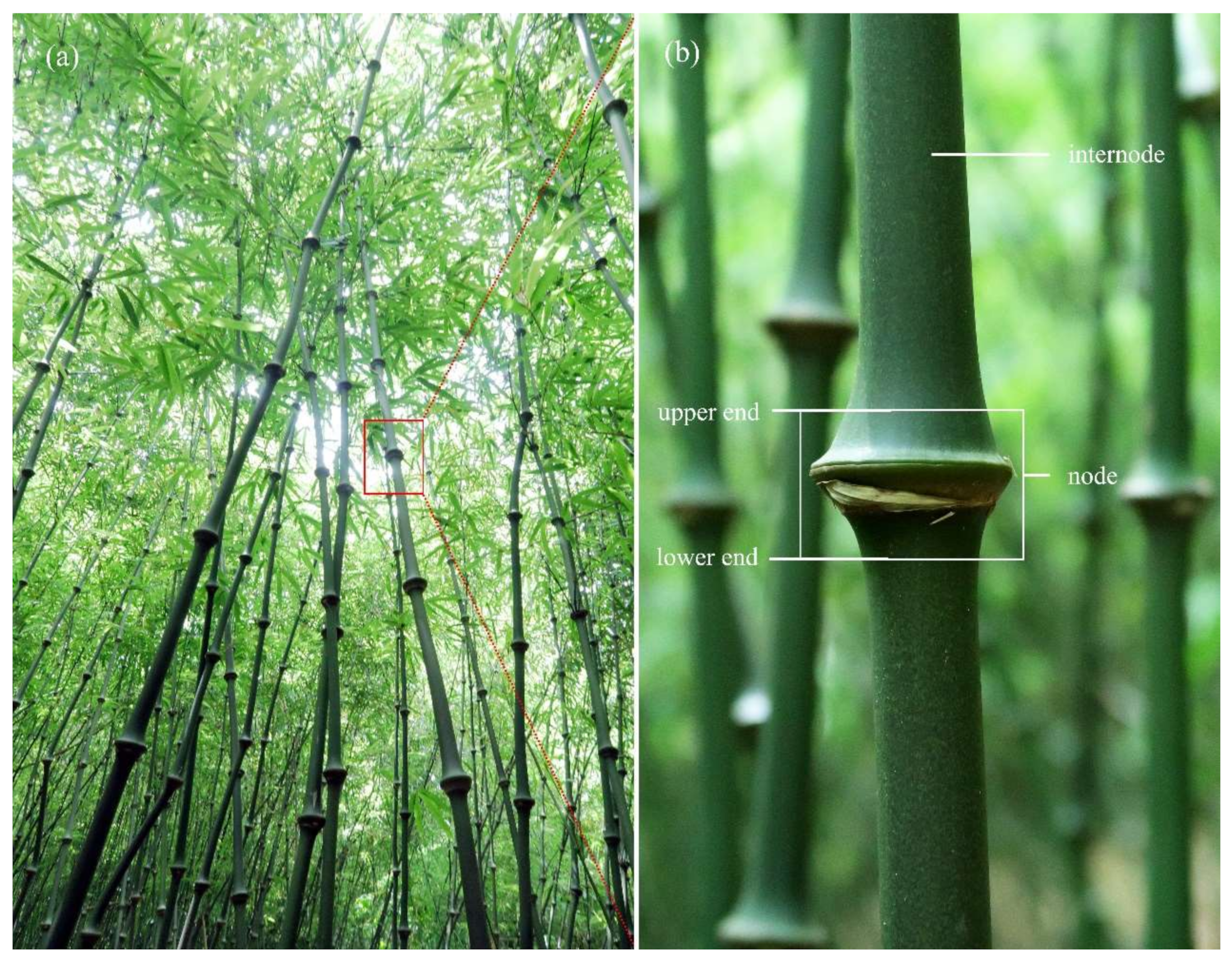
2.1. Materials
2.2. CT Scanning
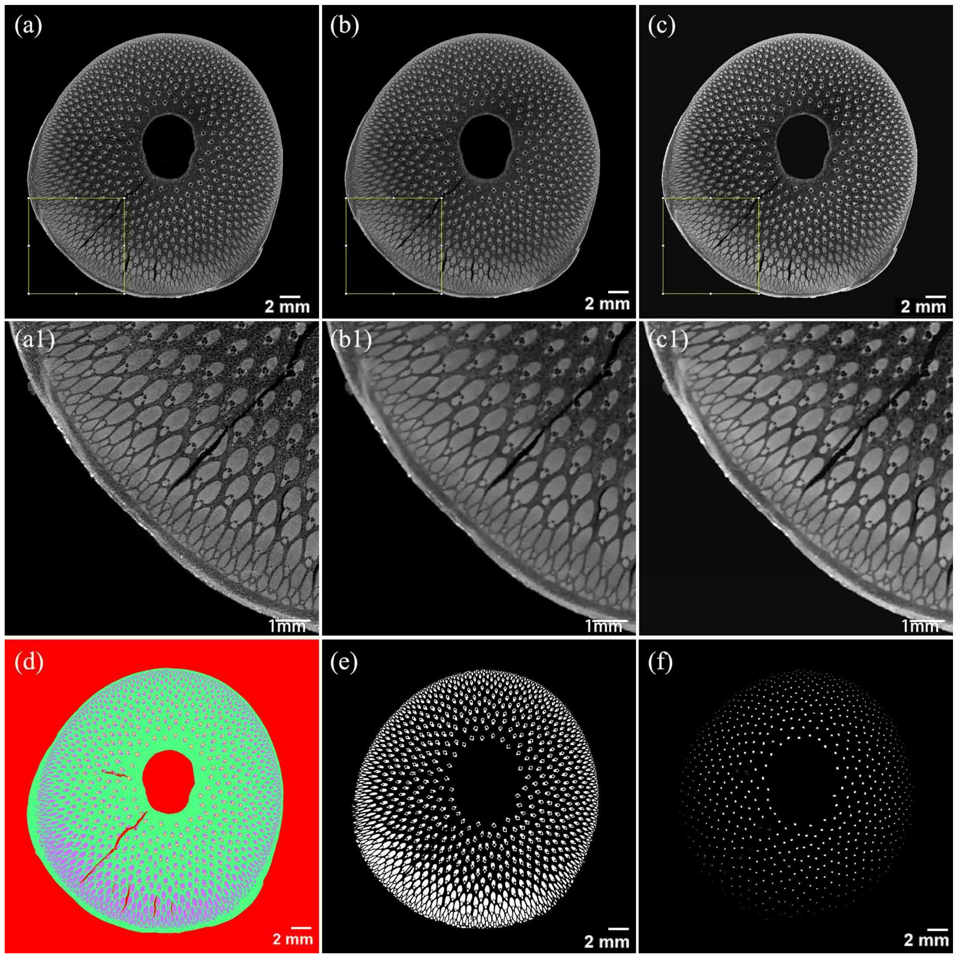
2.3. Image Processing and 3D Reconstruction
2.4. Statistical Analysis
3. Results and Discussion
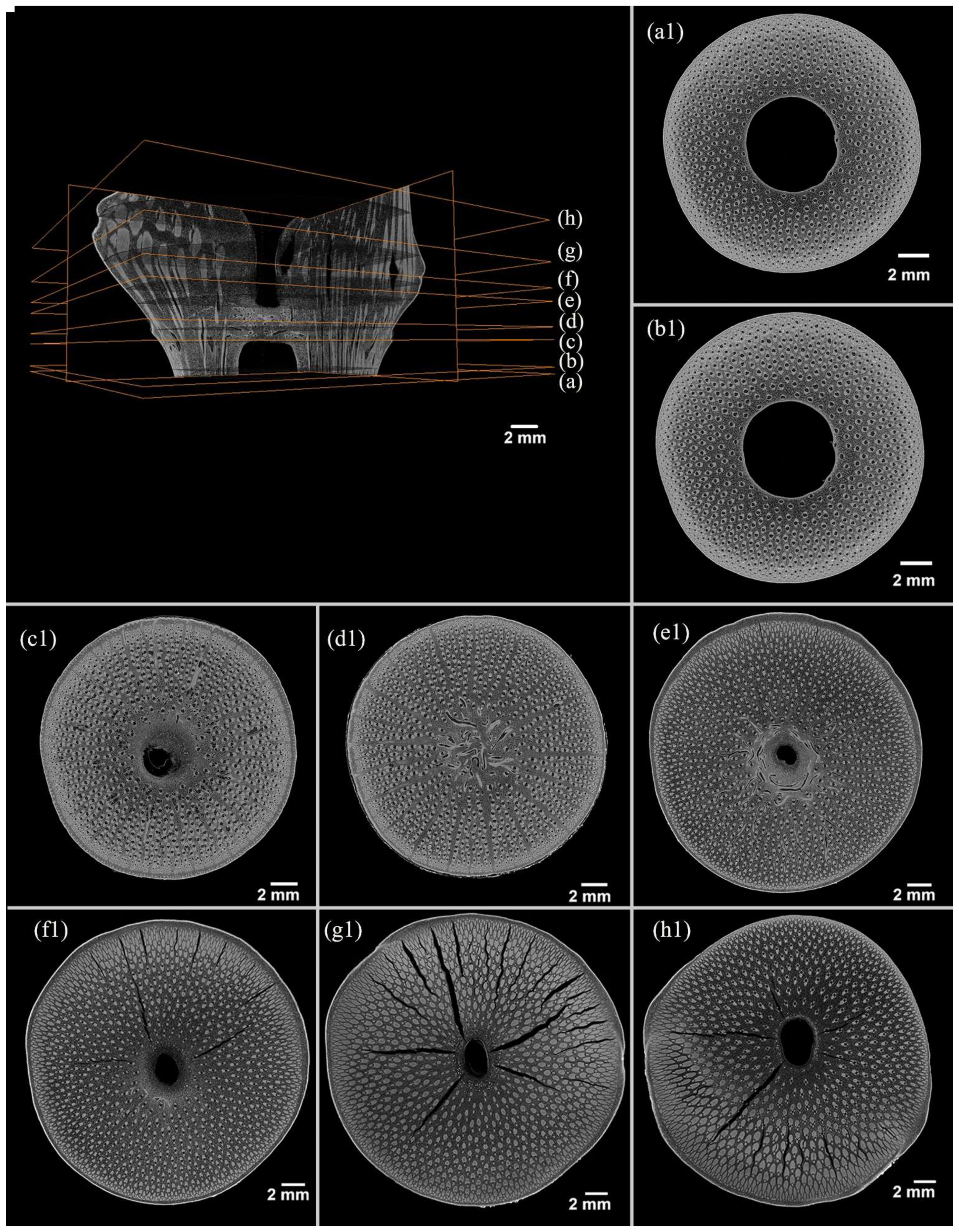
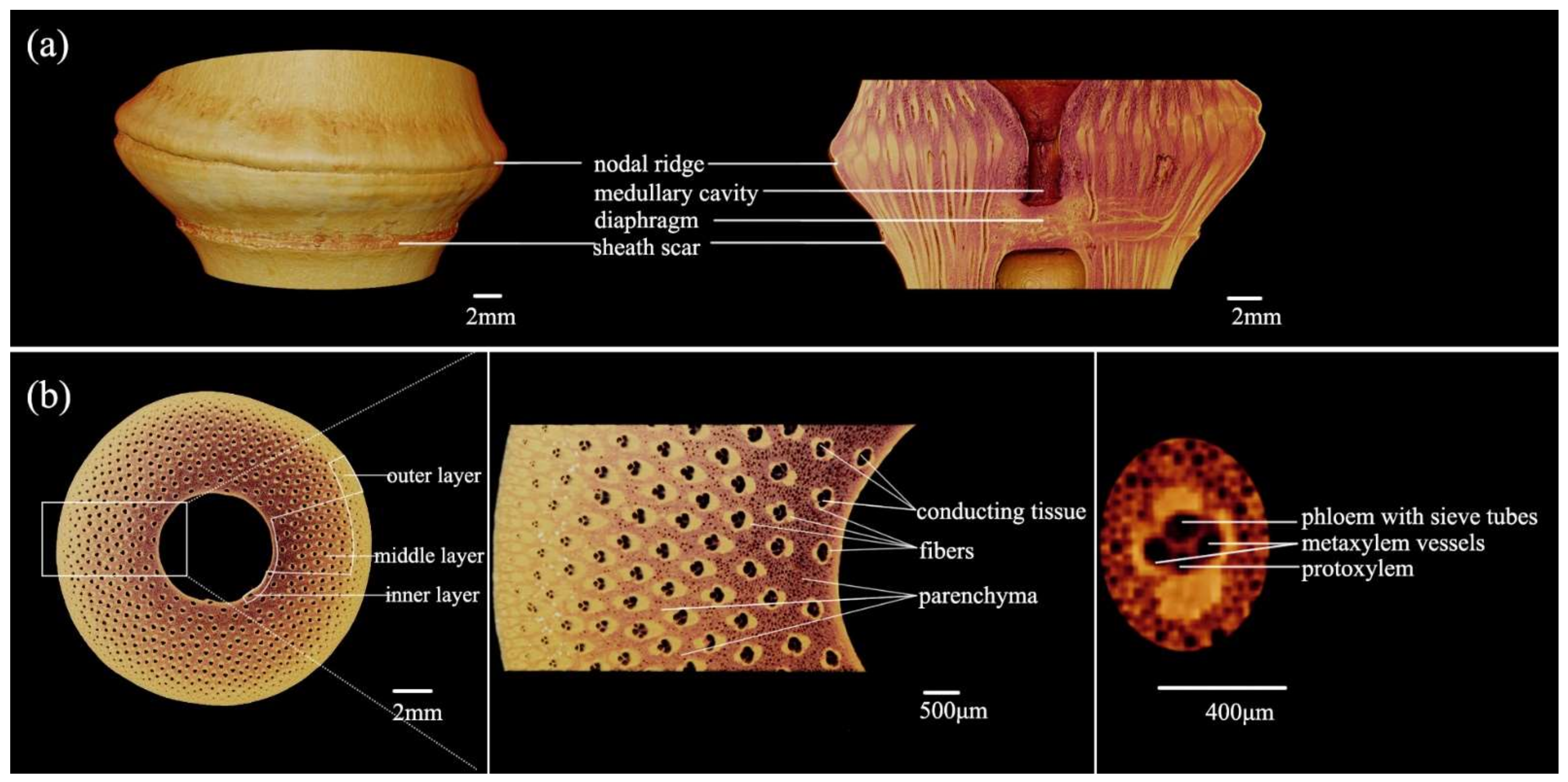
3.1. Morphology of the Bamboo Node
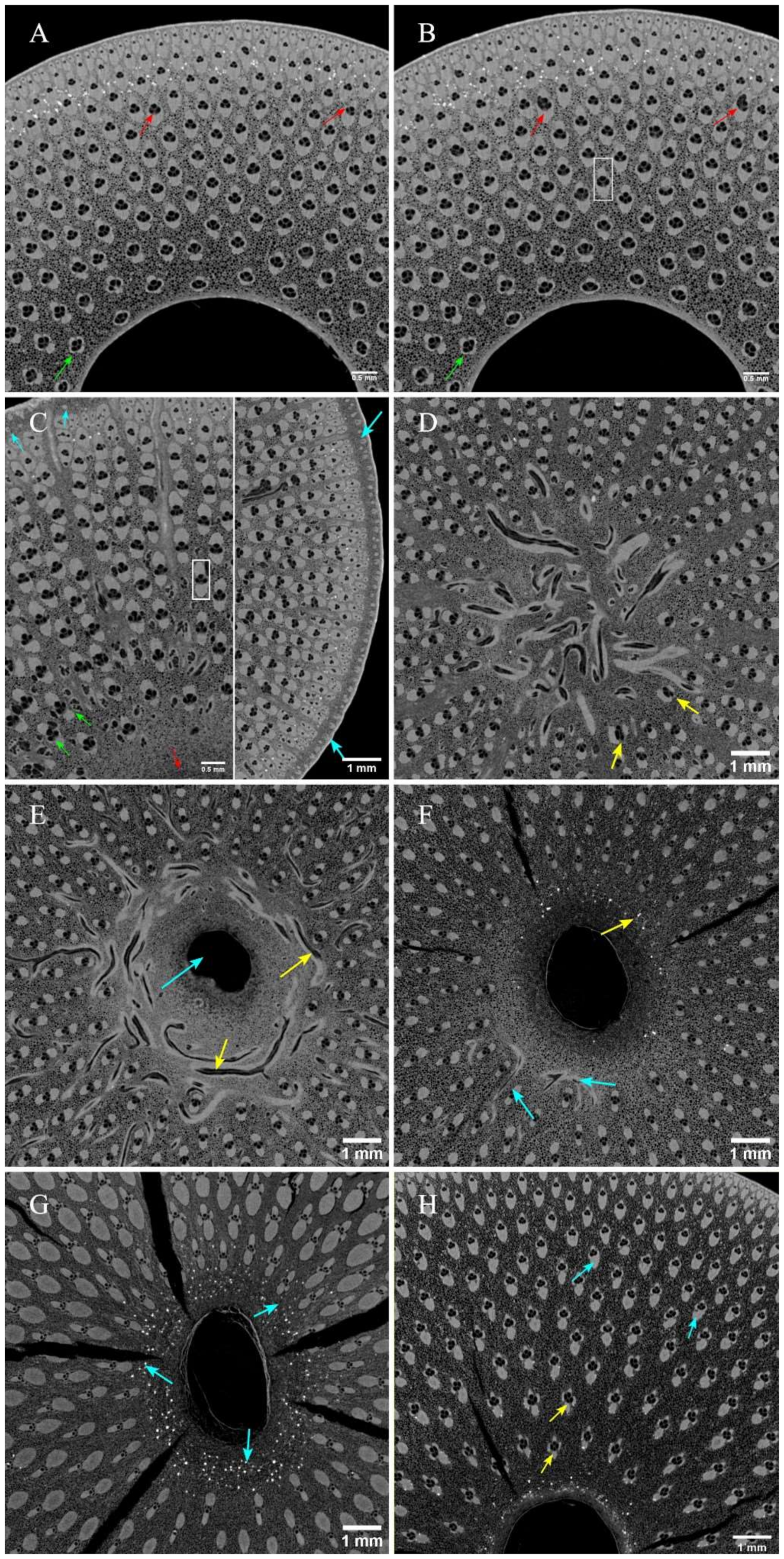
3.2. Anatomical Characteristics of Vascular Bundles in Bamboo Nodes
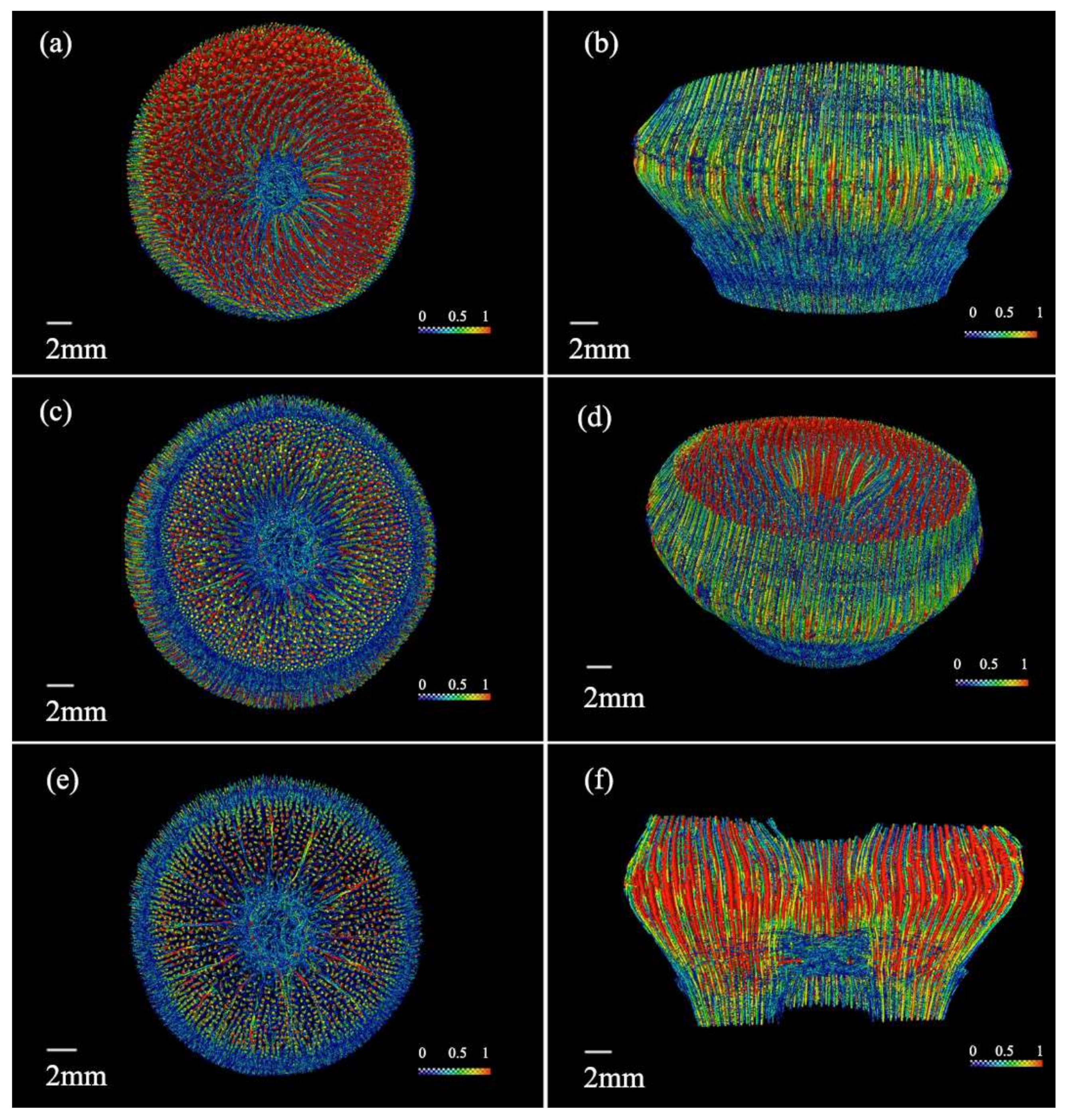
3.2.1. Morphological Changes in Vascular Bundles
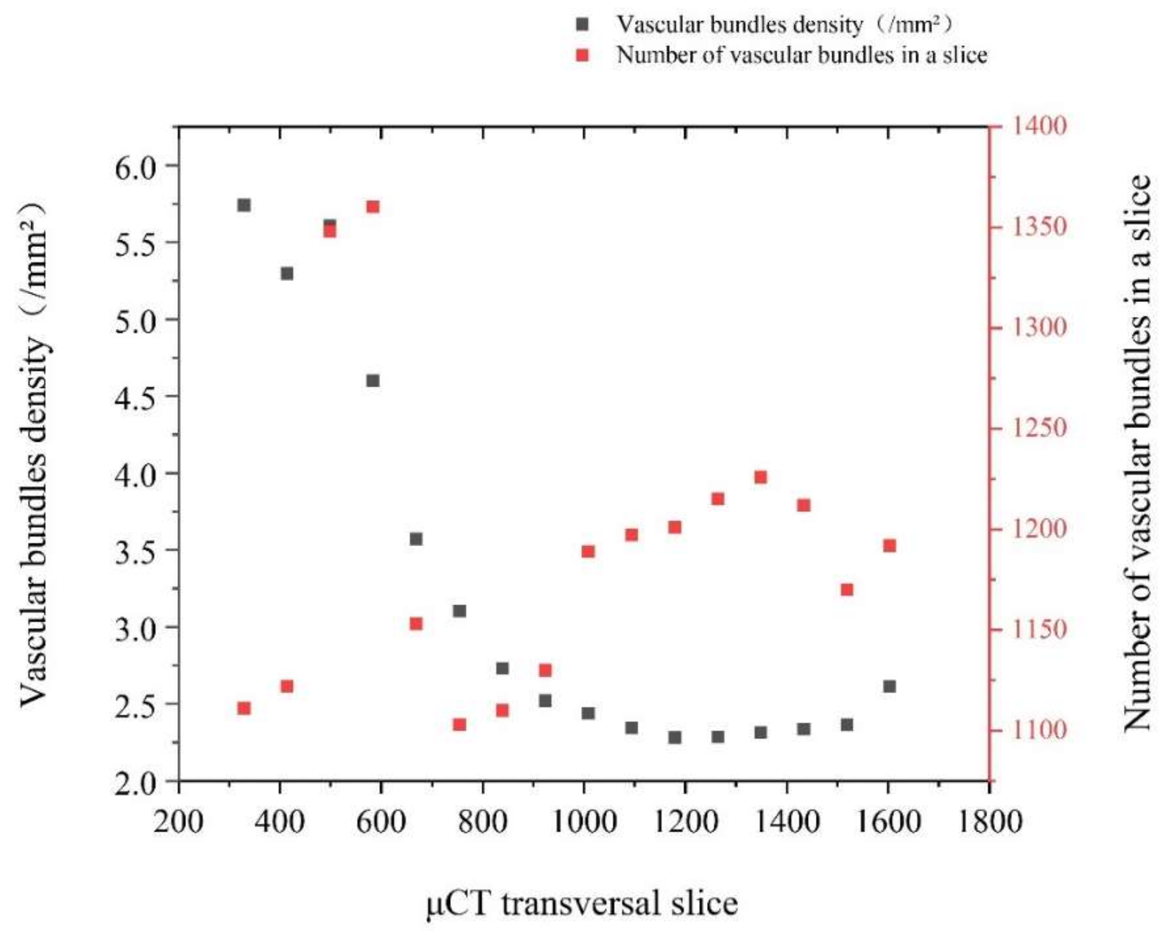
3.2.2. Size and Quantity of Axial Vascular Bundles
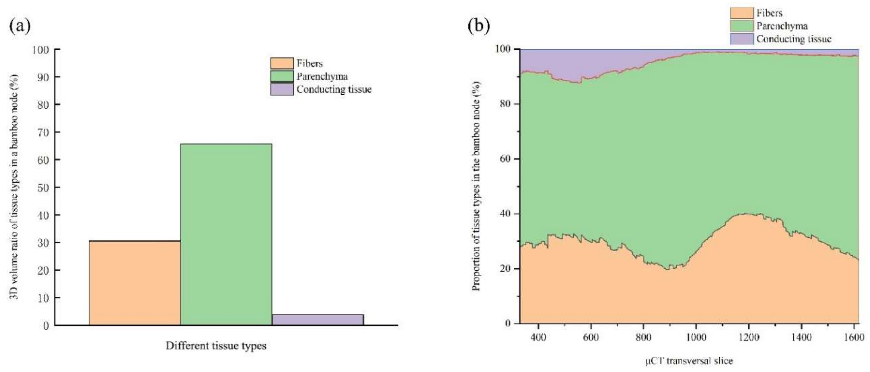
3.2.3. Tissue Proportion
3.3. Three-Dimensional Reconstruction of the Bamboo Node
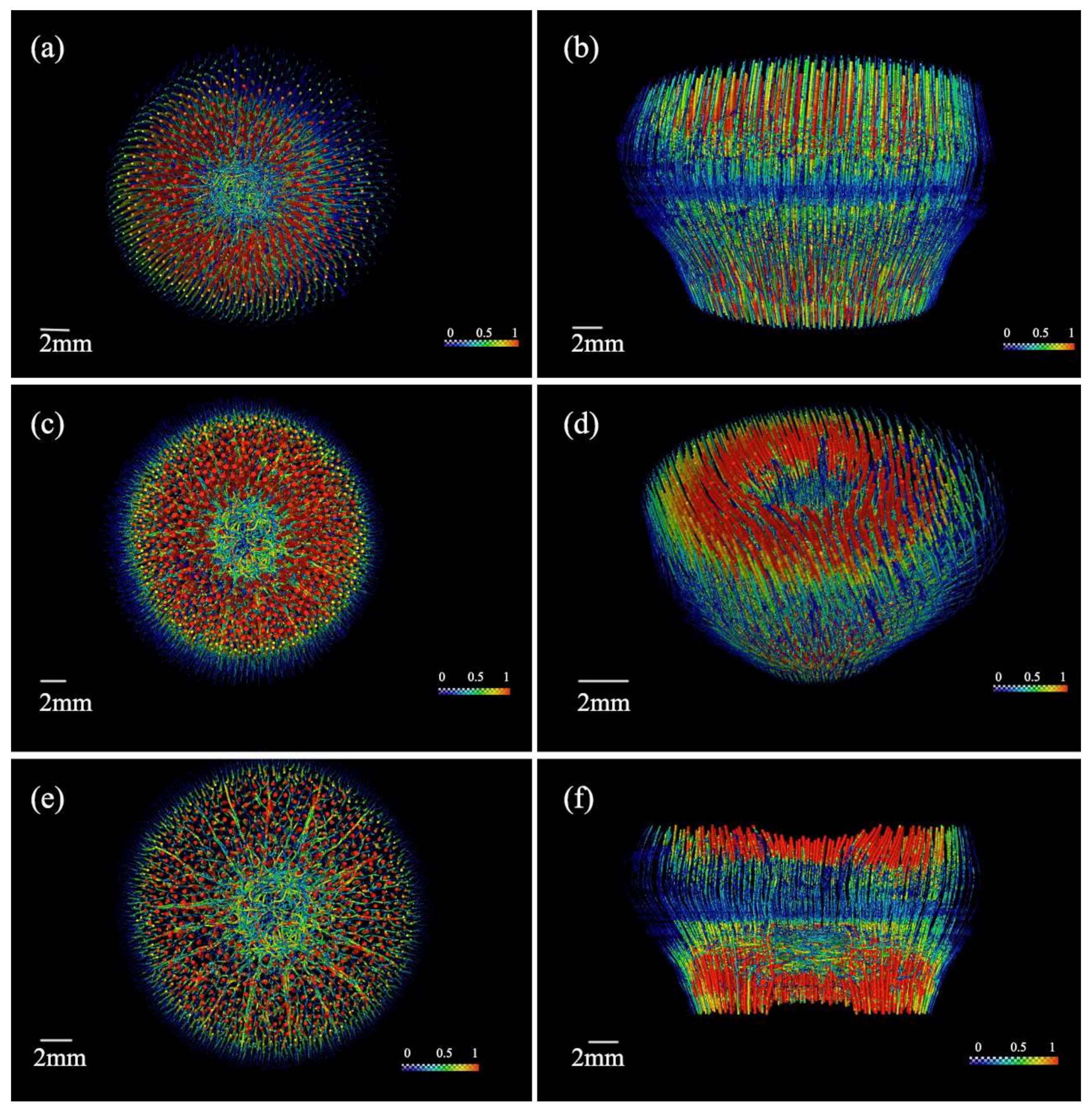
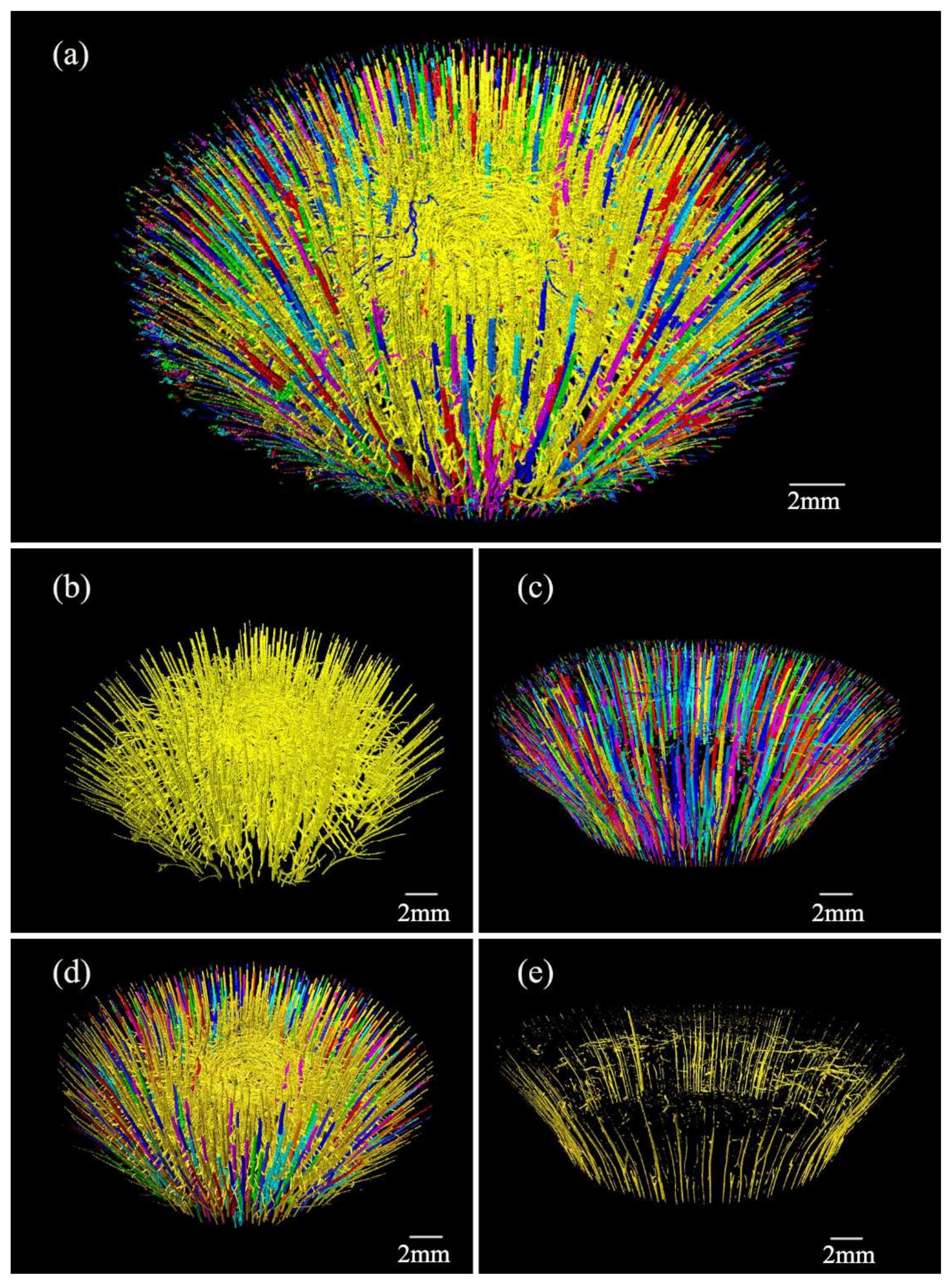
3.3.1. Three-Dimensional Morphometry of the Conducting Tissue
3.3.2. Three-Dimensional Morphometry of the Fibers
4. Discussion
5. Conclusions
- (1)
- X-ray microtomography was an ideal tool for visualizing and quantitatively analyzing the internal structure of the bamboo node. Furthermore, a large quantity of data of the quantitative indicators such as volume, area, tissue ratio, and connectivity of the bamboo node could be obtained with high speed by segmentation of μCT images combined with 3D visualization software such as AVIZO and ImageJ.
- (2)
- The anatomical structure of Q. tumidinoda with enlarged node was acquired. It was the first time that the conducting tissue and the fibers from the completed node’s vascular bundle were separated to observe its morphology, and the reconstruction of the skeleton made the morphology more intuitive. The 3D distribution patterns of the conducting tissue of the bamboo and those of the fibers were similar, but their thickness changed in the opposite pattern. The curved and branched conducting tissues in the bamboo node formed an intricate and highly connected 3D network structure, which was efficient at the transportation and circulation of nutrients inside the bamboo.
Author Contributions
Funding
Institutional Review Board Statement
Informed Consent Statement
Data Availability Statement
Acknowledgments
Conflicts of Interest
References
- Liu, S.; Tong, Z.; Tang, Z.; Yang, L.; Zhang, Z. Bionic design modification of non-convex multi-corner thin-walled columns for improving energy absorption through adding bulkheads. Thin Wall Struct. 2015, 88, 70–81. [Google Scholar] [CrossRef]
- Zou, M.; Xu, S.; Wei, C.; Wang, H.; Liu, Z. A bionic method for the crashworthiness design of thin-walled structures inspired by bamboo. Thin Wall Struct. 2016, 101, 222–230. [Google Scholar] [CrossRef]
- Wang, F.; Shao, Z.; Wu, Y.; Wu, D. The Toughness Contribution of Bamboo Node to the Mode I Interlaminar Fracture Toughness of Bamboo. Wood Sci. Technol. 2014, 48, 1257–1268. [Google Scholar] [CrossRef]
- Ding, Y.L.; Liese, W. On the nodal structure of bamboo. Bamboo Res. 1995, 14, 24–32. [Google Scholar]
- Burgert, I. Exploring the Micromechanical Design of Plant Cell Walls. Am. J. Bot. 2006, 93, 1391–1401. [Google Scholar] [CrossRef]
- Wansi, F.; Zhangrong, Z.; Wang, H.; Jianbo, Z. Research on finite element model for parallel to bamboo culms axial shear. Appl. Mech. Mater. 2014, 477, 986–989. [Google Scholar]
- Ghysels, P.; Samaey, G.; Tijskens, B.; Liedekerke, P.V.; Ramon, H.; Roose, D. Multi-scale simulation of plant tissue deformation using a model for individual cell mechanics. Phys. Biol. 2009, 6, 016009. [Google Scholar] [CrossRef]
- Lee, C.L.; Chin, T.C. Anatomical Studies Of Some Chinese Bamboos. J. Integr. Plant Biol. 1960, 9, 76–97. [Google Scholar]
- Ding, Y.L.; Fang, R.W.; Huang, J.S. Development and Ultrastructure of the Phloem Ganglion in Bamboo Node. Acta Bot. Sin. 2000, 42, 1009–1013. [Google Scholar]
- Xiong, W.Y.; Qiao, S.Y.; Li, Y.F. The Anatomical Structure Of Culms Of Phyllostachys pubescens Mazel ex H.de Lehaie. Acta Bot. Sin. 1980, 22, 343–348. [Google Scholar]
- Grosser, D.; Liese, W. On the Anatomy of Asian Bamboos, with Special Reference to Their Vascular Bundles. Wood Sci. Technol. 1971, 5, 290–312. [Google Scholar] [CrossRef]
- Xiong, W.Y.; Ding, Z.F.; Li, Y.F. Intercalary Meristem And Internodal Elongation Of Bamboo Plants. Sci. Silvae Sin. 1980, 16, 81–89. [Google Scholar]
- Peng, G.Y.; Jiang, Z.H.; Liu, X.E.; Fei, B.H.; Yang, S.M. Detection of complex vascular system in bamboo node by X-ray μct imaging technique. Holzforschung 2014, 68, 223–227. [Google Scholar] [CrossRef]
- Xiang, E.; Yang, S.; Cao, C.; Liu, X.; Peng, G.; Shang, L.; Tian, G.; Ma, Q.; Ma, J. Visualizing complex anatomical structure in bamboo nodes based on x-ray microtomography. J. Renew. Mater. 2021, 9, 1531–1540. [Google Scholar] [CrossRef]
- Huang, P.; Chang, W.S.; Ansell, M.P.; Chew, Y.; Shea, A. Density distribution profile for internodes and nodes of phyllostachys edulis (moso bamboo) by computer tomography scanning. Constr. Build. Mater. 2015, 93, 197–204. [Google Scholar] [CrossRef]
- Huang, P.; Chang, W.S.; Ansell, M.P.; John, C.; Shea, A. Porosity estimation of phyllostachys edulis (moso bamboo) by computed tomography and backscattered electron imaging. J. Wood Sci. Technol. 2017, 51, 11–27. [Google Scholar] [CrossRef] [Green Version]
- Palombini, F.L.; Kindlein, W.; De Oliveira, B.F.; Ernesto, D.A.M.J. Bionics and design: 3d microstructural characterization and numerical analysis of bamboo based on x-ray microtomography. Mater. Charact. 2016, 120, 357–368. [Google Scholar] [CrossRef]
- Palombini, F.L.; Nogueira, F.M.; Junior, K.W.; Paciornik, S.; Mariath, J.E.D.A.; Olivwira, B.F.D. Biomimetic Systems and Design in the 3D Characterization of the Complex Vascular System of Bamboo Node Based on X-Ray Microtomography and Finite Element Analysis. J. Mater. Res. 2020, 35, 842–854. [Google Scholar] [CrossRef] [Green Version]
- Palombini, F.L.; Lautert, E.L.; Mariath, J.; Oliveira, B. Combining numerical models and discretizing methods in the analysis of bamboo parenchyma using finite element analysis based on x-ray microtomography. Wood Sci. Technol. 2020, 54, 161–186. [Google Scholar] [CrossRef]
- Ignacio, A.C.; Verena, K.; Curtis, R.; Eliceiri, K.W.; Johannes, S.; Albert, C.; Seung, H.S. Trainable weka segmentation: A machine learning tool for microscopy pixel classification. Bioinformatics. Bioinformatics 2017, 33, 2424–2426. [Google Scholar]
- Fouard, C.; Malandain, G.; Prohaska, S.; Westerhoff, M. Blockwise processing applied to brain microvascular network study. IEEE Trans. Med. Imaging 2006, 25, 1319–1328. [Google Scholar] [CrossRef] [Green Version]
- Hege, H.C.; Stalling, D.; Seebass, M.; Zockler, M. A Generalized Marching Cubes Algorithm Based on Non-Binary Classifications; Preprint SC 97-05; ZIB: Berlin, Germany, 1997. [Google Scholar]
- Wen, T.H.; Zhou, W.W. A Report on the Anatomy of the Vascular Bundle of Bamboos from China. Bamboo Res. 1984, 3, 1–21. [Google Scholar]
- Peng, G.Y. Study on Detection of Structure Characteristics for Wood and Bamboo Using Computed Tomography. Ph.D. Thesis, Chinese Academy of Forestry, Beijing, China, 2010. [Google Scholar]
- Robischon, M.; Du, J.; Groover, M.A. The populus class iii hd zip, poprevoluta, influences cambium initiation and patterning of woody stems. Plant Physiol. 2011, 155, 1214–1225. [Google Scholar] [CrossRef] [Green Version]
- Marinho, N.P.; Nisgoski, S.; Muñiz, G.I.B.d. Avaliação das dimensões das fibras de colmos de bambu, dendrocalamus giganteus (wall) munro, em diferentes idades. Cienc. Florest. 2014, 24, 251–256. [Google Scholar] [CrossRef] [Green Version]
- Mrad, A.; Johnson, D.M.; Love, D.M.; Domec, J.C. The Roles of Conduit Redundancy and Connectivity in Xylem Hydraulic Functions. New Phytol. 2021, 231, 996–1007. [Google Scholar] [CrossRef]
- Wu, W.; Xia, R. 3D Microstructure Reconstruction of Bamboo Fiber and Parenchyma Cell. J. Nat. Fibers 2019, 18, 1119–1127. [Google Scholar] [CrossRef]
- Brodersen, C.R.; Lee, E.F.; Choat, B.; Jansen, S.; Matthews, M.A. Automated analysis of three-dimensional xylem networks using high-resolution computed tomography. New Phytol. 2011, 191, 1168–1179. [Google Scholar] [CrossRef]
- Page, G.; Liu, J.; Grierson, P.F. Three-dimensional xylem networks and phyllode properties of co-occurring acacia. J. Plant Cell Environ. 2011, 34, 2149–2158. [Google Scholar] [CrossRef]
- Bulcke, J.V.D.; Boone, M.A.; Dhaene, J.; Loo, D.V.; Hoorebeke, L.V.; Boone, M.N.; Wyffels, F.; Beeckman, H.; Acker, J.V.; Mil, T.D. Advanced x-ray ct scanning can boost tree-ring research for earth-system sciences. Ann. Bot-Lond. 2019, 124, 837–847. [Google Scholar] [CrossRef] [PubMed]


| Sample | Slice Position | Mean Length (mm) | Mean Width (mm) | Mean Aspect Ratio |
|---|---|---|---|---|
| Slice 339 | the end of the node | 0.41 ± 0.12 e | 0.26 ± 0.09 ih | 1.70 ± 0.44 |
| Slice 387 | the lower end of the node | 0.44 ± 0.13 e | 0.27 ± 0.09 hg | 1.73 ± 0.46 |
| Slice 579 | the lower end of the sheath scar | 0.55 ± 0.14 d | 0.28 ± 0.06 hg | 2.00 ± 0.34 |
| Slice 669 | the middle of the diaphragm | 0.56 ± 0.12 d | 0.27 ± 0.05 hg | 2.11 ± 0.36 |
| Slice 849 | the upper end of the diaphragm | 0.59 ± 0.16 d | 0.22 ± 0.04 i | 2.67 ± 0.76 |
| Slice 944 | the lower end of the nodal ridge | 0.75 ± 0.14 b | 0.24 ± 0.04 ih | 3.13 ± 0.63 |
| Slice 1129 | the middle of the nodal ridge | 0.85 ± 0.30 a | 0.32 ± 0.11 f | 2.71 ± 0.86 |
| Slice 1429 | the upper end of the nodal ridge | 0.70 ± 0.30 c | 0.31 ± 0.11 gf | 2.25 ± 0.76 |
Publisher’s Note: MDPI stays neutral with regard to jurisdictional claims in published maps and institutional affiliations. |
© 2021 by the authors. Licensee MDPI, Basel, Switzerland. This article is an open access article distributed under the terms and conditions of the Creative Commons Attribution (CC BY) license (https://creativecommons.org/licenses/by/4.0/).
Share and Cite
Li, S.; Yang, S.; Shang, L.; Liu, X.; Ma, J.; Ma, Q.; Tian, G. 3D Visualization of Bamboo Node’s Vascular Bundle. Forests 2021, 12, 1799. https://doi.org/10.3390/f12121799
Li S, Yang S, Shang L, Liu X, Ma J, Ma Q, Tian G. 3D Visualization of Bamboo Node’s Vascular Bundle. Forests. 2021; 12(12):1799. https://doi.org/10.3390/f12121799
Chicago/Turabian StyleLi, Shan, Shumin Yang, Lili Shang, Xinge Liu, Jianfeng Ma, Qianli Ma, and Genlin Tian. 2021. "3D Visualization of Bamboo Node’s Vascular Bundle" Forests 12, no. 12: 1799. https://doi.org/10.3390/f12121799
APA StyleLi, S., Yang, S., Shang, L., Liu, X., Ma, J., Ma, Q., & Tian, G. (2021). 3D Visualization of Bamboo Node’s Vascular Bundle. Forests, 12(12), 1799. https://doi.org/10.3390/f12121799