Article

Diplodia fraxini and Diplodia subglobosa: The Main Species Associated with Cankers and Dieback of Fraxinus excelsior in North-Eastern Italy

by
Benedetto T. Linaldeddu
1,*,
Francesco Bottecchia
1,
Carlo Bregant
1,
Lucia Maddau
2 and
Lucio Montecchio
1
1
Dipartimento Territorio e Sistemi Agro-Forestali, Università degli Studi di Padova, Viale dell’Università 16, 35020 Legnaro, Italy
2
Dipartimento di Agraria, Università degli Studi di Sassari, Viale Italia, 39, 07100 Sassari, Italy
*
Author to whom correspondence should be addressed.
Forests 2020, 11(8), 883; https://doi.org/10.3390/f11080883
Submission received: 17 July 2020 / Revised: 6 August 2020 / Accepted: 11 August 2020 / Published: 14 August 2020
(This article belongs to the Special Issue Tree Care in 2020, the International Year of Plant Health)

Abstract

:
In Italy, after the first report in Friuli-Venezia Giulia along the border with Slovenia in 2009, ash dieback has successively been reported in Veneto, Tuscany and Trentino-Alto Adige. Given its alarming expansion in European ash formations along the sub-montane belt of north-eastern Italy and the limited information about the associated fungal microorganisms; since 2017, a study has been conducted in order to isolate and characterize the fungal species involved in the aetiology of the disease. The surveys were conducted in six ash–maple forests distributed along the Veneto and Friuli-Venezia Giulia pre-Alpine regions (Italy). In each site, the health status of ash trees was assessed and a sample (shoot or branch) with the typical symptoms of the disease was taken from ten trees to isolate the associated pathogens. The fungal colonies developed were identified using morphological features and DNA sequences. The 60 samples processed yielded a total of 109 fungal isolates belonging to 9 families including: Botryosphaeriaceae (62 isolates), Diaporthaceae (18), Nectriaceae (10), Didymellaceae (9), Helotiaceae (5), Diatrypaceae (2), Didymosphaeriaceae (1), Phaeosphaeriaceae (1) and Valsaceae (1). In particular, three species—Diplodia subglobosa, Diplodia fraxini and Diaporthe eres—were isolated with high frequency, while Hymenoscyphus fraxineus was isolated from only five plants distributed in four sites. The pathogenicity tests, conducted on 3-year-old seedlings, detached branches (3–4 cm diameter), and leaves of Fraxinus excelsior, showed that Diplodia fraxini is the most virulent species and the only one able to reproduce the symptoms observed in nature. Overall, the results obtained emphasize that several fungal pathogens are involved in the aetiology of the disease, many of which belong to the Botryosphaeriaceae family.

1. Introduction

Since the early 1990s, European ash (Fraxinus excelsior L.) has been impacted by a severe disease commonly known as ash dieback [1,2,3]. The disease was observed for the first time in north-eastern Poland [4], and has now become widespread in forest ecosystems and nurseries in over 20 European countries [5,6]. In Italy, the disease was detected in 2009 in Friuli-Venezia Giulia along the border with Slovenia and in a few years, it spread quickly to the Alpine valleys of Veneto and Trentino, and in ash formations of the Apennines [7,8,9].
Affected trees show a broad range of symptoms such as leaf necrosis, shoot blight, inner bark discolorations, sunken canker and epicormic shoots [3,5]. This range of symptoms is generally attributed to infections by the ascomycetous fungus Hymenoscyphus fraxineus [10,11], an invasive species initially described in 2006 as Chalara fraxinea [1]. Since the discovery of this pathogen in Europe several researches have focused on its biology, ecology and genetics to understand the pattern and process of infections and its ecological impact [12,13,14].
Hymenoscyphus fraxineus is considered a hemibiotroph with an initial intracellular infection phase and late necrotrophic stage [15]. It is well-known for its ability to produce a broad array of secondary metabolites in vitro, including the phytotoxin hyfraxinic acid [16] and furanosteroid viridiol, a non-specific toxin with a wide spectrum of biological activities spanning from antifungal to antibacterial also produced by some saprotrophs, such as Trichoderma virens and Hymenoscyphus albidus [17,18,19].
Hymenoscyphus is a large genus within Helotiaceae, which includes principally saprophytic species able to colonize dead plant material such as leaves, petioles, twigs, fruits and wood in the litter [20]. To date, Hymenoscyphus fraxineus seems to be the only member of this genus reported as an aggressive plant pathogen. The fungus is probably native to East Asia, where it occurs as an endophyte or latent pathogen on Fraxinus spp. [21,22].
In addition to H. fraxineus, various studies have demonstrated that other fungal species are potentially involved in the aetiology of ash dieback [4,23]. In particular, the plurivorous pathogens Diaporthe eres and Diplodia mutila have consistently been isolated from symptomatic ash trees in different countries and their pathogenicity towards European ash has been demonstrated [24,25]. Furthermore, another species Diplodia fraxini has been isolated in Italy, Portugal and Spain from declining narrow-leaved ash (Fraxinus angustifolia Vahl) trees showing the same symptoms reported on European ash [26,27]. The name D. fraxini was recently re-instated to accommodate several D. mutila-like isolates from Fraxinus spp. It is an aggressive pathogen known to produce host-specific phytotoxins towards ash [28].
Therefore, given the growing expansion of ash dieback in several forests in north-eastern Italy [29], and the still limited information available about the pathogens involved, a study was conducted to isolate, identify and characterize the main pathogens associated with the disease.

2. Materials and Methods

2.1. Study Sites, Field Surveys and Sampling Procedure

Field surveys were conducted in six mixed ash–maple forests distributed along the pre-Alps of two regions—Veneto and Friuli-Venezia Giulia in north-eastern Italy—where ash dieback symptoms were previously observed (Table 1) [29].
From spring 2017 to spring 2018 in each site, a circular monitoring plot (MP) of 10 m in diameter was established and geographical coordinates recorded with a portable GPS. At each MP, the number of European ash trees present was detected and their health status assessed based on the presence of typical ash dieback symptoms such as shoot blight, branch cankers and epicormic shoots (Figure 1). Disease incidence (DI = n/N × 100) was estimated as the number of symptomatic trees (n) out of the total number of trees (N), whereas mortality (M = d/N × 100) was estimated as the number of dead trees (d) out of the total number of trees (N).
In order to ascertain the causing agents of the main symptoms observed on European ash (shoot blight, brown inner bark discolorations and sunken cankers), at each MP, ten symptomatic trees were randomly chosen for sampling. From each tree, a single symptomatic sample of twig or branch was taken to be processed in the laboratory within 24 h (Table 1). Basically, three types of samples were examined: shoots with dieback, branches with inner bark discolorations and branches with sunken cankers that in cross-section showed a wedge-shaped necrotic sector (Table 1).

2.2. Fungal Isolation and Identification

Fungal isolation was performed from 60 symptomatic samples (Table 1). Samples were taken to the laboratory, carefully checked for the presence of pycnidia or ascomata and then used for fungal isolation. For each sample, the outer bark was cut away with a sterile scalpel and longitudinal and transversal sections were made to observe any disease symptom. Isolations were performed from ten chips (5 mm2) of inner bark and xylem tissues cut aseptically from the margin of the necrotic lesions. In particular, the branch samples with inner bark discoloration chips were taken from the underlying xylem. All chips were placed on 90 mm Petri dishes containing potato dextrose agar (PDA, Oxoid Ltd., Basingstoke, UK). After incubation at 20 ± 1 °C for 5–7 days in the dark, hyphal tips from the emerging fungal colonies were sub-cultured onto half-strength PDA and kept on the laboratory bench at 20–25 °C, where they received indirect sunlight to enhance sporulation. Fungal isolates were initially grouped in morphotypes on the basis of colony growth characteristics including surface and reverse colony appearance observed after 7 days of incubation on PDA at 25 °C in the dark and morpho-biometric data of conidia. Measurements of conidia were taken with the software Motic Images Plus 3.0 paired with a Moticam 10+ camera connected to a Motic BA410E microscope (Motic, Wetzlar, Germania).
Representative isolates of each species were stored on PDA slants under oil at 10 °C in the dark in the culture collection of the Dipartimento Territorio e Sistemi Agro-Forestali, Università degli Studi di Padova.

2.3. DNA Extraction, PCR Amplification and Sequencing

Molecular analysis was used to confirm identification of all isolates at species level. Instagene Matrix (BioRad Laboratories, Hercules, CA, USA) was used to extract genomic DNA from mycelium of 5-day-old cultures grown on PDA and incubated at 25 °C in the dark. The primers ITS1 and ITS4 [30] were used to amplify and sequence the internal transcribed spacer regions (ITS), including the complete 5.8S gene. Polymerase chain reaction (PCR) mixtures and amplification conditions were as described by Linaldeddu et al. [31]. The PCR products were purified using a EUROGOLD gel extraction kit (EuroClone S.p.A., Pero, Italy) following the manufacturer’s instructions. ITS regions were sequenced in both directions with the primers used for amplification by the BMR Genomics DNA sequencing service (BMR Genomics, Padova, Italy). The nucleotide sequences were read and edited with FinchTV 1.4.0 (Geospiza, Inc., Seattle, WA, USA) and then compared with reference sequences (ex-type culture or representative strains) retrieved in GenBank using the BLAST search function [32]. Isolates were assigned to a species when their sequences were at least 99.8% identical to the sequence of type material or representative isolates (Table 2). ITS sequences from representative isolates obtained in this study were deposited at GenBank (Table 2).

2.4. Pathogenicity Test

The pathogenicity of the main species obtained in this study was tested using three different assays. In the first assay one isolate of each species was tested on 3-year-old European ash seedlings grown in plastic pots (10 cm diameter, 1 L volume). Nine seedlings were inoculated with each isolate, and 5 seedlings were used as controls. The inoculated region of the stem was surface-disinfected with 90% ethanol and a small piece of outer bark (3 × 3 mm) was removed with a flamed scalpel and inoculated with an agar-mycelium plug, of the same size, taken from the margin of an actively growing colony on PDA. The inoculation site was covered with cotton wool soaked in sterile water and wrapped in a piece of aluminium foil. Controls were inoculated with a sterile PDA plug applied as described above. All inoculated seedlings were kept in an unheated greenhouse from November 2017 to April 2018. During the experimental period temperature ranged from −1 to 32 °C. At the end of this period, seedlings were checked for the presence of disease symptoms; the outer bark was carefully removed with a scalpel and the length of necrotic lesion surrounding each inoculation point was measured.
The second assay was conducted on detached branches (50 cm long and 3–4 cm diameter) taken from asymptomatic European ash trees in a natural forest in spring 2018. The branches were immediately transferred to the laboratory, marked and the bottom and top ends sealed with a synthetic grafting resin to prevent drying and contamination. The bark surface was initially disinfected with 90% ethanol and then inoculated on the wound (3 × 8 mm) made with a sterile scalpel on the middle of each branch. In the wound was placed an agar-mycelium plug of the same size, taken from the margin of a 5-day-old colony growing actively on PDA with the aerial mycelium facing the inner bark. The inoculation site was covered with cotton wool soaked in sterile water and wrapped with a piece of aluminium foil. Five branches per isolate were used, whereas five were inoculated with a sterile PDA plug and used as controls. The branches were enclosed in plastic bags for 21 days, and kept in thermostats at 20 °C. At the end of the experiment, the length of necrotic lesions caused by each tested isolate was measured after removal of the outer bark.
Finally, the third assay was conducted on young leaves. This bioassay was performed using healthy leaves detached from asymptomatic 3-year-old seedlings in May 2018. Before inoculation, leaves were gently disinfected with 70% ethanol for 5 s, rinsed in sterile distilled water and placed onto 140 mm Petri dish containing a sterile wet paper. The bioassay was performed on wounded leaves gently punctured on the adaxial side with a sterile needle. Leaves were inoculated on the middle of the wound with an agar-mycelium plug (5 mm diameter) taken from the margin of a 5-day-old colony growing actively on PDA with the aerial mycelium facing the leaf surface. Control leaves were treated with a sterile PDA plug. Five leaves were used per isolate and five leaves as controls. The test was repeated twice. The Petri dishes were placed inside sterile sealed bags and incubated at 20 °C. After 7 days, the occurrence of a necrotic lesion was assessed, and the size of necrotic area measured using Assess v. 2.0 [33].
For each bioassay, the re-isolation of fungal species was attempted by transferring 10 pieces of inner bark or leaf tissues taken around the margin of the necrotic lesions onto PDA. Growing colonies were sub-cultured onto PDA, incubated in the dark at 20 °C and identified by morphological and molecular analysis (ITS region).

2.5. Data Analysis

Pathogenicity assay data were checked for normality, then subjected to analysis of variance (ANOVA). Significant differences among mean values were determined using Fisher’s least significant differences multiple range test (p = 0.05) after one-way ANOVA using XLSTAT 2008 software (Addinsoft, Paris, France).

3. Results

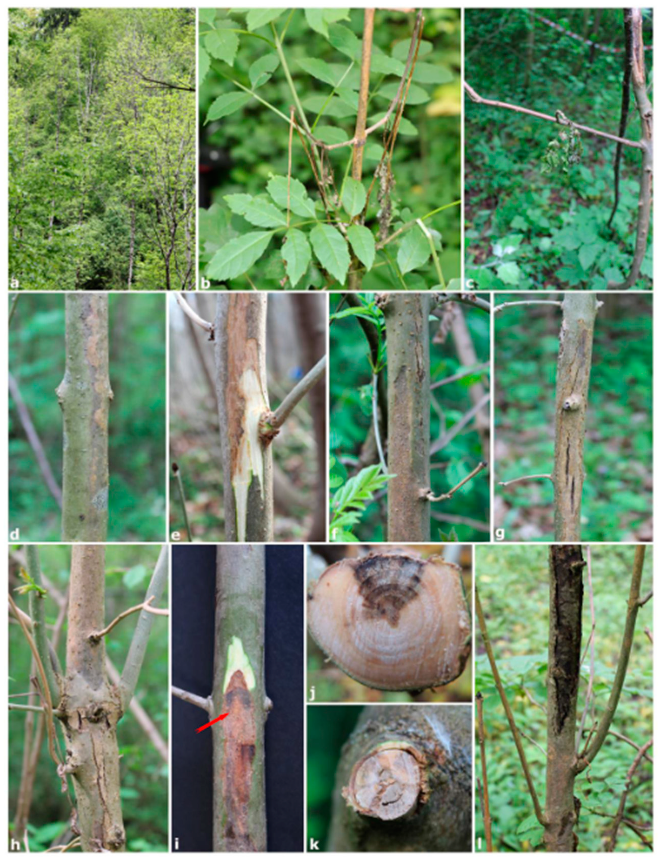
3.1. Symptomatology and Disease Incidence

Field surveys conducted in six mixed ash–maple forests along the pre-Alps in north-eastern Italy showed the widespread presence of typical ash dieback symptoms in all sites (Figure 1). Disease incidence (estimated as number of symptomatic plants out of the total number of plants in each MP) ranged from 33 (site 5) to 82% (site 1) and mortality rate from 0 (site 5) to 23% (site 3). Both young and old European ash trees showed shoot blight, brown discoloration, extensive dieback, sunken cankers and epicormic shoots on the stem and branches (Figure 1).
Shoot blight symptoms were very frequent on young plants (Figure 1b,c). In some cases, the bark necrotic lesions on twigs and branches were superficial, and developed from the inner bark to the xylem (Figure 1d,e), whereas more often they spread to the woody tissues (Figure 1f,i). On this type of lesion, the bark was collapsed and the canker appeared with a typical sunken lesion. When examined in cross-section, these cankers showed a characteristic “V” shaped sector (Figure 1k). Epicormic shoots near sunken cankers progressively died during the growing season. Furthermore, along the margin of some sunken cankers several pycnidia and ascomata belonging to Botryosphaeriaceae species were observed (Figure 1i and Table 2).

3.2. Fungal Isolation and Identification
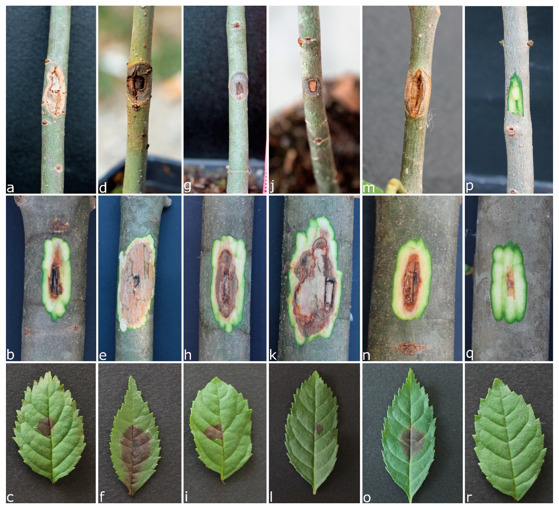
Isolation performed from 60 symptomatic shoot and branch samples yielded a total of 109 fungal colonies belonging to ascomycetous fungi. A total of 13 genera belonging to 9 families were identified; in particular, Botryosphaeriaceae and Diaporthaceae were the most abundant taxonomic groups, with 62 and 18 isolates, respectively.
A total of 22 fungal species were identified based on DNA sequence data and morphological features, of which 17 were from shoot blight symptoms, 7 were from inner bark discolorations on branches, and 16 were from woody tissues taken from sunken cankers (Table 2). The most frequently isolated fungal species were Diplodia subglobosa and Diplodia fraxini, followed by Diaporthe eres and Diplodia mutila (Table 2). They were isolated from all three types of symptoms examined. Diaporthe eres preferentially from shoot blight, while D. subglobosa, D. fraxini and D. mutila especially from sunken cankers.
Diplodia subglobosa was isolated at every site sampled, whereas the other dominant species were isolated from five sites. The other 18 taxa, represented by a limited number of isolates, were often obtained from few sites. Hymenoscyphus fraxineus was isolated from only five plants distributed in four sites—particularly from branch samples with inner bark discolorations.
On branches with sunken cankers, the presence of several pycnidia belonging to D. fraxini was detected near the margin of the necrotic lesion (Figure 1). Ascomata of Diplodia seriata were also observed on three branches.
Botryosphaeriaceous fungi were the largest taxonomic group with four genera (Botryosphaeria, Diplodia, Dothiorella and Neofusicoccum) and nine species. BLAST searches in GenBank showed 100% identity with reference sequences of representative isolates including those of ex-type cultures. For D. mutila, the comparison of the GenBank sequences of some strains isolated from Fraxinus spp. revealed some incorrect species name assignments (Table 3). A re-evaluation of these sequences indicated that in recent years, many reports attributed to D. mutila on Fraxinus spp. should have actually been attributed to D. fraxini (Table 3).

3.3. Pathogenicity

In artificial inoculation trials, representative isolates from the five species were shown to be pathogenic to European ash but they had marked differences in aggressiveness. At the end of the experimental period, all seedlings inoculated with D. eres, D. fraxini, D. mutila, D. subglobosa and H. fraxineus displayed dark brown inner bark lesions that spread up and down from the inoculation site (Figure 2). The average lesion length differed significantly among species (Table 4). The lesions caused by D. fraxini were significantly larger than those caused by other species. In addition, lesions caused by D. fraxini, D. subglobosa and D. mutila were characterized by the collapse of bark tissues, a symptom congruent with field observations. Control seedlings inoculated with sterile PDA plugs remained symptomless. Five months after inoculation, all species, except H. fraxineus, were successfully re-isolated from the margin of necrotic inner bark lesions on ash seedlings, thus fulfilling Koch’s postulates (Table 4).
In the detached branch inoculation tests, necrotic lesions in the inner bark and xylem tissues of European ash caused by D. fraxini were consistently larger compared with those of other fungal species and controls, confirming its aggressiveness to this host (Figure 2 and Table 4). Diplodia subglobosa was similarly aggressive, producing similar lesions to those produced by D. fraxini. The other three species caused only a small lesion restricted to the inoculation point. The results obtained with the detached branches were in accordance with those obtained with ash seedlings. In this test, H. fraxineus was successfully re-isolated from three of the five branches inoculated (Table 4).
Finally, all fungi tested induced a dark-brown necrosis with an irregular margin on the inoculated ash leaves. Necrosis usually appeared beneath the inoculum within 24 h after inoculation, and spread radially into leaf tissues. Symptoms induced by the different species tested were visibly indistinguishable, although on leaves inoculated with D. fraxini the necrosis stretched along the leaf petiole (Figure 2). Furthermore, in this bioassay, significant variation in lesion sizes was detected among the species tested. In particular, D. fraxini proved to be the most aggressive species (Table 4).

4. Discussion

The findings obtained in this study allowed us to characterize the fungal pathogens associated with ash dieback in Italy, contributing to expand knowledge on the aetiology of this emerging disease in Europe.
A total of 22 fungal species were isolated from six different sites located across the pre-Alpine forests in north-eastern Italy. The fungal community associated with symptomatic plant tissues was characterized by a few dominant species and several sporadic species usually present on a single type of symptom or survey site. This fungal assemblage is similar to that found in other studies conducted on common ash trees with dieback symptoms [4,23,25,38]. Many of the sporadic species such as Epicoccum nigrum and Diaporthe foeniculina are well known endophytes of various forest trees.
In this study, D. subglobosa and D. fraxini were detected as the main fungal species associated with sunken canker samples of European ash. Additionally, both species were isolated from branches with inner bark discolorations and symptomatic shoots.
Diplodia subglobosa is a recently described species and for which, prior to this study, only four isolates were known to occur worldwide [26]. In our study, it was isolated at every site showing a wide geographical distribution in north-eastern Italy. Diplodia subglobosa proved to be a pathogen of European ash, although less aggressive than D. fraxini. In a previous study, its pathogenicity was demonstrated on manna ash, although it was identified as D. mutila [41].
In 2014, the name D. fraxini was re-instated to accommodate several Diplodia isolates from sunken cankers of declining narrow-leaved ash in Italy and Portugal and an isolate from European ash in the Netherlands (strain CBS 431.82), and a neotype was designated for this species [26]. Diplodia fraxini is a heterothallic species and, until now, only the MAT1-1-1 idiomorph was known [42]. Currently, an extensive study is in progress to establish occurrence, frequency ratio and pathogenicity of idiomorphs of this pathogen in Italy (Linaldeddu, unpublished).
The results obtained in this study (field survey and re-evaluation of GenBank sequences) indicate that D. fraxini has a much broader host range than previously reported and that the geographic distribution encompasses several European countries where ash dieback symptoms have been reported during the last few decades (Table 3). In particular, sequence comparison showed that D. fraxini has been long confused with D. mutila. This error in the identification of D. fraxini could also explain the contradictory results regarding the pathogenicity of D. mutila on Fraxinus spp. reported by various authors [23,24]. In this regard, the results of the pathogenicity test conducted in our study clear demonstrate that D. fraxini is more aggressive than D. mutila on European ash. An interesting issue in this study is related to the nature of symptoms from which this pathogen was preferentially isolated—the sunken cankers with a characteristic wedge-shaped necrotic sector in the wood. This symptom is usually associated with infection of several Botryosphaeriaceae species on woody hosts [43,44,45,46]. In the pathogenicity assay conducted on 3-year-old seedlings, it was reproduced only by the three Diplodia species tested.
Results obtained confirm D. mutila as one of the most commonly occurring fungi on twigs and branches of European ash with dieback symptoms. This fungus occurred on all types of necrotic lesions examined, often in co-habitation with D. subglobosa or D. fraxini, suggesting a possible synergistic interaction. Diplodia mutila is the type species of the genus Diplodia [47,48]; silver poplar was type host of anamorph, whereas manna ash was type host of the sexual morph of D. mutila. Unfortunately, no live cultures linked to the holotypes of D. mutila are extant and an epitype specimen with an associated ex-epitype culture was established only recently [26]. Over the last few decades, the name D. mutila has been applied to a number of cryptic species, including D. corticola, D. subglobosa and also, as demonstrated in this study, D. fraxini.
Diaporthe eres has emerged as an opportunistic and potentially invasive fungal pathogen in Europe, Asia and North America [49,50,51]. The frequency of its attacks on woody hosts of forests and agriculture has increased significantly over recent decades [50]. In this study, D. eres was the most abundant species obtained from shoots, which agrees with a recent report where this pathogen was consistently isolated from necrotic bark lesions on declining one-year-old European ash plants in a forest stand in Montenegro [25].
Hymenoscyphus fraxineus is well-known to occur in many organs and tissues of European ash trees; it was recorded on leaves, buds, shoots, stems and roots, but also on rachises in the litter, demonstrating a high adaptability to different ecological niches [12,21,38]. In this study, it was isolated only sporadically and preferentially from branches with inner bark discolorations. Low isolation frequency of H. fraxineus from declining European ash trees was also obtained in several other studies [4,23,25,35]. In the pathogenicity assays, it proved to be less aggressive than D. fraxini, although a strain selected for its virulence and ability to produce phytotoxic metabolites was used [16]. This result agrees with the observation of Kowalski et al. [24], who reported an average length of necrotic lesions caused by D. mutila that was slightly greater than those caused by H. fraxineus. Similarly to what was also reported in other researches [24,25], the re-isolation frequency of this pathogen from artificially infected tissues was very low in this study. The pathogenicity test on seedlings was conducted in the period November–April, precisely to evaluate its ability to persist in the host’s tissues during the winter, but it was not possible to re-isolate this pathogen from any of the inoculated seedlings.
Among the sporadic species obtained in this study, several belonged to the genus Dothiorella. Dothiorella species are considered weak pathogens or endophytes of different woody plants worldwide [50]; however, some species, such as Dothiorella iberica and D. omnivora can cause branch cankers and dieback [44,52]. In this study, D. omnivora, D. parva and D. sempervirentis are reported for the first time on European ash.

5. Conclusions

The findings obtained in this study highlight that D. subglobosa and D. fraxini play an important role in the aetiology of ash dieback, and in particular that D. fraxini is the main species involved in the aetiology of sunken cankers. Therefore, in addition to H. fraxineus, other species, many of which belong to the Botryosphaeriaceae family, could contribute to the onset of this complex disease characterized by different symptoms. Whether the fungal species isolated in this study can act in a synergic way remains to be clarified.
Finally, the co-occurrence of multiple pathogens in the onset of European ash dieback suggests that management strategies to preserve these important and vulnerable ecosystems in the future should take this aetiological complexity into due consideration.

Author Contributions

Conceptualization, B.T.L.; formal analysis, F.B., C.B. and B.T.L.; investigation, F.B., C.B. and B.T.L.; resources, B.T.L. and L.M. (Lucio Montecchio); data curation, B.T.L., L.M. (Lucio Montecchio) and L.M. (Lucia Maddau); writing—original draft preparation, B.T.L.; writing—review and editing, B.T.L., L.M. (Lucio Montecchio) and L.M. (Lucia Maddau); funding acquisition, B.T.L. and L.M. (Lucio Montecchio) All authors have read and agreed to the published version of the manuscript.

Funding

This research was funded by the project “Ruolo di Diplodia fraxini e Diplodia subglobosa nell’eziologia del deperimento del frassino in Italia”, grant number DOR2021428/20.

Conflicts of Interest

The authors declare no conflict of interest.

References

  1. Kowalski, T. Chalara fraxinea sp. nov. associated with dieback of ash (Fraxinus excelsior) in Poland. For. Pathol. 2006, 36, 264–270. [Google Scholar] [CrossRef]
  2. Davydenko, K.; Vasaitis, R.; Stenlid, J.; Menkis, A. Fungi in foliage and shoots of Fraxinus excelsior in eastern Ukraine: A first report on Hymenoscyphus pseudoalbidus. For. Pathol. 2013, 43, 462–467. [Google Scholar] [CrossRef]
  3. Bengtsson, S.B.K.; Barklund, P.; von Brömssen, C.; Stenlid, J. Seasonal pattern of lesion development in diseased Fraxinus excelsior infected by Hymenoscyphus pseudoalbidus. PLoS ONE 2014, 9, e76429. [Google Scholar] [CrossRef] [Green Version]
  4. Przybył, K. Fungi associated with necrotic apical parts of Fraxinus excelsior shoots. For. Pathol. 2002, 32, 387–394. [Google Scholar] [CrossRef]
  5. Gross, A.; Holdenrieder, O.; Pautasso, M.; Queloz, V.; Sieber, T.N. Hymenoscyphus pseudoalbidus, the causal agent of European ash dieback. Mol. Plant Pathol. 2014, 15, 5–21. [Google Scholar] [CrossRef] [PubMed]
  6. Pautasso, M.; Aas, G.; Queloz, V.; Holdenrieder, O. European ash (Fraxinus excelsior) dieback—A conservation biology challenge. Biol. Conserv. 2013, 158, 37–49. [Google Scholar] [CrossRef]
  7. Ogris, N.; Hauptman, T.; Jurc, D.; Floreancig, V.; Marsich, F.; Montecchio, L. First report of Chalara fraxinea on common ash in Italy. Plant Dis. 2010, 94, 133. [Google Scholar] [CrossRef]
  8. Giongo, S.; Oliveira Longa, C.M.; Dal Maso, E.; Montecchio, L.; Maresi, G. Evaluating the impact of Hymenoscyphus fraxineus in Trentino (Alps, Northern Italy): First investigations. iForest 2017, 10, 871–878. [Google Scholar] [CrossRef] [Green Version]
  9. Luchi, N.; Ghelardini, L.; Santini, A.; Migliorini, D.; Capretti, P. First record of ash dieback caused by Hymenoschyphus fraxineus on Fraxinus excelsior in the Apennines (Tuscany, Italy). Plant Dis. 2016, 100, 535. [Google Scholar] [CrossRef]
  10. Bakys, R.; Vasaitis, R.; Barklund, P.; Ihrmark, K.; Stenlid, J. Investigations concerning the role of Chalara fraxinea in declining Fraxinus excelsior. Plant Pathol. 2009, 58, 284–292. [Google Scholar] [CrossRef]
  11. Baral, H.O.; Queloz, V.; Hosoya, T. Hymenoscyphus fraxineus, the correct scientific name for the fungus causing ash dieback in Europe. IMA Fungus 2014, 5, 79–80. [Google Scholar] [CrossRef] [PubMed]
  12. Gross, A.; Zaffarano, P.L.; Duo, A.; Grünig, C.R. Reproductive mode and life cycle of the ash dieback pathogen Hymenoscyphus pseudoalbidus. Fungal Genet. Biol. 2012, 49, 977–986. [Google Scholar] [CrossRef] [PubMed]
  13. Dal Maso, E.; Fanchin, G.; Mutto Accordi, S.; Scattolin, L.; Montecchio, L. Ultrastructural modifications in common ash tissues colonised by Chalara fraxinea. Phytopathol. Mediterr. 2012, 51, 599–606. [Google Scholar]
  14. Burokiene, D.; Prospero, S.; Jung, E.; Marciulyniene, D.; Moosbrugger, K.; Norkute, G.; Rigling, D.; Lygis, V.; Schoebel, C.N. Genetic population structure of the invasive ash dieback pathogen Hymenoscyphus fraxineus in its expanding range. Biol. Invasions 2015, 17, 2743–2756. [Google Scholar] [CrossRef]
  15. Mansfield, J.; Brown, I.; Papp-Rupar, M. Life at the edge the cytology and physiology of the biotroph to necrotroph transition in Hymenoscyphus fraxineus during lesion formation in ash. Plant Pathol. 2019, 68, 908–920. [Google Scholar] [CrossRef]
  16. Masi, M.; Di Lecce, R.; Tuzi, A.; Linaldeddu, B.T.; Montecchio, L.; Maddau, L.; Evidente, A. Hyfraxinic acid, a phytotoxic tetrasubstituted octanoic acid, produced by the Ash (Fraxinus excelsior L.) pathogen Hymenoscyphus fraxineus together with viridiol and some of its analogues. J. Agric. Food Chem. 2019, 67, 13617–13623. [Google Scholar] [CrossRef]
  17. Andersson, P.F.; Johansson, S.B.K.; Stenlid, J.; Broberg, A. Isolation, identification and necrotic activity of viridiol from Chalara fraxinea, the fungus responsible for dieback of ash. For. Pathol. 2010, 40, 43–46. [Google Scholar] [CrossRef]
  18. Howell, C.R.; Stipanovic, R.D. Phytotoxicity to crop plants and herbicidal effects on weeds of viridiol produced by Gliocladium virens. Phytopathology 1984, 74, 1346–1349. [Google Scholar] [CrossRef]
  19. Junker, C.; Mandey, F.; Pais, A.; Ebel, R.; Schulz, B. Hymenoscyphus pseudoalbidus and Hymenoscyphus albidus: Viridiol concentration and virulence do not correlate. For. Pathol. 2014, 44, 39–44. [Google Scholar] [CrossRef]
  20. Zheng, H.D.; Zhuang, W.Y. Five new species of Hymenoscyphus (Helotiaceae, Ascomycota) with notes on the phylogeny of the genus. Mycotaxon 2015, 130, 1017–1038. [Google Scholar] [CrossRef]
  21. Drenkhan, R.; Solheim, H.; Bogacheva, A.; Riit, T.; Adamson, K.; Drenkhan, T.; Maaten, T.; Hietala, A.M. Hymenoscyphus fraxineus is a leaf pathogen of local Fraxinus species in the Russian Far East. Plant Pathol. 2016, 66, 490–500. [Google Scholar] [CrossRef]
  22. McMullan, M.; Rafiqi, M.; Kaithakottil, G.; Clavijo, B.; Bilham, B.; Orton, E.; Percival-Alwyn, L.; Ward, B.J.; Edwards, A.; Saunders, D.G.; et al. The ash dieback invasion of Europe was founded by two individuals from a native population with huge adaptive potential. Nat. Ecol. Evol. 2018, 2, 1000–1008. [Google Scholar] [CrossRef] [PubMed] [Green Version]
  23. Bakys, R.; Vasaitis, R.; Barklund, P.; Thomsen, I.M.; Stenlid, J. Occurrence and pathogenicity of fungi in necrotic and non-symptomatic shoots of declining common ash (Fraxinus excelsior) in Sweden. Eur. J. For. Res. 2009, 128, 51–60. [Google Scholar] [CrossRef]
  24. Kowalski, T.; Bilański, P.; Kraj, W. Pathogenicity of fungi associated with ash dieback towards Fraxinus excelsior. Plant Pathol. 2017, 66, 1228–1238. [Google Scholar] [CrossRef]
  25. Vemić, A.; Tomšovský, M.; Jung, T.; Milenković, I. Pathogenicity of fungi associated with ash dieback symptoms of one-year-old Fraxinus excelsior in Montenegro. For Path. 2019, 49, e12539. [Google Scholar] [CrossRef]
  26. Alves, A.; Linaldeddu, B.T.; Deidda, A.; Scanu, B.; Phillips, A.J.L. The complex of Diplodia species associated with Fraxinus and some other woody hosts in Italy and Portugal. Fungal Divers. 2014, 67, 143–156. [Google Scholar] [CrossRef]
  27. Elena, G.; León, M.; Abad-Campos, P.; Armengol, J.; Mateu-Andrés, I.; Güemes-Heras, J. First Report of Diplodia fraxini causing dieback of Fraxinus angustifolia in Spain. Plant Dis. 2018, 102, 2645. [Google Scholar] [CrossRef]
  28. Cimmino, A.; Maddau, L.; Masi, M.; Linaldeddu, B.T.; Pescitelli, G.; Evidente, A. Fraxitoxin, a new isochromanone isolated from Diplodia fraxini. Chem. Biodivers. 2017, 14, e1700325. [Google Scholar] [CrossRef] [Green Version]
  29. Dal Maso, E.; Montecchio, L. Risk of natural spread of Hymenoscyphus fraxineus with environmental niche modelling and ensemble forecasting technique. Forest Res. 2014, 3, 4. [Google Scholar] [CrossRef]
  30. White, T.J.; Bruns, T.; Lee, S.; Taylor, J. Amplification and Direct Sequencing of Fungal Ribosomal RNA 502 Genes for Phylogenetics. In PCR Protocols, a Guide to Methods and Applications; Innis, M.A., Gelfand, D.H., Sninsky, J.J., White, T.J., Eds.; Academic Press: San Diego, CA, USA, 1990; pp. 315–322. [Google Scholar]
  31. Linaldeddu, B.T.; Alves, A.; Phillips, A.J.L. Sardiniella urbana gen. et sp. nov., a new member of the Botryosphaeriaceae isolated from declining Celtis australis trees in Sardinian streetscapes. Mycosphere 2016, 7, 893–905. [Google Scholar] [CrossRef]
  32. Altschul, S.F.; Gish, W.; Miller, W.; Myers, E.W.; Lipman, D.J. Basic local alignment search tool. J. Mol. Biol. 2010, 215, 403–410. [Google Scholar] [CrossRef]
  33. Lamari, L. Assess: Image Analysis Software for Plant Disease Quantification; APS Press: St Paul, MN, USA, 2002. [Google Scholar]
  34. Kranjec Orlović, J.; Andrić, I.; Bulovec, I.; Diminić, D. Mycobiota in the seeds of narrow-leaved ash (Fraxinus angustifolia Vahl). Šumar. List. 2019, 3–4, 103–110. [Google Scholar] [CrossRef]
  35. Żółciak, A.; Nowakowska, J.A.; Pacia, A.; Keča, N.; Oszako, T. Fungi isolated from shoots showing ash dieback in the Wolica Nature Reserve in Poland and artificially inoculated seedlings with Hymenoscyphus fraxineus. Folia For. Pol. Ser. A 2019, 61, 42–50. [Google Scholar] [CrossRef] [Green Version]
  36. Kosawang, C.; Amby, D.B.; Bussaban, B.; McKinney, L.V.; Xu, J.; Kjær, E.D.; Collinge, D.B.; Nielsen, L.R. Fungal communities associated with species of Fraxinus tolerant to ash dieback, and their potential for biological control. Fungal Biol. 2018, 122, 110–120. [Google Scholar] [CrossRef] [PubMed]
  37. Langer, G. Collar rots in forests of Northwest Germany affected by ash dieback. Balt. For. 2017, 23, 4–19. [Google Scholar]
  38. Kowalski, T.; Kraj, W.; Bednarz, B. Fungi on stems and twigs in initial and advanced stages of dieback of European ash (Fraxinus excelsior) in Poland. Eur. J. For Res. 2016, 135, 565–579. [Google Scholar] [CrossRef] [Green Version]
  39. Cleary, M.R.; Arhipova, N.; Gaitnieks, T.; Stenlid, J.; Vasaitis, R. Natural infection of Fraxinus excelsior seeds by Chalara fraxinea. For. Pathol. 2012, 43, 83–85. [Google Scholar]
  40. Kraj, W.; Kowalski, T.; Zarek, M. Structure and genetic variation of Diplodia mutila on declining ashes (Fraxinus excelsior) in Poland. J. Plant Pathol. 2013, 95, 499–507. [Google Scholar]
  41. Sidoti, A.; Granata, G. L’orniello (Fraxinus ornus): Nuovo ospite di Diplodia mutila. Inform. Fitopatol. 2004, 2, 49–51. [Google Scholar]
  42. Lopes, A.; Linaldeddu, B.T.; Phillips, A.J.L.; Alves, A. Mating type gene analyses in the genus Diplodia: From cryptic sex to cryptic species. Fungal Biol. 2018, 122, 629–638. [Google Scholar] [CrossRef]
  43. Linaldeddu, B.T.; Maddau, L.; Franceschini, A.; Alves, A.; Phillips, A.J.L. Botryosphaeriaceae species associated with lentisk dieback in Italy and description of Diplodia insularis sp. nov. Mycosphere 2016, 7, 962–977. [Google Scholar] [CrossRef]
  44. Smahi, H.; Belhoucine-Guezouli, L.; Berraf-Tebbal, A.; Chouih, S.; Arkam, M.; Franceschini, A.; Linaldeddu, B.T.; Phillips, A.J.L. Molecular characterization and pathogenicity of Diplodia corticola and other Botryosphaeriaceae species associated with canker and dieback of Quercus suber in Algeria. Mycosphere 2017, 8, 1261–1272. [Google Scholar] [CrossRef]
  45. Savocchia, S.; Steel, C.C.; Stodart, B.J.; Somers, A. Pathogenicity of Botryosphaeria species isolated from declining grapevines in sub-tropical regions of Eastern Australia. Vitis 2007, 46, 27–32. [Google Scholar]
  46. Barradas, C.; Phillips, A.J.L.; Correia, A.; Diogo, E.; Bragança, H.; Alves, A. Diversity and potential impact of Botryosphaeriaceae species associated with Eucalyptus globulus plantations in Portugal. Eur. J. Plant Pathol. 2016, 146, 245–257. [Google Scholar] [CrossRef]
  47. Montagne, J.F.C. Notice sur les plantes cryptogames récemment découvertes en France contenant aussi l’indication précis des localités de quelques espèces les plus rares de la flore française. Ann. Sci. Nat. Bot. Sér. 1834, 2, 295–307. [Google Scholar]
  48. Fries, E.M. Systema Mycologicum; Ex Office Berlingiana: Lundin, Sweden, 1823; Volume 2, pp. 276–620. [Google Scholar]
  49. Dissanayake, A.J.; Zhang, W.; Liu, M.; Hyde, K.D.; Zhao, W.; Li, X.H.; Yan, J.Y. Diaporthe species associated with peach tree dieback in Hubei, China. Mycosphere 2017, 8, 533–549. [Google Scholar] [CrossRef]
  50. Dissanayake, A.J.; Phillips, A.J.L.; Hyde, K.D.; Yan, J.Y.; Li, X.H. The current status of species in Diaporthe. Mycosphere 2017, 8, 1106–1156. [Google Scholar] [CrossRef]
  51. Ali, S.; Renderos, W.; Bevis, E.; Hebb, J.; Abbasi, P.A. Diaporthe eres causes stem cankers and death of young apple rootstocks in Canada. Can. J. Plant Pathol. 2020, 42, 218–227. [Google Scholar] [CrossRef]
  52. Váczy, K.Z.; Németh, M.Z.; Csikós, A.; Kovács, G.M.; Kiss, L. Dothiorella omnivora isolated from grapevine with trunk disease symptoms in Hungary. Eur. J. Plant Pathol. 2018, 150, 817–824. [Google Scholar] [CrossRef]
Figure 1. Main disease symptoms detected on European ash trees: extensive canopy dieback (a), shoot blight (b,c); branches with inner bark discolorations (d,e); active sunken cankers on branches with Diplodia fraxini pycnidia (arrow) (fi); cross-section of branches showing wedge-shaped necrotic sectors (j,k); epicormics shoots below a branch canker (l).
Forests 11 00883 g001
Figure 2. From top to bottom symptoms observed on seedlings, detached branches and leaves after inoculation with Diaporthe eres (ac), Diplodia fraxini (df), Diplodia mutila (gi), Diplodia subglobosa (jl), Hymenoscyphus fraxineus (mo). Controls (pr).
Forests 11 00883 g002
Table 1. Study sites information and number of symptomatic shoots (S), branches with brown inner bark discolorations (B) and branches with sunken cankers (C) used for fungal isolation.
Study SitesElevation
(m a.s.l.)
Geographic CoordinatesNumber of Samples
121745°50′30″ N11°58′47″ E3 (S), 2 (B), 5 (C)
224745°50′19″ N11°58′23″ E5 (S), 1 (B), 4 (C)
350846°06′29″ N12°21′36″ E7 (S), 0 (B), 3 (C)
445746°12′54″N12°43′10″ E3 (S), 4 (B), 3 (C)
538146°02′09″ N12°02′51″ E3 (S), 2 (B), 5 (C)
674446°04′51″ N12°12′20″ E4 (S), 1 (B), 5 (C)
Table 2. Accession numbers deposited in GenBank and number of isolates of each species obtained from shoots (S), branches with brown inner bark discolorations (B) and branches with sunken canker (C). The number of branches with pycnidia or ascomata is reported in the last column.
Fungal Species (Strain Number)Accession
Number
Type of SamplesNumber of SitesPycnidia/Ascomata
(S)(B)(C)
Botryosphaeria dothidea (FB4)MT7577732001-
Diaporthe eres (FB5)MT75777410235-
Diaporthe foeniculina (FB34)MT7577871001-
Diaporthe sp. 1 (FB12)MT7577760011-
Diaporthe sp. 2 (FB18)MT7577801001-
Diatrypella sp. (FB62)MT7577901011-
Diplodia fraxini (FB1)MT75777143958
Diplodia mutila (FB29)MT7577853355-
Diplodia seriata (FB21)MT75778200223
Diplodia subglobosa (FB2)MT7577727396-
Dothiorella omnivora (FB32)MT7577861001-
Dothiorella parva (FB69)MT7577910011-
Dothiorella sempervirentis (F39)MT7577881211-
Epicoccum nigrum (FB20)MT7577816033-
Fusarium avenaceum (FB47)MT7577892135-
Fusarium lateritium (FB16)MT7577781011-
Hymenoscyphus fraxineus (CHA1)MN4280711314-
Neofusicoccum parvum (FB7)MT7577752042-
Neonectria sp. (FB26)MT7577840022-
Phaeosphaeriopsis glaucopunctata (FB14)MT7577771001-
Pseudopithomyces sp. (FB23)MT7577830011-
Valsa sp. (FB17)MT7577791001-
Table 3. Re-evaluation of ITS rDNA sequences erroneously associated with Diplodia mutila in GenBank.
Species Name in
GenBank
Accession
no.
HostCountryReferenceRevised Species Name
Diplodia mutilaMH137758,Fraxinus angustifoliaCroatia[34]Diplodia fraxini
D. mutilaKX618487,Fraxinus excelsiorPoland[35]D. fraxini
D. mutilaLC171705Fraxinus chinensisDenmark[36]D. fraxini
D. mutilaLC171694Fraxinus pennsylvanicaDenmark[36]D. fraxini
D. mutilaKU712211F. excelsiorGermany[37]D. fraxini
D. mutilaKT004548F. excelsiorPoland[38]D. fraxini
D. mutilaJQ765661F. excelsiorLatvia[39]D. fraxini
D. mutilaKF225519F. excelsiorPoland[40]D. fraxini
D. mutilaFJ228165F. excelsiorSweden[23]D. fraxini
Table 4. Lesion sizes ± standard deviation caused by the five fungal species on European ash and number of positive re-isolations from seedlings (S), detached branches (B) and leaves (L).
Fungal SpeciesMean Lesion Length (cm) *Leaf Lesion Size (mm2) *Positive Re-Isolation
SeedlingsDetached BranchesSBL
Diaporthe eres (FB5)0.7 ± 0.4 b3.4 ± 2.2 bc31.8 ± 19.7 b(8/9)(5/5)(4/5)
Diplodia fraxini (FB1)1.3 ± 0.4 a5.9 ± 3.8 a137.6 ± 62.6 a(9/9)(5/5)(5/5)
Diplodia mutila (FB29)0.7 ± 0.3 b2.7 ± 1.5 bc10.0 ± 7.3 b(9/9)(5/5)(4/5)
Diplodia subglobosa (FB2)0.7 ± 0.3 b3.6 ± 2.0 ab9.5 ± 8.9 b(9/9)(5/5)(4/5)
Hymenoscyphus fraxineus (CHA1)0.7 ± 0.2 b2.1 ± 1.4 bc49.5 ± 42.1 b(0/9)(3/5)(4/5)
Control-1.3 ± 0.1 c----
* Values with the same letter do not differ significantly at p = 0.05, according to LSD multiple range test.

