Article

Aspects Determining the Dominance of Fomitopsis pinicola in the Colonization of Deadwood and the Role of the Pathogenicity Factor Oxalate

Institute of Earth Sciences, Friedrich-Schiller-University, Burgweg 11, D-07749 Jena, Germany
Forests 2020, 11(3), 290; https://doi.org/10.3390/f11030290
Submission received: 10 January 2020 / Revised: 25 February 2020 / Accepted: 28 February 2020 / Published: 3 March 2020
(This article belongs to the Section Forest Ecology and Management)

Abstract

:
Carbon and mineral cycling in sustainable forest systems depends on a microbiome of basidiomycetes, ascomycetes, litter-degrading saprobes, ectomycorrhizal, and mycoparasitic fungi that constitute a deadwood degrading consortium. The brown rot basidiomycete Fomitopsis pinicola (Swartz: Fr.) P. Karsten (Fp), as an oxalate-producing facultative pathogen, is an early colonizer of wounded trees and fresh deadwood. It replaces basidiomycetous white rot fungi and non-basidiomycetous fungal phyla in the presence of its volatilome, but poorly in its absence. With the goal of determining its dominance over the most competitive basidiomycetes and its role in fungal successions within the forest microbiome in general, Fp was exposed to the white rot fungus Kuehneromyces mutabilis (Schaeff.: Fr.) Singer & Smith (Km) in aseptic dual culture established on fertilized 100 mm-long wood dust columns in glass tubes with the inclusion of their volatilomes. For the mycelia approaching from the opposite ends of the wood dust columns, the energy-generating systems of laccase and manganese peroxidase (MnP), the virulence factor oxalate, and the exhalation of terpenes were determined by spectrophotometry, High Pressure Liquid Chromatography (HPLC), and Gas Chromatography-Mass Spectrometry (GC-MS). Km mycelia perceived the approaching Fp over 20 mm of non-colonized wood dust, reduced the laccase activity to 25%, and raised MnP to 275%–500% by gaining energy and presumably by controlling oxalate, H2O2, and the dropping substrate pH caused by Fp. On mycelial contact, Km stopped Fp, secured its substrate sector with 4 mm of an impermeable barrier region during an eruption of antimicrobial bisabolenes, and dropped from the invasion mode of substrate colonization into the steady state mode of low metabolic and defensive activity. The approaching Fp raised the oxalate production throughout to >20 g kg−1 to inactivate laccase and caused, with pH 1.4–1.7, lethal conditions in its substrate sector whose physiological effects on Km could be reproduced with acidity conditions incited by HCl. After a mean lag phase of 11 days, Fp persisting in a state of high metabolic activity overgrew and digested the debilitated Km thallus and terminated the production of oxalate. It is concluded that the factors contributing to the competitive advantage of F. pinicola in the colonization of wounded trees and pre-infected deadwood are the drastic long-term acidification of the timber substrate, its own insensitivity to extremely low pH conditions, its efficient control of the volatile mono- and sesquiterpenes of timber and microbial origin, and the action of a undefined blend of terpenes and allelopathic substances.

1. Introduction

Undisturbed forest systems, as sites of biodiversity, depend on a microbiome in which basidiomycete fungi play a major part. With their active contribution to the growth of timber and their efficacy in its complete mineralization, the ecological groups of ectomycorrhizal, tree root and stem pathogenic, to slash and plant residue-degrading basidiomycetes give a unique example of familiar cooperation. The release of oxidoreductases, which contribute to the opening of aromatic rings in lignin [1,2,3], characterizes more or less all ecological groups [4,5,6,7]. This enables proteobacteria and ascomycetous and some zygomycetous soft rot fungi to take part in the degradation of lignin moieties and humic colloids in the completion of the carbon and mineral nutrient cycle [8,9,10].
The maintenance of a respective microbiome with a balanced incidence of macrofungi presupposes the presence of standing and downed deadwood resources, e.g., at estimated >60 m3 ha−1 in German Fagus sylvatica forests [11]. Molecular analyses identified 110–398 operational taxonomic units (OTUs) in coniferous and broad-leaved stem wood samples after 6–12 years of outdoor storage. Ascomycota and Basidiomycota dominated the early stages of decay, accompanied, or followed by, litter degrading saprotrophs, ectomycorrhizal, endophytic, mycoparasitic, and lichen mycobiotic species [12,13,14]. Rules determining their dominance and succession in the forest microbiome are poorly understood. They may be elucidated on the level of interactions between early colonizers, such as Fomitopsis pinicola (Swartz: Fr.) P. Karsten, and single competitors in sterile dual culture.
In spite of a host range of 82 softwood and 42 hardwood spp. [15], the brown rot basidiomycete F. pinicola is not regarded as a serious pathogen in managed Eurasian forests due to its low and primarily hidden incidence in living trees. It is an important disease agent of wounded conifer spp. and pome and stone fruit orchards in North America, as well as in protected old-growth forests [16,17], and forms basidiomes as pioneer invader of softwoods within one year after bark beetle outbreaks [18]. The fungus can survive in deadwood for up to 41 years [19], leaving poorly modified lignin cores that resist mineralization for further decades. This dominance and persistence in the field was expressed, too, in aseptic dual cultures with 28 wood decay fungi when the fungal volatilomes were included [20]. This is contrasted by a drop in the competitive ability of F. pinicola in well-aerated systems and peaks in any lack of mold resistance of its soft-spawn mycelia.
The metabolome and the stress-activated gene expression during different microbial interactions, as well as the rules of fungal succession, are frequently studied in sterile dual cultures established on solid malt agar or pre-inoculated and paired wood blocks [21,22,23,24,25]. Placed on the opposite ends of an agar plate, the growth of the approaching mycelia may gradually slow down due to the long-distance effects of diffusible, inhibitory factors or volatile organic compounds (VOCs) emanated by the microbial competitors [26,27]. The mycelia may be arrested at the junction point (deadlock) and form a melanized zone or barrier (barrage) line [25] with the contribution of fungal laccase [28,29]. The dominating mycelium may overgrow its competitor immediately or after stalling [30,31], autotoxification [32,33], or debilitation of the latter by the metabolites of the dominator. The results of dual interactions and the blend of accompanying volatiles with their widely unknown significance change with pH, moisture, temperature, substrate, culture age [23,34] and the test arrangement itself.
Accordingly, the inclusion of fungal volatiles seemed to determine the outcome of interactions between the cultivated white rot fungus Kuehneromyces mutabilis (Schaeff.: Fr.) Singer & Smith (Km 10) and the brown rot fungus F. pinicola (Foma 4). Km 10 is transmitted to the stem sections of leafwood at a 100% rate with a plastic sterile spawn and tolerates traces of gram-negative bacteria and mitosporic fungi as commensals. In contrast, Foma 4 represents a set of brown rot fungi whose sterile spawn molds upon any contact with stem sections but holds substrates once colonized sterile [35]. In their dixenic cultures established on fertilized beech wood dust in aerated Petri dishes, Foma 4 was regionally overgrown by the competitively superior Km 10, failed to colonize wood dust dominated by the mycoparasitic Trichoderma, but overgrew a bacterial suspension. In glass tubes bearing wood dust columns of 100 mm whose poorly aerated interior could preserve the volatile fungal exhalations, Foma 4 overgrew all of these competitors [20].
In soil with comparable structural conditions, volatile (hydro) carbon compounds <300 Da easily spread across gas and water filled pores and impair the composition of microbial and plant communities [36,37,38] in high but not low concentrations [39,40,41]. Volatiles comprise the emanations from plant roots with 80% of their exudates [42] beside those from bacteria and fungi that are released upon the degradation of organics.
Similar to the 300 volatiles identified from fungal pure cultures [34,43,44,45,46] and those from bacteria (http://bioinformatics.charite.de/mvoc/#), the 1700 plant exhalations mainly comprise C6 to C18 saturated and unsaturated aliphatic hydrocarbons and their derivatives such as acids, alcohols, aldehydes, ketones, esters, ethers, and exceptionally N- and S-containing compounds, as well as variable mixtures of terpenoids and aromatics from plant essential oils which make up the odor of the plant [47,48,49]. In their role as nutrients, toxins, antibiotics, hormones, and signaling agents, volatiles interfere with all physiological processes of microorganisms and plants as well as the integrity of walls and organelles of the cell [47,48,50,51,52].
In this study, dixenic cultures of K. mutabilis and F. pinicola were established on fertilized beech wood dust in the closed systems of glass tubes and packing jars. Thereby, the activities of the energy-generating systems of Km 10 oxidoreductases were monitored in their exposure to the virulence factor oxalate of Foma 4 [53,54], overlaid by fungal terpenoids.
In K. mutabilis, the initial one-electron oxidation (e/H+ abstraction) of aromatic compounds from the recalcitrant plant lignin to simple phenols and anilines proceeds with laccases of 64 and 72 kDa and manganese peroxidases (MnPs) [55,56]. Phenolic and non-phenolic aromatics are transformed to reactive radicals that stabilize themselves in subsequent polymerizations, disproportionations, and fragmentations of the lignin polymer (1–3 for reviews). Pairing fungal isolates on agar or liquid media can alter the titer of laccase enzymes and increase the activity of MnP [57,58,59].
In F. pinicola, several accessions bear genes encoding laccase and several H2O2 generating carbohydrate oxidases similar to white rot fungi but at lower activity. Class II peroxidases, such as MnP, are lacking [15,60,61]. This could explain the marginal modification of lignin in the brown rot wood decay [62,63] whose biochemistry is still hypothetical [63]. A non-enzymatic attack on the holocellulose component is ascribed to the hydroxyl radical (•OH), the ultimate oxidant with a redox potential of E0 = 1.8–2.7 V [64] formed by Fenton chemistry: Fe2+ + H2O2 → Fe3+ + OH + •OH. The hydroxyl radical is small enough to penetrate the cell walls, degrade their S2 layer and facilitate the hydrolysis of cellulose and hemicellulose components [61,63,65].
Unlike white rot fungi, brown rot basidiomycetes share the accumulation of oxalic acid as a host-nonspecific toxin with plant pathogenic necrotrophes, such as Botrytis cinerea and Sclerotinia sclerotiorum [53,54,66,67,68]. Its role in pathogenicity is not well defined. The organic acid forms crystals of insoluble oxalates with Ca, Co, Cu, and Zn cations and soluble complexes with Fe3+ [68,69,70]. Oxalate sequesters the cell wall, stabilizing Ca2+ cations from Ca pectate in the middle lamellae and activates the pectin hydrolysing polygalacturonase by lowering pH and by its obligate cooperation with the enzyme [71,72]. Deficits in oxalate production or applications of oxalate-precipitating CaCl2 to culture filtrates from necrotrophic pathogens [73] reduce the plant tissue maceration in vitro [66]. Non-disssociated oxalate applied to wounded host tissue results in cell death [74,75] due to low pH conditions. Xylophilous bacteria [76] and the food-spoiling pathogen Bacillus cereus [77] experience lethal shocks at pH values of 3.5 and 4.5, respectively.
In the expected interaction of oxidoreductases with oxalate, white rot fungi do not only dispose of oxalate decarboxylase to transform oxalate to formate and CO2 [78]. The abiotic oxidant Mn3+ in the redox cycle of MnP transforms oxalate to CO2 and CO2 upon its reduction to Mn2+. The resulting formate radical CO2 is reduced to CO2 by the oxidation of O2 to O2, a precursor of H2O2 [79]. To the best of our knowledge, no reports document that oxalate could damage the MnP glycoprotein by sequestering its stabilizing two Ca2+ cations [80,81], whereas laccases are inactivated via the abstraction of the catalytic Cu cations by the oxalate chelant [82].
In this study, the dual cultures were established in closed systems. It was the goal to refer the production of oxidoreductases and volatiles as vitality indicators of K. mutabilis with the oxalate production of F. pinicola. Oxalate is its most probable means to gain advantage over the competitively stronger white rot isolate Km 10. The results should contribute to improving the poor understanding of the role fungal oxidoreductases, sesquiterpenes and oxalates play in the mutual replacement of fungi. The economic consequences of the resulting prevalence of the normally mold-susceptible F. pinicola in forestry linked with the recent dry seasons in 2018/2019 should be appraised.

2. Materials and Methods

2.1. Fungal Pure Cultures

The isolate of Kuehneromyces mutabilis Km 10 was obtained from the plectenchyma of a basidiome placed on 2.5% malt extract agar in 1971. Fomitopsis pinicola Foma 4 was derived from progressively decayed Picea abies (L.) Karst. timber in 1968. For daily access, the strains were maintained in test tubes Ø 16 mm containing barked leafwood branch sections Ø 1.5–3 × 10–14 mm submerged in 2.5% malt extract agar and stored at 2 °C, a method challenging and, thus, retaining the fungal metabolic systems completely.

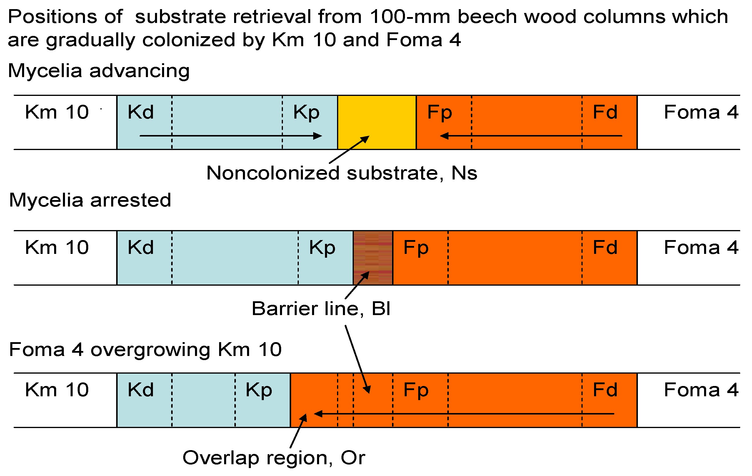
2.2. Substrate Arrangements

Industrial sawdust from European beech (Fagus sylvatica L.), devoid of antimicrobial wood extractives, was used as the common substrate for dual fungal cultures. It was amended with 100 g saccharose, 50 g wheat flour kg−1, and water to 200% by dry weight (DW) to improve the normally poor expression of fungal metabolites. The substrate was placed as a 4 mm layer in Petri dishes Ø 90 mm, as a column of Ø 20 × 100 mm in glass tubes 200 mm long, cotton-stoppered at both open ends, and at 6 g DW in 0.3 L packing jars Ø 80 × 125 mm with Teflon-coated screw caps. The vessels were autoclaved at 121 °C for 1 h prior to inoculation. Ascertaining the impact of soluble oxalate on fungal interactions in glass tubes, beech wood dust with an intrinsic Ca content of 1.4 g kg−1 DW was amended with 3 and 10 g kg−1 Ca, respectively, by CaCl2 × 2H2O to immobilize oxalate with calcium.

2.3. Fungal Interactions in Glass Tubes 200 mm

The beech dust columns Ø 20 × 100 mm were aseptically inoculated at the opposite ends with Km 10 and Foma 4, respectively, at up to 20 replications per treatment and incubated at 20 °C. The advancing mycelia initiated the formation of a melanized and hardened barrier layer of 4 mm at the junction points (Figure 1) that resisted the further progress of Foma 4 for a mean of 11 d. Finally, Foma 4 started overgrowing the Km 10 colonized region completely at a 100% rate. In the stages of approaching, contacting, arrested, and overlapping mycelia, the entire substrate columns were dissected.
Twelve mm sections of the columns were taken as substrate samples from the distal (Kd, Fd) and proximal ends (Kp, Fp) of the current mycelial colonies for the determination of fungal metabolites (Figure 1).

2.4. Oxidoreductase Enzymes in Substrate Samples

Activities of the polyphenoloxidase, laccase (EC 1.10.3.2) were determined from the wood dust suspended in aqueous 0.1 M KH2PO4 buffer pH 5.0 (0.033 M) and 2,2′-azinobis-(3-ethylbenzthiazoline-6-sulfonate (ABTS, 0.4 mM) as increase in absorbance at A420420 = 36,000 M−1 cm−1) due to the formation of ABTS cation radical (ABTS•+) [83]. The activities of manganese peroxidase (MnP, EC 1.11.1.13) were determined in a reaction mixture of 0.15 mM H2O2, 0.2 mM MnSO4 × H2O, and 50 mM malonate at pH 4.5 as increase in A265270 = 11,590 M−1 cm−1) due to the formation of Mn(III) malonate [84]. The values were determined at least in triplicate over 10 min.

2.5. Oxalate Quantification

The supernatants of the aqueous wood substrate suspensions were acidified to pH 1.4–1.6 with H2SO4. Carboxylic acids were quantified using a Shimadzu SCL-10A model with a SPD-M10Avp diode array detector (Shimadzu Corp., Kyoto, Japan) and a Chrompack Organic acids column 300 × 6.5 mm (Varian, Mulgrave, Australia), under isocratic conditions. The mobile phase (0.6 mL min−1) consisted of 0.005 M H2SO4 in bideionized water. The working conditions included 10 μL of sample injection, running time 20 min, column temperature 40 °C, and UV detection from 210 to 500 nm. In a method selective to oxalate, 1.5 mL of centrifuged aqueous beech dust extracts in 1 cm cuvettes were amended with 25 mg CaCl2 × 2H2O to obtain the white Ca oxalate precipitates within 30 min at 23 °C. After vigorous shaking, the increase in turbidity was determined densiometrically at 650 nm (Helios Beta; Unicam UV-Vis; Cambridge, UK) and referred to a calibration plot. Oxalate was detected with a limit of 0.63 mg L−1.

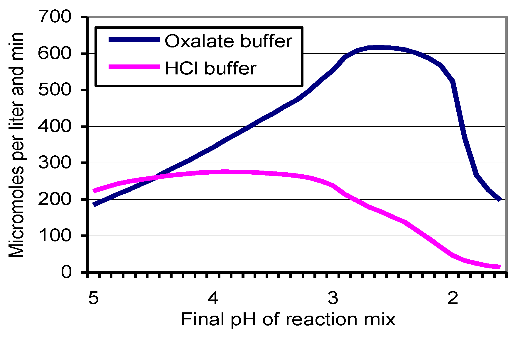
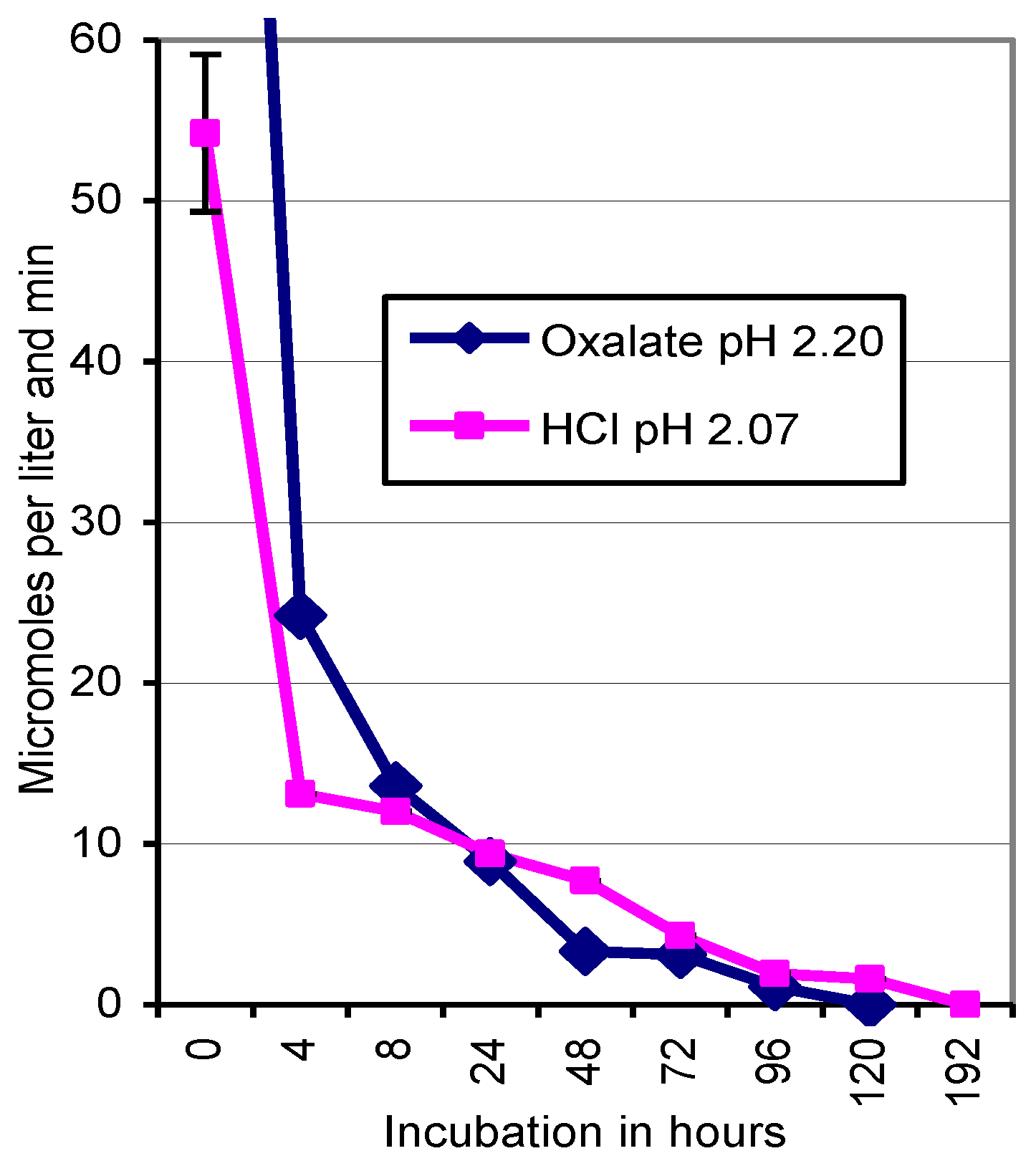
2.6. Inactivation of Laccase by Oxalate and HCl

Kuehnerommyces mutabilis Km 10 was incubated on 20 g L−1 malt extract and 5 g L−1 pancreatic peptone in aseptic liquid culture at 20 °C for 35 d. Using culture fluid aliquots to determine the pH dependence of ABTS oxidation by laccase, the reaction mixtures, starting at pH 5.0 (0.1 M KH2PO4), were successively acidified with oxalate crystals or HCl down to pH 1.6. To determine laccase damage by long-term exposure to low pH conditions, fungal culture fluids were pre-incubated in phosphate-oxalate (pH 2.20) or phosphate-HCl buffer (pH 2.07) for 0 to 192 h at 20 °C in triplicate before ABTS was added.

2.7. Oxalate Tolerance of K. mutabilis Mycelia

Quadruplicate test tubes Ø16 mm were filled with crude beech wood dust of 1 g in DW. The substrates were moistened with 2 mL deionized water that contained 0, 5, 10, and 50 mg oxalic acid, respectively. The autoclaved substrates were aseptically inoculated with Km 10 mycelia from malt agar plates and incubated at 20 °C.

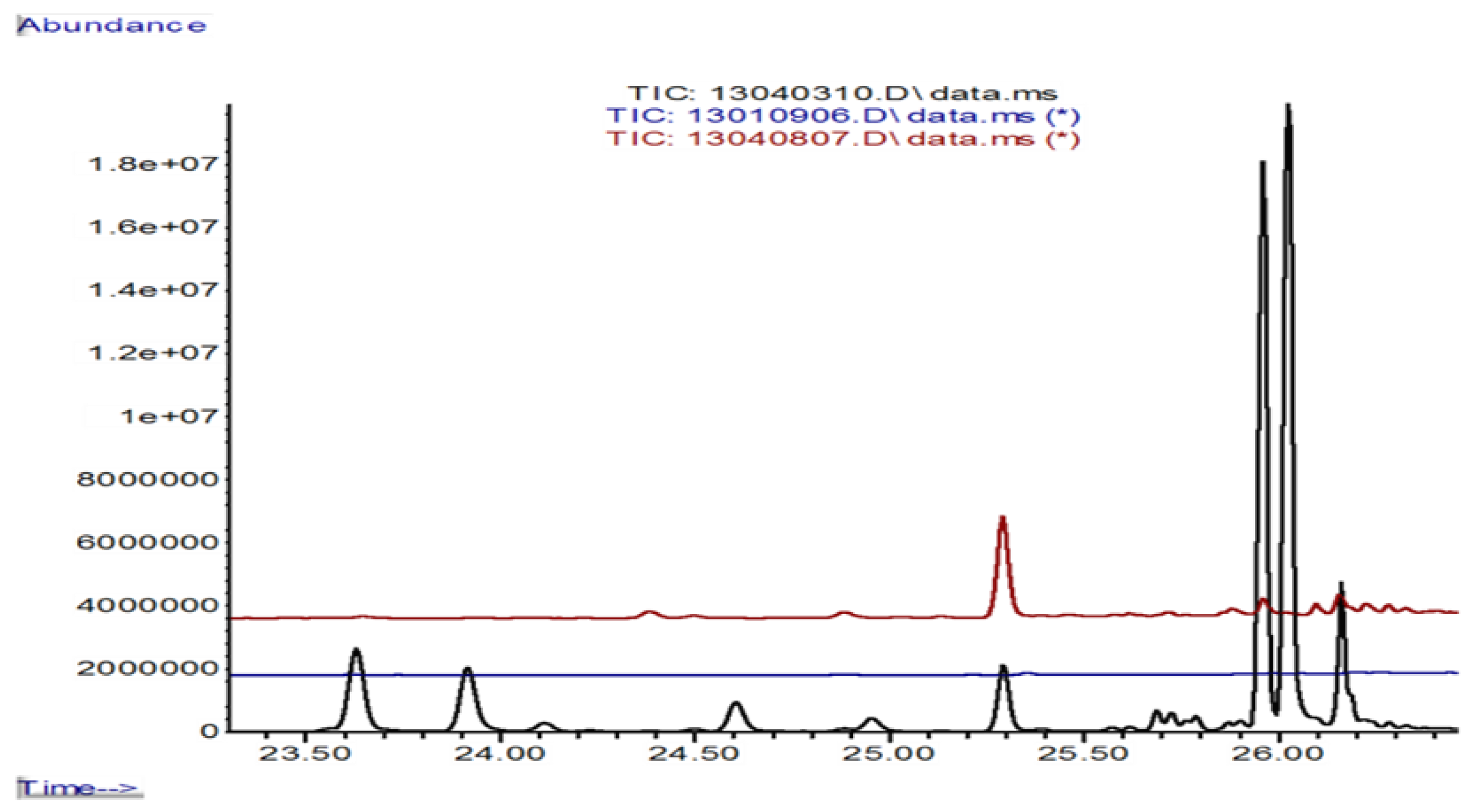
2.8. Collection of Volatile Terpenes from 0.3 L Packing Jars

At the desired target stage of the fungal colonies, the internally aseptic packing jars were opened to filtered ambient air for 10 min, 24 h prior to examination. To collect the volatiles subsequently emitted within 24 h, the de-Teflonized screw caps were pierced (Ø 1 mm) to insert the Solid Phase Micro Extraction (SPME) unit (polydimethylsiloxane 100 µm, Supelco Analytics, Sigma Aldrich) for solid phase extraction over 45 min at 23 °C. The collected terpenes were identified with the GC-MS system Agilent 6890 Series (Agilent Technologies, Inc., Wilmington, DE 19808-1610, USA), equipped with an Agilent 5973 mass-selective detector and the column ZB-5MSi, Øi 0.25 mm × 30 m, 0.25 µm film thickness (Phenomenex Inc., D-63741 Aschaffenburg, Germany), using helium at 1 mL min−1 as the carrier gas.
After splitless injection at 22 °C, a temperature of 40 °C, held for 3 min, was raised at 5 °C min−1 to 150 °C, and at 60 °C min−1 to 300 °C, held for 2 min, to a total of 29.5 min. Reported mono- and sesquiterpenes concur with the library spectra of Wiley 275.L in match qualities > 93% and show identical retention times in measurements of different culture treatments. The spectra of main compounds, such as bisabolenes, were ascertained in comparison with those of the original chemicals.

2.9. Statistical Treatments

SPSS 8.0 software (SPSS Inc., Chicago, IL, USA) was used to calculate the standard deviations (SD) of two- to ten-fold replicates in the determination of fungal metabolites. Furthermore, we calculated confidence intervals to designate significant differences in the concentration ranges of oxalate, laccase, and MnP within the proximate substrate sectors of the advancing mycelia (Table 1). The abundance values of volatiles in Tables and Figures indicate the relative maximum concentrations of terpenes emitted from fungal cultures into a 0.3 L headspace within 24 h. Values were elected among two to three replicate samples.

3. Results

3.1. Dual Cultures in Glass Tubes

3.1.1. Oxalate Production

The sole Km 10 mycelia growing in the columns of 100 mm beech wood at pH 4.4 acidified the substrate by 1–1.2 units. They released occasional oxalate traces of 13–35 mg kg−1 wood (DW) in the proximal Kp, rather than in the distal Kd region (Figure 1). The sole Foma 4 mycelia concentrated 15–21 g kg−1 oxalate around the advancing hyphal tips (Fp) and held as little as 2 g kg−1 in the older part of the fungal colony (Fd) (Table 1). The mycelia of Km 10, approaching those of Foma 4 by 22–19 mm in dual culture, fundamentally incited alterations in the distribution of oxalate in the thallus of the brown rot fungus. Concentrations of 9–13 g kg−1 in Fp were linked with 16–19 g kg−1 in the older Fd region.
On mycelial contact, Km 10 formed a melanized barrier line region of 4 mm which was held at pH 3.4 and arrested the progress of both mycelia for a mean of 11 days. The barrier prevented any diffusions of Foma 4 oxalate that had been accumulated to concentrations of 24–30 g kg−1 in a substrate acidified to pH 1.4–1.75 into the sector of Km 10.
During the subsequent spread of Foma 4 over the barrier line and the entire Km 10 region, oxalate concentrations of 16–29 g kg−1 around the hyphal tips dropped to traces of 16–126 mg kg−1 in the older mycelial parts (Table 1), leaving the entire colonized wood dust in the 2.64–3.34 pH range. Attempts to retrieve Km 10 from its former substrate sector failed.

3.1.2. Production of Oxidoreductases by K. mutabilis Km 10

On the beech wood substrates pH 3.2–4.4, the sole thalli of Km 10 concentrated the activities of laccase and manganese peroxidase in the Kp region of the hyphal tips. Negligible values were found in Kd and in the substrate Ns prior to its colonization (Table 1). The mycelia of Foma 4, approaching those of Km 10 by 22–0 mm in dual culture, gave rise to a reduction in laccase activities by one fourth; however, the mycelia of Foma 4 gave rise to three- to five-fold increases in MnP activities. In the state of mutual arrest at the junction point, laccase and traces of MnP were concentrated within the barrier section of 4 mm to vanish in the substrates overgrown by Foma 4 (Table 1).

3.2. Altering Oxalate Release with Calcium

In tests on the impact of soluble oxalate on fungal interactions in glass tubes, the beech wood dust with an intrinsic Ca content of 1.4 g kg−1 DW was amended with 3 and 10 g kg−1 Ca, respectively, by CaCl2 × 2H2O. In general, equimolar oxalate solutions around pH 1.0 amended with CaCl2 dropped by 0.43 pH units upon the formation of the insoluble Ca oxalate, whereas CaCO3 supplements increased pH by 0.1 units.
In analogy to Table 1 values, amendments of 3 and 10 g kg−1 Ca as chloride did not alter the pH conditions across the Kd to Fd sections of the 100 mm wood dust columns. Values of soluble oxalate generally dropped to 5–51 mg kg−1. Laccase activities persisted in the order of those of the non-amended Table 1 substrates, whereas activities of MnP exceeded 3200–3700 µmol kg−1 wood DW in treatments of 10 g kg−1 Ca during mycelial contact (data not shown). The weekly growth rates at 20 °C amounted to 10.2 ± 1.3/11.4 ± 1.0/6.7 ± 0.9 mm for Km 10, and 19.3 ± 1.0/16.2 ± 2.1/6.9 ± 1.2 mm for Foma 4 on the substrates amended with 0/3/10g kg−1 Ca, respectively. Substrates amended with 3 g kg−1 Ca did not show the brown pigmented barrier zones at the mycelial junction points, whereas Foma 4 overgrew Km 10 in dozens of replicates. In substrates amended with 10 g kg−1 Ca, the depressed advance of Foma 4 predominantly ended at the mycelial junction points.

3.3. Stability of Km 10 Laccase at Low pH Conditions

Low pH conditions caused by HCl and oxalate interfered with the persistence and activity of the non-purified laccase as part of the fungal culture fluid. Acidifying a 0.1 M phosphate buffer in the reaction mixture with HCl revealed a maximum of ABTS oxidation at pH 3.8–4.0 (276 ± 14 µmol L−1 min−1) and a drop to 152 ± 7 and 15 ± 2 µmol L−1 min−1 at pH 2.5 and 1.6, respectively (Figure 2). Using oxalate as a source of protons, the conversion rate of ABTS peaked at pH 2.4 (617 ± 54 µmol L−1 min−1) and displayed identical activities of 190 ± 7 µmol L−1 min−1 at pH 5.0 and 1.6. Peaking at pH 2.4 could point to possible interferences with MnP, activated by H2O2 that could have been formed during the decarboxylation of oxalate in the complex mycelial culture fluid (Figure 2). Pre-incubation for 0 to 192 h of the Km 10 culture fluid in phosphate-oxalate and phosphate-HCl buffers acidified to pH 2.20 and 2.07, respectively, reduced the oxidation of the subsequently added ABTS dramatically. The enzyme activities reached zero after incubation for 120 h in the oxalate, and for 192 h in the HCl treatment (Figure 3).

3.4. Colonization of Oxalate-Amended Beech Wood Dust by K. Mutabilis

The colonization of quadruplicate sets of beech wood dust in test tubes by the agar-based inocula of Km 10 was pH dependent. In the presence of 0 and 5 mg g−1 oxalate and initial pH values of 5.08 and 3.10, respectively, all samples were overgrown. Oxalate was metabolized, raised the hyphal density to optically estimated 175%, and increased the pH value of the colonized substrate to that of the control (Table 2). The substrates acidified to pH 2.61 and 1.68, respectively, repelled 7 out of 8 inocula.

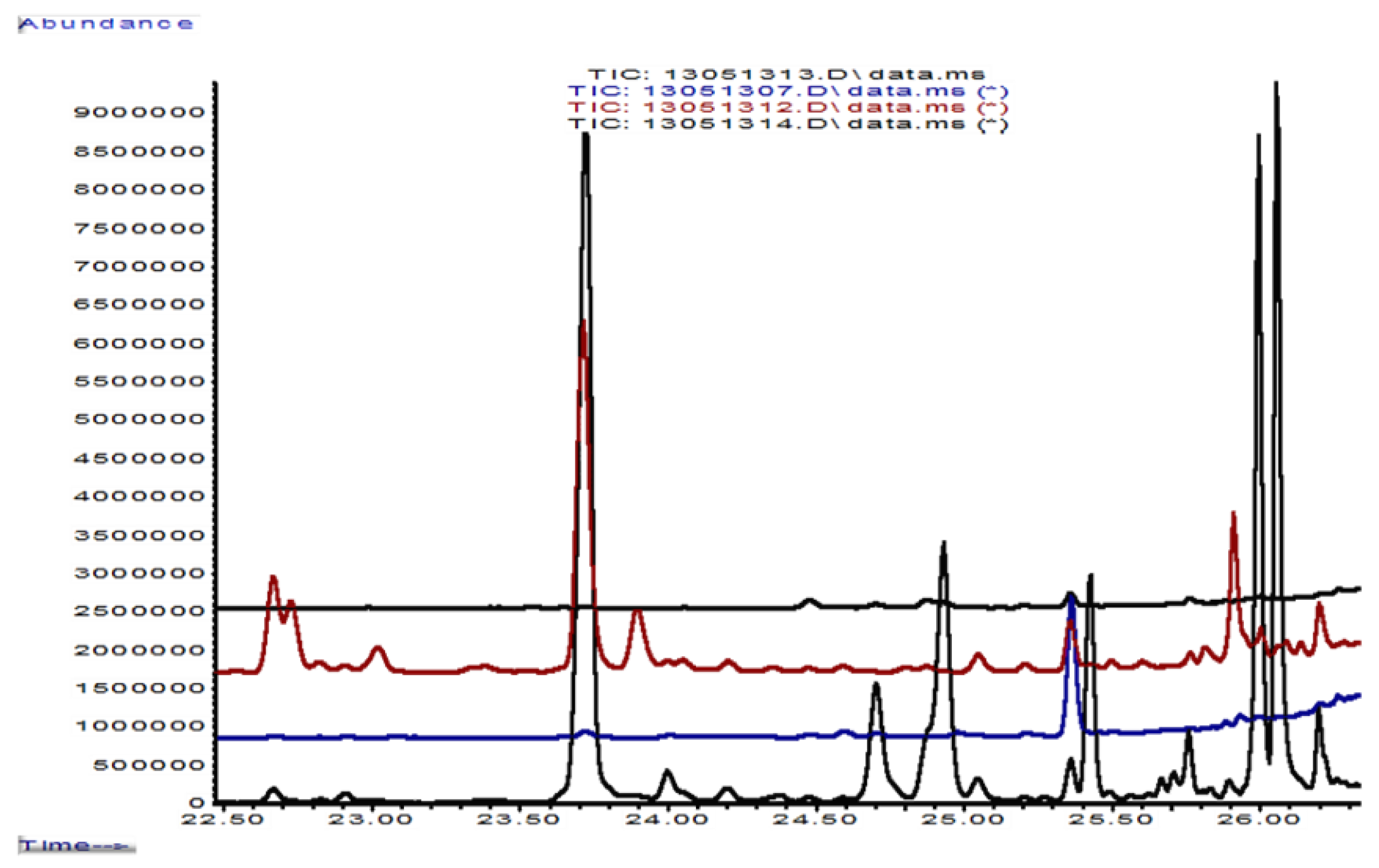
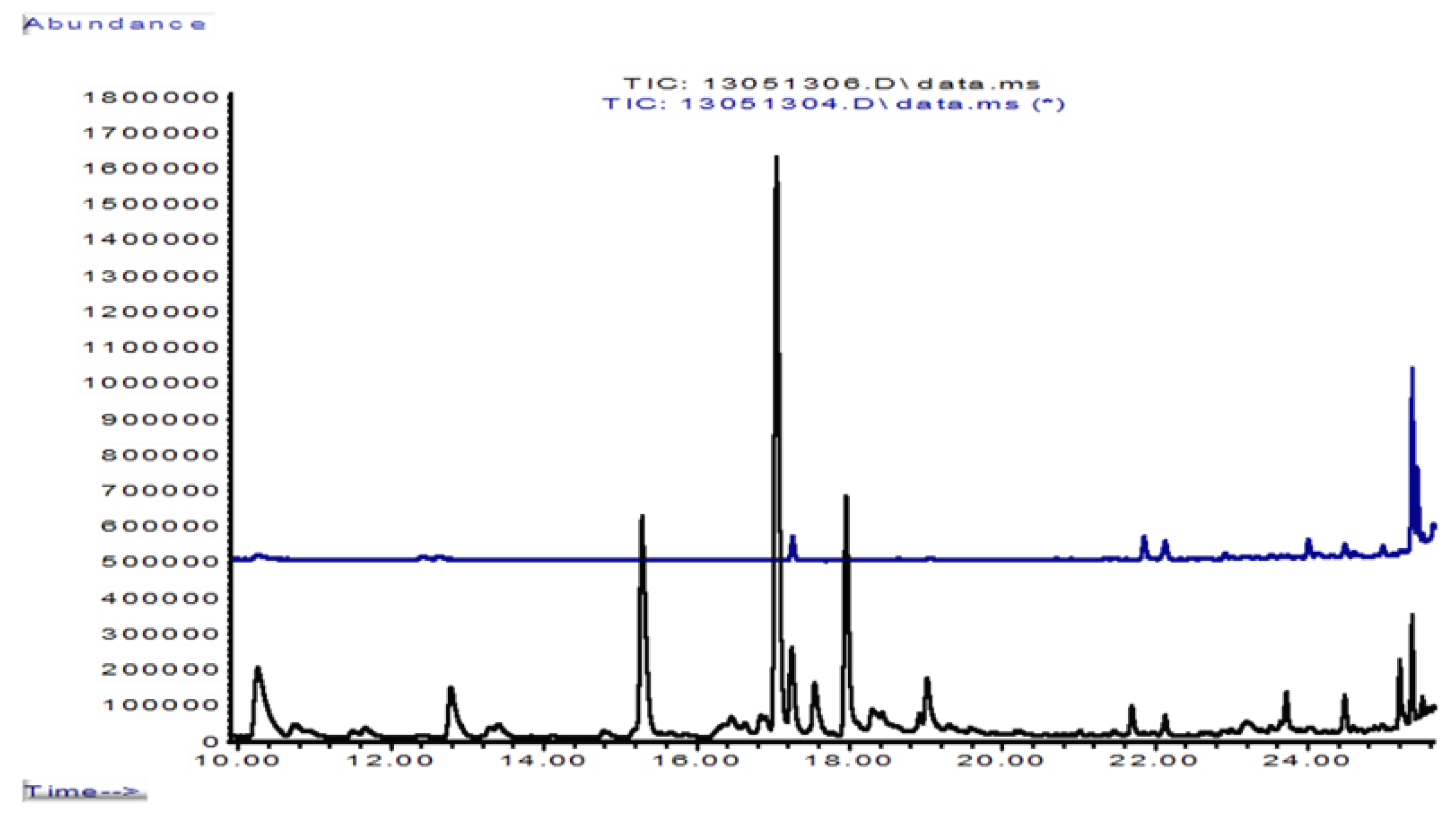
3.5. Formation of Volatile Sesquiterpenes in 0.3 L Jars

Fertilized and autoclaved beech wood dust of 6 g released a set of tentatively identified and confirmed monoterpenes, oxygenated compounds and sesquiterpenes into the 0.3 L headspaces of packing jars within 24 h (Table 3). Inoculated with single fungal cultures, the volatile production changed considerably with culture age and apparently with the depletion of nutrients. Actively growing Km 10 mats displayed traces of junipene and alpha-muurolene and removed any C10 to C12 compounds from the headspace air. Single cultures of Foma 4 released seven sesquiterpenes (C15H24) in low concentrations, too. The first contact of Km 10 and Foma 4 mycelia in dual culture incited an eruption of volatiles in abundances of 35 × 106 (Table 3, column 8; Figure 4 with more details). The dominating and safely identified trans-alpha- (12.5 × 10⁶) and beta-bisabolenes (16 × 106) were accompanied by beta-cubebene (1.9 × 10⁶), gamma-muurolene (1.1 × 10⁶), and delta-cadinene (2.4 × 10⁶). The abundance of emitted sesquiterpenes doubled to 69 × 106 with the formation of a melanized barrier line at the junction point of the mycelia. Accordingly, trans-alpha-bisabolene (26 × 106), beta-bisabolene (30 × 106) and delta-cadinene (5.6 × 10⁶) reached top values within 24 h of emission into the 0.3 L headspace (Table 3, column 9). Traces of gamma-muurolene remained when the section of Km 10 had been overgrown by Foma 4. The application of 6 ml HCl solution pH 1.95 to growing mats of Km 10 mimicked the eruption of volatiles, especially of gamma-muurolene, trans-alpha-bisabolene and beta-bisabolene to a total abundance of 24 × 106 (Table 3, column 7; Figure 5 with more details). Apart from the solution of methyloxalate (pH 2.45), those of oxalate (pH 1.95) and the neutral water control had no effect (Figure 5).
The potential response of Foma 4 colonies to volatile emanations was ascertained with their exposure to 6 g of a non-sterile beech wood dust/soil mix (Figure 6). The fungus removed monoterpenes, oxygenated compounds and most of the sesquiterpenes from the headspace air.

4. Discussion

4.1. Dynamics of Interaction

The closed test system of glass tubes conferred the expected dominance of the mold-susceptible F. pinicola over the competitively strong K. mutabilis strain [20]. With the production of the virulence factor oxalate [54,66,67], Foma 4 acidified its wood substrate sector to the normally lethal pH 1.4–1.7 [66,74,76,77] without impairing its own vitality. Km 10 could secure its substrate sector and the optimum pH range of 3.2–3.4 by erecting a 4 mm wide melanized barrier zone and via an eruption of antimicrobial volatiles, at least at the start of a mean lag phase of 11 days. With the temporary security of its vegetative thallus, Km 10 switched from an invasion mode of highest metabolic activity into a steady state mode [85]. This comprised activity losses of its manganese peroxidase but not of its laccase in the barrier zone and a drop of its laccase (11×) and MnP activities (37×) in the adjacent Kp sector during the stationary lag phase (Table 1).
Foma 4 perceived the approaching mycelium of its competitor over 19–22 mm of non-colonized residual substrate that had been enriched with traces of laccase, MnP (Table 1), and apparently with VOCs (Table 3, Figure 4) in the gaseous or solute-bound state in advance of the Km 10 mycelium. The resulting accumulation of oxalate in the whole thallus of Foma 4 gave rise to substrate pH values below 2.0 (Table 1) but questioned somewhat the role of this low-pKa acid as a virulence factor in plant pathogenesis. The continuous accumulation of oxalate indicated that Foma 4 persisted in the invasion mode until Km 10 was finally overgrown (Table 1). The production of laccase ascribed to certain F. pinicola accessions [15,60,86] could not be recorded.
Km 10 also sensed the approaching Foma 4 mycelium at least over the spectrum of its VOCs (Table 3, Figure 4) and compounds of a higher molecular weight. Comparable long-distance interactions, mediated by VOCs and diffusible factors, are also reported for dual cultures on agar plates [26,27,87]. Junctions of different wood-decaying basidiomycetes are frequently linked with changes in laccase and rising MnP activities in the interaction zones [57,58,88,89]. The laccase activity of Km 10 in dual culture dropped to 25% of the values recorded for the monoxenically growing hyphal tips. Laccase contributes to the formation of the melanized barrier zone of 4 mm by the oxidation of precursors [28,29] such as tyrosine, DOPA, catechol, and several phenolics to brown and black granular deposits on the inner and outer fungal cell walls and in the cytoplasm [90].
With its wide impermeability to fungal metabolites, it blocked the insinuation of oxalate from the Foma 4 sector, stabilized the Km 10 compatible pH conditions to around 3.4 and confined its oxalate content to the 35 mg kg−1 wood DW produced by Km 10 itself in Kp regions (Table 1, Figure 1). In microbial interactions, laccase is not biocidal [91] but catalyzes, e.g., the formation of antimicrobial reactive quinones and inhibitory radical scavengers from phenolic moieties [92].
The activity of MnP peaked in its first mycelial contact with a timber substrate containing 260 mg kg−1 manganese as the enzymes’ co-factor but abated and was no longer needed as a pioneer catalyst when mycelial spread terminated in the widest impermeable barrier region (Table 1). It also had become less important for the degradation of oxalate [79] and H2O2 formed by Foma 4 and implicated in the interaction prior to the formation of the blocking barrier region.
There were no clues for MnP inactivation via the abstraction of its stabilizing Ca2+ cations [80,81] by the oxalate chelant. As its catalytic cycle includes the reduction of H2O2 + 2 H+ to 2H2O [1,2,3], MnP may act in analogy to plant peroxidase in the control of this stress-induced active oxygen species [93] and reduce the acidity of the substrate. Peroxide itself is primarily seen as a signaling agent rather than a toxicant [94].

4.2. Volatile Sesquiterpene Components in the Interaction

The eruption of the gaseous sesquiterpenes beta-bisabolene > trans-alpha-bisabolene > delta-cadinene > beta-cubebene > gamma-curcumene and others upon mycelial contact has been observed for the first time (Table 3, Figure 4) and is solely ascribed to Km 10 (Figure 5). Its time course coincided with the formation of the melanized barrier zone and could be understood to ensure this process. Several bisabolene isoforms and their alcohols are constituents of the antimicrobial essential oils from plant tissues and seeds [95,96]. Beta-bisabolene constitutes up to 80.5% of the essential oil of Daucus carota L. seeds [97] and makes up 4.7–25% of several other essential oils [98,99,100]. Cis-alpha-bisabolene comes to 22.7% in the essential oil of the perennial herb Helichrysum rugulosum (Asteraceae) [98]. Bisabolenes show antibacterial, antifungal [97,98,99,100] and antioxidant properties [100,101]. Wounding grand fir trees (Abies grandis L.) induce the synthesis of (E)-alpha-bisabolene from farnesyl diphosphate by (E)-alpha-bisabolene synthase, directed against insect herbivores and fungal pathogens [102]. The highly concentrated delta-cadinene emitted during the mycelial interaction was not inhibitory to the tree pathogen Phytophthora ramorum [103]. Not recorded were the antimicrobial sesquiterpenes kuehneromycin A and B [104].
The Foma 4 thallus efficiently cleaned the 0.3 L headspace of the packing jars within 24 h from monoterpenes, oxygenated compounds, and sesquiterpenes (Figure 6), presumably after their sorption to mycelium and the substrate [105,106], rather than via respiratory mechanisms. Microbial VOCs receptors have not yet been described [37]. In general, VOCs are stimulatory at low, and inhibitory at high concentrations [39,40,41]. Their temporary mass production (Table 3) could primarily confine the energy resources of Km 10 and feed its competitor.

4.3. The Role of Oxalate

The drastic accumulation of oxalate by Foma 4 triggered by the approaching Km 10 mycelium and its melanized barrier region (Section 3.1.1) led to pH values below 2.0. These pH conditions repelled Km 10 inocula from oxalate amended wood dust (Table 2) and resulted in cell death and wilting of (wounded) plant tissue [74,107]. The non-purified laccase as part of the Km 10 culture fluid lost its entire catalytic potential to ABTS if the phosphate buffer was gradually acidified with HCl, but not with oxalate to pH values below 2.0 (Figure 2). In contrast, both the completely dissociated HCl (pKa = −8.0) and the partially HC2O4 dissociated oxalate (pKa1 = 1.23; pKa2 = 4.9) inactivated laccase definitively at pH around 2.0 within 5–8 days of pre-incubation in the respective buffer solution (Section 3.3, Figure 3). Activity losses down to 3–9.5% are reported for Trametes versicolor and Pycnoporus cinnabarinus laccases incubated for 4–6 h in citrate and B&R buffer of pH 2.0 and 2.2, respectively [108,109], resembling the data of Figure 3. A 92 kDa laccase from the pH insensitive F. pinicola was stable from pH 1.5–11 [86]. In the current stability tests, the inactivation of laccase by the sequestration of its catalytic Cu cations, at least by oxalate [82], was not addressed.
The precipitation of oxalate with CaCl2 down to soluble rest concentrations in the mg range further acidified the substrate, prevented the pigmentation of a barrier zone in spite of high laccase activities (Section 3.2), did not confine the ability of Foma 4 to overgrow Km 10, and stressed that all oxalate effects in this fungal interaction can be mimicked by (HCl generated) acidity. In unbarked leafwood blocks 5 × 5 × 12 mm, living parenchyma cells of cambium and outer sapwood were killed by 24 h incubations in oxalic and citric acid solutions of pH 1.8–2.1 and by sulphuric and nitric acid solutions at pH 1.2. The necrobiotic effects vanished with their widest dissociation at pH 3.9–6.0 (unpublished). Oxalate even failed to trigger the eruption of sesquiterpene volatiles in Km 10 while the dissociated HCl did so (Figure 5). Nevertheless, 0.05 M CaCl2 applied to culture filtrates of the pathogen Sclerotium rolfsii indirectly reduced plant tissue maceration in vitro [73] by the oxalate dependent polygalacturonase [66] to underline the significance of oxalate, at least in plant pathogenesis rather than in the current interaction trial.
The present data do not unambiguously confirm that oxalate or even the extreme pH conditions account for the breakdown of, and have access to, the widely impermeable barrier zone of Km 10 and the laccase of its interior. But with the formation of the barrier zone itself, the thallus of Km 10 believed its substrate sector secured against further attacks. Thus, it terminated the phase of exuberant metabolic, enzymatic and defensive activities that characterized the fungal invasion mode during colonization of a juvenile substrate. This is indicated by the drop of laccase (11×) and MnP (37×) titers in the Kp region (Table 1 and Table 3). Similarly, in technical inoculations of European beech sections with K. mutabilis or Trametes versiolor, the mycelia rapidly cause a DW loss of 30% by exhausting the easily accessible carbohydrates in the invasive phase of colonization. In the following steady state mode, the fungi drastically reduce the rate of metabolic activity and substrate breakdown to their current and very low needs in nutrients [85].
Additionally, the implication of further compounds in the fungal interaction (Section 4.4), stalling [30,31] and autotoxification effects [32,33] could have resulted in debilitation of the arrested Km 10 mycelium. Contemporarily, Foma 4 persisted in the invasion mode of undiminished oxalate production and vitality. It overgrew and digested a metabolically slackening Km 10 in the steady state mode after the mean lag phase of 11 days. From its overgrown substrate sector, Km 10 could no longer be retrieved. Thereby, several (but not all) mold-resistant basidiomycetous white rot and ground fungi overgrew and killed fungal and bacterial contaminants and used them as a nutrient source [35,110]. The cultivated mushroom Agaricus bisporus has expressively been shown to use the microbial biomass formed during substrate composting as primary source of nutrition [111].

4.4. Further Volatile Biocidal Terpenes of F. pinicola

Ethnopharmacological studies with organic solvent extracts from basidiomes of the polypores Fomes fomentarius, Fomitopsis pinicola, Laricifomes officinalis, Laetiporus sulphureus, and Piptoporus betulinus yielded polysaccharides and up to 100 triterpenoids with anti-inflammatory, cytotoxic, and antimicrobial effects [112,113,114,115]. The lanosterol derived triterpenoids, mainly lanostanes, account for nearly 75% of the secondary metabolites in polypores. They form acids, alcohols, aldehydes, esters, ethers, glycosides, ketones, lactones, peroxides, and miscellaneous compounds, primarily with 30 carbon atoms and a tetracyclic skeleton [113]. Lanostanoid derivatives from the F. pinicola sporophore such as polyporenic acid C, 3R-acetyloxylanosta-8,24-dien-21-oic acid, pinicolic acid A, trametenolic acid B, and fomitopsic acid were antimicrobial against Bacillus subtilis [115]. Data regarding the biocidal activity of its diterpenes cembrene A, (Z)-biformene, abietatriene, and 7,13-abietadiene [116] were not found. To date, the contribution of these antimicrobial secondary metabolites to the long-term interactions of basidiomyceteous mycelia or their early production by the mycelia of F. pinicola during the colonization of timber in the invasion mode has not been reported but cannot be ignored.

5. Conclusions

In the primary phase of substrate colonization, K. mutabilis, with its exuberant production of laccase, MnP, and antimicrobial bisabolenes was able to stop the progress of F. pinicola temporarily by the formation of a melanized barrier zone. The barrier secured its substrate sector of pH 3.4 against the sector of Foma 4 with its excess oxalate and the lethal pH conditions down to 1.4 which the brown rot fungus itself tolerated and used in the microbial competition. With the drastic reduction in enzymatic activity, substrate breakdown [85], and the production of antimicrobials in the second phase of wood decay by the stationary and debilitated Km 10 mycelium in the steady state mode, the lethal pH conditions, rather than the oxalate itself, may have gained the potential to inactivate metabolites such as laccase and to kill and overgrow the thallus of Km 10 by Foma 4. Thereby, pH conditions below 2.0 generated by oxalate or strong mineral acids such as HCl had identical lethal effects.
The current results indicate that F. pinicola owes its competitive advantage in the colonization of wounded trees and deadwood to the drastic long-term acidification of the timber substrate, its own insensitivity to extremely low pH conditions, its efficient control of volatile mono- and sesquiterpenes of timber and microbial origin, and the action of an undefined blend of terpenes and allelopathic substances [112,113,114,115,116]. These factors account for a competitive superiority that is expressed in closed systems, such as compact timber, and is the most lacking under aerated conditions. With mass declines of softwoods, as observed during the dry seasons 2018/2019 in Central Europe, that comprised at least 114,000 ha in Germany in 2018 [117] and preliminary 5.24 Mio. m3 timber solely for Thuringia in 2019 [118], the fungus may become epidemic on these preferred host species and depreciate the timber as a building material and pulpwood feedstock within one season [18]. Moreover, with the displacement of the lignin-degrading white rot fungi and the production of highly recalcitrant lignin remnants, F. pinicola delays the mineralization of deadwood and soil formation.

Conflicts of Interest

The author declares no conflicts of interest.

References

  1. Gramss, G. Potential contributions of oxidoreductases from alfalfa plants to soil enzymology and biotechnology: A review. J. Nat. Sci. Sust. Technol. 2012, 6, 169–223. [Google Scholar]
  2. Hofrichter, M.; Ullrich, R.; Pecyna, M.J.; Liers, C.; Lundell, T. New and classic families of secreted fungal heme peroxidases. Appl. Microbiol. Biotechnol. 2010, 87, 871–897. [Google Scholar] [CrossRef] [PubMed]
  3. Wong, D.W.S. Structure and action mechanism of ligninolytic enzymes. Appl. Biochem. Biotechnol. 2008, 157, 174–209. [Google Scholar] [CrossRef] [PubMed]
  4. Baldrian, P. Fungal laccases – occurrence and properties. FEMS Microbiol. Rev. 2006, 30, 215–242. [Google Scholar] [CrossRef] [PubMed] [Green Version]
  5. Chen, D.M.; Taylor, A.F.S.; Burke, R.M.; Cairney, J.W.G. Identification of genes for lignin peroxidases and manganese peroxidases in ectomycorrhizal fungi. New Phytol. 2001, 152, 151–158. [Google Scholar] [CrossRef]
  6. Steffen, K.; Hofrichter, M.; Hatakka, A. Mineralisation of 14C-labelled synthetic lignin and ligninolytic enzyme activities of litter-decomposing basidiomycetous fungi. Appl. Microbiol. Biotechnol. 2000, 54, 819–825. [Google Scholar] [CrossRef]
  7. Steffen, K.; Hofrichter, M.; Hatakka, A. Purification and characterization of manganese peroxidases from the litter-decomposing basidiomycetes Agrocybe praecox and Stropharia coronilla. Enzym. Microb. Technol. 2002, 30, 550–555. [Google Scholar] [CrossRef]
  8. Brink, D.; Ravi, K.; Lidén, G.; Gorwa-Grauslund, M.F. Mapping the diversity of microbial lignin catabolism: Experiences from the eLignin database. Appl. Microbiol. Biotechnol. 2019, 103, 3979–4002. [Google Scholar] [CrossRef] [Green Version]
  9. Gramss, G.; Ziegenhagen, D.; Sorge, S. Degradation of soil humic extract by wood- and soil-associated fungi, bacteria, and commercial enzymes. Microb. Ecol. 1999, 37, 140–151. [Google Scholar] [CrossRef]
  10. Janusz, G.; Pawlik, A.; Sulej, J.; Świderska-Burek, U.; Jarosz-Wilkolazka, A.; Paszczyński, A. Lignin degradation: Microorganisms, enzymes involved, genomes analysis and evolution. FEMS Microbiol. Rev. 2017, 41, 941–962. [Google Scholar] [CrossRef] [Green Version]
  11. Müller, J.; Engel, H.; Blaschke, M. Assemblages of wood-inhabiting fungi related to silvicultural management intensity in beech forests in southern Germany. Eur. J. For. Res. 2007, 126, 513–527. [Google Scholar] [CrossRef]
  12. Kubartová, A.; Ottosson, E.; Stenlid, J. Linking fungal communities to wood density loss after 12 years of log decay. FEMS Microbiol. Ecol. 2015, 91. [Google Scholar] [CrossRef] [PubMed] [Green Version]
  13. Leonhardt, S.; Hoppe, B.; Stengel, E.; Noll, L.; Moll, J.; Bässler, C.; Dahl, A.; Buscot, F.; Hofrichter, M.; Kellner, H. Molecular fungal community and its decomposition activity in sapwood and heartwood of 13 temperate European tree species. PLoS ONE 2019, 14, e0212120. [Google Scholar] [CrossRef] [PubMed]
  14. Ottoson, E. Succession of Wood-Inhabiting Fungal Communities. Diversity and Species Interactions During the Decomposition of Norway Spruce. Ph.D. Thesis, Swedish University of Agricultural Sciences, Uppsala, Swedish, 2013. [Google Scholar]
  15. Wu, B.; Gaskell, J.; Held, B.W.; Toapanta, C.; Vuong, T.; Ahrendt, S.; Lipzen, A.; Zhang, J.; Schilling, J.S.; Master, E.; et al. Substrate-specific differential gene expression and RNA editing in the brown rot fungus Fomitopsis pinicola. Appl. Environ. Microbiol. 2018, 84, e00991-18. [Google Scholar] [CrossRef] [Green Version]
  16. Allard, G.; FAO; Overview of forest pests Kyrgyz Republic; Forest Resources Development Service; Forest Management Division; Forestry Department. Forest Health & Biosecurity Working Papers; Working Paper FBS/29E; FAO: Rome, Italy, 2007. Available online: http://www.fao.org/forestry/12277-0ac81b7e1d2a6d6861bbab1599f7244b9.pdf (accessed on 19 December 2019).
  17. Plantwise Knowledge Bank. Species Page Brown Crumbly Rot, Fomitopsis pinicola. Available online: https://www.plantwise.org/knowledgebank/datasheet/24310 (accessed on 19 December 2019).
  18. Vogel, S.; Alvarez, B.; Bässler, C.; Müller, J.; Thorn, S. The red-belted bracket (Fomitopsis pinicola) colonizes spruce trees early after bark beetle attack and persists. Fungal Ecol. 2017, 27, 182–188. [Google Scholar] [CrossRef]
  19. Ruokolainen, A.; Shorohova, E.; Penttilä, R.; Kotkova, V.; Kushnevskaya, H. A continuum of dead wood with various habitat elements maintains the diversity of wood-inhabiting fungi in an old-growth boreal forest. Eur. J. For. Res. 2018, 137, 707–718. [Google Scholar] [CrossRef]
  20. Gramss, G. Invasion of wood by basidiomycetous fungi. IV. Microbiological approach to the role of kratovirulence in the expression of pathovirulence. J. Basic Microbiol. 1987, 27, 241–251. [Google Scholar] [CrossRef]
  21. Arfi, Y.; Levasseur, A.; Record, E. Differential Gene Expression in Pycnoporus coccineus during interspecific mycelial interactions with different competitors. Appl. Environ. Microbiol. 2013, 79, 6626–6636. [Google Scholar] [CrossRef] [Green Version]
  22. Holmer, L.; Renvall, P.; Stenlid, J. Selective replacement between species of wood-rotting basidiomycetes, a laboratory study. Mycol. Res. 1997, 101, 714–720. [Google Scholar] [CrossRef]
  23. O’Leary, J.; Hiscox, J.; Eastwood, D.C.; Savoury, M.; Langley, A.; McDowell, S.W.; Rogers, H.J.; Boddy, L.; Müller, C.T. The whiff of decay: Linking volatile production and extracellular enzymes to outcomes of fungal interactions at different temperatures. Fungal Ecol. 2019, 39, 336–348. [Google Scholar] [CrossRef]
  24. Ujor, V.C.; Monti, M.; Peiris, D.; Clements, M.O.; Hedger, J.N. The mycelial response of the white-rot fungus, Schizophyllum commune to the biocontrol agent, Trichoderma viride. Fungal Boil. 2012, 116, 332–341. [Google Scholar] [CrossRef] [PubMed]
  25. Ujor, V.C.; Adukwu, E.C.; Okonkwo, C.C. Fungal wars: The underlying molecular repertoires of combating mycelia. Fungal Boil. 2018, 122, 191–202. [Google Scholar] [CrossRef] [PubMed]
  26. Giles, R.L.; Zackeru, J.C.; Galloway, E.R.; Elliott, G.D.; Parrow, M.W. Single versus simultaneous species treatment of wood with Ceriporiopsis subvermispora and Postia placenta for ethanol applications, with observations on interspecific growth inhibition. Int. Biodeterior. Biodegradation 2015, 99, 66–72. [Google Scholar] [CrossRef]
  27. Minerdi, D.; Bossi, S.; Gullino, M.L.; Garibaldi, A. Volatile organic compounds: A potential direct long-distance mechanism for antagonistic action of Fusarium oxysporum strain MSA 35. Environ. Microbiol. 2009, 11, 844–854. [Google Scholar] [CrossRef]
  28. Baldrian, P. Interactions of heavy metals with white-rot fungi. Enzym. Microb. Technol. 2003, 32, 78–91. [Google Scholar] [CrossRef]
  29. Rühl, M. Laccases and Other Ligninolytic Enzymes of the Basidiomycetes Coprinopsis cinerea and Pleurotus ostreatus. Ph.D. Thesis, Georg-August-University, Göttingen, Germany, December 2009. [Google Scholar]
  30. Arora, D.K.; Upadhyay, R.K. Effect of fungal staling growth substances on colony interaction. Plant Soil 1978, 49, 685–690. [Google Scholar] [CrossRef]
  31. Park, D.; Robinson, P.M. Isolation and bioassay of a fungal morphogen. Nature 1964, 203, 988–989. [Google Scholar] [CrossRef]
  32. Gramss, G. Role of soil mycelium in nutrition of wooddestroying basidiomycetous fungi on inoculated wood blocks in soil. J. Basic Microbiol. 1979, 19, 143–145. [Google Scholar]
  33. Rudman, P. Some observations on the biological activity of the metabolic products of the wood rotting fungus Lentinus lepideus. Holzforschung 1963, 17, 149–151. [Google Scholar] [CrossRef]
  34. Korn, V.; Inamdar, A.A. Are some fungal volatile organic compounds (VOCs) mycotoxins? Toxins 2015, 7, 3785–3804. [Google Scholar]
  35. Gramss, G. The Influence of the concomitant microflora on establishment and dieback of decay fungi in standing timber. J. Phytopathol. 1987, 120, 205–215. [Google Scholar] [CrossRef]
  36. Gao, Y.; Jin, Y.; Li, H.-D.; Chen, H.-J. Volatile organic compounds and their roles in bacteriostasis in five conifer species. J. Integr. Plant Boil. 2005, 47, 499–507. [Google Scholar] [CrossRef]
  37. Bohm, K.; Martín-Sánchez, L.; Garbeva, P. Microbial volatiles: Small molecules with an important role in intra- and inter-kingdom interactions. Front. Microbiol. 2017, 8. [Google Scholar]
  38. Yuan, J.; Zhao, M.; Li, R.; Huang, Q.; Raza, W.; Rensing, C.; Shen, Q. Microbial volatile compounds alter the soil microbial community. Environ. Sci. Pollut. Res. 2017, 24, 22485–22493. [Google Scholar] [CrossRef]
  39. Gramss, G. Approach to the nature of volatile compounds that dominate the ecological niche of basidiomycetous ground fungi in the edaphosphere of grassland. Zentralblatt für Mikrobiologie 1985, 140, 597–606. [Google Scholar] [CrossRef]
  40. Quintana-Rodríguez, E.; Rivera-Macias, L.; Adame-Álvarez, R.M.; Torres, J.M.; Heil, M. Shared weapons in fungus-fungus and fungus-plant interactions? Volatile organic compounds of plant or fungal origin exert direct antifungal activity in vitro. Fungal Ecol. 2018, 33, 115–121. [Google Scholar] [CrossRef]
  41. Tortella, G.; Rubilar, O.; Stenström, J.; Cea, M.; Briceno, G.; Quiroz, A.; Diez, M.C.; Parra, L. Using volatile organic compounds to enhance atrazine biodegradation in a biobed system. Biodegradation 2013, 24, 711–720. [Google Scholar] [CrossRef]
  42. McDougall, B.M. Movement of 14C-photosynthate into the roots of wheat seedlings and exudation of 14C from intact roots. New Phytol. 1970, 69, 37–46. [Google Scholar] [CrossRef]
  43. Booth, E.; Strobel, G.; Knighton, B.; Sears, J.; Geary, B.; Avci, R. A rapid column technique for trapping and collecting of volatile fungal hydrocarbons and hydrocarbon derivatives. Biotechnol. Lett. 2011, 33, 1963–1972. [Google Scholar] [CrossRef]
  44. Fischer, G.; Schwalbe, R.; Möller, M.; Ostrowski, R.; Dott, W. Species-specific production of microbial volatile organic compounds (MVOC) by airborne fungi from a compost facility. Chemosphere 1999, 39, 795–810. [Google Scholar] [CrossRef]
  45. Matysik, S.; Herbarth, O.; Mueller, A. Determination of microbial volatile organic compounds (MVOCs) by passive ampling onto charcoal sorbents. Chemosphere 2009, 76, 114–119. [Google Scholar] [CrossRef] [PubMed]
  46. Wilkins, K.; Larsen, K.; Simkus, M. Volatile metabolites from mold growth on building materials and synthetic media. Chemosphere 2000, 41, 437–446. [Google Scholar] [CrossRef]
  47. Benchaar, C.; Calsamiglia, S.; Chaves, A.V.; Fraser, G.; Colombatto, D.; McAllister, T.A.; Beauchemin, K. A review of plant-derived essential oils in ruminant nutrition and production. Anim. Feed. Sci. Technol. 2008, 145, 209–228. [Google Scholar] [CrossRef]
  48. Fisher, R.F. Allelopathy. In Plant Disease, How Pathogens Induce Disease; Horsfall, J.G., Cowling, E.B., Eds.; Academic Press: New York, USA, 1979; pp. 313–330. [Google Scholar]
  49. Hamilton-Kemp, T.R.; Andersen, R.A. Volatiles from winter wheat: Identification of additional compounds and effects of tissue source. Phytochemistry 1985, 25, 241–243. [Google Scholar] [CrossRef]
  50. Gramss, G.; Bergmann, H. Role of plants in the vegetative and reproductive growth of saprobic basidiomycetous ground fungi. Microb. Ecol. 2008, 56, 660–670. [Google Scholar] [CrossRef] [PubMed]
  51. Huffer, S.; Clark, M.E.; Ning, J.C.; Blanch, H.W.; Clark, U.S. Role of Alcohols in growth, lipid composition, and membrane fluidity of yeasts, bacteria, and archaea. Appl. Environ. Microbiol. 2011, 77, 6400–6408. [Google Scholar] [CrossRef] [PubMed] [Green Version]
  52. Sikkema, J.; A De Bont, J.; Poolman, B. Mechanisms of membrane toxicity of hydrocarbons. Microbiol. Rev. 1995, 59, 201–222. [Google Scholar] [CrossRef]
  53. Krokene, P.; Nagy, N.E.; Solheim, H. Methyl jasmonate and oxalic acid treatment of Norway spruce: Anatomically based defense responses and increased resistance against fungal infection. Tree Physiol. 2008, 28, 29–35. [Google Scholar] [CrossRef] [Green Version]
  54. Walz, A.; Zingen-Sell, I.; Theisen, S.; Kortekamp, A. Reactive oxygen intermediates and oxalic acid in the pathogenesis of the necrotrophic fungus Sclerotinia sclerotiorum. Eur. J. Plant Pathol. 2007, 120, 317–330. [Google Scholar] [CrossRef]
  55. Gramss, G.; Kirsche, B.; Voigt, K.-D.; Günther, T.; Fritsche, W. Conversion rates of five polycyclic aromatic hydrocarbons in liquid cultures of fifty-eight fungal species and the concomitant production of oxidative enzymes. Mycol. Res. 1999, 103, 1009–1018. [Google Scholar] [CrossRef]
  56. Leonowicz, A.; Malinowska, M. Purification of the constitutive and inducible forms of laccase of the fungus Pholiota mutabilis by affinity chromatography. Acta Biochim. Pol. 1982, 29. [Google Scholar]
  57. Chen, Q.; Krügener, S.; Hirth, T.; Rupp, S.; Zibek, S. Co-cultured production of lignin-modifying enzymes with white-rot fungi. Appl. Biochem. Biotechnol. 2011, 165, 700–718. [Google Scholar]
  58. Chi, Y.; Hatakka, A.; Maijala, P. Can co-culturing of two white-rot fungi increase lignin degradation and the production of lignin-degrading enzymes? Int. Biodeterior. Biodegrad. 2007, 59, 32–39. [Google Scholar] [CrossRef]
  59. Velázquez-Cedeño, M.; Farnet, A.M.; Mata, G.; Savoie, J.-M. Role of Bacillus spp. in antagonism between Pleurotus ostreatus and Trichoderma harzianum in heat-treated wheat-straw substrates. Bioresour. Technol. 2008, 99, 6966–6973. [Google Scholar] [CrossRef]
  60. Kersten, P.J.; Cullen, D. Copper radical oxidases and related extracellular oxidoreductases of wood-decay Agaricomycetes. Fungal Genet. Boil. 2014, 72, 124–130. [Google Scholar] [CrossRef]
  61. Riley, R.; Salamov, A.A.; Brown, D.W.; Nagy, L.G.; Floudas, D.; Held, B.W.; Levasseur, A.; Lombard, V.; Morin, E.; Otillar, R.; et al. Extensive sampling of basidiomycete genomes demonstrates inadequacy of the white-rot/brown-rot paradigm for wood decay fungi. Proc. Natl. Acad. Sci. USA 2014, 111, 9923–9928. [Google Scholar] [CrossRef] [Green Version]
  62. Arantes, V.; Milagres, A.; Filley, T.R.; Goodell, B. Lignocellulosic polysaccharides and lignin degradation by wood decay fungi: The relevance of nonenzymatic Fenton-based reactions. J. Ind. Microbiol. Biotechnol. 2010, 38, 541–555. [Google Scholar] [CrossRef]
  63. Shah, F.; Mali, T.; Lundell, T. Polyporales brown rot species Fomitopsis pinicola: Enzyme activity profiles, oxalic acid production, and Fe3+-reducing metabolite secretion. Appl. Environ. Microbiol. 2018, 84, e02662-17. [Google Scholar] [CrossRef] [Green Version]
  64. Huang, K.-C.; Couttenye, R.A.; Hoag, G.E. Kinetcs of heat-assisted persulfate oxidation of methyl tert-butyl ether (MTBE). Chemosphere 2002, 49, 413–420. [Google Scholar] [CrossRef]
  65. Hastrup, A.C.S. Aspects of Cellulose Degradation by Brown Rot Fungi. Involvement of Enzymatic and Non-Enzymatic Decay Processes Including Oxalic Acid Regulation and Metal Sequestering. Ph.D. Thesis, University of Copenhagen, Copenhagen, Denmark, June 2011. [Google Scholar]
  66. Dutton, M.V.; Evans, C.S. Oxalate production by fungi: Its role in pathogenicity and ecology in the soil environment. Can. J. Microbiol. 1996, 42, 881–895. [Google Scholar] [CrossRef]
  67. Kunz, C.; Vandelle, E.G.G.; Rolland, S.; Poinssot, B.; Bruel, C.; Cimerman, A.; Zotti, C.; Moreau, E.; Vedel, R.; Pugin, A.; et al. Characterization of a new, nonpathogenic mutant of Botrytis cinerea with impaired plant colonization capacity. New Phytol. 2006, 170, 537–550. [Google Scholar] [CrossRef] [PubMed]
  68. Munir, E.; Yoon, J.-J.; Tokimatsu, T.; Hattori, T.; Shimada, M. New role for glyoxylate cycle enzymes in wood-rotting basidiomycetes in relation to biosynthesis of oxalic acid. J. Wood Sci. 2001, 47, 368–373. [Google Scholar] [CrossRef]
  69. Jarosz-Wilkołazka, A.; Gadd, G.M. Oxalate production by wood-rotting fungi growing in toxic metal-amended medium. Chemosphere 2003, 52, 541–547. [Google Scholar] [CrossRef]
  70. Schilling, J.S. Oxalate Production and Cation Translocation During Wood Biodegradation by Fungi. Electronic Theses and Dissertations. 2006. Available online: http://digitalcommons.library.umaine.edu/etd/336 (accessed on 19 December 2019).
  71. Bateman, D.F. An induced mechanism of tissue resistance to polygalacturonase in Rhizoctonia infected hypocotyls of bean. Phytopathology 1964, 54, 438–445. [Google Scholar]
  72. Bateman, D.F.; Beer, S.V. Simultaneous production and synergistic action of oxalic acid and polygalcturonase during pathogenesis by Sclerotium rolfsii. Phytopathology 1965, 55, 204–211. [Google Scholar]
  73. Punja, Z.; Huang, J.-S.; Jenkins, S. Relationship of mycelial growth and production of oxalic acid and cell wall degrading enzymes to virulence in Sclerotium Rolfsii. Can. J. Plant Pathol. 1985, 7, 109–117. [Google Scholar] [CrossRef]
  74. Kurian, P. The synergistic role of oxalic acid and endopolygalacturonase in bean leaves infected by Cristulariella pyramidalis. Phytopathology 1979, 69, 1301. [Google Scholar] [CrossRef]
  75. Gramss, G. Invasion of wood by basidiomycetous fungi. V. The role of fungal elicitors of plant cell necrobiosis in the expression of pathovirulence and kratovirulence. J. Basic Microbiol. 1989, 29, 563–579. [Google Scholar] [CrossRef]
  76. De Boer, W.; Folman, L.B.; Gunnewiek, P.J.K.; Svensson, T.; Bastviken, D.; Öberg, G.; Del Río, J.C.; Boddy, L. Mechanism of antibacterial activity of the white-rot fungus Hypholoma fasciculare colonizing wood. Can. J. Microbiol. 2010, 56, 380–388. [Google Scholar] [CrossRef]
  77. Desriac, N.; Broussolle, V.; Postollec, F.; Mathot, A.-G.; Sohier, D.; Coroller, L.; Leguerinel, I. Bacillus cereus cell response upon exposure to acid environment: Toward the identification of potential biomarkers. Front. Microbiol. 2013, 4. [Google Scholar] [CrossRef] [Green Version]
  78. Hayaishi, O.; Jakoby, W.B.; Ohmura, E. Enzymatic decarboxylation of oxalic acid. J. Boil. Chem. 1956, 222. [Google Scholar]
  79. Hofrichter, M.; Ziegenhagen, D.; Vares, T.; Friedrich, M.; Jäger, M.; Fritsche, W.; Hatakka, A. Oxidative decomposition of malonic acid as basis for the action of manganese peroxidase in the absence of hydrogen peroxide. FEBS Lett. 1998, 434, 362–366. [Google Scholar] [CrossRef]
  80. Sutherland, G.R.; Aust, S.D. The effects of calcium on the thermal stability and activity of manganese peroxidase. Arch. Biochem. Biophys. 1996, 332, 128–134. [Google Scholar] [CrossRef] [PubMed]
  81. Wang, N.; Ren, K.; Jia, R.; Chen, W.; Sun, R. Expression of a fungal manganese peroxidase in Escherichia coli: A comparison between the soluble and refolded enzymes. BMC Biotechnol. 2016, 16, 87. [Google Scholar] [CrossRef] [PubMed] [Green Version]
  82. Sato, M. Inhibition by oxalates of spinach chloroplast phenolase in unfrozen and frozen states. Phytochemistry 1980, 19, 1613–1617. [Google Scholar] [CrossRef]
  83. Sterjiades, R.; Dean, J.; Eriksson, K.-E.L. Laccase from sycamore maple (Acer pseudoplatanus) polymerizes monolignols. Plant Physiol. 1992, 99, 1162–1168. [Google Scholar] [CrossRef] [Green Version]
  84. Wariishi, H.; Valli, K.; Gold, M.H. Manganese(II) oxidation by manganese peroxidase from the basidiomycete Phanerochaete chrysosporium. Kinetic mechanism and role of chelators. J. Boil. Chem. 1992, 267, 23688–23695. [Google Scholar]
  85. Luthardt, W. Wood Inhabiting Fungi. Cultivation and Wood Mycology. In Holzbewohnende Pilze. Anzucht und Holzmykologie; A. Ziemsen: Wittenberg, Germany, 1969. [Google Scholar]
  86. Park, N.; Park, S.-S. Purification and characterization of a novel laccase from Fomitopsis pinicola mycelia. Int. J. Boil. Macromol. 2014, 70, 583–589. [Google Scholar] [CrossRef]
  87. Evans, J.A.; Eyre, C.A.; Rogers, H.; Boddy, L.; Muller, C. Changes in volatile production during interspecific interactions between four wood rotting fungi growing in artificial media. Fungal Ecol. 2008, 1, 57–68. [Google Scholar] [CrossRef]
  88. Flores, C.; Vidal, C.; Trejo-Hernandez, M.R.; Galindo, E.; Serrano-Carreón, L. Selection of Trichoderma strains capable of increasing laccase production by Pleurotus ostreatus and Agaricus bisporus in dual cultures. J. Appl. Microbiol. 2009, 106, 249–257. [Google Scholar] [CrossRef]
  89. Hiscox, J.; Baldrian, P.; Rogers, H.; Boddy, L. Changes in oxidative enzyme activity during interspecific mycelial interactions involving the white-rot fungus Trametes versicolor. Fungal Genet. Boil. 2010, 47, 562–571. [Google Scholar] [CrossRef]
  90. Bell, A.A.; Wheeler, M.H. Biosynthesis and functions of fungal melanins. Annu. Rev. Phytopath. 1986, 24, 411–451. [Google Scholar] [CrossRef]
  91. Baldrian, P. Wood-inhabiting ligninolytic basidiomycetes in soils: Ecology and constraints for applicability in bioremediation. Fungal Ecol. 2008, 1, 4–12. [Google Scholar] [CrossRef]
  92. Jeon, J.-R.; Baldrián, P.; Murugesan, K.; Chang, Y.-S. Laccase-catalysed oxidations of naturally occurring phenols: From in vivo biosynthetic pathways to green synthetic applications. Microb. Biotechnol. 2012, 5, 318–332. [Google Scholar] [CrossRef] [Green Version]
  93. Decros, G.; Baldet, P.; Beauvoit, B.; Stevens, R.; Flandin, A.; Colombié, S.; Gibon, Y.; Pétriacq, P. Get the balance right: ROS homeostasis and redox signalling in fruit. Front. Plant Sci. 2019, 10, 1091. [Google Scholar] [CrossRef]
  94. Silar, P. Peroxide accumulation and cell death in filamentous fungi induced by contact with a contestant. Mycol. Res. 2005, 109, 137–149. [Google Scholar] [CrossRef]
  95. González-Mas, M.C.; Rambla, J.L.; López-Gresa, M.P.; Blázquez, M.A.; Granell, A. Volatile compounds in citrus essential oils: A comprehensive review. Front. Plant Sci. 2019, 10, 12. [Google Scholar] [CrossRef]
  96. Martins, C.D.M.; Nascimento, E.A.D.; De Morais, S.A.L.; De Oliveira, A.; Chang, R.; Cunha, L.C.S.; Martins, M.M.; Martins, C.H.G.; Moraes, T.D.S.; Rodrigues, P.V.; et al. Chemical constituents and evaluation of antimicrobial and cytotoxic activities of Kielmeyera coriacea Mart. & Zucc. essential oils. Evid.-Based Complement. Altern. Med. 2015, 2015, 1–9. [Google Scholar]
  97. Rokbeni, N.; Mrabet, Y.; Dziri, S.; Chaabane, H.; Jemli, M.; Fernandez, X.; Boulila, A. Variation of the chemical composition and antimicrobial activity of the essential oils of natural populations of Tunisian Daucus carota L. (Apiaceae). Chem. Biodivers. 2013, 10, 2278–2290. [Google Scholar] [CrossRef]
  98. Bougatsos, C.; Meyer, J.J.M.; Magiatis, P.; Vagias, C.; Chinou, I.B. Composition and antimicrobial activity of the essential oils of Helichrysum kraussii Sch. Bip. and H. rugulosum Less. from South Africa. Flavour Fragr. J. 2003, 18, 48–51. [Google Scholar] [CrossRef]
  99. Carneiro, N.S.; Alves, C.C.F.; Alves, J.M.; Egea, M.B.; Martins, C.H.G.; Silva, T.S.; Bretanha, L.C.; Balleste, M.P.; Caon, T.; Silveira, E.V.; et al. Chemical composition, antioxidant and antibacterial activities of essential oils from leaves and flowers of Eugenia klotzschiana Berg (Myrtaceae). Anais da Academia Brasileira de Ciências 2017, 89, 1907–1915. [Google Scholar] [CrossRef]
  100. Kazemi, M.; Rostami, H. Chemical composition, antimicrobial and antioxidant activities of the essential oil of Psammogeton canescens. Nat. Prod. Res. 2014, 29, 277–280. [Google Scholar] [CrossRef]
  101. De Melo, N.I.; Magalhães, L.G.; De Carvalho, C.E.; Wakabayashi, K.A.L.; Aguiar, G.D.P.; Ramos, R.C.; Mantovani, A.L.L.; Turatti, I.C.C.; Rodrigues, V.; Groppo, M.; et al. Schistosomicidal activity of the essential oil of Ageratum conyzoides L. (Asteraceae) against adult Schistosoma mansoni Worms. Molecules 2011, 16, 762–773. [Google Scholar] [CrossRef]
  102. Bohlmann, J.; Crock, J.; Jetter, R.; Croteau, R. Terpenoid-based defenses in conifers: cDNA cloning, characterization, and functional expression of wound-inducible (E)-α-bisabolene synthase from grand fir (Abies grandis). Proc. Natl. Acad. Sci. USA 1998, 95, 6756–6761. [Google Scholar] [CrossRef] [Green Version]
  103. Manter, D.K.; Kelsey, R.G.; Karchesy, J.J. Antimicrobial activity of extractable conifer heartwood compounds toward Phytophthora ramorum. J. Chem. Ecol. 2007, 33, 2133–2147. [Google Scholar] [CrossRef]
  104. Erkel, G.; Lorenzen, K.; Anke, T.; Velten, R.; Gimenez, A.; Steglich, W. Kuehneromycins A and B, two new biological active compounds from a Tasmanian Kuehneromyces sp. (Strophariaceae, Basidiomycetes). Z. Naturforsch. 1995, 50, 1–10. [Google Scholar] [CrossRef]
  105. Malhautier, L.; Khammar, N.; Bayle, S.; Fanlo, J.-L. Biofiltration of volatile organic compounds. Appl. Microbiol. Biotechnol. 2005, 68, 16–22. [Google Scholar] [CrossRef]
  106. Schenkel, D.; Macia-Vicente, J.G.; Bissell, A.; Splivallo, R. Fungi indirectly affect plant root architecture by modulating soil volatile organic compounds. Front. Microbiol. 2018, 9, 1847. [Google Scholar] [CrossRef] [Green Version]
  107. Noyes, R.; Hancock, J. Role of oxalic acid in the Sclerotinia wilt of sunflower. Physiol. Plant Pathol. 1981, 18, 123–132. [Google Scholar] [CrossRef]
  108. Pardo, I.; Rodríguez-Escribano, D.; Aza, P.; De Salas, F.; Martinez, M.J.; Camarero, S. A highly stable laccase obtained by swapping the second cupredoxin domain. Sci. Rep. 2018, 8, 15669. [Google Scholar] [CrossRef] [Green Version]
  109. Zhang, S.; Wu, Z.; Chen, G.; Wang, Z. An improved method to encapsulate laccase from Trametes versicolor with enhanced stability and catalytic activity. Catalysts 2018, 8, 286. [Google Scholar] [CrossRef] [Green Version]
  110. Barron, G.L. Microcolonies of bacteria as a nutrient source for lignicolous and other fungi. Can. J. Bot. 1988, 66, 2505–2510. [Google Scholar] [CrossRef]
  111. Kertesz, M.; Thai, M. Compost bacteria and fungi that influence growth and development of Agaricus bisporus and other commercial mushrooms. Appl. Microbiol. Biotechnol. 2018, 102, 1639–1650. [Google Scholar] [CrossRef]
  112. Dresch, P.; D’aguanno, M.N.; Rosam, K.; Grienke, U.; Rollinger, J.; Peintner, U. Fungal strain matters: Colony growth and bioactivity of the European medicinal polypores Fomes fomentarius, Fomitopsis pinicola and Piptoporus betulinus. AMB Express 2015, 5, 687. [Google Scholar] [CrossRef] [Green Version]
  113. Grienke, U.; Zöll, M.; Peintner, U.; Rollinger, J.M. European medicinal polypores – A modern view on traditional uses. J. Ethnopharmacol. 2014, 154, 564–583. [Google Scholar] [CrossRef]
  114. Rösecke, J.; König, W.A. Steroids from the fungus Fomitopsis pinicola. Phytochemistry 1999, 52, 1621–1627. [Google Scholar]
  115. Zjawiony, J. Biologically Active Compounds from Aphyllophorales (Polypore) Fungi⊥. J. Nat. Prod. 2004, 67, 300–310. [Google Scholar] [CrossRef]
  116. Rösecke, J.; Pietsch, M.; König, W.A. Volatile constituents of wood-rotting basidiomycetes. Phytochemistry 2000, 54, 747–750. [Google Scholar]
  117. Reuter, A. Über 100,000 Hektar müssen Wieder Aufgeforstet Werden. Available online: https://www.tagesschau.de/inland/wald-stuerme-borkenkaefer-101.html (accessed on 10 January 2020).
  118. Waldschäden: 2019 Bereits mehr als 5 Millionen Kubikmeter Schadholz | in Südthüringen.de. Available online: https://www.insuedthueringen.de/region/thueringen/thuefwthuedeu/Waldschaeden-2019-bereits-mehr-als-5-Millionen-Kubikmeter-Schadholz;art83467,7014843 (accessed on 10 January 2020).
Figure 1. Denotation of the 1.2-cm substrate columns retrieved from situations distal (d) or proximal (p) to the advancing mycelial fronts, from central non-colonized substrate, the melanized barrier line sector and from overlap regions. The arrows indicate the direction of the mycelial advance.
Forests 11 00290 g001
Figure 2. Oxidation of ABTS (µmol L−1 min−1) by culture filtrate of Km 10 in phosphate buffer pH 5, increasingly acidified with oxalate or HCl to result in final pH values of the reaction mixtures down to 1.6 (n = 2).
Forests 11 00290 g002
Figure 3. Oxidation of ABTS (µmol L−1 min−1) finally added to culture fluid samples of Km 10 which had been pre-incubated at pH 2.20/2.07 in phosphate-oxalate/phosphate-HCl buffer for up to 192 h. Initial enzyme activity in phosphate-oxalate buffer at zero hours, 628 ± 70 µmol L−1 min−1. Error bar, ± SD (n = 3).
Forests 11 00290 g003
Figure 4. Emission of volatile terpenes by F. pinicola and K. mutabilis in single and in their dual culture during the initial mycelial contact (from above to below; compare Table 3, columns 5; 6; and 8). Main compounds, below: 23.632 (min), alpha-longipinene; 23.918, beta-cubebene; 24.608, gamma-muurolene; 25.295, (±)-gymnomitrene; 25.963, trans-alpha-bisabolene; 26.028, beta-bisabolene; 26.164, delta-cadinene. Medium line (Km 10, blue): 26.189, beta-sesquiphellandrene. Above (Foma 4, red): 24.387, junipene; 24.887, germacrene B; 25.293, (±)-gymnomitrene; 25.964, alpha-muurolene; 26.099, gamma-cadinene; 26.160, delta-cadinene; 26.284, alpha-cadinene.
Forests 11 00290 g004
Figure 5. Volatile terpenes of K. mutabilis elicited by pH 2 solutions of HCl (below); oxalate; methyloxalate; and the neutral water control (from below to above; compare Table 3, column 7). Main compounds, below (HCl, black): 23.725 (min), alpha-longipinene; 24.000, beta-cubebene; 24.705, gamma-muurolene; 25.048, germacrene D; 25.427, (+)-sativene; 25.996, trans-alpha-bisabolene; 26.059, beta-bisabolene; 26.200, delta-cadinene. Third lowest (oxalate, blue): 25.365, (±)-gymnomitrene. Second lowest (methyloxalate, red): 25.051, germacrene D; 25.361, (±)-gymnomitrene; 25.912, gamma-curcumene; 26.203, delta-cadinene. Above (water control): 24.478, junipene; 24.873, gamma-muurolene; 25.355, (±)-gymnomitrene.
Forests 11 00290 g005
Figure 6. Removal of volatile terpenes emitted from a non-sterile beechwood/soil mix (below) by monoxenic F. pinicola (above). Main compounds, below: 10.255 (min), alpha-pinene; 12.783, (+)-3-carene; 15.287, fenchone; 17.047, camphor; 17.545, trans-3-pinanone; 17.959, cis-3-pinone; 23.715, (+)-alpha-longipinene; 24.478, junipene; 25.365, (+)-gymnomitrene. Above: 21.859, alpha-gurjunene; 25.366, (±)-gymnomitrene; 25.432, sativene.
Forests 11 00290 g006
Table 1. Ranges of variation: production of soluble oxalate (g kg−1 wood dry weight (DW)) and activities of laccase and MnP (µmol min−1 kg−1 wood DW) by monoxenic and interacting mycelia of Km 10 and Foma 4 growing in 100 mm long beech wood columns of glass tubes. Refer to Figure 1 for the location of substrate samples.
Variant (No. of Replicates)Retrieval atpHaqu 1:2.5 w:vOxalateLaccase (ABTS)MnPDistan-ce d-p 1
Km, monoxenic (2)Kd3.21–3.47ND22–750–1271–77
Kp4.42–4.440.0345610–5690 a251–365 a
Ns4.36–4.38ND48–490
Foma, monoxenic (2)Fd3.32–3.401.76–2.300065
Fp1.97–2.215–21.3 a00
Ns2.45–2.941.95–6.1100
Km and Foma approaching;
Ns, 19-22 mm (3)
Kd3.49–3.62ND5.4–2820–53
Kp3.92–4.15ND1400–1430 b788–901 b29–42
Ns4.42–4.610.043–0.05132–684.9–7.8
Fp2.29–2.539.85–12.8 b0021
Fd1.95–2.0416.2–17.100
Km and Foma contacting (4)Kd3.28–3.32ND2.3–4.627–141
Kp3.54–3.630.022–0.035995–1944 b1146–1983 b,c46
Fp2.06–2.239.27–9.75 b,c0.7–1030
Fd1.74–2.1312.3–19.300
Km and Foma arrested for 11 d;
barrier line
4 mm. (6)
Kd3.13–3.290.013–0.02619–394.6–16
Kp3.15–.310–0.027130–175 c15–53 d44–48
Bl3.21–3.400.019–0.035724–282015–140
Fp1.69–1.7523.8–25.1 d0031–40
Fd1.40–1.5624–29.800
Foma overgrowing Km
by 17–26 mm (5)
Kd3.12–3.670.029–0.0400–3.70
Kp2.96–3.160.017–0.0450–12 d017–29
Or1.50–2.3416.3–29.300
Fp1.78–2.713.92–16.1 a,d0034–40
Fd2.65–3.630.016–0.12600
Km overgrown by Foma for 23 d (4)Or2.70–3.340.015–0.0320088
Fp2.64–2.690.015–0.019 e00
Fd2.67–2.850.017–0.0570034
1 Midpoint distance of the neighbored substrate samples K distal (Kd), K proximal (Kp) or Fd–Fp given in mm. ND (not detected), oxalate concentration below the detection limit of 0.00063 g L−1. Superscript letters a to e indicate significant differences between consecutive concentration ranges in Fp sections for oxalate (p ≤ 0.05), and in Kp sections for laccase and MnP, respectively (p ≤ 0.01).
Table 2. Colonization of oxalate amended beech wood samples by agar mycelia of K. mutabilis Km 10.
Oxalate Amendment in mg g−1Number of Samples ColonizedInitial Substrate pHpH of the Colonized Substrate
04 of 45.08 ± 0.014.04 ± 0.03
54 of 43.10 ± 0.144.02 ± 0.02
101 of 42.61 ± 0.064.06
500 of 41.68 ± 0.10
Table 3. Tentative and confirmed identification of the main terpenes released within 24 h by monoxenic and dual fungal cultures of Km 10 and Foma 4 growing on 6 g of fertilized beech wood dust in 0.3-L glasses. Volatiles were arranged in the approximate sequence of elution. Values represent relative maximum abundances displayed within 24 h from 2 to 3 replicate samples. Blanks are compounds that were absent or in identity below the probability level of 93%.
Rt, elution sequenceIsoprenoidFormula6 g of Beech DustKm 10Foma 4Km 10/HCl SolutionKm/Foma, First ContactKm/Foma, Barrier LineFoma over-Grew Km
10.094Alpha-pineneC10H16220,000 1
10.574CampheneC10H1640,000 1
12.608Delta-3-careneC10H16240,000 1
13.106Para-cymeneC10H1630,000
13.221LimoneneC10H1660,000 1
14.192Gamma-terpineneC10H1627,000 1
16.832CamphorC10H16O47,000 1
17.385PinocarvoneC10H14O57,000
17.8464-TerpineolC10H18O36,000
18.392MyrtenalC10H14O110,000
18.778Berbenone (Verbenone)C10H14O200,000
20.946(-)-Bornyl acetateC12H20O270,000 1
21.66–21.86Alpha-gurjuneneC15H24 65,00095,000 1 33,000
22.71–22.83Alpha-cubebeneC15H2440,000 1 310,000 1680,000 1
23.44Alpha-copaeneC15H24 170,000 1
23.85Alpha-cedreneC15H24 470,000 1
23.80–24.00Beta-cubebeneC15H24 250,000 11,900,000 12,500,000 1
24.00–24.20(+)-SativeneC15H24 2,400,000300,000 1350,000 1
24.25Iso-longifoleneC15H24140,000 1
24.38–24.48Junipene C15H24 8,00060,000 170,000 140,000 1
24.61–24.89Gamma-muuroleneC15H24 10,100,000 11,100,000 1120,00060,000
24.88–25.05Germacrene B and DC15H24 175,000 1360,000380,000 1700,000 1
25.64–26.01Alpha-amorpheneC15H2455,000 1
25.67–25.91Gamma-curcumeneC15H24 300,0002,000,000
25.91–25.96Alpha-muuroleneC15H2490,000 120,000235,000 1
25.97Trans-alpha-bisaboleneC15H24 4,750,000 112,500,000 126,000,000 1
26.06Beta-bisaboleneC15H24 35,0004,900,000 116,000,000 130,000,000 1
26.15–26.20Delta-cadineneC15H24100,000 1 50,000 1950,000 12,400,000 15,600,000 1
26.25Cis-alpha-bisaboleneC15H24 1,000,000 1
26.28Alpha-cadineneC15H24 110,000 1
Total abundance of terpenes1,562,00028,000730,00024,345,00035,230,00069,153,00060,000
Probable identity of the listed compounds (match quality) based on the library Wiley 275.L ≥ 93%, 1 match quality ≥ 97%.

