Article

The Dark Septate Endophytes and Ectomycorrhizal Fungi Effect on Pinus tabulaeformis Carr. Seedling Growth and their Potential Effects to Pine Wilt Disease Resistance

1
State Key Laboratory of Conservation and Utilization of Subtropical Agro-bioresources (South China Agricultural University), Guangdong Key Laboratory for Innovative Development and Utilization of Forest Plant Germplasm, College of Forestry and Landscape Architecture, South China Agricultural University, Guangzhou 510642, China
2
College of Biological Resource and Food Engineering, Center for Yunnan Plateau Biological Resources Protection and Utilization, Qujing Normal University, Qujing 655011, Yunnan, China
3
College of Forestry, Northwest A&F University, Yangling 712100, China
4
Department of Biology, Brigham Young University, Provo, UT 84602, USA
*
Author to whom correspondence should be addressed.
Forests 2019, 10(2), 140; https://doi.org/10.3390/f10020140
Submission received: 8 January 2019 / Revised: 23 January 2019 / Accepted: 3 February 2019 / Published: 8 February 2019

Abstract

:
Pine wilt disease (PWD), a worldwide threat to pine forests, has caused tremendous damage to conifer forest in the world. However, little research has been conducted on the relationship between symbiosis functions of root associated fungi and pine wilt disease. In this study, we assessed the influence of seven ectomycorrhizal fungi (ECMF) and five dark septate endophytic fungi (DSE) on the growth traits and root morphology as well as the correlation of these parameters to the cumulative mortality and the morbidity rates in Pinus tabulaeformis Carr.showed the lowest cumulative mortality rates. We propose that the ECMF/DSE symbiosis enhanced the resistance of pine wilt disease via mitigation the dysfunction of water caused by PWN infection. Our research provided evidence that inoculation of ECMF/DSE could be a potential way for pine wilt disease prevention. To find highly efficient fungi for pine wilt disease management, more ECMF and DSE species should be tested.

1. Introduction

Pine wilt disease (PWD), one of the most serious worldwide conifer diseases, causes a significant economic and environmental damage to the affected countries, such as Japan, China, and Korea in Asia, and Portugal and Spain in Europe [1,2]. Once outbreaks occur, it leads to huge annual losses of timber, increased costs in management and disease control, and irreversible changes to the forest ecosystems, including loss of biodiversity and wildlife habitat destruction [1,2]. The disease was first discovered in the early 20th century in Japan. However, it was not until 1971 when pinewood nematode (PWN), Bursaphelenchus xylophilus (Steiner & Buhrer) Nickle [3,4], was found to be the causal agent of the disease [5]. B. xylophilus, first described in 1934 in Louisiana, is originally from North America [3]. Under natural conditions, the parasite is transferred by the pine sawyer beetle (Monochamus spp. Dejean), which disperses the nematode from wilt-killed trees to healthy ones during feeding or oviposition [6,7,8,9,10]. Despite the pathogenic mechanism and the spread model of PWD, this disease has been studied for many years and has made many significant advances [1,11,12,13]; however, is still hard to treat effectively, and, just like human “cancer”, it is difficult to cure once it occurs [1].
Previous studies showed Pinaceae trees were obligate to ectomycorrhizal fungi (ECMF) [14], whereas recent research reported that dark septate endophytic fungi (DSE) were also the main root-associated fungi of pine [15,16,17]. It is well known that ECMF had tremendous effects on plants, such as facilitating water and nutrient uptake of host plants, improving plant growth, aiding in the accumulation of metabolites, and conferring resistance for host plants to pathogens and other abiotic stresses [14,18,19,20,21]. DSE has a wide range of ecosystem distribution and variable effects on the growth of host plants [22,23,24]. Much research has indicated that DSE has a similar function to mycorrhizal fungi [22,25].
Several reports have shown that early inoculation by ECMF could improve the performance of tree seedlings by increasing growth and survival ratios [17,26]. At the same time, DSE and ECMF have been demonstrated to protect host plants from attacks by soil pathogens and even aboveground pathogens in the greenhouse or the field [27,28,29,30,31,32]. However, the different species of ECMF and DSE had varying levels of tolerance or resistance to different diseases [33,34]. So far, information about the influence of ECMF and DSE symbiosis on P. tabulaeformis infected by PWN in shoots was little known. The study of Akema and Futai (2005) indicated that the ectomycorrhizal abundance was negatively related to the damage of PWD in the field [35]. Nakashima et al., (2016) investigated the survival rates and ectomycorrhizal composition as well as associated fungi on four kinds of different resistant Japanese black pine seedlings. They found that the abundance of ectomycorrhizal types differed even though seedlings were grown sympatrically in the same areas. Ectomycorrhizae formed by Astraeus sp., Atheliaceae, Boletaceae, and Thelephoraceae of pine seedlings showed the highest survival and growth rates regardless of the variety of black pine [32]. However, Ugawa et al., (2009) showed that the rates of ectomycorrhizal root tips were similar between heavily and lightly damaged pine stands, but a higher number of ECMF sporocarp and lower relative abundance of Cenococcum geophilum Fr. were observed in the lightly damaged pine stands [36]. In addition, our previous study found that PWD could alter the community of root-associated fungi and reduce the colonization rate of ECMF and DSE [14,16]. To understand the influence of ECMF and DSE symbiosis on P. tabulaeformis infected by PWN, we carried out a study to investigate (1) whether the different ECMF/DSE strains have different impacts on the growth of pine seedlings; (2) whether those impacts influence the death rate and tolerance of pine seedlings suffering from an infection of PWN.

2. Materials and Methods

2.1. Plant and Fungal Materials

The fungi applied in the experiment were obtained from Northwest A&F University College of Forestry microbiology laboratory. DSE species of Gaeumannomyces cylindrosporus D. Hornby, Slope, Gutter. & Sivan (Gc), Paraphoma chrysanthemicola (Hollós) Gruyter (Pc), Phialophora mustea Neerg. (Pm), Exophiala salmonis Carmich. (Es), and Cladosporium cladosporioides Fres. (Cc) were isolated from the roots of Astragalus adsurgens Pall, which grew naturally on Qiandongshan lead-zinc mine tailings, Fengxian county, Shaanxi province, China (106°38′ E, 33°49′ N) [37]. ECMF species of Suillus lactifluus A.H. Sm. & Thiers. (Sl), Suillus bovinus L. (Sb), Suillus tomentosus (Kauffman) Singer (St), Handkea utriformis Bull. (Hu), Amanita vaginata (Bull.) Lam. (Av), Suillus laricinus (Sla), and Schizophyllum sp. Fr. (Ss) were isolated from sporocarps harvested from Huoditang forest region in Qinling Mountains in China (108°21′–108°29′ E, 33°18′–33°18′ N) [38]. These ECMF and DSE were selected because of their availability and record of intensive study [17,18,24,37,38,39]. All the ECMF and DSE strains were subcultured in a Petri-dish of half strength potato dextrose agar (PDA) solid medium with a disk of mycelia agar (5 mm) from a culture stored at 4 °C. Five agar disks of mycelium (5 mm) from each of the 12 subcultured strains were used to inoculate one 250 mL Erlenmeyer flask containing 80 mL potato dextrose broth (PDB) and 5 replicates per strain. Then, flasks were incubated on a shaker at 120 rpm at 25 °C for 20 days in the dark. Chinese pine seeds, which were provided by the Forestry Department of Shaanxi Province, were surface sterilized for 30 min in 30% hydrogen peroxide, and washed 4 times with sterilized distilled water. After that, the seeds emerged in 40 °C sterilized distilled water for 24 h. A magnetic stirrer was used to maintain a constant temperature and stirring. The seeds were pre-germinated on sterile gauze in Petri dishes (15 cm) in room temperature. During the incubation period, seeds were rinsed with sterilized water twice a day. Seedlings with fully developed cotyledons were transferred to incubation plates containing autoclaved vermiculite, and incubated in a climate chamber at 25 °C (RQH-250, Jing Hong laboratory instrument Co., Ltd., Shanghai, China).

2.2. Inoculation Method

Twenty days after fungal incubation, the mycelium was filtered out of the PDB using a fine, sterile mesh (230 mesh, 63 µm). The mycelium was then rinsed with sterile distilled water 5 times. We prepared fungal inoculums in a laminar flow hood by transferring the mycelium to a blender cup (Joyoung JYL-C022, Joyoung Company Limited, Jinan, China) and disrupting for 40 s in 500 mL of sterile, distilled water. Before disruption, the blender cup and blade were sterilized with 50% household bleach (4.13% hypochlorite), and rinsed 3 times with sterile distilled water. The mycelium suspensions were used to inoculate pine seedlings. The vitality of each inoculum was tested by plating onto PDA to ensure the mycelium had remained alive after the processing in every case.
Seedlings with a similar size were selected and individually transplanted into plastic cups (88 × 60 × 110 mm) containing 450 ml of growth substrate, which was totally mixed with autoclaved sand, vermiculite, and soil (1:1:1). Soil was collected from the top layer (0–15 cm) in the Nursery of Forestry college, Northwest A&F University in Yangling, Shaanxi Province, China. The soil was air-dried and passed through a 2 mm sieve to remove large stones and plant debris. The sand, collected from Wei River Side near the campus, was washed five times using tap water and dried in the open air before being mixed with soil. The mix was then autoclaved (121 °C, 2 h) to eliminate all microorganisms. During seedlings transplanting, 5 mL mycelium suspension was added to each cup (80 cups seedlings per strains, 80 × (7 ECMF + 5 DSE + 1 control group) total of 1024 cups). All the remaining mycelium suspension of all strains were mixed and autoclaved, and 5 mL mixture were added to control group.
The experiment was conducted in the greenhouse of Northwest A&F University in Shaanxi province of China (34° 15′ 59′′ N, 108° 03′ 39′′ E) from May to September, 2013. The temperature was between 24 and 35 °C, the day light was 12–14 h, and the relative air humidity was 55%–78%. Each cup was irrigated once a week with 50 ml Hoagland nutrition solution [40] throughout the growth period. Five weeks after transplanting, 70 cups of uniform growth seedlings of each treatment were taken for the experiment. All cups were arranged in a randomized complete block design.

2.3. Parameter Measurement and Harvest

Four months after transplantation, 15 seedlings of each treatment were randomly sampled for assessment of the colonization rate, biomass, chlorophyll, root morphological characters, and root activity. Seedling roots were carefully washed with tap water to remove all soil particles, and then dried with paper towels. Five seedlings were used for recording of the fresh weight of the shoot and root, and oven-dried at 80 °C to constant weight (about 48 h) to calculate the dry weight, water content, and ratio of root to shoot. Roots of five seedlings were applied for root activity measurement by triphenyl tetrazolium chloride (TTC) reduction. The shoot of the seedlings was used for chlorophyll content determination. Fresh roots (0.25 g) were cut into 1 cm segments, put into a test tube with 5 mL of 0.4% (m/v) TTC and 5 mL of 0.1 mol/L Na2HPO4-KH2PO4, and incubated for 1 h at 37 °C. Then, 2 mL of 2 mol/L H2SO4 was added. After that, the roots were taken out, dried with paper towels, and ground with quartz sand and 10 mL of acetic ether. The absorbance of the extract was recorded at 485 nm. Root activity was expressed as TTC reduction intensity (mg g−1 h−1) [41].
TTC   reduction   intensity = TTC   reduction   mass   ( mg ) Root   fresh   mass ( g ) × time ( h )
The concentration of chlorophyll a, chlorophyll b, and carotenoids were calculated according to Chappelle’s method (1992) [42]. Fresh leaf samples were cleaned with sterilized distilled water to remove any surface contamination and 100 mg fresh sample was homogenized in 15 mL dimethyl sulfoxide in the dark at room temperature for 12 h. A UV/V spectrophotometer was used to measure chlorophyll concentration at 648, 664, and 470 nm. Chlorophyll concentrations and carotenoids were calculated using the following formula:
Chlorophyll   a = 12.25 A 664 nm 2.79 A 648 nm FW
Chlorophyll   b = 21.50 A 648 nm 5.10 A 664 nm FW
Carotenoid = 1000 A 470 nm 1.82 chl   a 85.02 chl   b 198
A = absorbance.
The remaining five seedlings were scanned, and the images of roots were stored in the computer via a digital scanner (STD1600 Epson, Long Beach, CA, USA)". Total root length, root surface area, root volume, average diameter, tip number, and number of forks were determined, and root was classified (diameter 0–0.2 mm, 0.2–0.4 mm, 0.4–0.6 mm, 0.6–0.8 mm, and >1.0 mm) by the scanner supporting WinRhizo 5.0B (Regent Instrument Inc, Quebec, Canada) root analysis system software (Regent Instrument Inc, Quebec, Canada). After the image scanned, roots were stained with trypan blue [43], and the colonization rate of fungi we applied was measured by the gridline intercept method [44].
Pinewood nematode was obtained by Baermann funnels from P. tabulaeformis samples collected from an area with pine wilt disease: Zhashui county, Shangluo city, Shaanxi province, China. The morphology of the nematodes separated from P. tabulaeformis was observed by a compound microscope (Olympus BX51, Olympus Corporation, Tokyo, Japan). The nematodes were cultured on PDA plates covered with a colony of Botrytis cinerea at 25 °C for 5 days, and isolated by Baermann funnels, then collected by centrifugation at 3000 rpm for 5 min. The nematodes were washed three times with sterile distilled water before inoculation. The density of nematode suspension was counted using the compound microscope.
The remaining pine seedlings of each treatment (7 ECMF + 5 DSE + control) were divided into two groups. One group (50 seedlings of each treatment) was inoculated with 500 pinewood nematodes of each seedling, the other group (5 seedlings of each treatment) was inoculated with the same volume of sterile water as a control treatment. Nematodes suspension/sterile distilled water (5 μL) was piped to wounds at the fourth needle of P. tabulaeformis seedlings cut by a razor. To make sure the wilting was caused by PWN, after the mortality rate was measured, the nematode was re-isolated from seedlings that were inoculated with PWN for three weeks. The wilting symptoms development of P. tabulaeformis was observed every 12 h after PWN inoculation. The number of dead seedlings and wilting seedlings was counted according to the serious wilting symptom (Figure 1). The mortality rates and morbidity rates were calculated as follows:
Mortality   rate ( % ) = number   of   dead   seedlings Total   number   of   seedlings   × 100 %
Morbidity   rate ( % ) = number   of   wilting   seedling + number   of   dead   seedlings Total   number   of   seedlings × 100 %

2.4. Statistical Analysis

Analyses were performed using SPSS statistical software package (Version 16.0) (International Business Machines Corp., Chicago, IL, USA). All data were presented as the mean value ± standard deviation (SD) of five replicates. The pictures in the article were drawn using Prism 7.0 (GraphPad Software, CA, USA). The significances of experimental effects of DSE and ECMF on P. tabulaeformis seedlings were tested by one-way analysis of variance (Dancon’s test, p < 0.05). Pearson’s correlation analysis was performed at α = 0.05. For principal component analysis (PCA), all data were standardized and subsequently computed. PCA was implemented to reduce all the parameters to the fewest dimensions keeping the eigenvalue >1. The figure was drawn with the scores on the three dimensions.

3. Results

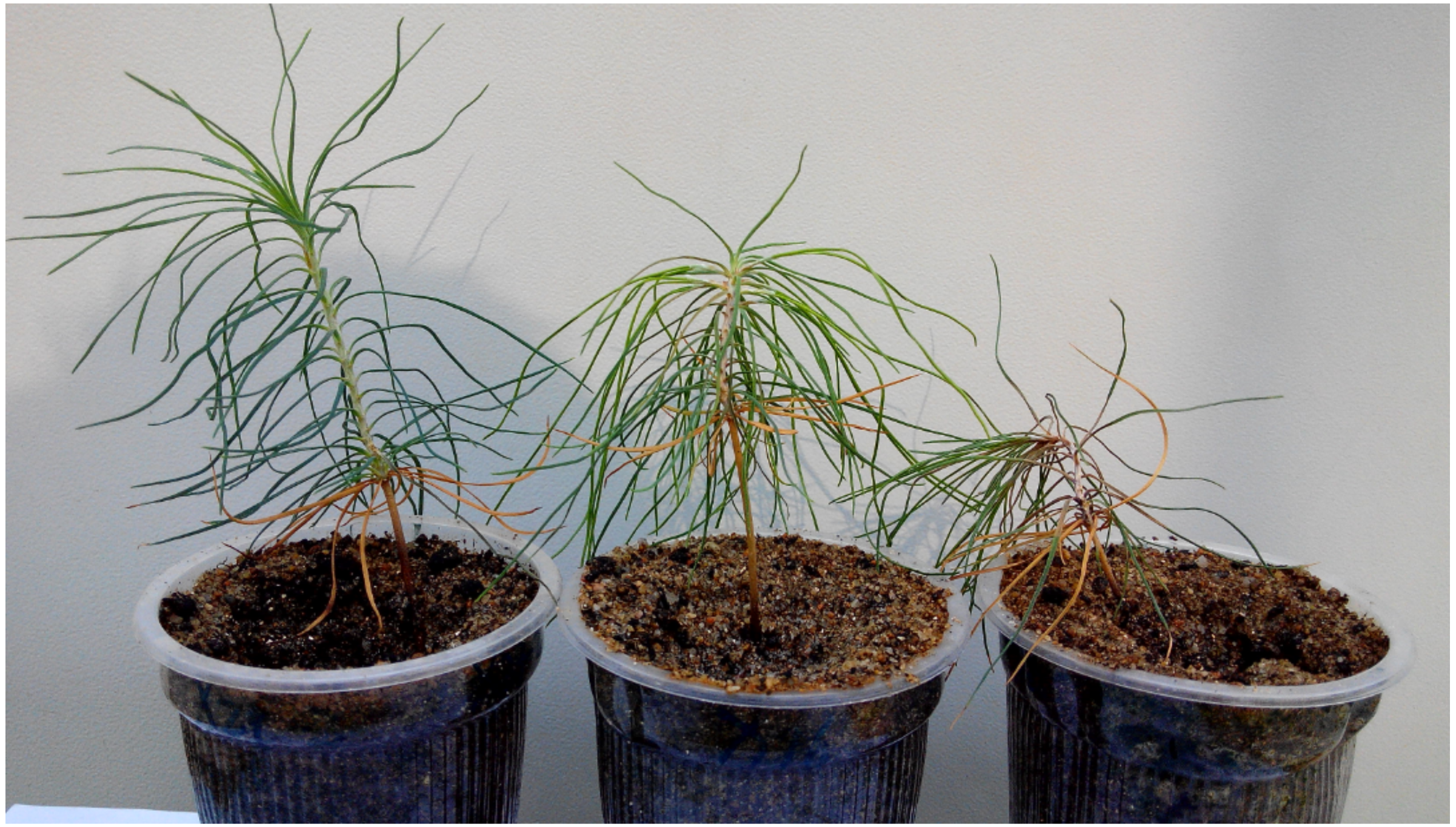
3.1. P. tabuliformis Seedling Growth and Fungal Colonization

Root colonization of fungi were found in all tested seedling roots. Fungal mantle formed by ECMF and microsclerotia and/or dark hyphae formed by DSE were observed in the roots of the pine seedlings. The dominant forms of ECMF and DSE were dichotomous branching and monoaxial branching, club-shaped mycorrhizas were fewer (data were not shown in this study). Different strains had different colonization rates, but there were no significant differences between ECMF and DSE (Table 1). In our experiment, noninoculated plants also exhibited very little mycelium colonization; it might come from the environment or the water we used. According to ANOVA analysis of the seedling growth, height, basal stem diameter, dry weight, root activity, water content, and root shoot ratio of seedlings varied significant (p < 0.05) among each treatment. Fungal colonization had a significant effect on seedlings’ growth in this study. Compared to the control group, Av, Sla, and Pm significantly improved the seedlings’ height; Av, Cc, and Gc significantly increased seedlings’ diameter; Av and Pm significantly improved the root activity of the seedlings; Av, Sla, Cc, and Pc significantly increased seedlings’ dry weight; Av, Sla, Cc, and Es significantly improved seedlings’ water content. However, not only positive effects were observed for plant growth, but also negative effects of seedlings growth were observed. Strains, such as Sb and Sl, decreased the dry weight; Ss, Hu, Sb, Es, and Pm decreased the basal stem diameter compared to the control group. There was no apparent difference on seedlings’ growth between ECMF and DSE (Table 1).

3.2. Effects of Inoculation ECMF and DSE on Roots Parameters

According to ANOVA results of the root morphological analysis, different effects of ECMF and DSE were observed in root morphological characters. Compared to the control group, Es significantly increased seedlings’ root length, Hu and Es significantly increased root tip numbers, and Ss significantly increased the average root diameter. No significant effects on the root length, root surface area, average root diameter, root volume, number of root tips, and root forks were observed among the other different treatments. Inoculation of ECMF/DSE altered the percentage of root length in different diameter classes of pine seedling roots. Compared to the control group, Gc and Hu significantly increased the percentage of root length in the 0–0.2 mm diameter class; Es, Gc, Pm, and Sl significantly increased the percentage of root length in the 0.2–0.4 mm diameter class; Es significantly increased the percentage of root length in the 0.4–0.6 mm diameter class; Ss significantly increased the percentage of root length in the 0.6–1 mm diameter class. However, Av and Cc significantly decreased the percentage of root length in the 0–0.2 mm diameter class; Ss significantly decreased the percentage of root length in the 0.2–0.4 mm diameter class; and both Es and Gc significantly decreased the percentage of root length in the 0.6–0.8 mm and D > 1 mm root diameter classes (Table 2; Figure 2).

3.3. Pigment Parameters of Pine Needles in Different ECMF and DSE Inoculation Treatments

Pigments extracted from the seedlings’ needles showed that seedlings inoculated with ECMF and DSE significantly increased the concentration of chlorophyll a and chlorophyll b. Compared to the control group, the content of carotenoids significantly increased in all strains except Sla and Es. The ratio of chlorophyll a/b decreased in the Av, Hu, Sb, St, Cc, Gc, Pc, and Pm treatments significantly (Table 3).

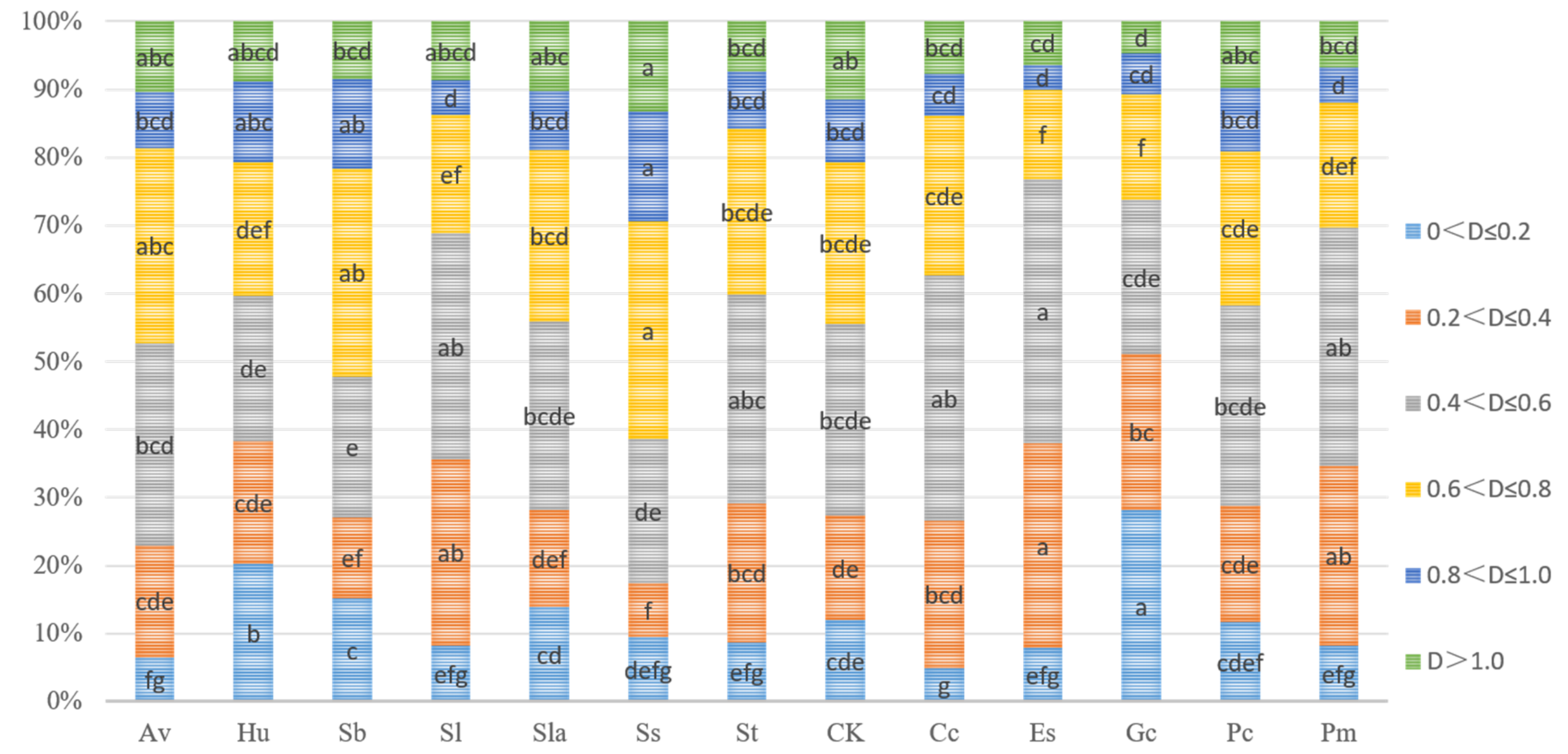
3.4. Comparison Among Strains in Principal Component Analysis

PCA was performed using all the treatments’ experimental data sets. All parameters were reduced to the fewest and most representative dimensions. The PCA figure could be interpreted as a measure of distance among different treatments, sacrificing the information on specific parameters for a global view. Thus, the closer the treatments in a PCA figure, the closer the similarity of the overall status. Among the treatments, principal component 1 (PC1), principal component 2 (PC2), and principal component 3 (PC3) accounted for 35.36%, 27.76%, and 12.08% of the variance, respectively (Figure 2). Among them, three principal components divided the strains into many groups. In detail, the Es, Sl, and Hu were closed to CK and separated from other treatments, indicating that the growth P. tabulaeformis seedlings inoculated with Es, Sl, and Hu was similar to the non-mycorrhizal plants. While Av and Cc, Sla and Pc, Gc, Pm, Sb, St, and Ss were separated from CK by PC1, respectively, which suggested the colonization of these strains differed from the control group. ECMF and DSE treatments were not separated by the principal component, indicating that the ECMF and DSE had no obvious performance differences on seedling parameters (Figure 3).

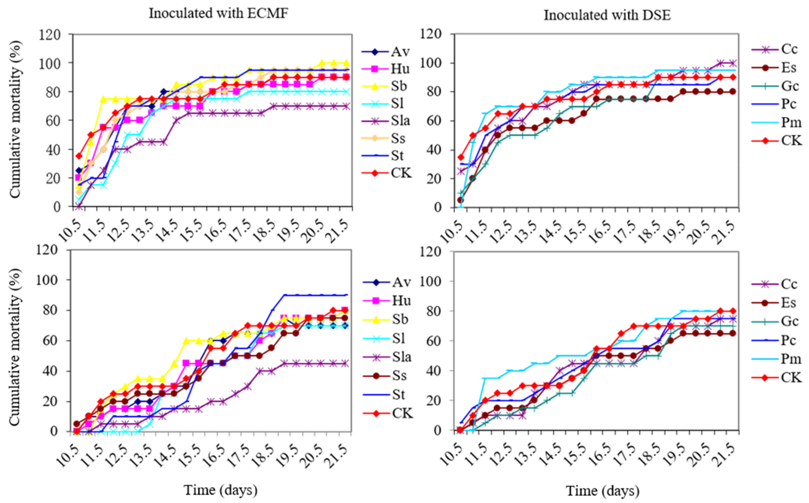
3.5. Cumulative Morbidity and Mortality of ECMF and DSE Pine Seedlings Caused by PWN

Ten days after the seedlings were inoculated with PWN, wilting and death began to appear. The wilting of seedlings burst in the 11th to 13th day after being inoculated with PWN, and the death of seedlings burst in the 14th to 18th day after being inoculated with PWN. Three weeks after PWN inoculated, the cumulative mortality of Sla, Sl, Es, Gc, Av, and Hu were lower than the control group (80%). However, only Sla, Sl, Hu, Es, Pc, and Gc treatments of the cumulative morbidity were not higher than the control group (90%). Seedlings colonized with Sla had the lowest cumulative mortality (50%) and morbidity (70%), followed by Sl, Es, and Gc, indicating that Sla was the best strain for P. tabulaeformis seedlings’ protection against pine wilt disease in this study (Figure 4). In all cases, PWN could be re-isolated from the death and wilting seedlings. The five non-inoculated seedlings of all treatments were healthy.

3.6. Correlation Analysis of Variables with Cumulative Mortality and Morbidity

The results of correlation analysis showed that the root length, root surface area, and number of root forks was negatively correlated with cumulative morbidity and tissue water content was negatively correlated with cumulative mortality. However, the carotenoids were positively correlated with cumulative morbidity (Table 4).

4. Discussion

In this study, the symbiotic efficiency of seven ECMF and five DSE in P. tabulaeformis seedlings, growth traits and root morphology among P. tabuliformis seedlings colonized with different ECMF/DSE strains as well as the relationship between these parameters and cumulative mortality rates caused by the PWN infection were studied. Both positive and negative effects on the growth occurred in different colonization treatments. For example, seedlings inoculated with ECMF and DSE could significantly increase the concentration of chlorophyll a and b in all treatments. S. laricinus and P. mustea significantly improved seedling height; A. vaginata, C. cladosporioides, and G. cylindrosporus significantly increased seedling diameter; A. vaginata and P. mustea significantly improved the root activity of the seedlings; A. vaginata, S. laricinus, C. cladosporioides, and P. chrysanthemicola significantly increased the dry weight of seedlings. On the other hand, some of the colonized strains significantly decreased or had no effects on these growth parameters, which may result from the excessive carbohydrate demand of the strains from plants [45,46]. In fact, both DSE and ECMF inoculation could range from parasitic to mutualistic [22,45,46,47,48]. Different symbiosis functions of ECMF and DSE on root morphological characters were observed in this study too. For example, E. salmonis significantly increased seedling root length, H. utriformis and E. salmonis significantly increased root tip numbers, and Schizophyllum sp. significantly increased average root diameter. However, no significantly positive or negative effects on the root length, root surface area, average root diameter, root volume, number of root tips, and root forks were observed among other treatments compared to the control group (Table 2). What is more, inoculation of ECMF/DSE altered the percentage of root length in different diameter classes of pine seedlings roots (Figure 2). These could be due to the colonization by ECMF/DSE that could facilitate water and nutrient uptake of host plants and reduce the formation of root for saving carbohydrates formed by photosynthesis of the host [49,50,51]. Principal component analysis showed that three principal components divided the strains into many groups (Figure 3). Different groups divided by principal components may have different plant traits [52], which indicated that the symbiotic function on seedlings of the ECMF and DSE strains applied in this study differed among fungal species.
Many studies showed that the colonization of ECMF and DSE could confer pathogen resistance to host plants [20,53,54,55], and this phenomenon was confirmed in our study too. In the study, the rates of mortality caused by PWN infection were reduced in most of ECMF and DSE treatments (Figure 4). P. tabulaeformis seedlings colonized with S. laricinus had a lowest cumulative mortality (50%) and cumulative morbidity (70%) in the first three weeks after being inoculated with PWN. Other studies also showed that S. laricinus could inhibit the growth of the pathogen and improve resistance to disease [17,46]. Kikuchi et al. (1991) reported that inoculation of Pinus densiflora with ectomycorrhizal fungi, Suillus luteus and Rhizopogon rubescens, improved seedling growth, thereby decreasing seedling mortality caused by B. xylophilus [55]. Nakashima et al., (2016) found that the seedlings that had plentiful white ectomycorrhizae showed the highest survival and growth rates regardless of the variety of black pine [32]. Not all symbiosis of mycorrhizal could confer a positive effect to host plant [33,56,57] and some reports showed that mycorrhizal may have a neutral effect or even a negative effect on plants’ disease resistance [57,58]. In our study, the seedlings showed a different performance of the resistance to PWD, perhaps simply because of different species of ECMF and DSE having different symbiosis levels of resistance or tolerance to diseases [33,34]. A. vaginata seedlings exhibited the best protective effect against damping-off among the tested strains in our previous study [18,38,39], but in the present study, A. vaginata seedlings showed a relatively low tolerance to PWD.
The mechanism of PWN that caused pine tree death in a short time was that pinewood nematode feeds on the host’s resin duct parenchyma cells, leading to destruction of cambium, cortex, phloem, and resin duct tissues, and the formation of wound periderm in cortex parenchyma cells around resin after vector beetles transport PWNs to a healthy tree [59]. Then, the population of PWNs increases and continues to migrate and feed on host tissues, leading to the accumulation of suberin-like substances, xylem occlusion, cavitation, and embolism in xylem cells [11,12,13]. These modifications in the xylem translate into dysfunction of water conduction in the stem, which result in decreasing water potential, transpiration, and photosynthesis and further lead to the manifestation of external symptoms, wilt, and finally death [60,61,62]. The colonization of ECMF and DSE might alleviate the dysfunction of water conduction in the stem caused by PWNs infection through the adjustment of root morphology, such as length, root surface area, and number of root forks as well as the external mycelium help [63,64]. In our study, the treatments that had lower mortality showed a relatively higher tissue water content (Table 1). Pearson correlation analysis showed that the root length, root surface area, and number of root forks were significantly (p < 0.05) negatively correlated with cumulative morbidity and tissue water content was significantly (p < 0.05) negatively correlated with cumulative mortality (Table 4). This might be one of the reasons that the inoculation of ECMF/DSE reduced the cumulative morbidity and mortality. In addition, ECMF could facilitate water uptake, alleviating harm caused by water deficit of host plants under drought stress [65,66,67]. Furthermore, Akema and Futai (2005) studied the relationship between ectomycorrhizal development and mortality of PWD in a P. thunbergii stand on a slope, and suggested a correlation between mycorrhizal development and resistance to PWD. The abundant mycorrhizae, which mitigate drought stress, may have decreased the rate of tree mortality caused by PWD [35]. However, the carotenoids content was significant positively correlated with cumulative morbidity caused by PWD. This might allow carotenoids content to be used as an indicator for the resistance of pinewood nematode of pine seedlings. What is more, Nakashima et al., (2016) also indicated that P. thunbergii seedlings intra-specific physiological resistance to the PWD might be affected by ectomycorrhizal composition or by the specific ectomycorrhizal species [32].
To our knowledge, this is the first study on the relationship between symbiosis functions of dark septate endophytes/ectomycorrhizal fungi on P. tabulaeformis and PWD. In this study, the root morphology, growth traits, and pigment parameters were surveyed to associate them with cumulative morbidity and mortality caused by infection of PWN for the first time. It constituted an important step for future research to understand the interaction between PWD and root associated fungi, as well as the role of root associated fungi, especially ECMF and DSE, in the resistance of pine trees against PWD.

5. Conclusions

It can be concluded that the growth traits, root morphology, and resistance of pine seedlings to PWD generated by ECMF/DSE varied to different ECMF/DSE species. The resistance of pine wilt disease induced by symbiosis of ECMF/DSE might result from mitigation of the dysfunction of water caused by PWN infection. Our research provided evidence that inoculation of ECMF and DSE should be considered as a potential method for pine wilt disease prevention. More ECMF/DSE should be tested for tolerance of the pine wilt disease. Future research to understand ECMF and DSE in the resistance of pine trees against PWD should be studied.

Author Contributions

Conceptualization, H.C., C.W. and M.T.; Formal analysis, H.W.; Funding acquisition, C.W. and M.T.; Methodology, H.C., Z.L. and H.W; Supervision, M.T.; Writing—original draft, H.C.; Writing—review & editing, C.W., Z.L., Y.X., J.C. and M.T.

Funding

This research was funded by The National Key Research and Development Program of China, grant number 2018YFD0600203” and the National Natural Science Foundation of China (31300543, 31860057), the Fundamental Research Funds for the Central Universities (Z109021308).

Acknowledgments

We acknowledge the Yunnan Applied Basic Research Projects (2018FD079) and Yunnan Local Colleges Applied Basic Research Projects (2017FH001-037).

Conflicts of Interest

The authors declare no conflict of interest.

References

  1. Mota, M.M.; Vieira, P. Pine Wilt Disease: A Worldwide Threat to Forest Ecosystems; Springer: Heidelberg, Germany, 2008. [Google Scholar]
  2. Vicente, C.; Espada, M.; Vieira, P.; Mota, M. Pine wilt disease: A threat to European forestry. Eur. J. Plant Pathol. 2012, 133, 89–99. [Google Scholar] [CrossRef]
  3. Steiner, G.; Buhrer, E.M. Aphelenchoides xylophilus n. sp., a nematode associated with blue-stain and other fungi in timber. J. Agric. Res. 1934, 48, 949–951. [Google Scholar]
  4. Nickle, W.; Golden, A.; Mamiya, Y.; Wergin, W. On the taxonomy and morphology of the pine wood nematode, Bursaphelenchus xylophilus (Steiner &Buhrer 1934) Nickle 1970. J. Nematol. 1981, 13, 385–392. [Google Scholar] [PubMed]
  5. Kiyohara, T.; Tokushige, Y. Inoculation experiments of a nematode, Bursaphelenchus sp., onto pine trees. J. Jpn. For. Soc. 1997, 153, 210–218. [Google Scholar]
  6. Arakawa, Y.; Togashi, K. Newly discovered transmission pathway of Bursaphelenchus xylophilus from males of the beetle Monochamus alternatus to Pinus densiflora trees via oviposition wounds. J. Nematol. 2002, 34, 396–404. [Google Scholar]
  7. Linit, M. Nemtaode-vector relationships in the pine wilt disease system. J. Nematol. 1988, 20, 227–235. [Google Scholar] [PubMed]
  8. Mamiya, Y.; Enda, N. Transmission of Bursaphelenchus lignicolus (Nematoda: Aphelenchoididae) by Monochamus alternatus (Coleoptera: Cerambycidae). Nematologica 1972, 18, 159–162. [Google Scholar] [CrossRef]
  9. Morimoto, K.; Iwasaki, A. Role of Monochamus alternatus (Coleoptera: Cerambycidae) as a vector of Bursaphelenchus lignicolus (Nematoda: Aphelenchoididae). J. Jpn. For. Soc. 1972, 54, 177–183. [Google Scholar]
  10. Wingfield, M.; Blanchette, R. The pine-wood nematode, Bursaphelenchus xylophilus, in Minnesota and Wisconsin: Insect associates and transmission studies. Can. J. Forest Res. 1983, 13, 1068–1076. [Google Scholar] [CrossRef]
  11. Futai, K. Pine wood nematode, Bursaphelenchus xylophilus. Annu. Rev. Phytopathol. 2013, 51, 61–83. [Google Scholar] [CrossRef]
  12. Yazaki, K.; Takanashi, T.; Kanzaki, N.; Komatsu, M.; Levia, D.F.; Kabeya, D.; Tobita, H.; Kitao, M.; Ishida, A. Pine wilt disease causes cavitation around the resin canals and irrecoverable xylem conduit dysfunction. J. Exp. Bot. 2017, 69, 589–602. [Google Scholar] [CrossRef]
  13. Zhao, B.G.; Futai, K.; Sutherland, J.R.; Takeuchi, Y. Pine Wilt Disease; Springer: Tokyo, Japan, 2008; pp. 144–161. [Google Scholar]
  14. Smith, S.E.; Read, D.J. Mycorrhizal Symbiosis; Academic Press: New York, NY, USA, 2010. [Google Scholar]
  15. Chu, H.; Wang, C.; Wang, H.; Chen, H.; Tang, M. Pine wilt disease alters soil properties and root-associated fungal communities in Pinus tabulaeformis forest. Plant Soil 2016, 404, 237–249. [Google Scholar] [CrossRef]
  16. Wagg, C.; Pautler, M.; Massicotte, H.B.; Peterson, R.L. The co-occurrence of ectomycorrhizal, arbuscular mycorrhizal, and dark septate fungi in seedlings of four members of the Pinaceae. Mycorrhiza 2008, 18, 103–110. [Google Scholar] [CrossRef]
  17. Chu, H.; Tang, M.; Wang, H.; Wang, C. Pinewood nematode infection alters root mycoflora of Pinus tabulaeformis Carr. J. Appl. Microbiol. 2018, 125, 554–563. [Google Scholar] [CrossRef] [PubMed]
  18. Zhang, H.; Yu, H.; Tang, M. Prior contact of Pinus tabulaeformis with ectomycorrhizal fungi increases plant growth and survival from damping-off. New For. 2017, 48, 855–866. [Google Scholar] [CrossRef]
  19. Zhang, R.Q.; Tang, M.; Chen, H.; Tian, Z.Q. Effects of ectomycorrhizal fungi on damping-off and induction of pathogenesis-related proteins in Pinus tabulaeformis seedlings inoculated with Amanita vaginata. For. Pathol. 2011, 41, 262–269. [Google Scholar] [CrossRef]
  20. Nickel, U.; Winkler, J.; Mühlhans, S.; Buegger, F.; Munch, J.; Pritsch, K. Nitrogen fertilisation reduces sink strength of poplar ectomycorrhizae during recovery after drought more than phosphorus fertilisation. Plant Soil 2017, 419, 405–422. [Google Scholar] [CrossRef]
  21. Luo, Z.-B.; Janz, D.; Jiang, X.; Göbel, C.; Wildhagen, H.; Tan, Y.; Rennenberg, H.; Feussner, I.; Polle, A. Upgrading root physiology for stress tolerance by ectomycorrhizas: Insights from metabolite and transcriptional profiling into reprogramming for stress anticipation. Plant Physiol. 2009, 151, 1902–1917. [Google Scholar] [CrossRef]
  22. Jumpponen, A.; Trappe, J.M. Dark septate endophytes: A review of facultative biotrophic root-colonizing fungi. New Phytol. 1998, 140, 295–310. [Google Scholar] [CrossRef]
  23. Newsham, K.K. A meta-analysis of plant responses to dark septate root endophytes. New Phytol. 2011, 190, 783–793. [Google Scholar] [CrossRef]
  24. Ban, Y.; Xu, Z.; Yang, Y.; Zhang, H.; Chen, H.; Tang, M. Effect of dark septate endophytic fungus Gaeumannomyces cylindrosporus on plant growth, photosynthesis and Pb tolerance of maize (Zea mays L.). Pedosphere 2017, 27, 283–292. [Google Scholar] [CrossRef]
  25. Mandyam, K.G.; Jumpponen, A. Mutualism–parasitism paradigm synthesized from results of root-endophyte models. Front. Microbiol. 2015, 5, 776. [Google Scholar] [CrossRef] [PubMed]
  26. Trappe, J.M. Selection of fungi for ectomycorrhizal inoculation in nurseries. Annu. Rev. Phytopathol. 1977, 15, 203–222. [Google Scholar] [CrossRef]
  27. Flor-Peregrín, E.; Azcón, R.; Martos, V.; Verdejo-Lucas, S.; Talavera, M. Effects of dual inoculation of mycorrhiza and endophytic, rhizospheric or parasitic bacteria on the root-knot nematode disease of tomato. Biocontrol Sci. Techn. 2014, 24, 1122–1136. [Google Scholar] [CrossRef]
  28. Su, Z.Z.; Mao, L.J.; Li, N.; Feng, X.X.; Yuan, Z.L.; Wang, L.W.; Lin, F.C.; Zhang, C.L. Evidence for biotrophic lifestyle and biocontrol potential of dark septate endophyte Harpophora oryzae to rice blast disease. PLoS ONE 2013, 8, e61332. [Google Scholar] [CrossRef] [PubMed]
  29. Khastini, R.O.; Ohta, H.; Narisawa, K. The role of a dark septate endophytic fungus, Veronaeopsis simplex Y34, in Fusarium disease suppression in Chinese cabbage. J. Microbiol. 2012, 50, 618–624. [Google Scholar] [CrossRef] [PubMed]
  30. Scattolin, L.; Dal Maso, E.; Mutto Accordi, S.; Sella, L.; Montecchio, L. Detecting asymptomatic ink-diseased chestnut trees by the composition of the ectomycorrhizal community. For. Pathol. 2012, 42, 501–509. [Google Scholar] [CrossRef]
  31. Hohmann, P.; Messmer, M.M. Breeding for mycorrhizal symbiosis: Focus on disease resistance. Euphytica 2017, 213, 113. [Google Scholar] [CrossRef]
  32. Nakashima, H.; Eguchi, N.; Uesugi, T.; Yamashita, N.; Matsuda, Y. Effect of ectomycorrhizal composition on survival and growth of Pinus thunbergii seedlings varying in resistance to the pine wilt nematode. Trees 2016, 30, 475–481. [Google Scholar] [CrossRef]
  33. Whipps, J.M. Prospects and limitations for mycorrhizas in biocontrol of root pathogens. Can. J. Botany 2004, 82, 1198–1227. [Google Scholar] [CrossRef]
  34. Nara, K. Pioneer dwarf willow may facilitate tree succession by providing late colonizers with compatible ectomycorrhizal fungi in a primary successional volcanic desert. New Phytol. 2006, 171, 187–198. [Google Scholar] [CrossRef] [PubMed] [Green Version]
  35. Akema, T.; Futai, K. Ectomycorrhizal development in a Pinus thunbergii stand in relation to location on a slope and effect on tree mortality from pine wilt disease. J. For. Res-JNP. 2005, 10, 93–99. [Google Scholar] [CrossRef]
  36. Ugawa, S.; Ichihara, Y.; Fukuda, K.; Suzuki, K. Ectomycorrhizae and ectomycorrhizal fungal fruit bodies in pine stands differentially damaged by pine wilt disease. Mycoscience 2009, 50, 63–69. [Google Scholar] [CrossRef]
  37. Ban, Y.; Tang, M.; Chen, H.; Xu, Z.; Zhang, H.; Yang, Y. The response of dark septate endophytes (DSE) to heavy metals in pure culture. PLoS ONE 2012, 7, e47968. [Google Scholar] [CrossRef] [PubMed]
  38. Tang, M.; Zhang, R.Q.; Chen, H.; Zhang, H.H.; Tian, Z.Q. Induced hydrolytic enzymes of ectomycorrhizal fungi against pathogen Rhizoctonia solani. Biotechnol. Lett. 2008, 30, 1777–1782. [Google Scholar] [CrossRef] [PubMed]
  39. Zhang, R.Q.; Tang, M. Role of hydrogen peroxide and antioxidative enzymes in Pinus tabulaeformis seedlings inoculated with Amanita vaginata and/or Rhizoctonia solani. Eur. J. Plant Pathol. 2012, 134, 381–389. [Google Scholar] [CrossRef]
  40. Hoagland, D.R.; Arnon, D.I. The water-culture method for growing plants without soil. Calif. Agric. Exp. Stn. Grc. 1950, 347, 1–32. [Google Scholar]
  41. Clemensson-Lindell, A. Triphenyltetrazolium chloride as an indicator of fine-root vitality and environmental stress in coniferous forest stands: Applications and limitations. Plant Soil 1994, 159, 297–300. [Google Scholar] [CrossRef]
  42. Chappelle, E.W.; Kim, M.S.; McMurtrey, J.E. Ratio analysis of reflectance spectra (RARS): An algorithm for the remote estimation of the concentrations of chlorophyll a, chlorophyll b, and carotenoids in soybean leaves. Remote Sens. Environ. 1992, 39, 239–247. [Google Scholar] [CrossRef]
  43. Phillips, J.; Hayman, D. Improved procedures for clearing roots and staining parasitic and vesicular-arbuscular mycorrhizal fungi for rapid assessment of infection. Trans. Br. Mycol. Soc. 1970, 55, 158–161. [Google Scholar] [CrossRef]
  44. Giovannetti, M.; Mosse, B. An evaluation of techniques for measuring vesicular arbuscular mycorrhizal infection in roots. New Phytol. 1980, 84, 489–500. [Google Scholar] [CrossRef]
  45. Hibbett, D.S.; Gilbert, L.-B.; Donoghue, M.J. Evolutionary instability of ectomycorrhizal symbioses in basidiomycetes. Nature 2000, 407, 506–508. [Google Scholar] [CrossRef] [PubMed]
  46. Johnson, N.; Graham, J.H.; Smith, F. Functioning of mycorrhizal associations along the mutualism-parasitism continuum. New Phytol. 1997, 135, 575–585. [Google Scholar] [CrossRef] [Green Version]
  47. Reininger, V.; Sieber, T.N. Mycorrhiza reduces adverse effects of dark septate endophytes (DSE) on growth of conifers. PLoS ONE 2012, 7, e42865. [Google Scholar] [CrossRef] [PubMed]
  48. Hoeksema, J.D.; Chaudhary, V.B.; Gehring, C.A.; Johnson, N.C.; Karst, J.; Koide, R.T.; Pringle, A.; Zabinski, C.; Bever, J.D.; Moore, J.C.; et al. A meta-analysis of context-dependency in plant response to inoculation with mycorrhizal fungi. Ecol. Lett. 2010, 13, 394–407. [Google Scholar] [CrossRef] [PubMed] [Green Version]
  49. Cheng, L.; Chen, W.; Adams, T.S.; Wei, X.; Li, L.; McCormack, M.L.; DeForest, J.L.; Koide, R.T.; Eissenstat, D.M. Mycorrhizal fungi and roots are complementary in foraging within nutrient patches. Ecology 2016, 97, 2815–2823. [Google Scholar] [CrossRef] [PubMed]
  50. Liu, B.; Li, H.; Zhu, B.; Koide, R.T.; Eissenstat, D.M.; Guo, D. Complementarity in nutrient foraging strategies of absorptive fine roots and arbuscular mycorrhizal fungi across 14 coexisting subtropical tree species. New Phytol. 2015, 208, 125–136. [Google Scholar] [CrossRef] [Green Version]
  51. Chen, W.; Koide, R.T.; Adams, T.S.; DeForest, J.L.; Cheng, L.; Eissenstat, D.M. Root morphology and mycorrhizal symbioses together shape nutrient foraging strategies of temperate trees. Proc. Natl. Acad. Sci. USA 2016, 113, 8741–8746. [Google Scholar] [CrossRef]
  52. Li, Z.; Wu, N.; Liu, T.; Chen, H.; Tang, M. Effect of arbuscular mycorrhizal inoculation on water status and photosynthesis of Populus cathayana males and females under water stress. Physiol. Plantarum 2015, 155, 192–204. [Google Scholar] [CrossRef]
  53. Narisawa, K.; Tokumasu, S.; Hashiba, T. Suppression of clubroot formation in Chinese cabbage by the root endophytic fungus, Heteroconium chaetospira. Plant Pathol. 1998, 47, 206–210. [Google Scholar] [CrossRef]
  54. Mandyam, K.; Jumpponen, A. Seeking the elusive function of the root-colonising dark septate endophytic fungi. Stud. Mycol. 2005, 53, 173–189. [Google Scholar] [CrossRef] [Green Version]
  55. Kikuchi, J.; Tsuno, N.; Futai, K. The effect of mycorrhizae as a resistance factor of pine trees (Pinus densiflora) to the pinewood nematode. J. Jpn. For. Soc. 1991, 73, 216–218. [Google Scholar]
  56. Graham, J.H. What do root pathogens see in mycorrhizas? New Phytol. 2001, 149, 357–359. [Google Scholar] [CrossRef]
  57. Corrêa, A.; Strasser, R.; Martins-Loução, M. Are mycorrhiza always beneficial? Plant Soil 2006, 279, 65–73. [Google Scholar] [CrossRef]
  58. Davis, R.; Menge, J.; Erwin, D. Influence of Glomus fasciculatus and soil phosphorus on Verticillium wilt of cotton. Phytopathology 1979, 69, 453–456. [Google Scholar] [CrossRef]
  59. Nunes da Silva, M.; Lima, M.; Vasconcelos, M. Susceptibility evaluation of Picea abies and Cupressus lusitanica to the pine wood nematode (Bursaphelenchus xylophilus). Plant Pathol. 2013, 62, 1398–1406. [Google Scholar] [CrossRef]
  60. Fukuda, K. Physiological process of the symptom development and resistance mechanism in pine wilt disease. J. For. Res-JPN. 1997, 2, 171–181. [Google Scholar] [CrossRef]
  61. Ichihara, Y.; Fukuda, K.; Suzuki, K. The effect of periderm formation in the cortex of Pinus thunbergii on early invasion by the pinewood nematode. For. Pathol. 2000, 30, 141–148. [Google Scholar] [CrossRef]
  62. Hara, N.; Takeuchi, Y. Histological analysis for mechanism of pine wilt disease. J. Jpn. For. Soc. 2006, 88, 364–369. [Google Scholar] [CrossRef]
  63. Kumla, J.; Hobbie, E.A.; Suwannarach, N.; Lumyong, S. The ectomycorrhizal status of a tropical black bolete, Phlebopus portentosus, assessed using mycorrhizal synthesis and isotopic analysis. Mycorrhiza 2016, 26, 333–343. [Google Scholar] [CrossRef]
  64. Muhsin, T.; Zwiazek, J. Ectomycorrhizas increase apoplastic water transport and root hydraulic conductivity in Ulmus americana seedlings. New Phytol. 2002, 153, 153–158. [Google Scholar] [CrossRef]
  65. Kipfer, T.; Wohlgemuth, T.; van der Heijden, M.G.; Ghazoul, J.; Egli, S. Growth response of drought-stressed Pinus sylvestris seedlings to single-and multi-species inoculation with ectomycorrhizal fungi. PLoS ONE 2012, 7, e35275. [Google Scholar] [CrossRef] [PubMed]
  66. Bingham, M.A.; Simard, S. Ectomycorrhizal networks of Pseudotsuga menziesii var. glauca trees facilitate establishment of conspecific seedlings under drought. Ecosystems 2012, 15, 188–199. [Google Scholar] [CrossRef]
  67. Alvarez, M.; Huygens, D.; Fernandez, C.; Gacitua, Y.; Olivares, E.; Saavedra, I.; Alberdi, M.; Valenzuela, E. Effect of ectomycorrhizal colonization and drought on reactive oxygen species metabolism of Nothofagus dombeyi roots. Tree Physiol. 2009, 29, 1047–1057. [Google Scholar] [CrossRef] [PubMed]
Figure 1. Healthy, wilting, and dead Pinus tabulaeformis Carr. seedlings growing in the cup.
Forests 10 00140 g001
Figure 2. Percentage of root length in different diameter classes measured in roots colonized by different ECMF and DSE. Means labeled with different letters within each series are significantly different (p < 0.05, n = 5) by Duncan’s test. Av, Hu, Sb, Sl, Sla, Ss, and St represent P. tabulaeformis seedlings were inoculated with ECMF of A. vaginata, H. utriformis, S. bovinus, S. lactifluus, S. laricinus, Schizophyllum sp., and S. tomentosus ECMF, respectively; Cc, Es, Gc, Pc, and Pm represent the P. tabulaeformis seedlings were inoculated with DSE of C. cladosporioides, E. salmonis, G. cylindrosporus, P. chrysanthemicola, and P. mustea, respectively; and CK as the control group; D represents the diameter of the root.
Forests 10 00140 g002
Figure 3. Principal component analysis (PCA) plots of three principal components of different inoculation treatments. PC1, principal 1, PC2, principal 2, PC3, Principal 3. Av, Hu, Sb, Sl, Sla, Ss, and St represent P. tabulaeformis seedlings were inoculated with ECMF of A. vaginata, H. utriformis, S. bovinus, S. lactifluus, S. laricinus, Schizophyllum sp., and S. tomentosus ECMF, respectively; Cc, Es, Gc, Pc, and Pm represent the P. tabulaeformis seedlings were inoculated with DSE of C. cladosporioides, E. salmonis, G. cylindrosporus, P. chrysanthemicola, and P. mustea, respectively; and CK as the control group.
Forests 10 00140 g003
Figure 4. Cumulative morbidity and mortality of ECMF and DSE P. tabulaeformis seedlings after 10 days of inoculated with PWN. Av, Hu, Sb, Sl, Sla, Ss, and St represent P. tabulaeformis seedlings were inoculated with ECMF of A. vaginata, H. utriformis, S. bovinus, S. lactifluus, S. laricinus, Schizophyllum sp., and S. tomentosus, respectively; Cc, Es, Gc, Pc, and Pm represent the P. tabulaeformis seedlings were inoculated with DSE of C. cladosporioides, E. salmonis, G. cylindrosporus, P. chrysanthemicola, and P. mustea, respectively; and CK as the control group.
Forests 10 00140 g004
Table 1. ANOVA analysis of seedlings’ growth and the colonization of ECMF and DSE strains 3 months after transplant.
StrainsR/S RatioHeightDiameterWater ContentDry Weight
(g)
Root ActivityRoot Colonization Rate (%)
ECMFAv0.48 ± 0.07 cd5.34 ± 0.26 ab1.67 ± 0.06 a0.77 ± 0.04 a0.26 ± 0.035 bc8.49 ± 1.67 b48.88 ± 8.24 c
Hu0.53 ± 0.04 cd4.20 ± 0.17 d1.35 ± 0.06 ef0.63 ± 0.05 e0.19 ± 0.041 efg7.43 ± 1.49 bcde63.45 ± 7.24 b
Sb0.57 ± 0.05 cd4.20 ± 0.25 d1.26 ± 0.07 f0.62 ± 0.02 e0.17 ± 0.013 gf5.11 ± 1.76 f69.84 ± 9.38 ab
Sl0.37 ± 0.04 d5.22 ± 0.11 abc1.38 ± 0.06 cde0.69 ± 0.03 bcd0.14 ± 0.022 h7.95 ± 1.22 bcd62.53 ± 3.36 b
Sla0.62 ± 0.03 bcd5.36 ± 0.32 a1.44 ± 0.09 cd0.72 ± 0.03 ab0.34 ± 0.034 a7.46 ± 1.20 bcde64.33 ± 7.11 b
Ss0.61 ± 0.11 bcd4.88 ± 0.33 c1.31 ± 0.09 ef0.65 ± 0.02 cde0.24 ± 0.007 bcd2.98 ± 1.21 g33.33 ± 4.84 d
St0.88 ± 0.63 ab5.16 ± 0.32 abc1.39 ± 0.06cde0.54 ± 0.09 f0.19 ± 0.050 fg7.83 ± 0.97 bcd70.72 ± 5.71 ab
ControlCK0.65 ± 0.15 abcd4.80 ± 0.36 c1.45 ± 0.02 c0.64 ± 0.02 de0.22 ± 0.011 def6.56 ± 1.78 cdef6.48 ± 3.16 e
DSECc0.49 ± 0.05 cd4.90 ± 0.35 c1.55 ± 0.08 b0.72 ± 0.03 ab0.32 ± 0.017 a7.63 ± 0.80 bcd73.88 ± 7.92 ab
Es0.69 ± 0.21 abc4.84±0.30 c1.36 ± 0.03 de0.70 ± 0.01 bc0.22 ± 0.0197 def5.81 ± 0.54 ef48.53 ± 7.63 c
Gc0.53 ± 0.06 cd5.12 ± 0.33 abc1.56 ± 0.04 b0.67 ± 0.04 bcde0.18 ± 0.016 fg8.07 ± 1.56 bc79.28 ± 2.72 a
Pc0.91 ± 0.12 a5.14 ± 0.11 abc1.45 ± 0.08 cd0.63 ± 0.04 e0.27 ± 0.009 b6.18 ± 0.47 def23.33 ± 2.88 d
Pm0.57 ± 0.13 cd4.92 ± 0.39 bc1.33 ± 0.06 ef0.68 ± 0.03 bcde0.23 ± 0.024 bcde10.70 ± 0.55 a46.45 ± 5.91 c
ANOVAF2.8498.0314.759.9923.3810.8335.623
Sig.*************
Note: ECMF represent ectomycorrhizal fungi; DSE represent dark septate endophytic fungi; Av, Hu, Sb, Sl, Sla, Ss, St represent the seedlings were inoculated with Amanita vaginata, Handkea utriformis, Suillus bovinus, Suillus lactifluus, S. laricinus, Schizophyllum sp., and Suillus tomentosus, respectively; Cc, Es, Gc, Pc, and Pm represent the seedlings were inoculated with Cladosporium cladosporioides, Exophiala salmonis, Gaeumannomyces cylindrosporus, Paraphoma chrysanthemicola, and Phialophora mustea, respectively; and CK as the control group. Significantly differences among the inoculation treatments are shown by a different lowercase within each volume. F represent F-value; Sig. represent P-value. Data expressed as mean ± SD (n = 5). *, significant effect at p < 0.05; **, p < 0.01.
Table 2. Effect of inoculation ECMF and DSE on root morphological characters.
StrainsRoot Length (cm)Root Surface Area (cm2)Avg. Root Diameter (cm)Root Volume (cm3)No. of Root TipsNo. of Root Forks
Av228.3 ± 57.3 bc48.3 ± 13.0 ab0.68 ± 0.04 bcd0.81 ± 0.21 ab948.9 ± 442 de977.3 ± 219 a
Hu197.3 ± 8.1 cd41.4 ± 2.5 bc0.67 ± 0.02 cd0.69 ± 0.06 bc2172.5 ± 44 b778.0 ± 44 b
Sb108.9 ± 25.4 ef25.1 ± 8.2 fg0.73 ± 0.05 ab0.46 ± 0.18 de882.3 ± 59 def323.6 ± 55 d
Sl237.5 ± 13.6 b46.1 ± 5.2 ab0.62 ± 0.04 ef0.71 ± 0.12 b956.1 ± 74 de1177.1 ± 63 a
Sla212.7 ± 8.6 bcd46.7 ± 2.5 ab0.70 ± 0.02 bc0.82 ± 0.05 ab1529.9 ± 285 c982.0 ± 76 a
Ss87.8 ± 6.4 f21.0 ± 1.7 g0.76 ± 0.08 a0.40 ± 0.07 ef462.6 ± 52 gh318.6 ± 34 d
St144.0 ± 11.7 e28.4 ± 2.1 ef0.63 ± 0.06 de0.45 ± 0.07 e799.9 ± 70 defg419.1 ± 52 cd
CK233.1 ± 54.1 bc48.5 ± 7.8 ab0.68 ± 0.07 bcd0.81 ± 0.07 ab1179.4 ± 163 cd1073.4 ± 143 a
Cc186.4 ± 15.0 d36.9 ± 2.4 cd0.63 ± 0.03 de0.58 ± 0.03 cd559.9 ± 36 efgh740.1 ± 41 b
Es275.3 ± 29.8 a50.2 ± 5.0 a0.58 ± 0.03 efg0.73 ± 0.07 ab4086.0 ± 167 a1095.1 ± 49 a
Gc129.7 ± 31.8 e22.2 ± 6.0f g0.55 ± 0.01 g0.30 ± 0.08 f390.9 ± 62 h466.6 ± 67 cd
Pc236.6 ± 9.1 b50.4 ± 2.9 a0.68 ± 0.02 bcd0.86 ± 0.06 a1476.3 ± 127 c1082.5 ± 139 a
Pm180.0 ± 14.9 d32.7 ± 4.4 de0.57 ± 0.04 fg0.48 ± 0.10 de538.8 ± 120 fgh598.8 ± 71 bc
F21.3124.6913.5921.8061.2422.78
Sig.************
Note: Av, Hu, Sb, Sl, Sla, Ss, and St represent the seedlings were inoculated with ECMF of A. vaginata, H. utriformis, S. bovinus, S. lactifluus, S. laricinus, Schizophyllum sp., and S. tomentosus, respectively; Cc, Es, Gc, Pc, and Pm represent the seedlings were inoculated with DSE of C. cladosporioides, E. salmonis, G. cylindrosporus, P. chrysanthemicola, and P. mustea, respectively; and CK as the control group. Significant differences among the inoculation treatments are shown by a different lowercase within each volume. F represent F-value; Sig. represent p-value. Data expressed as mean ± SD (n = 5). *, significant effect at p < 0.05; **, p < 0.01.
Table 3. ANOVA analysis of pigment parameters of the seedling needles of different ECMF and DSE inoculation treatments.
StrainsChlorophyll A (mg/kg)Chlorophyll B (mg/kg)Chlorophyll A/BCarotenoids (mg/kg)
Av1.18 ± 0.04 a0.33 ± 0.01 a3.55 ± 0.01 f0.21 ± 0.009 a
Hu0.84 ± 0.02 f0.23 ± 0.01 f3.71 ± 0.04 de0.17 ± 0.001 cde
Sb0.99 ± 0.01 d0.27 ± 0.005 cd3.61 ± 0.02 ef0.19 ± 0.003 b
Sl0.87 ± 0.01 f0.20 ± 0.001 g4.23 ± 0.04a0.17 ± 0.005 cde
Sla1.05 ± 0.03 c0.26 ± 0.01 d4.02 ± 0.251 b0.16 ± 0.009 e
Ss1.01 ± 0.04 cd0.24 ± 0.02 ef4.26 ± 0.15 a0.19 ± 0.013 b
St1.03 ± 0.03 cd0.28 ± 0.01 c3.68 ± 0.03 ef0.20 ± 0.003 a
CK0.71 ± 0.07 g0.17 ± 0.02 h4.12 ± 0.16 ab0.17 ± 0.007 c
Cc1.13 ± 0.03 b0.30 ± 0.01 b3.73 ± 0.002 de0.21 ± 0.001 a
Es0.92 ± 0.05 e0.23 ± 0.02 f4.10 ± 0.06 b0.16 ± 0.013 de
Gc1.05 ± 0.03 c0.27 ± 0.01 cd3.90 ± 0.05 c0.21 ± 0.007 a
Pc1.04 ± 0.04 c0.27 ± 0.01 cd3.82 ± 0.03 cd0.17 ± 0.011 cd
Pm0.88 ± 0.02 ef0.24 ± 0.01 e3.61 ± 0.10 ef0.18 ± 0.004 bc
F61.4460.6834.1330.06
Sig.********
Note: Av, Hu, Sb, Sl, Sla, Ss, and St represent the seedlings were inoculated with ECMF of A. vaginata, H. utriformis, S. bovinus, S. lactifluus, S. laricinus, Schizophyllum sp., and S. tomentosus, respectively; Cc, Es, Gc, Pc, and Pm represent the seedlings were inoculated with DSE of C. cladosporioides, E. salmonis, G. cylindrosporus, P. chrysanthemicola, and P. mustea, respectively; and CK as the control group. Significant differences among the inoculation treatments are shown by a different lowercase within each volume. F represent F-value; Sig. represent P-value. Data expressed as mean ± SD (n = 5). *, significant effect at p < 0.05; **, p < 0.01.
Table 4. Pearson correlation coefficients (r) of variables with mortality and morbidity.
Root MorphologyRoot lengthRoot Surface AreaR/S RatioRoot ActivityAvg. Root DiameterRoot VolumeNo. of Root TipsNo. of Root ForksDiameter
Mortality−0.418−0.4110.311−0.1750.133−0.379−0.325−0.483−0.126
Morbidity−0.591 *−0.590 *0.064−0.1030.109−0.552−0.505−0.655*−0.074
Physiology parametersChorophyll AChorophyll BCarotenoidsA/BDryweightWater ContentHeightDiameterFresh Weight
Mortality0.0080.1690.491−0.409−0.360−0.564 *−0.363−0.126−0.369
Morbidity0.1350.3100.642 *−0.529−0.176−0.375−0.455−0.074−0.136
Note: *, significant effect at p < 0.05.

