Article

Genetic Analyses of the Laurel Wilt Pathogen, Raffaelea lauricola, in Asia Provide Clues on the Source of the Clone that is Responsible for the Current USA Epidemic

1
USDA-Forest Service, Southern Research Station, Forest Health Research and Education Center, Lexington, KY 40546, USA
2
College of Tropical Agriculture and Human Resources, University of Hawaii at Manoa, Hilo, HI 96720, USA
3
Tropical Research & Education Center, University of Florida, Homestead, FL 33031, USA
4
Peckerwood Garden Conservation Foundation, Hempstead, TX 77445, USA
5
School of Forest Resources and Conservation, University of Florida, Gainesville, FL 32611, USA
*
Author to whom correspondence should be addressed.
Forests 2019, 10(1), 37; https://doi.org/10.3390/f10010037
Submission received: 11 December 2018 / Revised: 21 December 2018 / Accepted: 28 December 2018 / Published: 8 January 2019
(This article belongs to the Special Issue Forest Invasive Species: Spread, Impact and Management)

Abstract

:
Laurel wilt is caused by the fungus Raffaelea lauricola T.C. Harr., Fraedrich and Aghayeva, a nutritional symbiont of its vector the redbay ambrosia beetle, Xyleborus glabratus Eichhoff. Both are native to Asia but appeared in Georgia in the early 2000s. Laurel wilt has since spread to much of the southeastern United States killing >300 million host trees in the Lauraceae plant family. The aims of this research were to elucidate the genetic structure of populations of R. lauricola, to examine its reproductive strategy, and determine how often the pathogen had been introduced to the USA. A panel of 12 simple sequence repeat (SSR) markers identified 15 multilocus genotypes (MLGs) in a collection of 59 isolates from the USA (34 isolates), Myanmar (18), Taiwan (6) and Japan (1). Limited diversity in the USA isolates and the presence of one MAT idiotype (mating type locus) indicated that R. lauricola was probably introduced into the country a single time. MLG diversity was far greater in Asia than the USA. Only three closely related MLGs were detected in the USA, the most prevalent of which (30 of 34 isolates) was also found in Taiwan. Although more work is needed, the present results suggest that a Taiwanese origin is possible for the population of R. lauricola in the USA. Isolates of R. lauricola from Myanmar were distinct from those from Japan, Taiwan and the USA. Although both MAT idiotypes were present in Myanmar and Taiwan, only the population from Taiwan had the genetic structure of a sexually reproducing population.

1. Introduction

Laurel wilt is a systemic vascular wilt of trees in the Lauraceae plant family. It is caused by the fungus Raffaelea lauricola T.C. Harr., Fraedrich and Aghayeva (Ophiostomatales), which is cultivated and transmitted by its symbiont, the redbay ambrosia beetle, Xyleborus glabratus Eichhoff (Curculionidae: Scolytinae) [1,2]. Xyleborus glabratus was first detected in the United States at Port Wentworth, GA in 2002 [3], and soon after laurel wilt began to kill Persea borbonia (L.) Spreng., a native tree in the Lauraceae [1]. To date, 14 species in the family are known hosts, most of which are American endemics [4].
Laurel wilt has spread throughout much of the southeastern USA and devastated forest and agricultural landscapes. It has killed an estimated 330 million redbay trees [5], and now affects sassafras (Sassafras albidum (Nutt.) Nees) outside of the range of redbay [6,7]. The potential for laurel wilt to continue to spread where sassafras is the predominant host is under investigation [8]. As it spreads, laurel wilt will have an unclear effect on the large reservoir of native Lauraceae that exists in tropical America. In addition, the disease affects commercial avocado (Persea americana Miller) production in Florida, and threatens larger producers of this important crop in California and Mexico [9,10].
Xyleborus glabratus has been reported in China, India, Bangladesh, Japan, Taiwan and Myanmar, where the beetle is associated with plants in the Lauraceae [3,11]. As the beetle’s primary symbiont [1,2], R. lauricola is probably found wherever X. glabratus occurs, presumably throughout southern Asia. Previously, Harrington et al. [12] regularly isolated the fungus from individuals of X. glabratus from Japan and Taiwan.
Although the pathogen and its vector occur in Asia, significant disease is not known on native members of the Lauraceae in the region [10]. Hulcr et al. [13] recovered the beetle and fungus from injured Machilus zuihoensis Hayata trees, but symptoms were localized to small stems near areas of X. glabratus attack and did not spread throughout the crown. It is hypothesized that the susceptibility of American hosts is due to a lack of coevolved resistance, which apparently occurs in Asian members of the family [10,14]. For example, the Asian camphortree (Cinnamomum camphora (L.) J. Presl.) tolerates R. lauricola infection in the USA [15], as do other Asian taxa that have been tested experimentally (Smith et al., unpublished data). Outside the USA, laurel wilt has only been reported on cultivated avocado in the Tuanggyi and Ywangan Districts of Myanmar [16]. Since X. glabratus is native to Myanmar [3] and R. lauricola is closely associated with its beetle symbiont, we assume that R. lauricola is also native to Myanmar.
Shortly after laurel wilt was described in the USA, it was suggested that X. glabratus and R. lauricola were each introduced into the country a single time [12]. Recently, this hypothesis was investigated with genetic markers in R. lauricola. Wuest et al. [17] used two simple sequence repeat (SSR), a single nucleotide polymorphism (SNP) and mating type markers with isolates from the USA, Japan and Taiwan, whereas Hughes et al. [5] used amplified fragment length polymorphism (AFLP) and SSR markers to examine isolates recovered early in the USA epidemic. Although the studies used a limited number of markers or isolates, Hughes et al. [5] and Wuest et al. [17] suggested that a single introduction of the pathogen had occurred in the USA.
The present study had several objectives. To robustly test the single introduction hypothesis, we developed a multiplexed panel of SSR markers based on those developed by Dreaden et al. [18] and used by others [5,17,19]. Isolates of R. lauricola from the USA and the only three countries in Asia from which isolates of the pathogen were available (Myanmar, Japan and Taiwan) were then assayed with SSR and mating type data (Table 1). In total, isolates from seven species of host tree and eight species of ambrosia beetles, seven of which acquired the fungus via lateral transfer [10,19], were represented. These data were used to assess clonality in the USA population of R. lauricola and whether a source of the putative clonal population in the USA was evident in Asia. In addition, we examined the hypothesis that R. lauricola reproduces sexually.

2. Materials and Methods

2.1. Isolates of Raffaelea lauricola

Isolates of R. lauricola were recovered by the authors or obtained from collaborators and culture collections (Table S1). A total of 59 isolates were examined from seven species of host tree, eight ambrosia beetle species, four countries (Myanmar, Japan, Taiwan and the USA) and five states in the USA.

2.2. Mating-Type Assay

Primers have been previously developed that amplify portions of the MAT1-1 genes (MAT1-1-3) [17] and MAT1-2 genes (MAT1-2-1) [20]. For MAT1-1-3 detection (Genbank MH365461) PCR primers LepMAT1F1 (5′-GKCCGATGARGAYTGC-3′) and RlrLpt13R (5′-ACCAGGATACATCTGCTTGTG-3′) were used with amplification conditions of 94 °C for 1 m followed by 35 cycles of 94 °C for 30 s, 58 °C for 30 s and 68 °C for 30 s with a 5 m final extension at 68 °C. For MAT1-2-1 detection (GenBank MH365460) PCR primers Oph-HMG1 (5′-CGYAAGGAYMAYCACAAGGC-3′) and Oph-HMG2 (5′-GGRTGAAGMMKCTCAACCTG-3′) were used with amplification conditions of 94 °C for 1 m followed by six touchdown cycles (94 °C for 15 s, 61 °C (−1°C per cycle) for 10 s and 68 °C for 7 s), then 33 cycles (94 °C for 15 s, 55 °C for 10 s and 68 °C for 7 s). All mating type PCRs were conducted in singleplex with 1.5 μL 10× PCR buffer, 1.5 μL 2 mM dNTPs, 0.75 μL 50 mM MgCl2, 0.075 μL Platinum Taq (Thermo Fisher Scientific Inc., Waltham, MA, USA), 0.4 μM each primer, 0.6 μL DNA and water to a total volume of 15 μL, visualized on 2% (w/v) agarose gels for the correct amplicon size and classified as either MAT1 or MAT2.

2.3. Microsatellite Markers

DNA was extracted as described by Hughes et al. [5] and Hulcr et al. [13]. SSR loci and primers were identified as described in Dreaden et al. [18] (Table S2). Multiplex Manager v1.2 [21] was used to design multiplexed PCRs of two to three loci, differentiated by amplicon size. Eight multiplex PCRs were selected that amplified 18 microsatellite loci (Table 1), using fluorescein-12-dUTP to label PCR amplicons following Drost et al. [22]. PCRs contained 1.5 μL 10× PCR buffer, 1.5 μL dNTPs (2 mM dATP, 1.5 mM dTTP, 2 mM dGTP, 2 mM dCTP, 0.08 mM fluorescein-12-dUTP), 0.9 μL 50 mM MgCl2, 0.075 μL Immolase Taq (Bioline USA Inc., Memphis, TN, USA), 0.4 μM each primer, 0.6 μL DNA and water to a total volume of 15 μL. Amplification conditions were 95 °C for 4 m followed by 35 cycles of 95 °C for 35 s, 65 °C for 30 s and 72 °C for 1 m with a 1 m final extension at 72 °C. Amplicon sizes were determined using an Applied Biosystem 3730 DNA Analyzer (University of Florida ICBR, Gainesville, FL, USA) and Geneious 9.1.8 software.
The ability to consistently genotype isolates was calculated with seven randomly selected samples. Original DNA extractions were re-amplified and analyzed with four biological replicates of subcultures of the original isolates. Technical and biological replicates along with all samples that were analyzed multiple times were analyzed to calculate genotyping error rate, which is reported as the number of incorrect alleles per number of repeated reactions (Table 1). All samples/loci that produced null alleles were amplified in singleplex PCRs and reanalyzed to confirm the presence of a null allele. Primer sequences, repeat motifs and amplicon sizes in isolate PL716 (GQ996063) are given in supplemental Table S2.

2.4. Statistical Analyses

Poppr [23,24] was used to analyze the microsatellite data, including null alleles. A genotype accumulation curve was constructed using 1000 random samples of n loci. Analyses of Molecular Variance (AMOVA) were determined among populations, and calculated with and without clone correction using Euclidean distances [25]. Significance of the variance component was tested with 1000 nonparametric permutations.
Discriminant Analysis of Principal Components (DAPC) was conducted with cross-validation to determine the appropriate number of principal components, as described by Jombart and Collins [26]. A Minimum Spanning Network (MSN) was constructed using Bruvo’s distance [27], and K-means hierarchical clustering was conducted, per Grunwald et al. [28]. An index of association (IA) was calculated for populations from Taiwan and Myanmar using 999 permutations [29].

3. Results

3.1. Mating-Type Assay

All isolates produced an amplicon for either the MAT1-1-3 or MAT1-2-1 genes, which were then classified as MAT1 or MAT2, respectively. The MAT2 idiomorph was found in 15 of 18 (83%) of the isolates from Myanmar, three of six (50%) from Taiwan, and all 34 isolates from the USA, whereas MAT1 was found in three isolates each from Mynamar and Taiwan and the single Japanese isolate (Figure 1, Table S1).

3.2. Microsatellite Markers

Of 18 SSR loci that were examined, six were not analyzed due either to difficulty in scoring (X21, 9V8) or high genotyping error rates (OCT, 46Z, QI5, REH) (Table 1). Null alleles that were found at four of the 12 remaining loci (0DS, ZWC, F81, KTR) were confirmed in singleplex PCRs and used in the population analyses. At each locus, a mean of four alleles (range = two–six) were detected (Table 1). A genotype accumulation curve indicated that the SSR panel was sufficient to distinguish clonal lineages in the examined populations (Figure 2).
The diversity of multilocus SSR genotypes (MLGs) was far greater in Asia than in the USA (Figure 1). A total of 15 MLGs were detected in the collection we studied, only three of which were found in the USA; 30 of the 34 USA isolates had MLG5, which was also detected in Taiwan (Figure 1, Table S1). Three USA isolates had MLG3, and another had MLG4 (Figure 1, Table S1); each of these less common MLGs shared an IFW allele that was not found in MLG5 or the Asian isolates. MLG4 also had a unique MNT allele that was not found in other USA isolates, but was present in four of six MLGs from Myanmar. With the exception of two SSR loci that had two alleles each, all other loci in the USA collection were monomorphic.

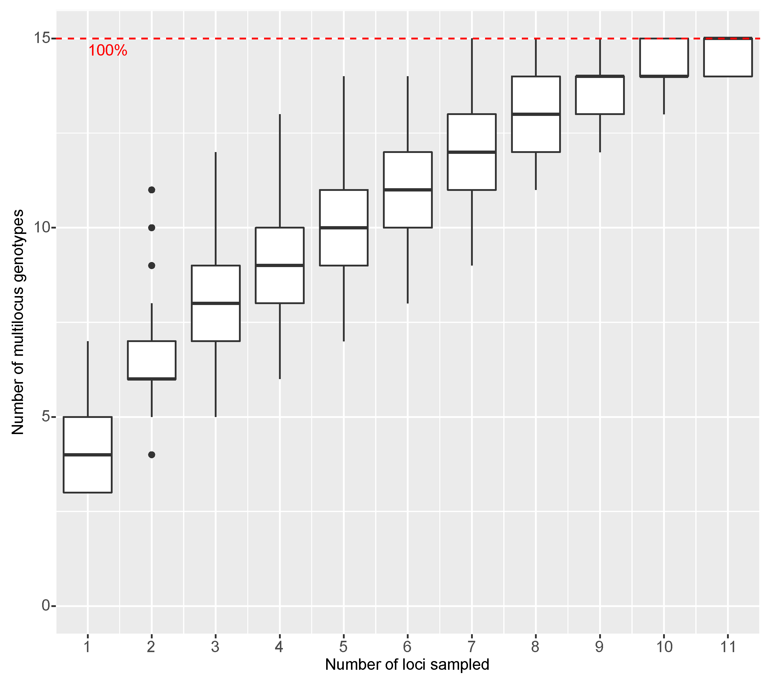
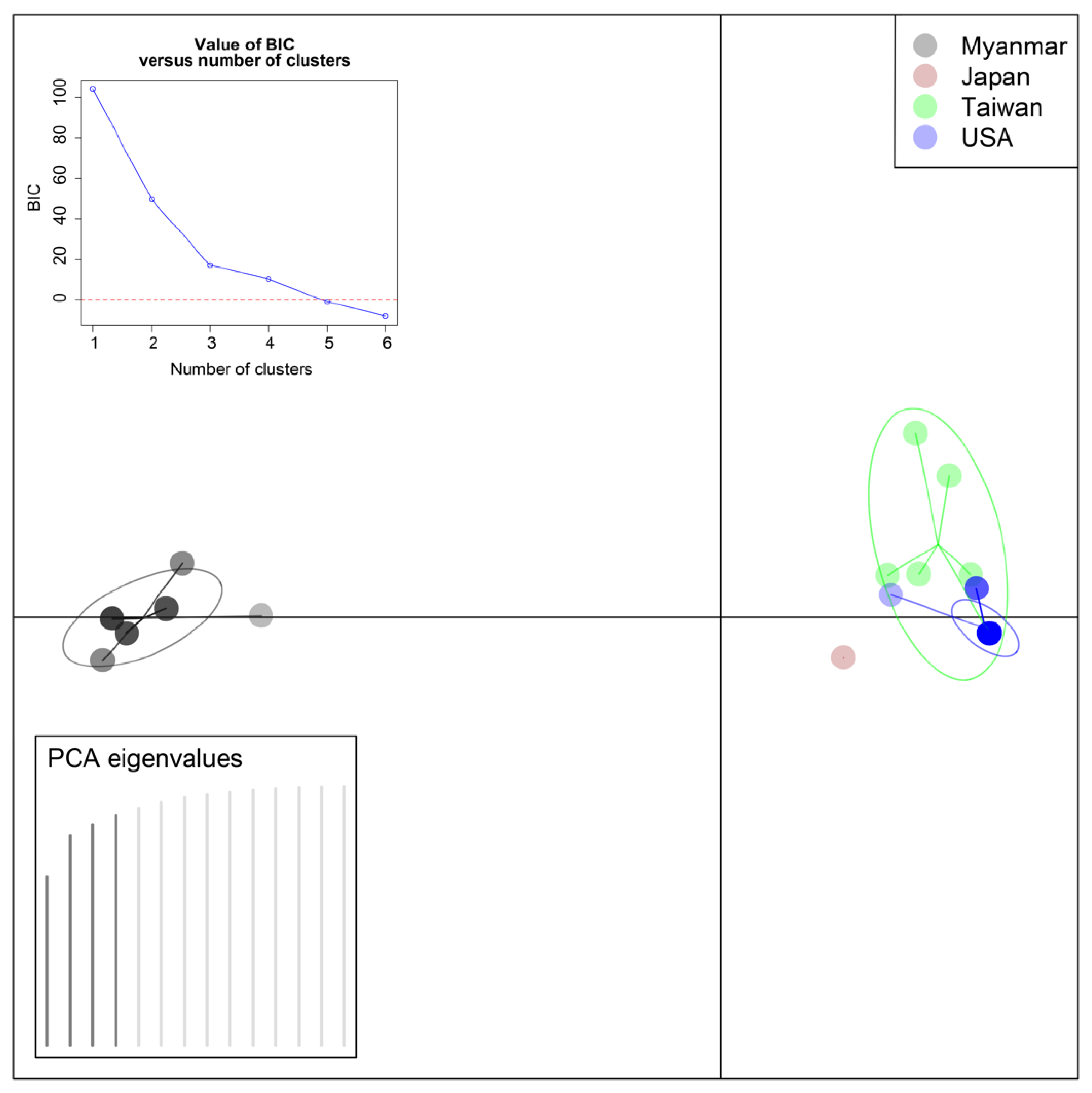
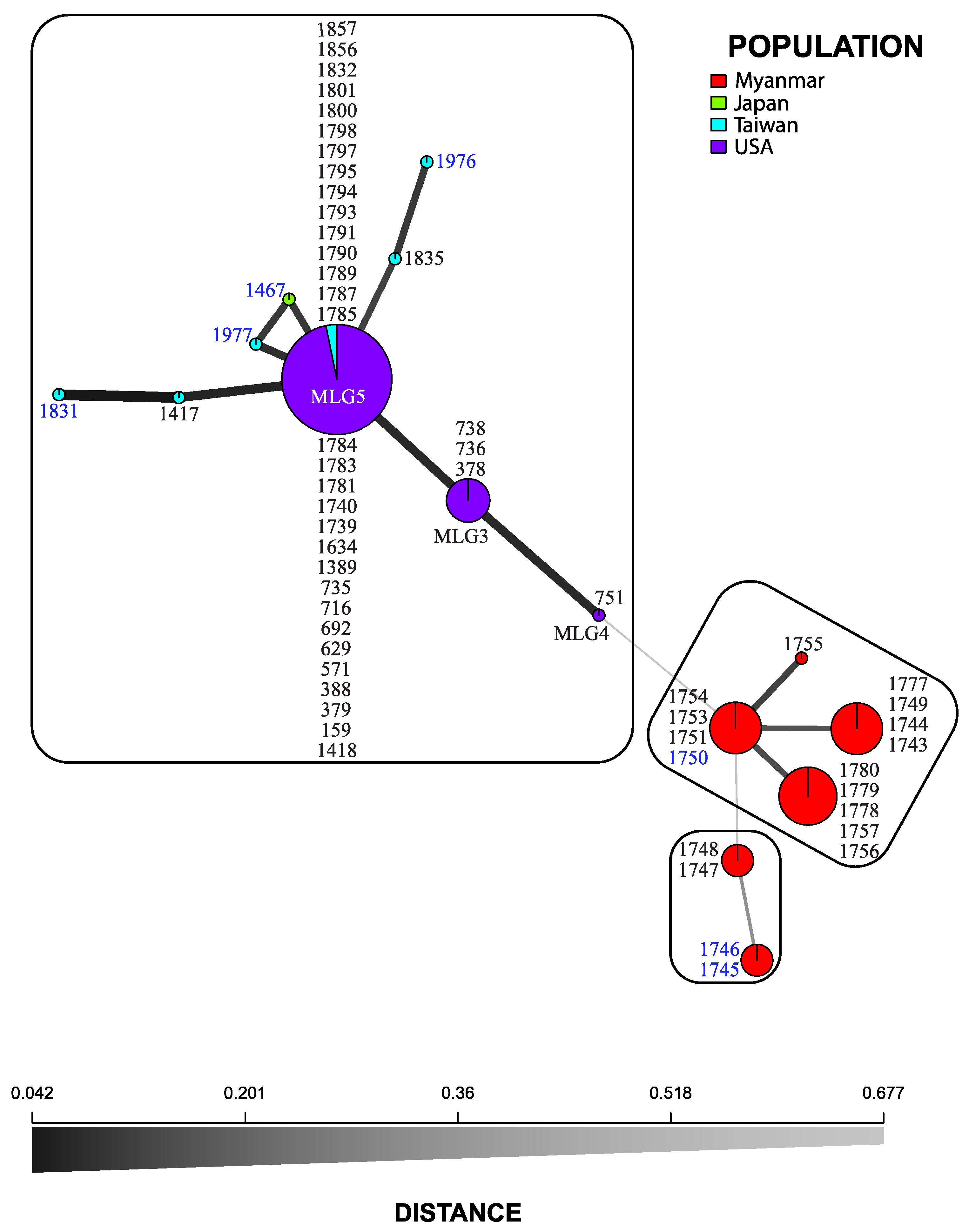
3.3. Statistical Analyses

In the AMOVA, most variance occurred among populations (p = 0.001), with and without clone correction (Table 2). Thus, there was evidence for differentiation, and the null hypothesis of random mating between populations could be rejected.
Discriminant Analysis of Principal Components utilized four Principal Component Analysis axes and three Discriminant Analyses. Although the Bayesian information criteria (BIC) supported three genetic groups, the scatter plot clearly separated the samples into two main groups, the first of which contained the isolates from Myanmar and the second of which included all other isolates (Figure 3). Minimum Spanning Network (MSN) separated the isolates from Myanmar from the remaining isolates, which in turn were separated into two groups (Figure 1). K-means hierarchical clustering also detected the three groups identified by the MSN (Figure 1).
The index of association for the Myanmar population was p = 0.001 for r ¯ d = 0.752 without clone correction and p = 0.001 for r ¯ d = 0.652 with correction. Thus, the null hypothesis of no linkage among markers could be rejected indicating that this population is mostly clonal. In contrast, the index of association for the Taiwan population was p = 0.566 for r ¯ d = 0.008 without clone correction and p = 0.532 for r ¯ d = 0.008 with correction. Thus, the null hypothesis could not be rejected, which indicated a sexually reproducing population.

4. Discussion

The AMOVA found evidence of differentiation among the Asian and USA populations. DAPC, K-means hierarchical clustering, and MSN were mostly congruent, and indicated that the population from Myanmar differed from those from Japan, Taiwan and the USA. Despite the high level of diversity, including the recovery of three MLGs from a single tree and the presence of both mating types, the index of association indicated that the pathogen in Myanmar had the structure of a clonally reproducing population. We recovered MLG14 (Table S1) from the three locations that were sampled in Myanmar, which provided further support for clonal reproduction in this population. In contrast, the index of association and presence of both mating types in Taiwan provided indirect evidence for sexual reproduction of the pathogen in that country. Wuest et al. [17] had suggested that R. lauricola could reproduce sexually in parts of its native range. Nevertheless, direct evidence for sexual reproductive in R. lauricola is lacking.
We detected limited genetic diversity and a single mating type in a collection of 34 isolates of R. lauricola from diverse geographic sources and ambrosia beetles in the USA. Thus, as suggested previously by Wuest et al. [17] and Hughes et al. [5], we conclude that the population of R. lauricola in the USA has gone through a genetic bottleneck. Unlike Wuest et al. [17], who found no geographic structure in their collection, we detected geographic clustering, mainly among isolates from Myanmar (Figure 1 and Figure 3). Although Wuest et al. [17] did not examine isolates from Myanmar, it is also possible that the markers they used played a role in this discrepancy.
Since isolates of R. lauricola from ambrosia beetles other than X. glabratus were all of MLG5 it did not appear that beetle species influenced MLG in the USA. Likewise, as found previously [5,17] isolates from different host trees in the USA were also genetically uniform. Although scant MLG diversity did not enable a good test of this relationship in the USA. We note that three MLGs were detected in a single tree of avocado in Myanmar (Table S1) and five MLGs were detected in a single tree of Cinnamomum osmophloeum Kaneh. in Taiwan [17]. This MLG diversity from a single tree is surprising as laurel wilt is generally considered a systemic disease in highly susceptible host like avocado [10].
Wuest et al. [17] and Hughes et al. [5] identified polymorphic isolates of R. lauricola from southeast Georgia and northeast Florida. Wuest et al. [17] identified isolates with an alternate IFW allele that was not found elsewhere. Three polymorphic isolates identified by Hughes et al. [5] with AFLPs were included in the present study, all of which were members of MLG5, the common SSR MLG in the USA. Although we did not have isolates used by Wuest et al. [17], the polymorphic isolates in the present study were recovered at the same times and from similar areas that they recovered the rare IFW alleles.
Minor differences were evident among the three MLGs that were detected in the USA. For example, isolates in MLG3 shared all its SSR alleles with MLG5, with the exception of one uncommon IFW allele. If a mutation at the IFW locus in MLG5 resulted in MLG3, a second mutation at the MNT locus in MLG3 could have resulted in MLG4. Two mutations in the predominant MLG5 would be consistent with the single introduction hypothesis for R. lauricola in the USA [5,17]. Since the prevalent MLG5 in the USA was also detected in Taiwan and no other MLG in the USA was detected in Asia, it is possible that the relatively homogeneous population of R. lauricola in the USA originated from a single introduction from Taiwan. Additional isolates from Asia would be needed to test this hypothesis.
Our ability to determine the origins and relatedness of populations of R. lauricola is limited by the small number of isolates that are available from the native range of the pathogen and its beetle symbiont, X. glabratus. As more isolates from more diverse geographic areas become available the multiplex microsatellite panel we developed could help elucidate R. lauricola’s genetic structure and possibly the source of the USA introduction. Clearly, better understandings of the pathogen’s global population structure could clarify unknown aspects of its biology, pathology and epidemiology.

5. Conclusions

An invasive fungus from Asia, R. lauricola, causes Laurel wilt, a lethal vascular disease of plants in the Lauraceae plant family. The hypothesis that the fungus was introduced into the USA a single time was corroborated with 12 simple sequence repeat (SSR) markers and a diverse collection of the pathogen from Asia (Myanmar, 18 isolates, Taiwan, 6 isolates, and Japan, 1 isolate) and the USA (34 isolates). The results indicate that a Taiwanese origin is possible for the population of R. lauricola in the USA. Sexual reproduction in the pathogen, which was statistically supported only in the population in Taiwan, needs to be confirmed empirically.

Supplementary Materials

The following are available online at https://www.mdpi.com/1999-4907/10/1/37/s1. Table S1. Collection information for isolates used in the R. lauricola population genetics study, Table S2. Primers and PCRs used in the R. lauricola population genetics study. Loci in bold were used in population genetic analyses.

Author Contributions

T.J.D., M.A.H., R.C.P., and J.A.S. conceptualized and designed the project. M.A.H., A.B., R.C.P., and J.A.S. collected and provided isolates. T.J.D. collected data, analyzed data, and wrote the original draft. R.C.P. and J.A.S. obtained funding. T.J.D., M.A.H., R.C.P., A.B. and J.A.S. reviewed and edited the manuscript.

Funding

Isolates of R. lauricola were collected in Myanmar by R.C.P. during a USAID (Winrock International)-sponsored mission. Funding support for this project was received from USDA Forest Service Southern Research Station, Forest Health Protection and USDA AFRI, Specialty Crops Research Initiative.

Acknowledgments

We thank Stephen Fraedrich (USDA Forest Service) for providing isolates and Carrie Harmon (Univ. Florida Plant Diagnostic Center, Gainesville) for her assistance in importing materials from Myanmar into the USA.

Conflicts of Interest

The authors declare no conflict of interest.

References

  1. Fraedrich, S.W.; Harrington, T.C.; Rabaglia, R.J.; Ulyshen, M.D.; Mayfield, A.E., III; Hanula, J.L.; Eickwort, J.M.; Miller, D.R. A fungal symbiont of the redbay ambrosia beetle causes a lethal wilt in redbay and other Lauraceae in the southeastern USA. Plant Dis. 2008, 92, 215–224. [Google Scholar] [CrossRef]
  2. Harrington, T.C.; Fraedrich, S.W.; Aghayeva, D.N. Raffaelea lauricola, a new ambrosia beetle symbiont and pathogen on the Lauraceae. Mycotaxon 2008, 104, 399–404. [Google Scholar]
  3. Rabaglia, R.J.; Dole, S.A.; Cognato, A.I. Review of American Xyleborina (Coleoptera: Curculionidae: Scolytinae) occurring North of Mexico, with an illustrated key. Ann. Entomol. Soc. Am. 2006, 99, 1034–1056. [Google Scholar] [CrossRef]
  4. Hughes, M.A.; Smith, J.A.; Ploetz, R.C.; Kendra, P.E.; Mayfield, A.E., III; Hanula, J.L.; Hulcr, J.; Stelinski, L.L.; Cameron, S.; Riggins, J.J.; et al. Recovery plan for laurel wilt on redbay and other forest species caused by Raffaelea lauricola and disseminated by Xyleborus glabratus. Plant Health Prog. 2015. [Google Scholar] [CrossRef]
  5. Hughes, M.A.; Riggins, J.J.; Koch, F.H.; Cognato, A.I.; Anderson, C.; Formby, J.P.; Dreaden, T.J.; Ploetz, R.C.; Smith, J.A. No rest for the laurels: Symbiotic invaders cause unprecedented damage to southern USA forests. Biol. Invasions 2017, 79, 22–36. [Google Scholar] [CrossRef]
  6. Fraedrich, S.W.; Johnson, C.W.; Menard, R.D.; Harrington, T.C.; Olatinwo, R.; Best, G.S. First report of Xyleborus glabratus (Coleoptera: Curculionidae: Scolytinae) and laurel wilt in Louisiana, USA: The disease continues westward on sassafras. Fla. Entomol. 2015, 98, 1266–1268. [Google Scholar] [CrossRef]
  7. Olatinwo, R.; Barton, C.; Fraedrich, S.W.; Johnson, W.; Hwang, J. First report of laurel wilt, caused by Raffaelea lauricola, on sassafras (Sassafras albidum) in Arkansas. Plant Dis. 2016, 100, 2331. [Google Scholar] [CrossRef]
  8. Formby, J.P.; Rodgers, J.C.; Koch, F.H.; Krishnan, N.; Duerr, D.D.; Riggins, J.J. Cold tolerance and invasive potential of the redbay ambrosia beetle (Xyleborus glabratus) in the eastern United States. Biol. Invasions 2018, 20, 995–1007. [Google Scholar] [CrossRef]
  9. Ploetz, R.C.; Hughes, M.A.; Kendra, P.E.; Fraedrich, S.W.; Carrillo, D.; Stelinski, L.L.; Hulcr, J.; Mayfield, A.E., III; Dreaden, T.J.; Crane, J.H.; et al. Recovery plan for laurel wilt of avocado, caused by Raffaelea lauricola. Plant Health Prog. 2017, 18, 51–77. [Google Scholar] [CrossRef]
  10. Ploetz, R.C.; Kendra, P.; Choudhury, R.A.; Rollins, J.; Campbell, A.; Garrett, K.; Hughes, M.; Dreaden, T. Laurel wilt in native and agricultural ecosystems: Understanding the drivers and scales of complex pathosystems. Forests 2017, 8, 48. [Google Scholar] [CrossRef]
  11. Hulcr, J.; Lou, Q.-Z. The redbay ambrosia beetle (Coleoptera: Curculionidae) prefers Lauraceae in its native range: Records from the Chinese national insect collection. Fla. Entomol. 2013, 96, 1595–1596. [Google Scholar] [CrossRef]
  12. Harrington, T.C.; Yun, H.Y.; Lu, S.S.; Goto, H.; Fraedrich, S.W. Isolations from the redbay ambrosia beetle, Xyleborus glabratus, confirm that the laurel wilt pathogen, Raffalea lauricola, originated in Asia. Mycologia 2011, 103, 1028–1036. [Google Scholar] [CrossRef] [PubMed]
  13. Hulcr, J.; Black, A.; Prior, K.P.; Chen, C.-Y.; Li, H.-F. Studies of ambrosia beetles in their native ranges help predict invasion impact. Fla. Entomol. 2017, 100, 257–261. [Google Scholar] [CrossRef]
  14. Ploetz, R.C.; Hulcr, J.; Wingfield, M.; de Beer, Z.W. Ambrosia and bark beetle-associated tree diseases: Black Swan events in tree pathology? Plant Dis. 2013, 95, 856–872. [Google Scholar] [CrossRef]
  15. Fraedrich, S.W.; Harrington, T.C.; Best, G.S. Xyleborus glabratus attacks and systemic colonization by Raffaelea lauricola associated with dieback of Cinnamomum camphora in the southeastern United States. For. Pathol. 2015, 45, 60–70. [Google Scholar] [CrossRef]
  16. Ploetz, R.C.; Thant, Y.Y.; Hughes, M.A.; Dreaden, T.J.; Konkol, J.L.; Kyaw, A.T.; Smith, J.A.; Harmon, C.L. Laurel wilt, caused by Raffaelea lauricola, is detected for the first time outside the southeastern USA. Plant Dis. 2016, 100, 2166. [Google Scholar] [CrossRef]
  17. Wuest, C.E.; Harrington, T.C.; Fraedrich, S.W.; Yun, H.-Y.; Lu, S.-S. Genetic variation in native populations of the laurel wilt pathogen, Raffaelea lauricola, in Taiwan and Japan and the introduced population in the United States. Plant Dis. 2017, 101, 619–628. [Google Scholar] [CrossRef]
  18. Dreaden, T.J.; Davis, J.M.; Harmon, C.L.; Ploetz, R.C.; Palmateer, A.J.; Soltis, P.S.; Smith, J.A. Development of multilocus PCR assays for Raffaelea lauricola, causal agent of laurel wilt disease. Plant Dis. 2014, 98, 379–383. [Google Scholar] [CrossRef]
  19. Ploetz, R.C.; Konkol, J.L.; Narvaez, T.; Duncan, R.E.; Saucedo, R.J.; Campbell, A.; Mantilla, J.; Carrillo, D.; Kendra, P.E. Presence and prevalence of Raffaelea lauricola, cause of laurel wilt, in different species of ambrosia beetles in Florida, USA. J. Econ. Entomol. 2017, 110, 347–354. [Google Scholar]
  20. Duong, T.A.; de Beer, Z.W.; Wingfield, B.D.; Wingfield, M.J. Mating type markers reveal high levels of heterothallism in Leptographium sensu lato. Fungal Biol. 2016, 120, 538–546. [Google Scholar] [CrossRef]
  21. Holleley, C.E.; Geerts, P.G. Multiplex Manager 1.0: A crossplatform computer program that plans and optimizes multiplex PCR. BioTechniques 2009, 46, 511–517. [Google Scholar] [CrossRef] [PubMed]
  22. Drost, D.R.; Novaes, E.; Boaventura-Novaes, C.; Benedic, C.I.; Brown, R.S.; Yin, T.; Tuskan, G.A.; Kirst, M. A microarray-based genotyping and genetic mapping approach for highly heterozygous outcrossing species enables localization of a large fraction of the unassembled Populus trichocarpa genome sequence. Plant J. 2009, 58, 1054–1067. [Google Scholar] [CrossRef] [PubMed]
  23. Kamvar, Z.N.; Tabima, J.F.; Grünwald, N.J. Poppr: An R package for genetic analysis of populations with clonal, partially clonal, and/or sexual reproduction. PeerJ 2014, 2, e281. [Google Scholar] [CrossRef] [PubMed]
  24. Kamvar, Z.N.; Brooks, J.C.; Grünwald, N.J. Novel R tools for analysis of genome-wide population genetic data with emphasis on clonality. Front. Genet. 2015, 6, 208. [Google Scholar] [CrossRef]
  25. Excoffier, L.; Smouse, P.E.; Quattro, J.M. Analysis of molecular variance inferred from metric distances among DNA haplotypes: Application to human mitochondrial DNA restriction data. Genetics 1992, 131, 479–491. [Google Scholar]
  26. Jombart, T.; Collins, C. A Tutorial for Discriminant Analysis of Principal Components (dapc) Using Adegenet 2.0.0. Available online: http://adegenet.r-forge.r-project.org/files/tutorial-dapc.pdf (accessed on 2 June 2018).
  27. Bruvo, R.; Michiels, N.K.; D’Souza, T.G.; Schulenburg, H. A simple method for the calculation of microsatellite genotype distances irrespective of ploidy level. Mol. Ecol. 2004, 13, 2101–2106. [Google Scholar] [CrossRef] [PubMed]
  28. Grünwald, N.J.; Kamvar, Z.N.; Everhart, S.E.; Tabima, J.F.; Knaus, B.J. Population Genetics and Genomics in R. Available online: http://grunwaldlab.github.io/Population_Genetics_in_R/ (accessed on 2 June 2018).
  29. Brown, A.H.D.; Feldman, M.W.; Nevo, E. Multilocus structure of natural populations of Hordeum spontaneum. Genetics 1980, 96, 523–536. [Google Scholar]
Figure 1. Minimum spanning network for SSR genotypes of Raffaelea lauricola from Asia and the USA. Node color indicates source country of a genotype, the relative node size indicates the number of clones that are represented by a given genotype, and the thickness of lines between nodes represents Bruvo’s (27) genetic distance, wherein thicker lines denote smaller genetic distances. Isolate numbers in blue are MAT1 and black are MAT2. Three groups detected by K-means hierarchical clustering are in separate boxes, and multilocus genotypes (MLG) in the USA are indicated.
Forests 10 00037 g001
Figure 2. Genotype accumulation curve for isolates of R. lauricola from the USA and Asia. The y axis gives the number of multilocus genotypes (MLGs), the x axis the number of SSR loci that were assayed (12 total), and the dashed red line represents 100% MLG resolution.
Forests 10 00037 g002
Figure 3. Discriminant Analysis of Principal Components (DAPC) of R. lauricola sampled from United States and Asian populations using four Principal Component Analysis axes and three Discriminant Analysis separates isolates from Myanmar from the rest of the isolates. Upper left inset, the Bayesian information criteria (BIC) supported three genetic groups.
Forests 10 00037 g003
Table 1. Genotyping statistics for microsatellite/simple sequence repeat (SSR) loci in isolates of Raffaelea lauricola from Asia and the USA.
Multiplex PCRSSR Loci xError Rate (Incorrect Calls/Total Calls)Alleles
1NB60/153
0DS0/154
ZWC1 */152
2OCT4/13
F8I0/133
3ZBI0/155
KTR0/156
4P860/143
46Z2/14
5QI51/17
V5I0/175
6EC60/134
03B0/134
IFW1 */136
7X21-
9V8-
8MNT1 */153
REH2/14
x Boldface loci were used in population genetic analyses. * Error due to null allele in multiplex PCRs. “-” Difficult to call, and omitted, alleles.
Table 2. Analysis of molecular variance (AMOVA) using twelve microsatellite loci from Raffaelea lauricola from Myanmar, Japan, Taiwan, and the United States.
No Clone CorrectionWith Clone Correction
dfSigma% VariancepdfSigma% Variancep
Between Populations32.98770.00131.66540.001
Within Population550.8622 121.4246
Total583.85 153.07

