1. Introduction
Variables such as microleakage, marginal gap, the oral environment, including force, thermal loading, and contact with the oral medium (saliva) influence the duration of indirect restoration (crown stability) and sealing ability
in vivo. Microleakage characterizes the marginal sealing [
1,
2], and the marginal gap characterizes the marginal adaptation [
3]. The depth of microleakage can be determined with the penetration test using silver staining [
2,
4,
5], or with fuchsine solution [
1]. The marginal gap depends on the surface characteristics and type of luting agent [
6]. In the literature, there are various methods for evaluating the marginal gap, including tactile and optical profilometry [
7].
Knowledge about the luting quality and characteristics of the different classes of materials for connecting the tested surface dentin or enamel with the full crown helps to achieve the long-term effect by cementation and to develop new classes of luting materials for improving clinical success [
3,
4,
5]. A luting agent is the dental cement, which is the active material, used to attach an indirect restoration to a prepared tooth, and it may aid in the marginal seal of restorations [
6,
7]. The sealing ability, the resistance to dissolution, and biological and physical attributes, such as bond and compressive strengths, dimensional change on setting, flexural modulus and no interfacial-gap incidence of different types of luting materials, are variable [
3,
8,
9,
10,
11]. These parameters, tooth preparation [
12], the type of restoration and the clinical situation influence the choice of proper luting agents [
13]. For clinical success in indirect restorations with minimal microleakage avoiding penetration of bacteria, the correct selection of luting agents with optimum physical properties is essential [
6].
For the optimal choice of luting material, it is important to collect comparative data about the quality of the marginal sealing and integrity of the different indirect restoration variants (regarding the size, geometry, cavity preparation, margin place, crown material, luting technique, and artificial aging model or intra-oral conditions simulation) [
2,
14,
15]. There is a practical need to improve the marginal integrity and adaptation for crowns [
2], and the assumption was that a resin and self-adhesive luting materials may help optimize the parameters for indirect restorations with full ceramic crowns. At the present time, there are a number of luting materials on the market. Clinically, it is of importance to know which resin-based, self-adhesive cements for cementation of full lithium disilicate CAD/CAM crowns should be preferred.
One study assessed the marginal adaptation and strength of esthetic ceramic CAD/CAM molar crowns due to four different self-adhesive resin luting agents [
4]. Other studies investigated the adhesive resin cements with respect to the marginal adaptation of ceramic inlays [
1], the partial ceramic crowns within dentin [
2], or the full-contour cast crowns made from high-gold alloy. Peutzfeldt
et al. [
10] tested Panavia F 2.0, RelyX Unicem 2 and MaxCem Elite cements, but the authors only examined the bonding strength of these materials [
5]. The luting materials and techniques used in these studies were different and were not appropriate for our comparison. In these studies, zinc-phosphate cement was used for comparision [
5,
10,
11]. To the authors’ knowledge, there is no comparative study about the marginal gap of full ceramic crowns due to self-adhesive resin cements within the enamel margin
versus within the dentin margin in the literature.
Therefore, in the present in vitro study, we evaluated marginal sealing and adaptation (regarding microleakage, marginal gap and absolute marginal gap) at the tooth surface/full-contour ceramic crowns interface after simulation of the oral conditions. Ceramic crowns were luted with different classes of luting materials and assessed, after simulation of the oral condition mimicking the thermal stress of body temperature in artificial saliva and hydrothermal stress of oral cavity in a thermocycler. The following parameters were examined: (1) microleakage and marginal gap after using various types of luting agents for cementing indirect restorations; (2) the influence of the margin location (within dentin or enamel) on the microleakage and marginal gap with respect to the different luting agents; and (3) the correlation between microleakage and marginal gaps in the full ceramic crowns.
The null hypothesis for the present study was that different chemical types of luting materials will not differ in their influence on the microleakage and marginal gap of full ceramic crowns with margin setting within dentin or enamel.
2. Materials and Methods
2.1. Tooth Collection
Tooth collection was approved by the ethical committee of Witten/Herdecke University (permission 116/2013). A total of 70 extracted human molar teeth were collected for this study. They were caries-free, lacked dental calculus and had completed root growth. Immediately after extraction, all teeth were stored in 0.9% NaCl containing 0.1% thymol at room temperature until use (maximum 1–3 months). The teeth were polished with a rotating brush and pumice.
2.2. Abutment Preparation
To avoid inter examiner differences, all specimen preparations were performed by the same operator which was calibrated prior to the study by one of the authors.
Teeth were prepared according to Goodacre [
12] for a full ceramic crown at a converging angle of approximately 6°. The reduction was approximately 1.0 mm in the axial and 1.5 mm in the occlusal surfaces, resulting in a residual height of least 4 mm. The preparation of the cervical margins was performed as circular chamfers, which were located from the vestibular and lingual margins in enamel, and from the mesial and distal margins in dentin. Torpedo-shaped burs (Busch, Engelskirchen, Germany; CD720120, CD72F012) were used for the circular chamfer, and pear-shaped diamonds (831/014, 8831/014) were used for the occlusal reduction.
2.3. Digital Data Acquisition
After drying, the prepared abutment teeth were covered with a thin layer of Powder Scan Spray (Vita Zahnfabrik, Bad Säckingen, Germany) to obtain an accurate digital impression. Digital data of the preparation were obtained using Cerec Bluecam (Sirona Dental GmbH, Bensheim, Germany). An occlusal recording and four angled images with an angle of 30° from each side of the tooth were made and matched together.
2.4. Manufacturing of Ceramic Crowns
The design of the ceramic full crowns was performed using the software 4.0 of Cerec chairside solution (Sirona Dental GmbH). Milling of lithium disilicate crowns (IPS e.max CAD Monolithic Solutions; LT A3/C14, Lot R64204, Ivoclar Vivadent, Schaan, Liechtenstein) was performed using a Cerec MC XL unit (Sirona Dental GmbH). The following milling parameters were defined: spacer 80 μm, and circular margin thickness 150 μm. Step bur 12S and cylinder pointed bur 12S were used according to the standard manufacturer’s parameters. Maintenance, cleaning and changing of the instruments was carried out strictly in accordance with the automatic wear message. Sintering of the milled crowns was provided in the oven Denta Star (imes-icore GmbH, Eiterfeld, Germany) according to the manufacturer’s instructions.
The fit of the crowns corresponding to the prepared abutment tooth was checked by a master dental technician. The marginal fit was verified under a stereomicroscope (×32 magnification), with an extra-fine probe (EXD5; Hu-Friedy, Chicago, IL, USA) and was improved where necessary. Marginal fitting of the crowns were improved using a silicone medium and a small bur. A logged control of all crowns was carried out before and after fitting with respect to the marginal fit as well as the retention and resistance.
2.5. Luting Materials
The luting materials are summarized in
Table 1.
2.6. Crown Cementation
To test the different luting agents, the pairs, consisting of the tooth and crown, were randomly divided into 7 test groups (n = 10).
The prepared abutment teeth were slightly air dried before cementation. No further conditioning of the abutment teeth, except for the use of Panavia F2.0 was applied. ED Primer (Kuraray, Tokyo, Japan) was used with Panavia. ED Primer A and B was mixed and this mixture was applied for 30 s on the prepared abutment tooth. Afterwards, the tooth was slightly air-dried.
Before cementation, the interior surface of all crowns was conditioned with IPS Ceramic Etching Gel containing hydrofluoric acid (Ivoclar Vivadent, Schaan, Liechtenstein) for 20 s and silanized (except the Harvard cement group) with Monobond Plus (Ivoclar Vivadent, Schaan, Liechtenstein) for 60 s.
All luting materials were handled, according to the manufacturers’ instructions, at room temperature. Harvard cement was manually mixed in a ratio of 1.5:1 powder-liquid on a cooled glass plate and Panavia F 2.0 at a ratio 1.0:1.0 on a mixing pad with a plastic spatula. All other materials were processed as automix variants.
Table 1.
Description of the luting agents used in this study.
Table 1.
Description of the luting agents used in this study.
| Materials | Type | Main Composition a | Adhesive System | Manufacturer |
|---|
| Harvard cement | Zinc-phosphate-cement | P: Zinc oxide, magnesia L: phosphoric acid | No adhesive system | Harvard, Germany |
| Panavia F 2.0 | Resin cement | MDP, dimethacrylate, silica, dl-Camphorquinone, barium glass filler, sodium fluoride | ED Primer | Kuraray, Japan |
| Maxcem Elite | Self-adhesive resin cement | HEMA, MEHQ, CHPO | No adhesive system | Kerr, USA |
| Perma Cem 2.0 | Self-adhesive resin cement | Bis-GMA-based Matrix of dental resin, Barium glass | No adhesive system | DMG, Germany |
| G-CEM Automix | Self-adhesive resin cement | UDMA, Camphorquinone, Hydroperoxide | No adhesive system | GC Corp., Japan |
| RelyX Unicem 2 Automix | Self-adhesive resin cement | Methacrylate monomers containing phosphoric acid groups, Methacrylate monomers, fillers, Initiator, Stabilizers, Rheological additives, Pigments | No adhesive system | 3MESPE, Germany |
| BifixSE | Self-adhesive resin cement | Bi-functional methacrylate, acid methacrylate, inorganic fillers | No adhesive system | VOCO, Germany |
The crowns were seated with finger pressure for 10 s and then axially loaded for 6 min with a device that ensured a defined axial load of 60 N. Light curing of all self-adhesive resin cements was performed according to the manufacturer’s instructions (Bluephase Style, Ivoclar Vivadent, Schaan, Liechtenstein). Excess cement was carefully removed with a foam pellet and scaler. The marginal fit of the crown was checked by visual inspection.
2.7. Experimental Procedure
To simulate the oral cavity medium and the influence of saliva on the luting characteristics of the tested materials, the specimens were stored at 37 °C in 100 mL artificial saliva (Dental center, Erfurt, Germany) [
16] for 4 weeks. To test the impact of alternating thermal stress on the characteristics of the different luting materials, the specimens were thermocycled for 5000 cycles at 5 and 55 °C (immersion time, 30 s, transfer time, 5 s) in a thermocycler THE1000 (SD Mechatronik, Feldkirchen-Westerham, Germany).
2.8. Assessment of Microleakage and Marginal Gap
To investigate the depth of the microleakage of the tooth-cement interface, the area of the root surface of the teeth was covered with two layers of nail varnish that ended approximately 1 mm below the crown margin. Subsequently, all test objects were immersed in 10% silver nitrate solution (Cristal, Fischer Scientific, Fairfield, NJ, USA) in a dark chamber for 6 h. Then, the specimens were rinsed carefully with distilled water and exposed in a 4 × 100-W floodlamp for 4 h. Then, the samples were immersed in a photochemical developer Periomat (Dürr Dental, Bietigheim-Bissingen, Germany) for 12 h.
All specimens were embedded in Technovit 9100 (Heraeus Kulzer, Wehrheim, Germany), and serial sections, in the buccolingual or mesiodistal direction with a thickness of 80 μm, were cut with a saw microtome (Leica SP 1600, Leica Microsystems, Wetzlar, Germany). All sections were mounded (fixation) with Technovit 7210 VLC (Heraeus Kulzer, Wehrheim, Germany) on acryl slides (Exakt, Hamburg, Germany) and the three central sections of the teeth were selected for further investigation. The sections were analyzed with a scanning electron microscope (SEM; Sigma VP, Zeiss, Oberkochen, Germany), at an acceleration voltage of 20 kV and pressure of 20 Pascal, and a backscattered electron detector. Electron dispersive spectroscopy (EDS) was performed with an EDAX Apollo XL system (EDAX Inc., Mahwah, NJ, USA), an active area of 30 mm
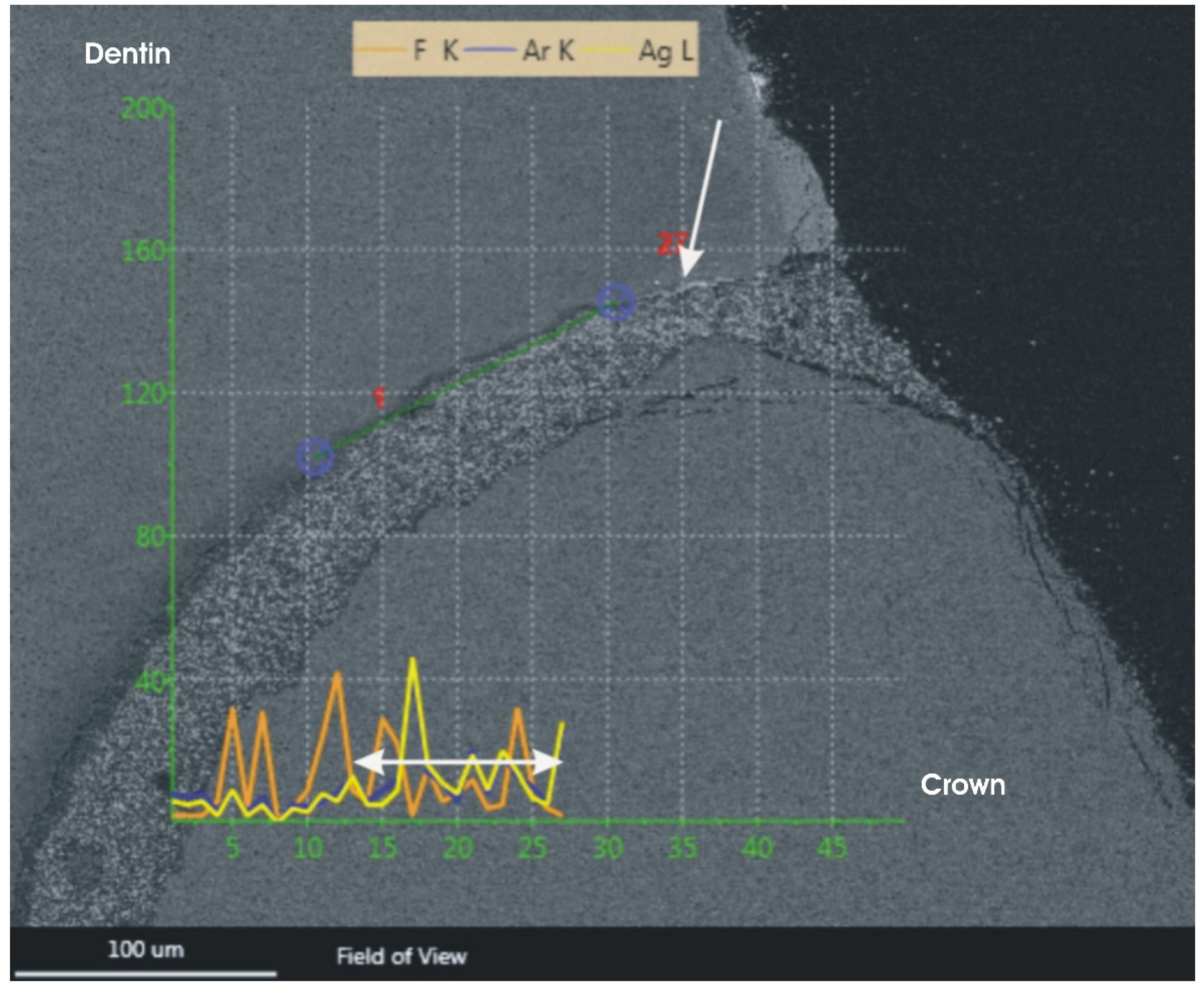
2 and the Team V3.3 software. The reading of the line scans was carried out with a dwell time of 25 msec and an amplification time of 12.8 µsec, using a distance between reading points of 1 μm. The depth of microleakage (μm) was measured after verification of the silver precipitation with EDS spectroscopy (
Figure 1). The length of the silver precipitation was measured by the calibrated investigator.
Figure 1.
Linescan of a silver precipitation within the gap of the microleakage. The silver precipitation is marked with an arrow. The depth of the silver precipitation is marked with a double-headed arrow along the yellow curve of the EDS scan.
Figure 1.
Linescan of a silver precipitation within the gap of the microleakage. The silver precipitation is marked with an arrow. The depth of the silver precipitation is marked with a double-headed arrow along the yellow curve of the EDS scan.
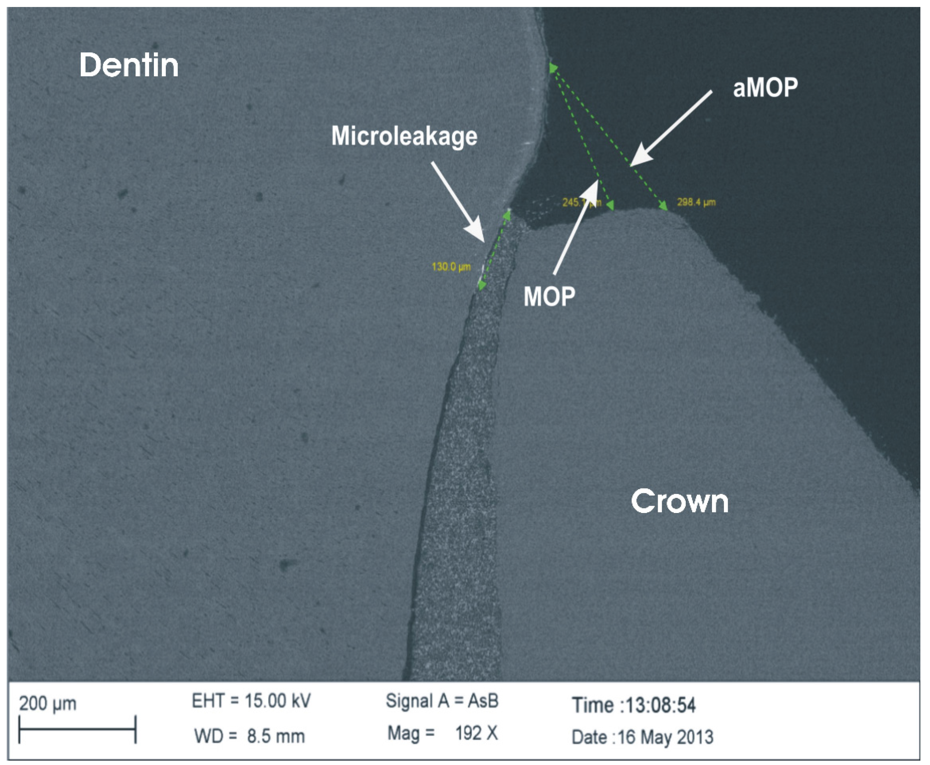
The absolute marginal gap was defined as the distance between the outer contour of the crown and dentin or enamel surface (aMOP). The marginal gap was defined as the distance between the edge of the crown and the dentin or enamel surface (MOP) [
17]. They were determined by measuring the distances between the respective margins (
Figure 2) with the measuring software SmartTiff Rel. 2 (Zeiss).
Figure 2.
Measurements of microleakage, marginal gap (MOP) and absolute marginal gap (aMOP).
Figure 2.
Measurements of microleakage, marginal gap (MOP) and absolute marginal gap (aMOP).
2.9. Statistical Analysis
The statistical analysis was carried out using the non-parametric Kruskal-Wallis test for related variables to determine differences between the materials and the Wilcoxon test for independent variables (p < 0.05) for the comparison of the different materials. Analysis of the correlation was performed with the Pearson correlation coefficient. The median, quartiles, minimum and maximum were calculated for descriptive analysis of the continuous data. The descriptive data are presented as boxplots.
4. Discussion
Luting with the self-adhesive resin cements reduces the number of application steps and technique sensitivity compared to conventional cements [
18,
19]. However, literature reports do not describe all luting variants for full contour ceramic crown with different self-adhesive resin cements. Few studies assess the compatibility of different self-adhesive resin luting agents for the indirect ceramic restorations with respect to marginal sealing (with the microleakage assessment) and adaptation (with the marginal gap assessment) [
1,
2,
10]. Schenke
et al. investigated luting of partial ceramic crowns [
2], Mörmann
et al. investigated ceramic CAD/CAM molar crowns, [
4] and Aschenbrenner
et al. investigated ceramic inlays [
1]. Piwowarczyk
et al. investigated luting of casted crowns made from high-gold alloy with two resin cements and one self-adhesive universal resin cement [
10]. The influence of microleakage using zinc-phosphate cement performing different crown preparations has also been described [
20]. The differences in the morphology and materials of these indirect restorations do not allow for a direct comparison of the luting characteristics of the tested materials.
For the longevity of dental restorations, “gap-free” continuous cement margins are important [
21], to avoid plaque accumulation and subsequent secondary caries the interface between restorative materials and tooth hard substances must be morphologically as perfect as possible [
22], even though no luting material is able to achieve a perfect marginal seal [
1,
23].
Margins of restorations have a large morphological variety [
24]. Roulet
et al. proposed a method to quantify the quality of dental restorations [
21]. Aschenbrenner
et al. examined the “perfect margin” and defined it as two adjoining surfaces (cement–ceramic, cement–tooth), which have no interruption of the continuous margin and merge into each other without any difference in level [
1]. The “perfect margins” can be analyzed by measuring the marginal gaps [
1,
21,
22,
24].
The present study was performed to compare the microleakage and marginal gap of five self-adhesive resin cements, one resin cement and one zinc-phosphate-cement (Harvard, Germany) as a control for full-contour molar ceramic crowns after simulation of intra-oral conditions with artificial saliva and thermal stress. To simulate the environment of the oral cavity during artificial aging, the molars with cemented full-contour crowns made out of lithium disilicate were stored in artificial saliva at 37 °C for 4 weeks, which was followed by thermal cycling. In previous studies on this topic, tap water was used for at least two weeks [
4], water at 37 °C for one week [
5], or distilled water at 37 °C for 90 days [
1,
25].
Usually, only visual methods for recognizing the depth of microleakage were applied, such as identifying silver with an image analyzing system (Optimas 6.1, Stemmer, München, Germany) [
2], or identifying fuchsine with a light microscope [
1]. In the present study, for more precise measurement, microleakage visualization was performed with scanning electron microscopy (SEM) energy-dispersive and X-ray spectroscopy (EDX). The EDX analysis was used for the conformation of silver penetration into the microleakage gap.
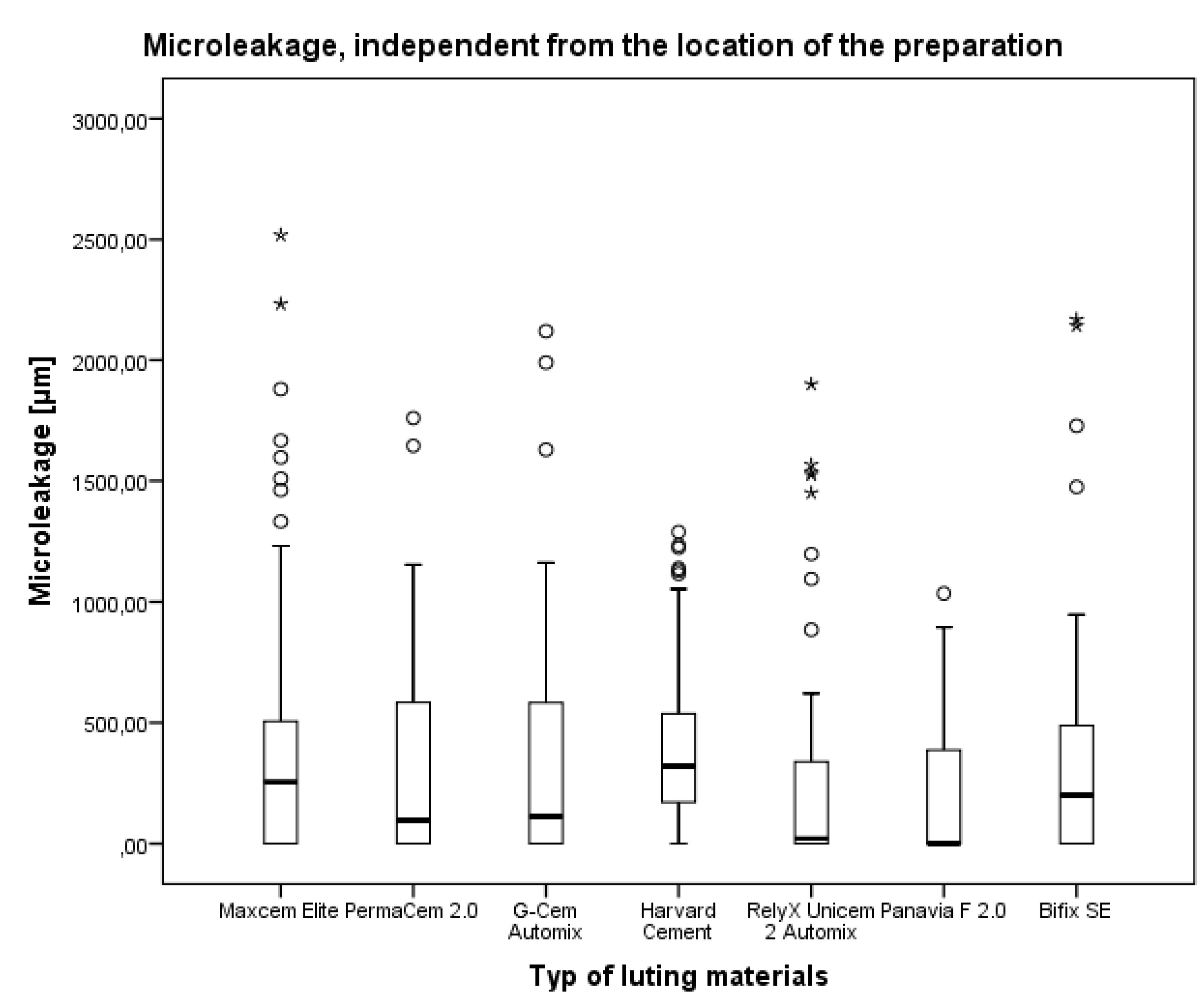
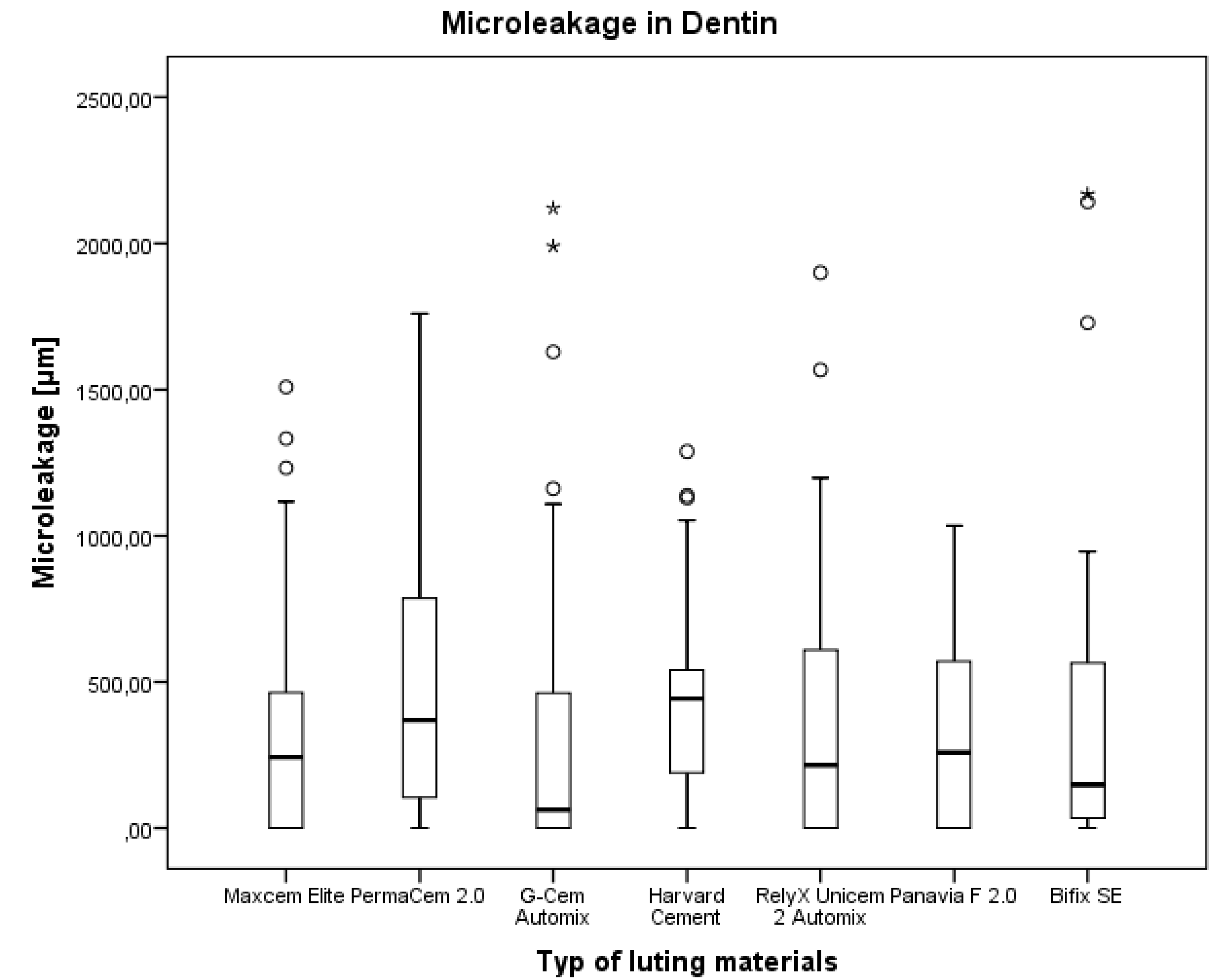
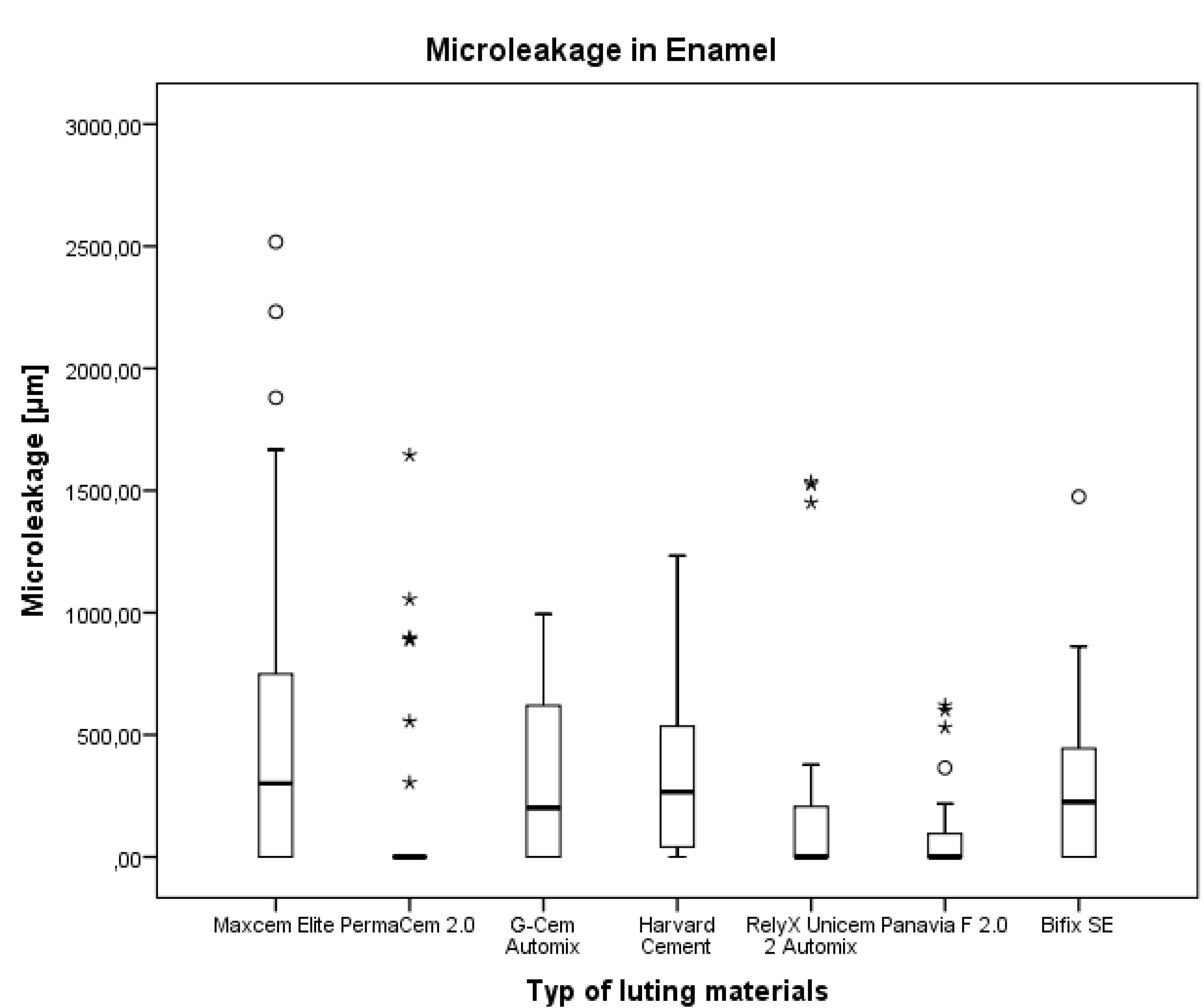
The comparison after the penetration test of all tested materials for luting CAD/CAM full-contour molar ceramic crowns demonstrated that significantly better marginal sealing independent of the location of the preparation (dentin/enamel) was achieved after applying resin cement Panavia F 2.0, self-adhesive resin cement RelyX Unicem 2 Automix, and self-adhesive resin cement G-CEM Automix; in enamel it was achieved after applying self-adhesive resin cements PermaCem 2.0 and self-adhesive resin cement RelyX Unicem 2 Automix, resin cement Panavia F 2.0; in dentin it was achieved after applying self-adhesive resin cement G-CEM Automix compared to the zinc-phosphate-cement Harvard Cement. The differences between the other tested materials were not significant.
These results are not in concordance with those reported by Aschenbrenner
et al. that, for dentin, margins Bifix SE showed one of the lowest mean values of dye penetration for the indirect restoration with a ceramic inlay [
1]. However, the differences in the morphology of the indirect ceramic restorations and some differences in the simulation of the intra-oral conditions do not allow for a direct comparison of the luting characteristics of the tested materials in these two studies.
This study also revealed remarkable differences in the microleakage between enamel and dentin and between the different materials. Bonding is dependent from the hydrophilicity and length of spacer chains as well as from the functional monomers [
23]. The remnants of the smear layer may have an influence on the bond strength of the different materials [
26]. All investigated materials differ in their properties regarding, hydrophilicity length of spacer chains and functional monomers. This may explain the differences found in this investigation.
The fact that the marginal adaptation (marginal gap) was not correlated with the marginal sealing for all test materials is in concordance with the results reported by Piwowarczyk
et al. [
10].
There are many publications about the size of marginal gaps available. They are summarized by Abduo
et al. [
27]. Reported ranges of marginal gaps are between <80 μm up to 150 μm, which are regarded as clinically acceptable [
28,
29,
30,
31,
32,
33]. The marginal gap for lithium disilicate crows which were manufactured with the Everest CAD/CAM system showed values of 28.1 ± 7.9 µm and manufactured with the Cerec inLab CAD (CAM system 40.2 ± 6.7 μm [
34]. Statistical analysis of the measured values of the marginal gap in the present study showed high deviations for milled crowns. Another study showed marginal gaps of the CAD/CAM zirconia crowns with a range from mean 49.32 to 91.20 micrometers with a standard deviation between 3.97 and 42.41 μm [
35]. The higher deviations of the investigated parameters in the present study compared to the results from the study of Alghazzawi
et al. may be due to the difference in the milling process, another material of the crown (zirconia) and the different evaluation methods [
35].
Limitations of the present study are the low number of test samples in each group. It may be the reason, that investigated parameters can be more affected by variables, such as age, history of the teeth, structure of hydroxyapatite [
1,
36], dentine tubule density, orientation, lumen size, thickness of highly mineralized cuff of peritubular dentin, intratubular dentin, containing mostly apatite crystals with little organic matrix, amount of intertubular dentin composed of a matrix of type I collagen reinforced by apatite, which also varies within location and age [
37]. Another limitation of this study is the fact that the measurements were not repeated in order to determine intra-rater reliability. These limitations may result in higher deviations in the statistical analysis.
Conclusion: This study showed that different chemical types of luting agents generally have better marginal sealing (regarding the microleakage) in the enamel than in dentin, and this is in concordance with the data about the specific properties of dentin, such as its tubular structure and intrinsic wetness. Clinically, it seems that there is no prevalence for the use of a specific luting material for the cementation of full lithium disilicate crowns. However, the comparison of marginal adaptation of all tested materials demonstrated no statistically significant difference in the marginal and absolute marginal gaps of full ceramic crowns with margin setting within dentin or enamel. Therefore, the null hypothesis was rejected with regard to marginal sealing; however it was not rejected with regard to adaptation (marginal gap).


