
A Comparative Finite Element Analysis of Titanium, Autogenous Bone, and Polyetheretherketone (PEEK)-Based Solutions for Mandibular Reconstruction

 ,
,  ,
,  ,
, 
Abstract
1. Introduction
2. Materials and Methods
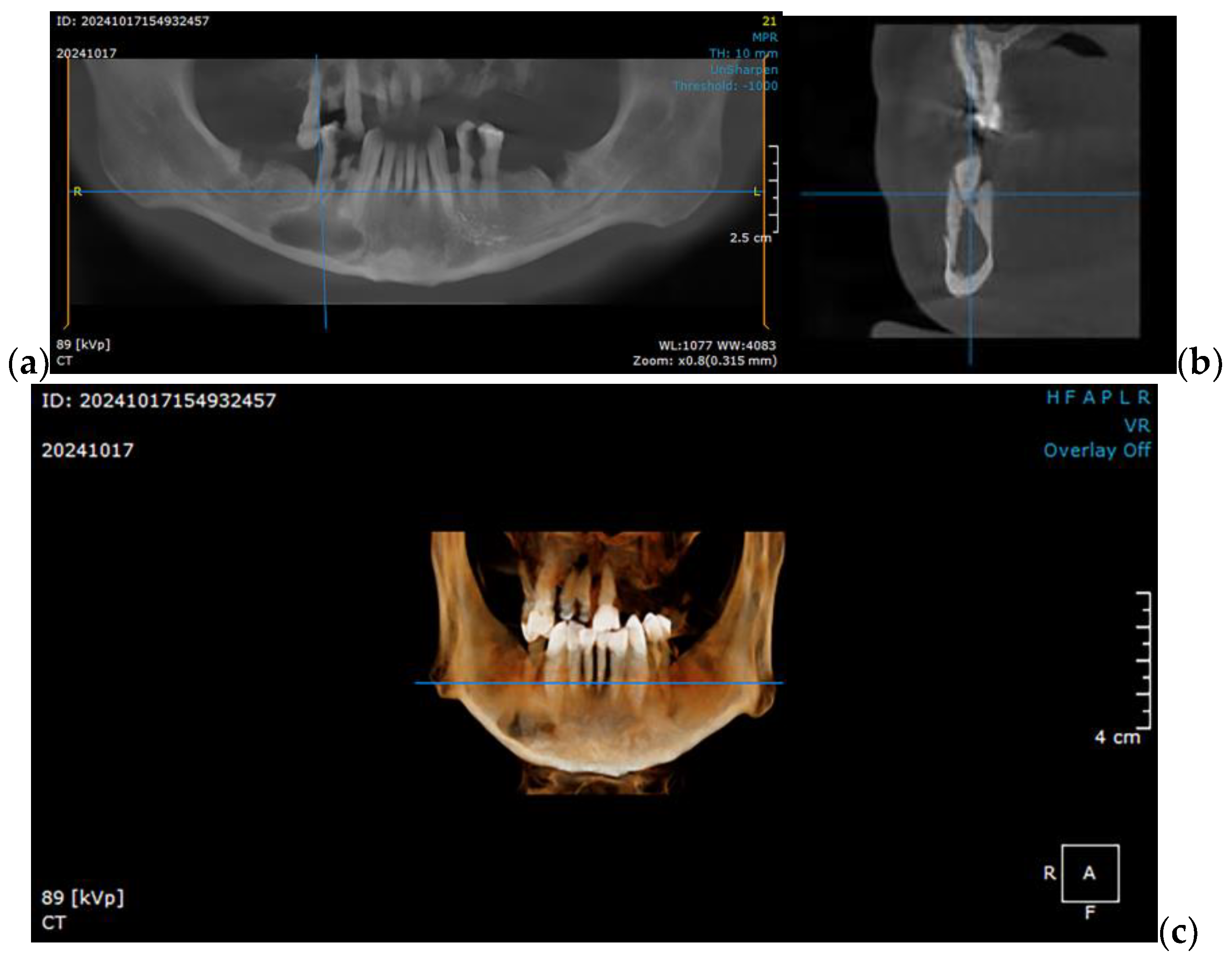
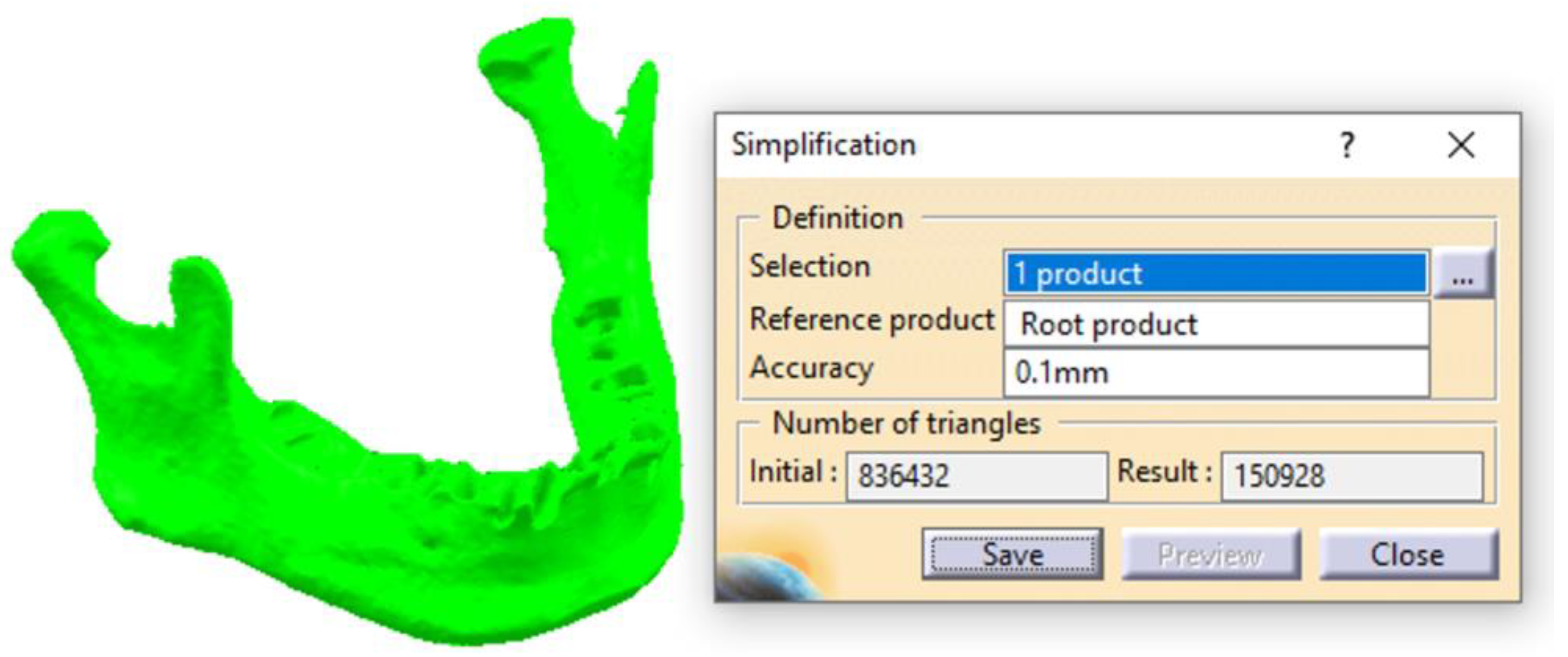
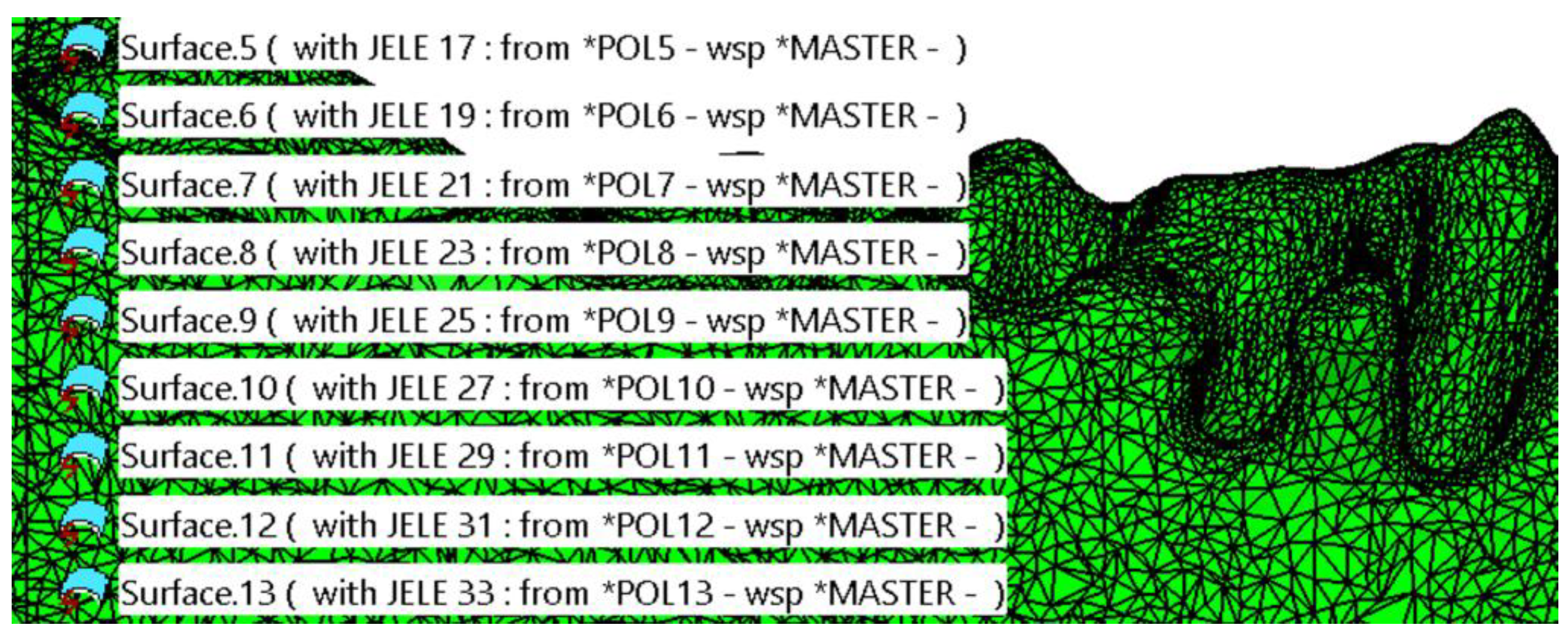
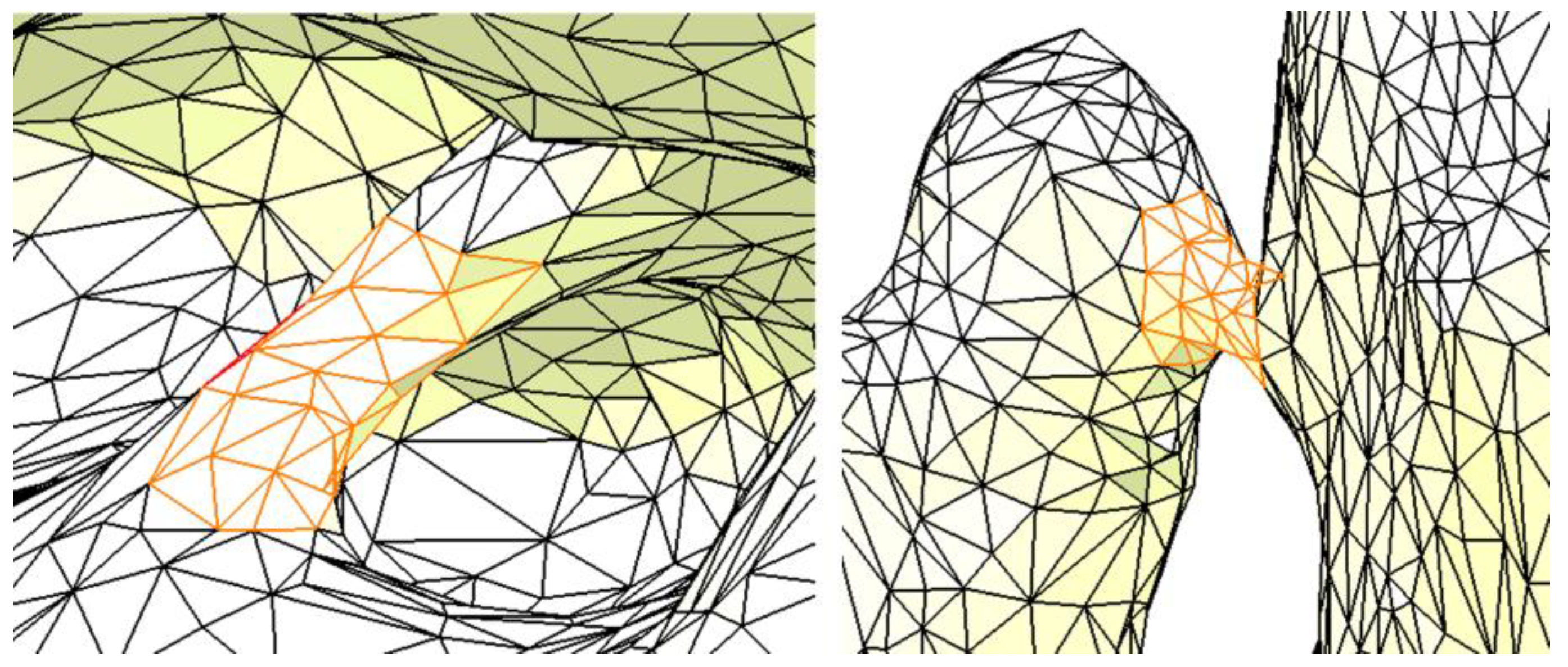
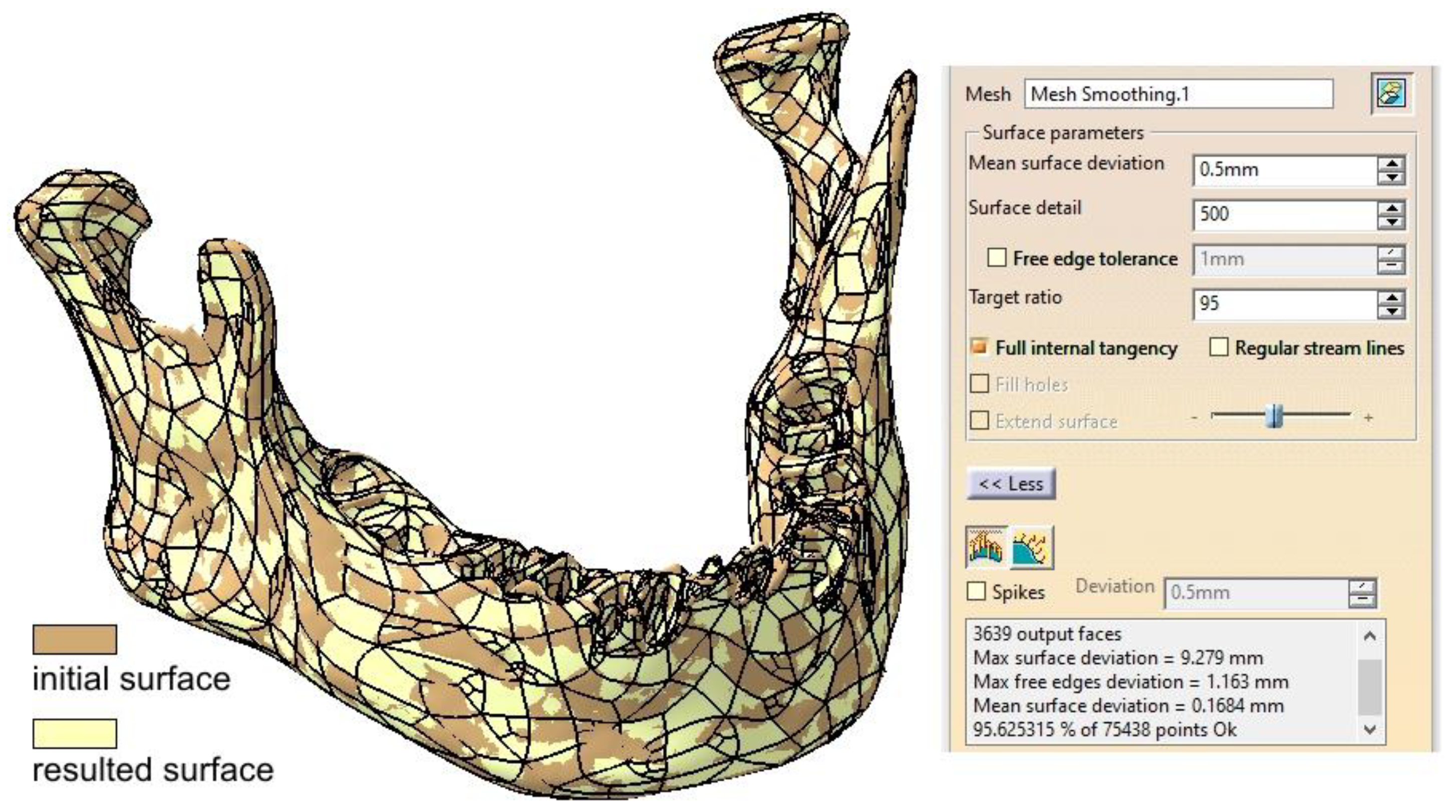
2.1. Optimizing the Mandibular Model for Simulation
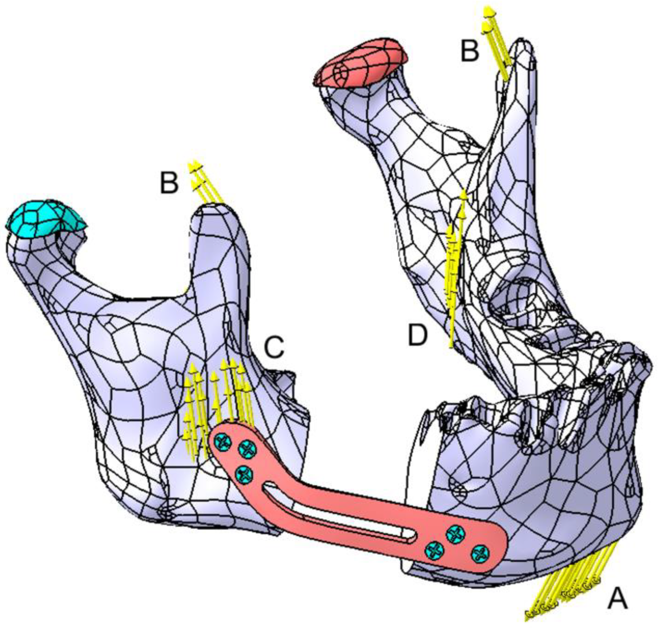
2.2. Post Tumor Ablation Defect Simulation
2.3. Reconstruction Simulation
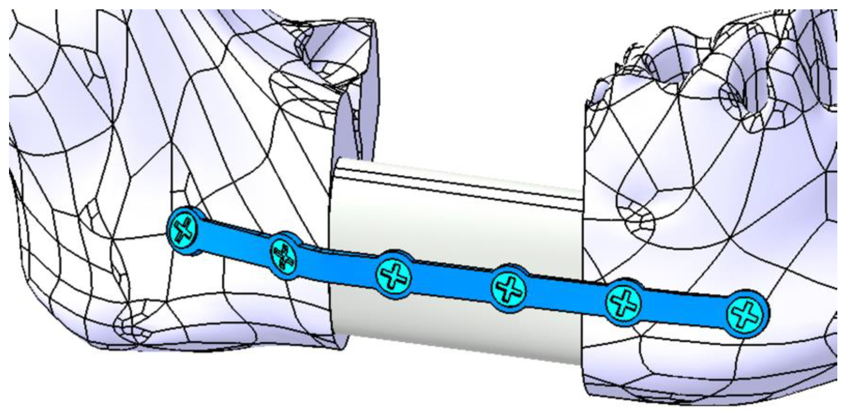
2.3.1. Fixation with a Titanium Osteosynthesis Plate
2.3.2. Reconstruction with a Fibular Autograft
2.3.3. Fixation with a Customized Polyetheretherketone (PEEK) Plate
2.4. Testing Assembly
3. Results
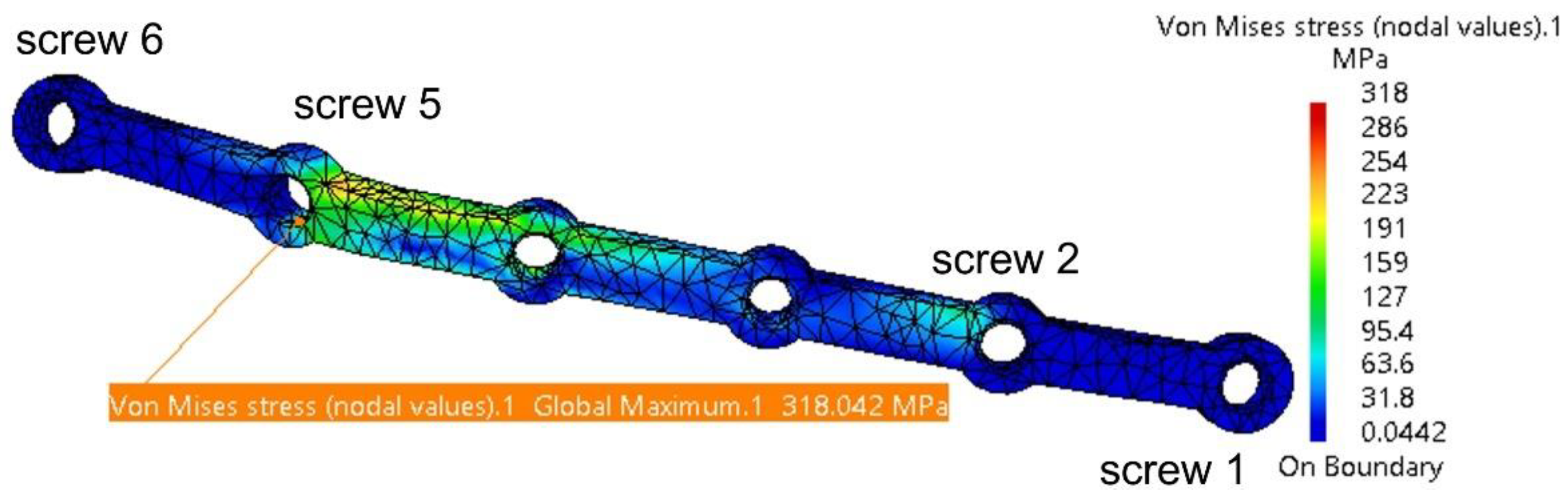
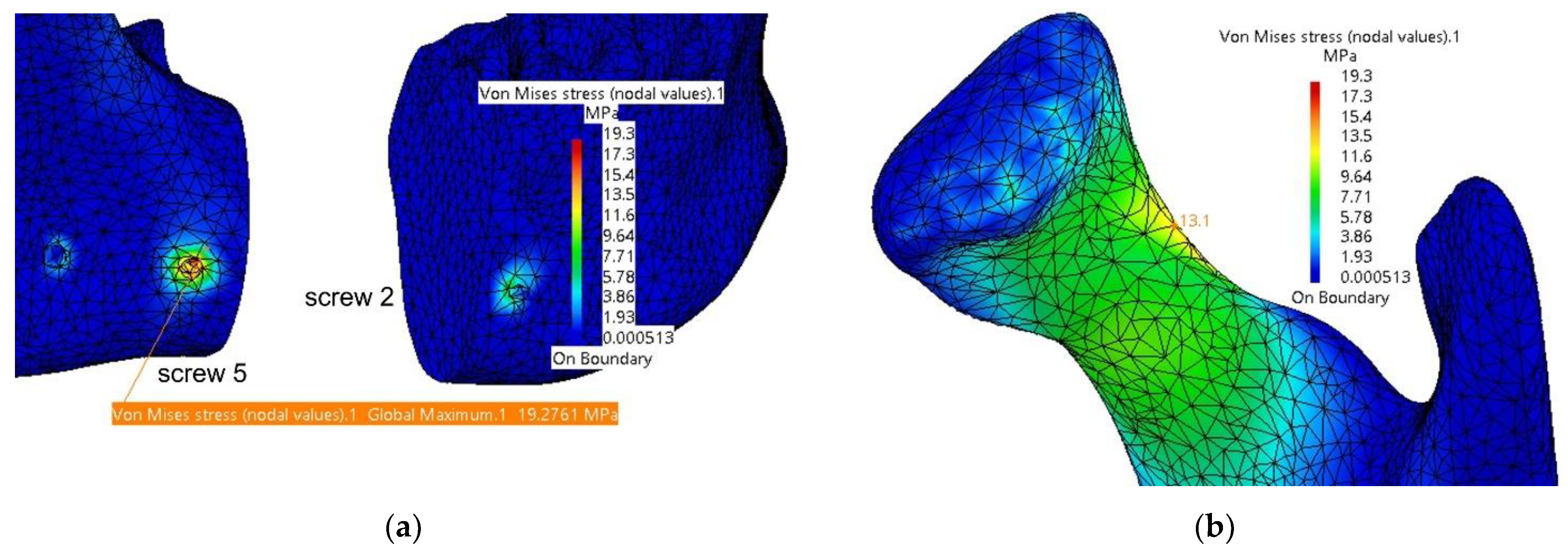
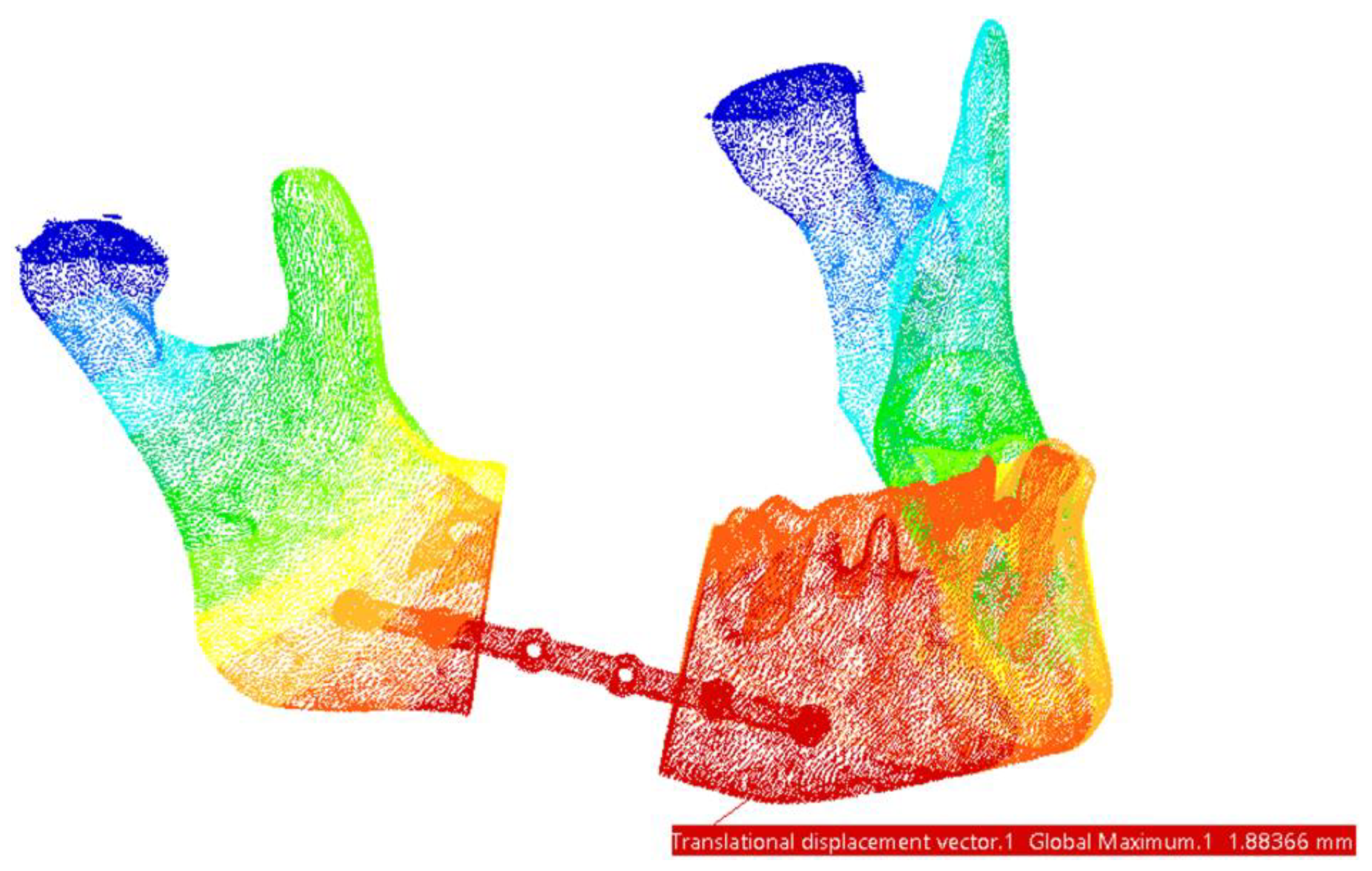
3.1. Fixation with a Titanium Osteosynthesis Plate
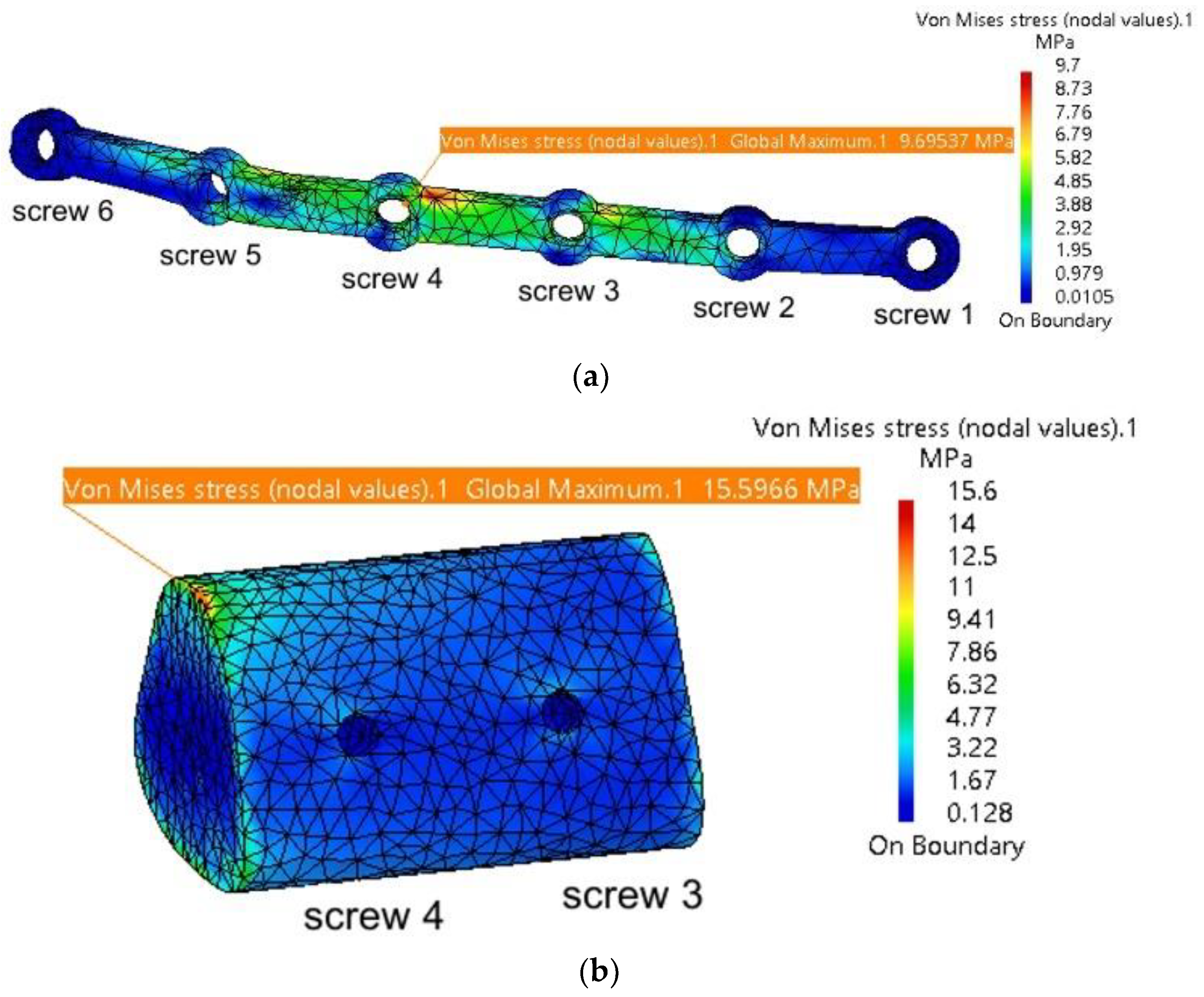
3.2. Reconstruction with a Fibular Autograft
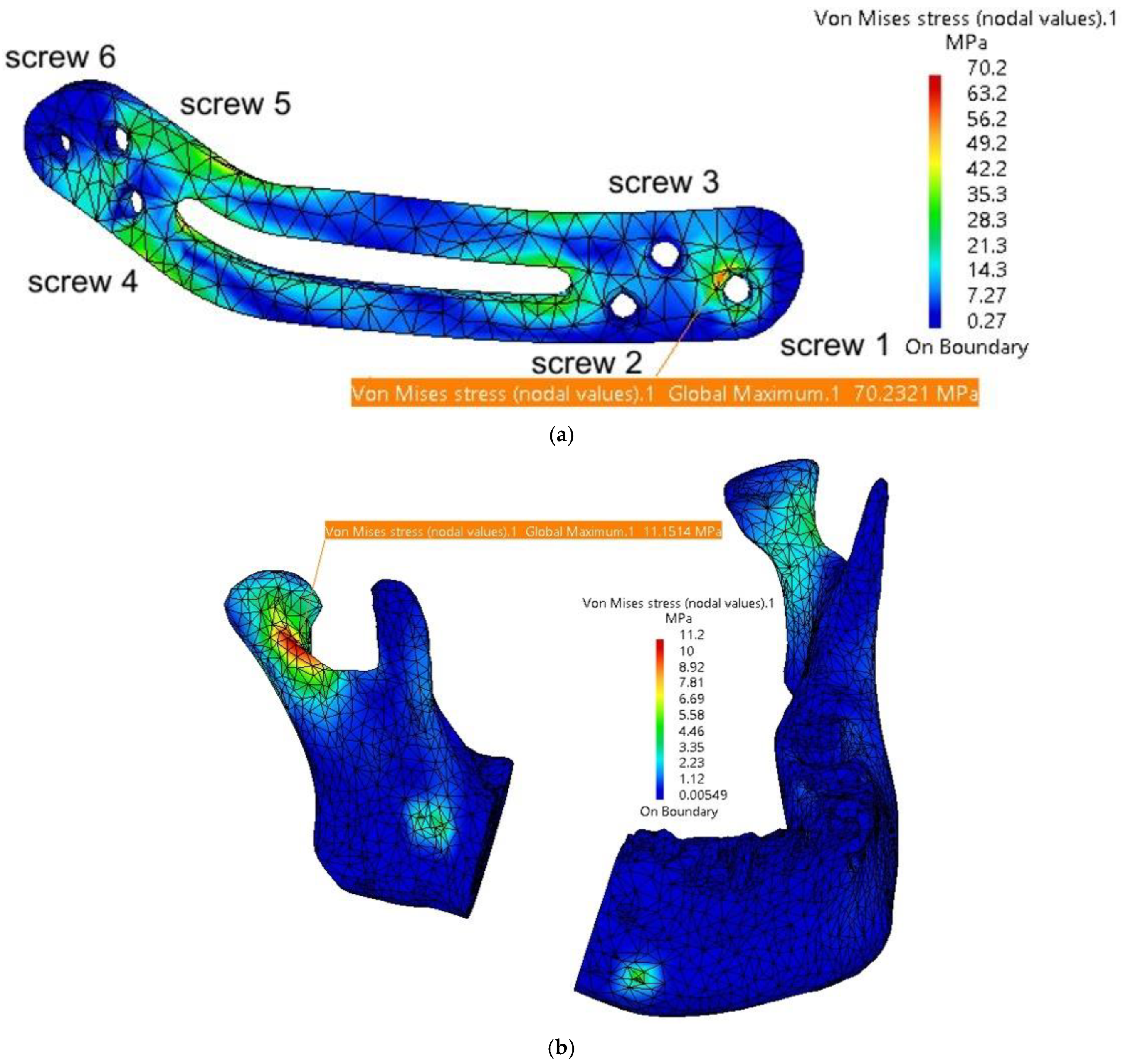
3.3. Fixation with a Customized PEEK Plate
4. Discussion
5. Conclusions
Supplementary Materials
Author Contributions
Funding
Institutional Review Board Statement
Informed Consent Statement
Data Availability Statement
Conflicts of Interest
References
- Sung, H.; Ferlay, J.; Siegel, R.L.; Laversanne, M.; Soerjomataram, I.; Jemal, A.; Bray, F. Global Cancer Statistics 2020: GLOBOCAN Estimates of Incidence and Mortality Worldwide for 36 Cancers in 185 Countries. CA Cancer J. Clin. 2021, 71, 209–249. [Google Scholar] [CrossRef] [PubMed]
- Aftabi, H.; Zaraska, K.; Eghbal, A.; McGregor, S.; Prisman, E.; Hodgson, A.; Fels, S. Computational models and their applications in biomechanical analysis of mandibular reconstruction surgery. Comput. Biol. Med. 2024, 169, 107887. [Google Scholar] [CrossRef] [PubMed]
- Sozzi, D.; Cassoni, A.; De Ponti, E.; Moretti, M.; Pucci, R.; Spadoni, D.; Canzi, G.; Novelli, G.; Valentini, V. Effectiveness of Resective Surgery in Complex Ameloblastoma of the Jaws: A Retrospective Multicenter Observational Study. Cancers 2022, 14, 4608. [Google Scholar] [CrossRef] [PubMed]
- Gupta, L.; Mishra, A.; Gurav, S.V.; Dholam, K.; Pal, A.; Kumar, A. Factors associated with mandibular deviation and proposed classification and treatment guidelines for applying mandibular guidance: A retrospective analysis of 185 patients with segmental mandibulectomy. J. Prosthet. Dent. 2024; Online ahead of print. [Google Scholar] [CrossRef]
- Pyne, J.M.; Davis, C.M.; Kelm, R.; Bussolaro, C.; Dobrovolsky, W.; Seikaly, H. Advanced mandibular reconstruction with fibular free flap and alloplastic TMJ prosthesis with digital planning. J. Otolaryngol.-Head Neck Surg. 2023, 52, s40463-023. [Google Scholar] [CrossRef]
- Weyh, A.M.; Fernandes, R.P. Narrative review: Fibula free flap, indications, tips, and pitfalls. Front. Oral Maxillofac. Med. 2021, 3, 1–7. [Google Scholar] [CrossRef]
- Hoffman, G.R.; Islam, S.; Eisenberg, R.L. Microvascular reconstruction of the mouth, jaws, and face: Experience of an australian oral and maxillofacial surgery unit. J. Oral Maxillofac. Surg. 2012, 70, e371–e377. [Google Scholar] [CrossRef]
- Zeller, A.N.; Neuhaus, M.T.; Weissbach, L.V.M.; Rana, M.; Dhawan, A.; Eckstein, F.M.; Gellrich, N.C.; Zimmerer, R.M. Patient-Specific Mandibular Reconstruction Plates Increase Accuracy and Long-Term Stability in Immediate Alloplastic Reconstruction of Segmental Mandibular Defects. J. Maxillofac. Oral Surg. 2020, 19, 609–615. [Google Scholar] [CrossRef]
- Saunders, J.R.; Hirata, R.M.; Jaques, D.A. Definitive mandibular replacement using reconstruction plates. Am. J. Surg. 1990, 160, 387–389. [Google Scholar] [CrossRef]
- Kumar, B.P.; Venkatesh, V.; Kumar, K.A.J.; Yadav, B.Y.; Mohan, S.R. Mandibular Reconstruction: Overview. J. Maxillofac. Oral Surg. 2015, 15, 425–441. [Google Scholar] [CrossRef]
- Kitami, R.; Izumi, M.; Taniguchi, M.; Kozai, Y.; Sakurai, T. Phantom study for CT artifacts of dental titanium implants and zirconia upper structures: The effects of occlusal plane angle setting and SEMAR algorithm. Oral Radiol. 2024, 40, 251–258. [Google Scholar] [CrossRef]
- Huber, F.A.; Sprengel, K.; Müller, L.; Graf, L.C.; Osterhoff, G.; Guggenberger, R. Comparison of different CT metal artifact reduction strategies for standard titanium and carbon-fiber reinforced polymer implants in sheep cadavers. BMC Med. Imaging 2021, 21, 1–11. [Google Scholar] [CrossRef] [PubMed]
- Midthun, P.; Kirkhus, E.; Østerås, B.H.; Høiness, P.R.; England, A.; Johansen, S. Metal artifact reduction on musculoskeletal CT: A phantom and clinical study. Eur. Radiol. Exp. 2023, 7, 46. [Google Scholar] [CrossRef] [PubMed]
- Lommen, J.; Schorn, L.; Sproll, C.; Kübler, N.R.; Nicolini, L.F.; Merfort, R.; Dilimulati, A.; Hildebrand, F.; Rana, M.; Greven, J. Mechanical Fatigue Performance of Patient-Specific Polymer Plates in Oncologic Mandible Reconstruction. J. Clin. Med. 2022, 11, 3308. [Google Scholar] [CrossRef] [PubMed]
- Panayotov, I.V.; Orti, V.; Cuisinier, F.; Yachouh, J. Polyetheretherketone (PEEK) for medical applications. J. Mater. Sci. Mater. Med. 2016, 27, 1–11. [Google Scholar] [CrossRef]
- Mehle, K.; Eckert, A.W.; Gentzsch, D.; Schwan, S.; Ludtka, C.M.; Knoll, W. Evaluation of a New PEEK Mandibular Reconstruction Plate Design for Continuity Defect Therapy by Finite Element Analysis. Int. J. New Technol. Res. 2016, 2, 263461. [Google Scholar]
- Vayvada, H.; Mola, F.; Menderes, A.; Yilmaz, M. Surgical Management of Ameloblastoma in the Mandible: Segmental Mandibulectomy and Immediate Reconstruction With Free Fibula or Deep Circumflex Iliac Artery Flap (Evaluation of the Long-Term Esthetic and Functional Results). J. Oral Maxillofac. Surg. 2006, 64, 1532–1539. [Google Scholar] [CrossRef]
- Gasparro, R.; Giordano, F.; Campana, M.D.; Aliberti, A.; Landolfo, E.; Dolce, P.; Sammartino, G.; di Lauro, A.E. The Effect of Conservative vs. Radical Treatment of Ameloblastoma on Recurrence Rate and Quality of Life: An Umbrella Review. J. Clin. Med. 2024, 13, 5339. [Google Scholar] [CrossRef]
- Chen, C.; Batstone, M.; Taheri, T.; Johnson, N. Surgical Resection and Reconstruction of Ameloblastoma: A 13-Year Retrospective Review. J. Oral Maxillofac. Surg. 2024, 82, 862–868. [Google Scholar] [CrossRef]
- Xue, R.; Lai, Q.; Xing, H.; Zhong, C.; Zhao, Y.; Zhu, K.; Deng, Y.; Liu, C. Finite element analysis and clinical application of 3D-printed Ti alloy implant for the reconstruction of mandibular defects. BMC Oral Health 2024, 24, 95. [Google Scholar] [CrossRef]
- Shen, Y.W.; Tsai, Y.S.; Hsu, J.T.; Shie, M.Y.; Huang, H.L.; Fuh, L.J. Biomechanical Analyses of Porous Designs of 3D-Printed Titanium Implant for Mandibular Segmental Osteotomy Defects. Materials 2022, 15, 576. [Google Scholar] [CrossRef]
- Kucukguven, M.B.; Akkocaolu, M. Finite element analysis of stress distribution on reconstructed mandibular models for autogenous bone grafts. Technol. Health Care 2020, 28, 249–258. [Google Scholar] [CrossRef] [PubMed]
- Melville, J.C.; Mañón, V.A.; Arribas, A.R.; Wong, M.E. Custom 3D printed titanium reconstruction plate with in-situ tissue engineering for the reconstruction and dental rehabilitation of a severely infected atrophic mandible. A review of technique. Dent. Rev. 2021, 1, 100005. [Google Scholar] [CrossRef]
- Chakraborty, S.; Guha, R.P.; Naskar, S.; Banerjee, R. Custom-Made 3D Titanium Plate for Mandibular Reconstruction in Surgery of Ameloblastoma: A Novel Case Report. Surg. Tech. Dev. 2022, 11, 98–104. [Google Scholar] [CrossRef]
- Tanaka, E. Biomechanical and tribological properties of the temporomandibular joint. Front. Oral Maxillofac. Med. 2021, 3, 15. [Google Scholar] [CrossRef]
- De Stefano, M.; Ruggiero, A. A Critical Review of Human Jaw Biomechanical Modeling. Appl. Sci. 2024, 14, 3813. [Google Scholar] [CrossRef]
- Ghionea, I.G.; Vatamanu, O.E.B.; Cristescu, A.M.; David, M.; Stancu, I.C.; Butnarasu, C.; Cristache, C.M. A Finite Element Analysis of a Tooth-Supported 3D-Printed Surgical Guide without Metallic Sleeves for Dental Implant Insertion. Appl. Sci. 2023, 13, 9975. [Google Scholar] [CrossRef]
- Pinheiro, M.; Willaert, R.; Khan, A.; Krairi, A.; Van Paepegem, W. Biomechanical evaluation of the human mandible after temporomandibular joint replacement under different biting conditions. Sci. Rep. 2021, 11, 14034. [Google Scholar] [CrossRef]
- Ghionea, I.G. CATIA V5 Practical Studies Using Finite Element Analysis; CRC Press: Boca Raton, FL, USA, 2024; ISBN 9781032711645. [Google Scholar] [CrossRef]
- Ashcroft, I.A.; Mubashar, A. Numerical Approach: Finite Element Analysis. In Handbook of Adhesion Technology; Springer: Berlin/Heidelberg, Germany, 2011; pp. 629–660. [Google Scholar] [CrossRef]
- Tang, Y.; Qing, H. Finite element formulation for higher-order shear deformation beams using two-phase local/nonlocal integral model. Arch. Appl. Mech. 2024, 94, 1243–1262. [Google Scholar] [CrossRef]
- Hilber, N.; Reichmann, O.; Schwab, C.; Winter, C. Finite Element Methods for Parabolic Problems. In Computational Methods for Quantitative Finance: Finite Element Methods for Derivative Pricing; Springer: Berlin/Heidelberg, Germany, 2013; pp. 27–45. [Google Scholar] [CrossRef]
- Kimura, A.; Nagasao, T.; Kaneko, T.; Tamaki, T.; Miyamoto, J.; Nakajima, T. Adaquate fixation of plates for stability during mandibular reconstruction. J. Cranio-Maxillofac. Surg. 2006, 34, 193–200. [Google Scholar] [CrossRef]
- Seebach, M.; Theurer, F.; Foehr, P.; von Deimling, C.; Burgkart, R.; Zaeh, M.F. Design of Bone Plates for Mandibular Reconstruction Using Topology and Shape Optimization. In Advances in Structural and Multidisciplinary Optimization; Springer: Cham, Switzerland, 2018; pp. 2086–2096. [Google Scholar] [CrossRef]
- Lang, J.J.; Bastian, M.; Foehr, P.; Seebach, M.; Weitz, J.; Von Deimling, C.; Schwaiger, B.J.; Micheler, C.M.; Wilhelm, N.J.; Grosse, C.U.; et al. Improving mandibular reconstruction by using topology optimization, patient specific design and additive manufacturing?—A biomechanical comparison against miniplates on human specimen. PLoS ONE 2021, 16, e0253002. [Google Scholar] [CrossRef] [PubMed]
- Kang, J.; Zhang, J.; Zheng, J.; Wang, L.; Li, D.; Liu, S. 3D-printed PEEK implant for mandibular defects repair—A new method. J. Mech. Behav. Biomed. Mater. 2021, 116, 104335. [Google Scholar] [CrossRef] [PubMed]
- Batstone, M.D. Reconstruction of major defects of the jaws. Aust. Dent. J. 2018, 63 (Suppl. S1), S108–S113. [Google Scholar] [CrossRef] [PubMed]
- Likhterov, I.; Roche, A.M.; Urken, M.L. Contemporary Osseous Reconstruction of the Mandible and the Maxilla. Oral Maxillofac. Surg. Clin. North Am. 2019, 31, 101–116. [Google Scholar] [CrossRef]
- Chen, L.; Gao, L.; Cui, H.; Guo, X.; Han, J.; Liu, J.; Yao, Y. Finite element comparison of titanium and polyetheretherketone materials for mandibular defect reconstruction. Am. J. Transl. Res. 2024, 16, 6097–6105. [Google Scholar] [CrossRef]
- Cassari, L.; Balducci, C.; Messina, G.M.L.; Iucci, G.; Battocchio, C.; Bertelà, F.; Lucchetta, G.; Coward, T.; Silvio, L.D.; Marletta, G.; et al. Polyetheretherketone Double Functionalization with Bioactive Peptides Improves Human Osteoblast Response. Biomimetics 2024, 9, 767. [Google Scholar] [CrossRef]
- Jones, E.A.; Sigurjónsson, Ó.E.; Weng, X.; Zhu, W.; Chai, H.; Wang, W.; Yuan, X.; Zhu, C. Bio-Activated PEEK: Promising Platforms for Improving Osteogenesis through Modulating Macrophage Polarization. Bioengineering 2022, 9, 747. [Google Scholar] [CrossRef]
- Dondani, J.R.; Iyer, J.; Tran, S.D. Surface Treatments of PEEK for Osseointegration to Bone. Biomolecules 2023, 13, 464. [Google Scholar] [CrossRef]
- Lommen, J.; Schorn, L.; Sproll, C.; Haussmann, J.; Kübler, N.R.; Budach, W.; Rana, M.; Tamaskovics, B. Reduction of CT Artifacts Using Polyetheretherketone (PEEK), Polyetherketoneketone (PEKK), Polyphenylsulfone (PPSU), and Polyethylene (PE) Reconstruction Plates in Oral Oncology. J. Oral Maxillofac. Surg. 2022, 80, 1272–1283. [Google Scholar] [CrossRef]


| Component | Finite Element Size, mm | Absolute Sag, mm | Finite Element Type | Material |
|---|---|---|---|---|
| Mandible | 1.5 | 0.8 | Parabolic | Bone |
| Grafted autogenous bone | 1.2 | 0.5 | Parabolic | Bone |
| TMJ disc | 0.8 | 0.1 | Parabolic | Cartilage |
| Fixing plate titanium | 1.5 | 0.2 | Parabolic | Ti-Grade-4 (TI75A) |
| Fixing plate PEEK | 1 | 0.5 | Parabolic | PEEK |
| Screws | 0.5 | 0.2 | Linear | Ti-Grade-4 (TI75A) |
| Component | Von Mises Stress, MPa | Translational Displacement Vector, mm | Component | Von Mises Stress, MPa | Translational Displacement Vector, mm |
|---|---|---|---|---|---|
| Plate thickness = 2 mm | |||||
| Mandible | 19.3 | 1.9 | Fixing plate | 257 | 1.89 |
| TMJ 1 | 2.58 | 0.197 | Screw 2 | 10.7 | - |
| TMJ 2 | 1.3 | 0.175 | Screw 5 | 20.4 | - |
| Plate thickness = 1.8 mm | |||||
| Mandible | 19.4 | 1.89 | Fixing plate | 307 | 1.89 |
| TMJ 1 | 2.63 | 0.199 | Screw 2 | 11.8 | - |
| TMJ 2 | 1.5 | 0.173 | Screw 5 | 27.1 | - |
| Plate thickness = 1.6 mm | |||||
| Mandible | 19.3 | 1.88 | Fixing plate | 318 | 1.88 |
| TMJ 1 | 2.67 | 0.201 | Screw 2 | 15.5 | - |
| TMJ 2 | 1.61 | 0.171 | Screw 5 | 27.3 | - |
| Plate thickness = 1.4 mm | |||||
| Mandible | 19.2 | 1.87 | Fixing plate | 349 | 1.86 |
| TMJ 1 | 2.69 | 0.204 | Screw 2 | 18 | - |
| TMJ 2 | 2.09 | 0.168 | Screw 5 | 26.5 | - |
| Plate thickness = 1.2 mm | |||||
| Mandible | 19 | 1.87 | Fixing plate | 431 | 1.86 |
| TMJ 1 | 2.69 | 0.204 | Screw 2 | 24.2 | - |
| TMJ 2 | 2.98 | 0.168 | Screw 5 | 33.1 | - |
| Component | Von Mises Stress, MPa | Translational Displacement Vector, mm | Component | Von Mises Stress, MPa | Translational Displacement Vector, mm |
|---|---|---|---|---|---|
| Plate thickness = 1.2 mm | |||||
| Mandible | 40.7 | 1.61 | Fixing plate | 12 | 1.53 |
| Cartilage 1 | 2.62 | 0.118 | Screw 2 | 2.11 | 1.41 |
| TMJ 2 | 1.17 | 0.191 | Screw 3 | 2.69 | 1.29 |
| Bone insert | 15.7 | 1.37 | Screw 4 | 2.29 | 1.17 |
| Plate thickness = 1.4 mm | |||||
| Mandible | 40.3 | 1.61 | Fixing plate | 10.8 | 1.53 |
| TMJ 1 | 2.68 | 0.118 | Screw 2 | 2.09 | 1.41 |
| TMJ 2 | 1.22 | 0.191 | Screw 3 | 2.49 | 1.29 |
| Bone insert | 15.6 | 1.37 | Screw 4 | 2.23 | 1.17 |
| Plate thickness = 1.6 mm | |||||
| Mandible | 40.2 | 1.61 | Fixing plate | 10.2 | 1.53 |
| TMJ 1 | 2.64 | 0.118 | Screw 2 | 1.77 | 1.41 |
| TMJ 2 | 1.34 | 0.191 | Screw 3 | 2.18 | 1.29 |
| Bone insert | 15.6 | 1.37 | Screw 4 | 2.19 | 1.17 |
| Plate thickness = 1.8 mm | |||||
| Mandible | 40.1 | 1.61 | Fixing plate | 9.7 | 1.53 |
| TMJ 1 | 2.78 | 0.118 | Screw 2 | 1.79 | 1.41 |
| TMJ 2 | 1.88 | 0.191 | Screw 3 | 2.16 | 1.29 |
| Bone insert | 15.6 | 1.37 | Screw 4 | 2.16 | 1.17 |
| Component | Von Mises Stress, MPa | Translational Displacement Vector, mm | Component | Von Mises Stress, MPa | Translational Displacement Vector, mm |
|---|---|---|---|---|---|
| Plate thickness = 2 mm | |||||
| Mandible | 11.2 | 1.14 | Screw 1 and 2 | 35 and 12.8 | max 0.11 |
| TMJ 1 | 2.98 | 0.2 | Screw 3 and 4 | 24.2 and 26.3 | max 0.09 |
| TMJ 2 | 3.31 | 0.2 | Screw 5 | 28.4 | 0.1 |
| Fixing plate | 70.2 | 1.12 | Screw 6 | 18.2 | 0.09 |
| Plate thickness = 1.8 mm | |||||
| Mandible | 16.8 | 1.32 | Screw 1 and 2 | 38 and 16.2 | max 0.13 |
| TMJ 1 | 3.1 | 0.2 | Screw 3 and 4 | 28.4 and 32.1 | max 0.11 |
| TMJ 2 | 3.49 | 0.2 | Screw 5 | 34.2 | 0.18 |
| Fixing plate | 81.29 | 1.29 | Screw 6 | 12 | 0.1 |
| Plate thickness = 1.6 mm | |||||
| Mandible | 20.6 | 1.46 | Screw 1 and 2 | 46 and 22.6 | max 0.16 |
| TMJ 1 | 3.48 | 0.22 | Screw 3 and 4 | 34 and 42.4 | max 0.14 |
| TMJ 2 | 3.88 | 0.22 | Screw 5 | 46 | 0.22 |
| Fixing plate | 92.7 | 1.38 | Screw 6 | 16 | 0.12 |
| Plate thickness = 1.4 mm | |||||
| Mandible | 32.6 | 1.85 | Screw 1 and 2 | 58 and 28.8 | max 0.23 |
| TMJ 1 | 3.79 | 0.24 | Screw 3 and 4 | 41.9 and 49.5 | max 0.2 |
| TMJ 2 | 4.22 | 0.22 | Screw 5 | 51.8 | 0.24 |
| Fixing plate | 130.4 | 1.7 | Screw 6 | 28.4 | 0.17 |
| Characteristic | Titanium Plates | PEEK Plates |
|---|---|---|
| Mechanical strength | High mechanical stability, with superior load-bearing capacity. | Sufficient strength for physiological loads, but thinner plates (<1.8 mm) risk plastic deformation. |
| Elastic modulus | Very high (104 GPa), leading to stress shielding and reduced bone remodeling. | Closer to cortical bone (3.9 GPa), minimizing stress shielding and promoting better load distribution. |
| Imaging artifacts [43] | Causes significant streaking and blooming artifacts in CT and MRI imaging. | Radiolucent with minimal imaging artifacts, allowing for a clear postoperative assessment. |
| Weight | Denser and heavier, which may reduce patient comfort. | Lightweight, improving patient comfort and reducing load on surrounding tissues. |
| Plate thickness | Minimum thickness recommended: 1.2–1.6 mm for mechanical stability. | Minimum thickness recommended: 1.8–2.0 mm to avoid plastic deformation under physiological loads. |
| Biocompatibility | Highly biocompatible but can enhance radiation dose in adjacent tissues during radiotherapy. | Biocompatible and inert; does not interfere with radiotherapy or adjacent tissue assessment. |
| Manufacturing | 3D printing or sintering; higher production cost. | Can be manufactured using additive techniques, offering reduced production costs and patient-specific design. |
Disclaimer/Publisher’s Note: The statements, opinions and data contained in all publications are solely those of the individual author(s) and contributor(s) and not of MDPI and/or the editor(s). MDPI and/or the editor(s) disclaim responsibility for any injury to people or property resulting from any ideas, methods, instructions or products referred to in the content. |
© 2025 by the authors. Licensee MDPI, Basel, Switzerland. This article is an open access article distributed under the terms and conditions of the Creative Commons Attribution (CC BY) license (https://creativecommons.org/licenses/by/4.0/).
Share and Cite
Ghionea, I.G.; Tarba, C.I.; Cristache, C.M.; Filipov, I.; Beuran, I.A. A Comparative Finite Element Analysis of Titanium, Autogenous Bone, and Polyetheretherketone (PEEK)-Based Solutions for Mandibular Reconstruction. Materials 2025, 18, 314. https://doi.org/10.3390/ma18020314
Ghionea IG, Tarba CI, Cristache CM, Filipov I, Beuran IA. A Comparative Finite Element Analysis of Titanium, Autogenous Bone, and Polyetheretherketone (PEEK)-Based Solutions for Mandibular Reconstruction. Materials. 2025; 18(2):314. https://doi.org/10.3390/ma18020314
Chicago/Turabian StyleGhionea, Ionut Gabriel, Cristian Ioan Tarba, Corina Marilena Cristache, Iulian Filipov, and Irina Adriana Beuran. 2025. "A Comparative Finite Element Analysis of Titanium, Autogenous Bone, and Polyetheretherketone (PEEK)-Based Solutions for Mandibular Reconstruction" Materials 18, no. 2: 314. https://doi.org/10.3390/ma18020314
APA StyleGhionea, I. G., Tarba, C. I., Cristache, C. M., Filipov, I., & Beuran, I. A. (2025). A Comparative Finite Element Analysis of Titanium, Autogenous Bone, and Polyetheretherketone (PEEK)-Based Solutions for Mandibular Reconstruction. Materials, 18(2), 314. https://doi.org/10.3390/ma18020314