Communication

Irradiation Effects of As-Fabricated and Recrystallized 12Cr ODS Steel Under Dual-Ion Beam at 973 K

1
National Institute for Fusion Science, Toki, Gifu 509-5292, Japan
2
Department of Fusion Science, The Graduate University for Advanced Studies, SOKENDAI, Toki, Gifu 509-5292, Japan
3
Institute of Advanced Energy, Kyoto University, Uji, Kyoto 611-0011, Japan
*
Author to whom correspondence should be addressed.
Materials 2025, 18(14), 3246; https://doi.org/10.3390/ma18143246
Submission received: 26 May 2025 / Revised: 6 July 2025 / Accepted: 8 July 2025 / Published: 10 July 2025
(This article belongs to the Special Issue Key Materials in Nuclear Reactors)

Abstract

The microstructure evolution and hardness variations of as-fabricated and recrystallized 12Cr oxide dispersion strengthened (ODS) steel after dual-ion (6.4 MeV Fe3+ and energy-degraded 1 MeV He+) irradiation at 973 K up to 10.6 displacements per atom (dpa) at peak damage and 8900 appm He are investigated. Results show that the oxide particles slightly shrink in the as-fabricated specimen, while they are stable in the recrystallized specimen. Furthermore, larger helium bubbles are trapped at the grain boundaries in the as-fabricated specimen, and the size of helium bubbles in the grains is almost the same for both as-fabricated and recrystallized specimens, indicating that reduction of grain boundaries would reduce the potential nucleation sites and suppress the helium segregation. Moreover, no obvious hardening occurs in the as-fabricated specimen, whereas the hardness increases a little in the recrystallized specimen. Based on the barrier model, the barrier strength factor of helium bubbles is calculated. The value is 0.077, which is much smaller and suggests that helium bubbles seem not to significantly induce irradiation hardening.

1. Introduction

Oxide dispersion strengthened (ODS) steel is considered as candidate structural material for blanket application in fusion reactors [1,2,3], where it will suffer from severe neutron irradiation doses up to 150–200 dpa, and helium concentration of ~10 appm/dpa [4], inducing irradiation hardening and helium embrittlement. Based on different design concepts, the maximum required temperature and radiation conditions are different, and higher temperatures give rise to higher efficiency. Hence, the stability of oxide particles under high-temperature and neutron irradiation conditions is essential to maintain adequate strength and system soundness for application in nuclear reactors. Many studies have been conducted to reveal helium irradiation effects on microstructure and mechanical properties of ODS steels [5,6,7,8,9,10,11,12,13,14,15,16]. Edmondson et al. [5] implanted helium ions into ODS steels at 673 K and analyzed helium bubble distributions on various interfaces, such as coarse precipitates, dislocations, grain boundaries and oxide particles, showing the oxide particles taking the dominant position. Moreover, some investigations [7,8,13] also demonstrated high density oxide particles can provide abundances of interfaces for trapping helium and effectively suppress the coarsening of helium bubbles. Song et al. [11] reported that even though oxide particles can enhance the swelling resistance of ODS steels, swelling percentage increased with increasing irradiation temperature from 573 K to 973 K. Furthermore, refining the grain size from several hundred nanometers to tens of nanometers leads to outstanding helium bubble suppression [14,15].
ODS steel is fabricated by powder metallurgy, including mechanical alloying, consolidation by hot extrusion/isostatic pressing, and the subsequent thermomechanical process. This process produces fine and elongated grains with a high density of nanoscale oxide particles and dislocations, and preferential orientations [17,18], which lead to low ductility and obviously anisotropic mechanical properties [19,20,21,22]. To enhance the ductility and suppress the anisotropy, a recrystallization process is suggested [23]. A novel thermomechanical process [24], namely multi-directional cold rolling followed by annealing, was developed to obtain recrystallization for 12Cr ODS steel. The improvement of ductility was confirmed for the recrystallized 12Cr ODS steel. Although it can be expected that irradiation hardening and swelling will become more evident because dislocation density and grain boundaries dramatically decreased after recrystallization compared with the as-fabricated state, the response of recrystallized 12Cr ODS steel under irradiation is still not clear. Furthermore, the recrystallization process changes the extremely complex microstructures into simple ones, reducing the high density of dislocations to almost free and obtaining coarse grains. This makes it possible to focus on the stability of oxide particles under irradiation excluding the effects of other defects, such as dislocations and grain boundaries. In addition, it offers a clear microstructure to observe irradiation-induced defects, characterize the interactions between oxide particles and irradiation-induced defects in future research, and finally provides fundamental insights for understanding the underlying mechanisms of oxide particle stability under irradiation. In the present study, we investigate the microstructure changes and irradiation hardening of the as-fabricated and recrystallized 12Cr ODS steel under Fe3+ and He+ ion irradiation at 973 K.

2. Material and Methods

In this study, 12Cr ODS steel with nominal composition of Fe-12Cr-2W-0.3Ti-0.25Y2O3 (wt%) was used. It was consolidated by hot extrusion at 1423 K. Then, hot forging was performed at 1423 K followed by annealing at 1373 K for 1 h. Afterward, cold rolling was conducted with 40% thickness reduction and final annealing was carried out at 1323 K for 1 h. Regarding the recrystallization process, a 3 mm thick specimen was cut from the as-fabricated plate. Then, cold rolling was applied on the original normal direction (ND)–transverse direction (TD) section with a 90% thickness reduction. Next, 3 mm diameter discs were punched out from the cold rolled sheet and annealed at 1373 K for 3 h in a vacuum. Before ion irradiation experiments, the as-fabricated and recrystallized specimens were mechanically ground with abrasive paper and electro-polished on one side by Struers Tenupol-5 in an electrolyte of 5 vol% perchloric acid and 95 vol% acetic acid at room temperature. Irradiation experiments were performed at 973 K via Dual-Beam Facility for Energy Science and Technology (DuET) at Kyoto University, using dual-beam 6.4 MeV Fe3+ and energy-degraded 1 MeV He+ with a flux of 6.37 × 10¹⁵ and 6.76 × 10¹⁶ ions m−2 s−1, respectively. Figure 1 shows the stopping and range of ions in matter (SRIM) calculation results. The irradiation dose is approximately 10.6 displacements per atom (dpa) at the damage peak of 1500 nm, and the helium concentration is approximately 8900 appm at the depth of 1000 nm.
After irradiation experiments, nanoindentation hardness tests were carried out up to a depth of 250 nm from the surface by a DUH-211S hardness tester (SHIMADZU, Kyoto, Japan). The indentation depth was selected from the effective depth where the plastic deformation zone can expand to ~4–5 times the depth of the nano-indenter [25]. The peak helium concentration was located around 1000 nm. It can be noted that Ha et al. [10] used the same facility for the irradiation experiments and found the maximum hardening at 250 nm. The average value was obtained by measuring 80 points for each specimen. The microstructure was characterized by JEM-2800 transmission electron microscope (TEM, JEOL, Tokyo, Japan) at an accelerating voltage of 200 kV. TEM specimens were prepared from the surface of the irradiated specimens by FB-2100 focused ion beam (FIB, HITACHI, Tokyo, Japan) system with Ga ions at 40 kV. To remove the layer damaged by Ga ions during FIB fabrication, flash electropolishing was conducted at approximately 238 K in an electrolyte of 10 vol% perchloric acid and ethanol. Counting the thickness fringes at the two-beam conditions was carried out to evaluate the thickness of the observed area. Approximately four TEM images and 930–1070 particles were measured for each condition.

3. Results and Discussion

3.1. Microstructure Before Dual-Beam Irradiation

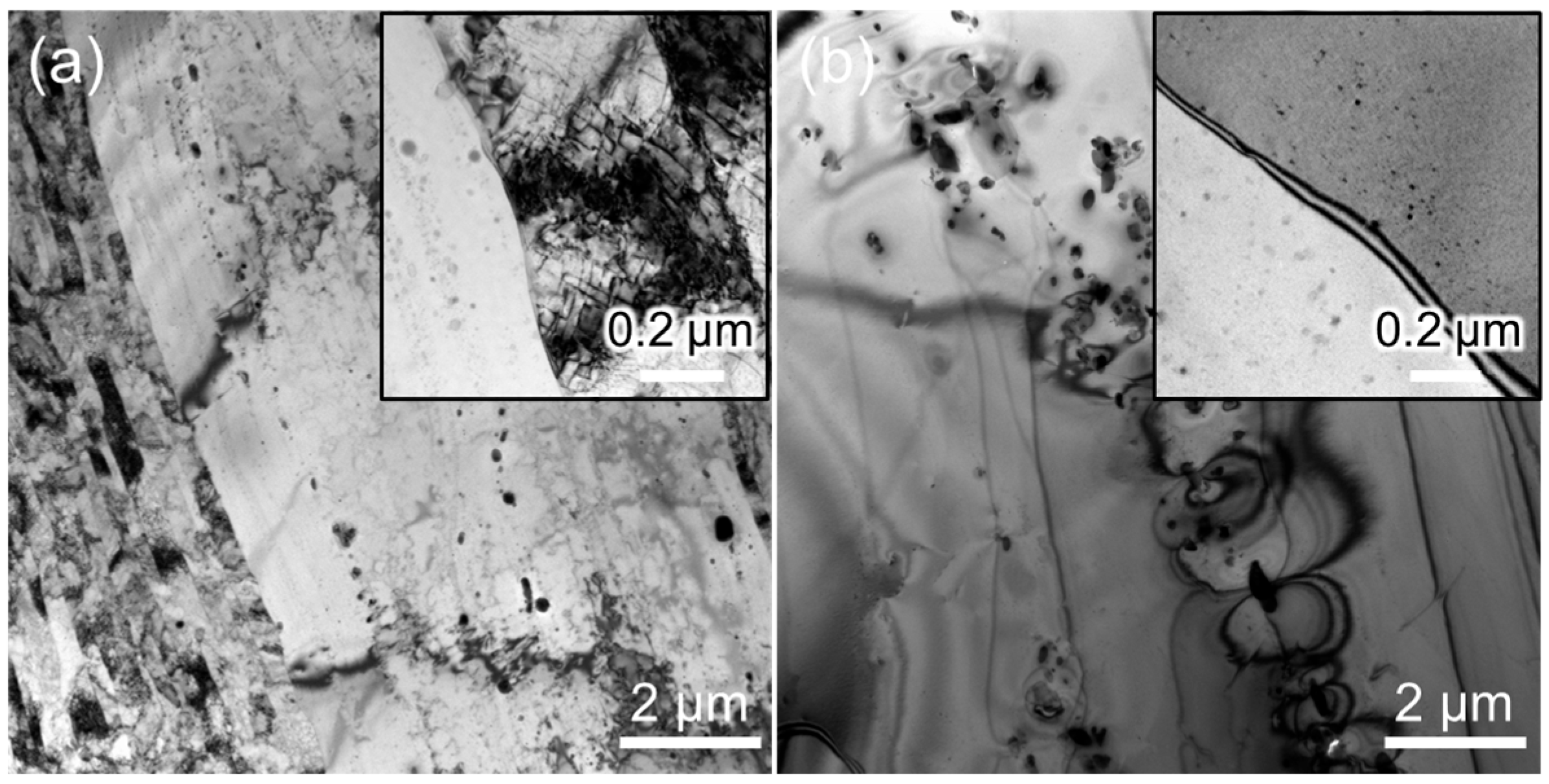
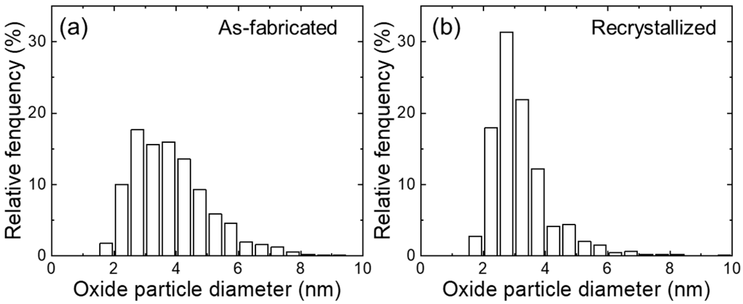
Figure 2 shows the microstructures of as-fabricated and recrystallized 12Cr ODS steel. For the as-fabricated specimen, non-uniform microstructure is observed, which contains fine and large grains [21]. The fine and elongated grains have a high density of dislocations and a width smaller than 500 nm (see the insert in Figure 2a), whereas the large grains are recrystallized during the fabrication process. Regarding the recrystallized specimen, large recrystallized grains contain much lower density of dislocations and more uniformly distributed nanoscale oxide particles (see the insert in Figure 2b). Additionally, coarse Ti-enriched precipitates are noted in both as-fabricated and recrystallized specimens. Figure 3 shows the size distribution of the oxide particles of as-fabricated and recrystallized specimens. The average diameter and number density of oxide particles in as-fabricated and recrystallized specimens are 3.8 ± 1.2 nm, 3.1 × 10²² m−3, and 3.2 ± 1.0 nm, 8.1 × 10²² m−3, respectively. This indicates that the oxide particles become smaller, and more oxide particles are precipitated after the recrystallization process, which are expected to supply more interfaces to absorb irradiation-induced defects.

3.2. Microstructure After Dual-Beam Irradiation

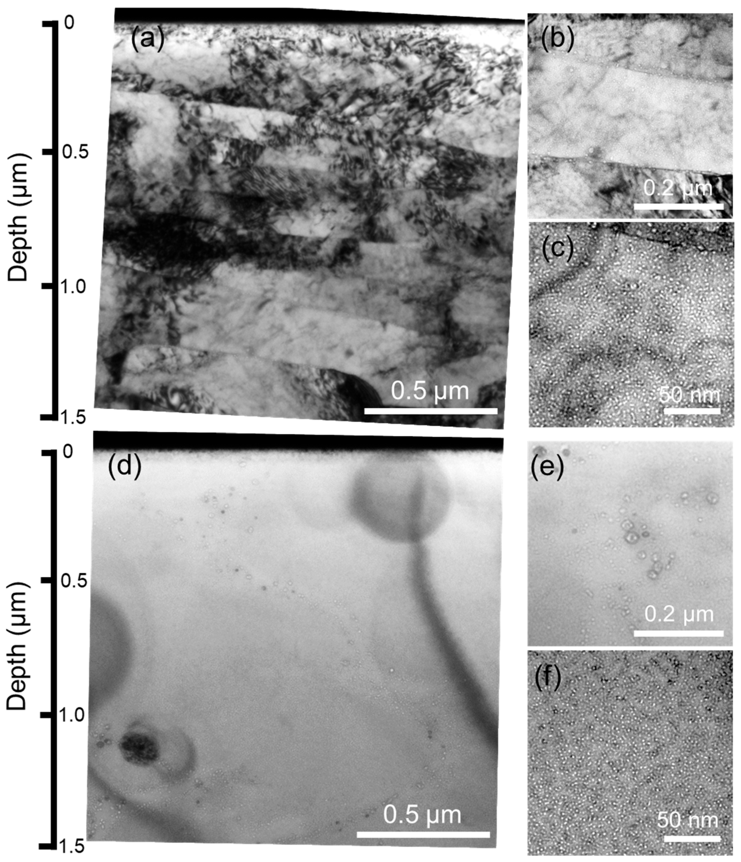
Figure 4 shows the TEM images of the as-fabricated and recrystallized specimens after Fe3+ and He+ ion irradiation at 973 K. Fine helium bubbles are observed in the irradiated regions. As for the as-fabricated specimen, Figure 4b shows the microstructure near the depth of 1000 nm, where helium bubbles are distributed at the grain boundaries and grain interior with an average diameter of 5.8 ± 1.3 nm and 1.6 ± 0.3 nm, respectively. The number density is ~3.8 × 10²² m−3. Note that the size of helium bubbles located at the grain boundaries is larger than that at the grain interiors, indicating that helium bubbles preferentially nucleate at the grain boundaries. Helium segregation at the grain boundaries would result in helium embrittlement [26,27,28]. In contrast, even though no grain boundaries are observed in the recrystallized specimen in Figure 4d, helium bubbles are preferentially trapped at the edge of the relatively large oxide particles. Furthermore, helium bubbles located in the grains have an average diameter of 1.7 ± 0.3 nm and a number density of ~3.1 × 10²² m−3, which are almost the same as the as-fabricated specimen. This suggests that the recrystallization process seems to have no significant effects on helium bubble formation inner grains during Fe3+ and He+ ion irradiation at 973 K. This can probably be ascribed to more uniformly distributed and finer oxide particle formation after the recrystallization process. In addition, reduction of grain boundaries would decrease the potential nucleation sites and suppress the helium segregation at the grain boundaries.
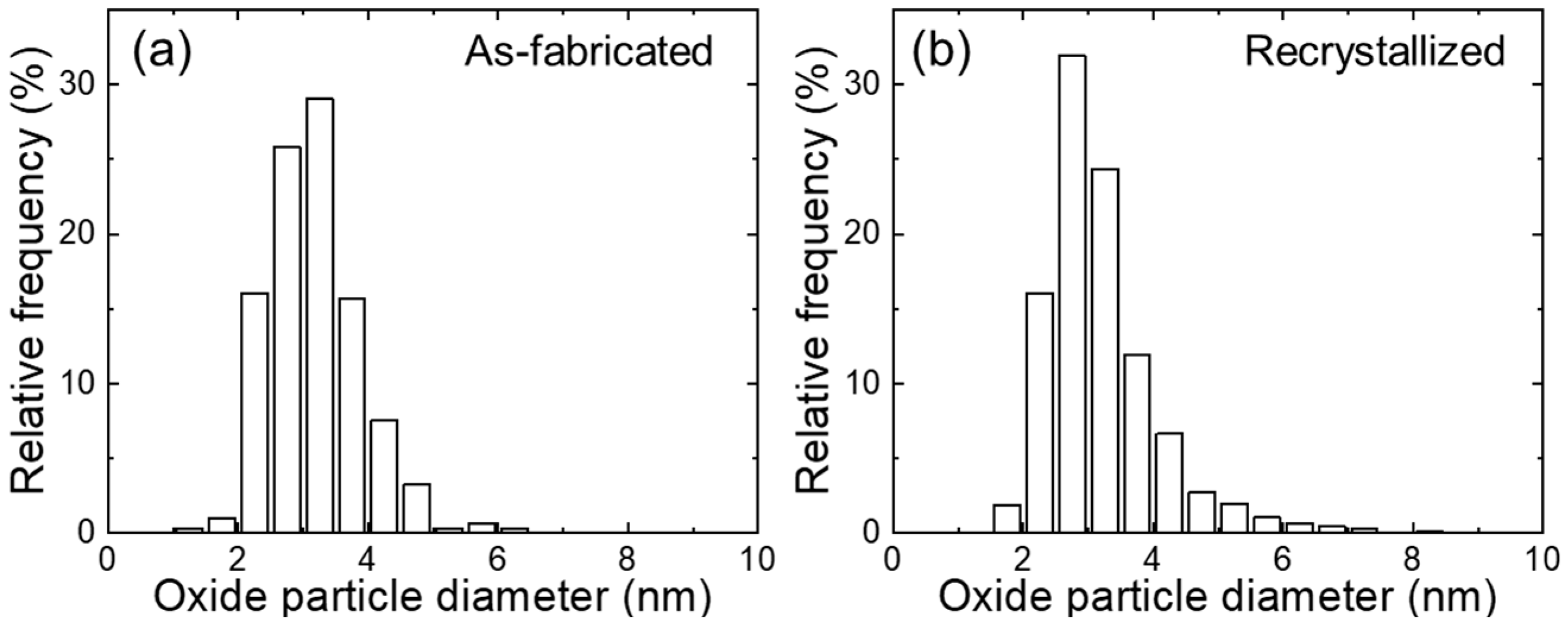
Figure 5 shows the size distribution of oxide particles in as-fabricated and recrystallized specimens after irradiation. The average diameter is 3.2 ± 0.7 nm and 3.2 ± 0.9 nm for as-fabricated and recrystallized specimens. Compared with the unirradiated specimens, the sizes of oxide particles are slightly smaller for the as-fabricated condition and the same as the recrystallized condition. Allen et al. [29] reported that the size of oxide particles decreased in 9Cr ODS steel at 773–973 K after Ni ion irradiation up to 150 dpa. Song et al. [30] revealed that oxide particles became slightly smaller in Y-Ti-ODS and Y-Al-Zr-ODS after Fe3+ and He+ irradiation up to 30 dpa at 823 K. Furthermore, in situ TEM study [31] indicated that larger oxide particles (>20 nm) were more evidently affected by ion irradiation than the smaller ones. Slight size reduction of oxide particles is probably caused by ballistic collisions [32,33], and the dissolved atoms more likely diffuse to the dislocations and/or grain boundaries in the as-fabricated specimen than the recrystallized specimen, which contains much lower density of dislocations and grain boundaries.

3.3. Irradiation Hardening

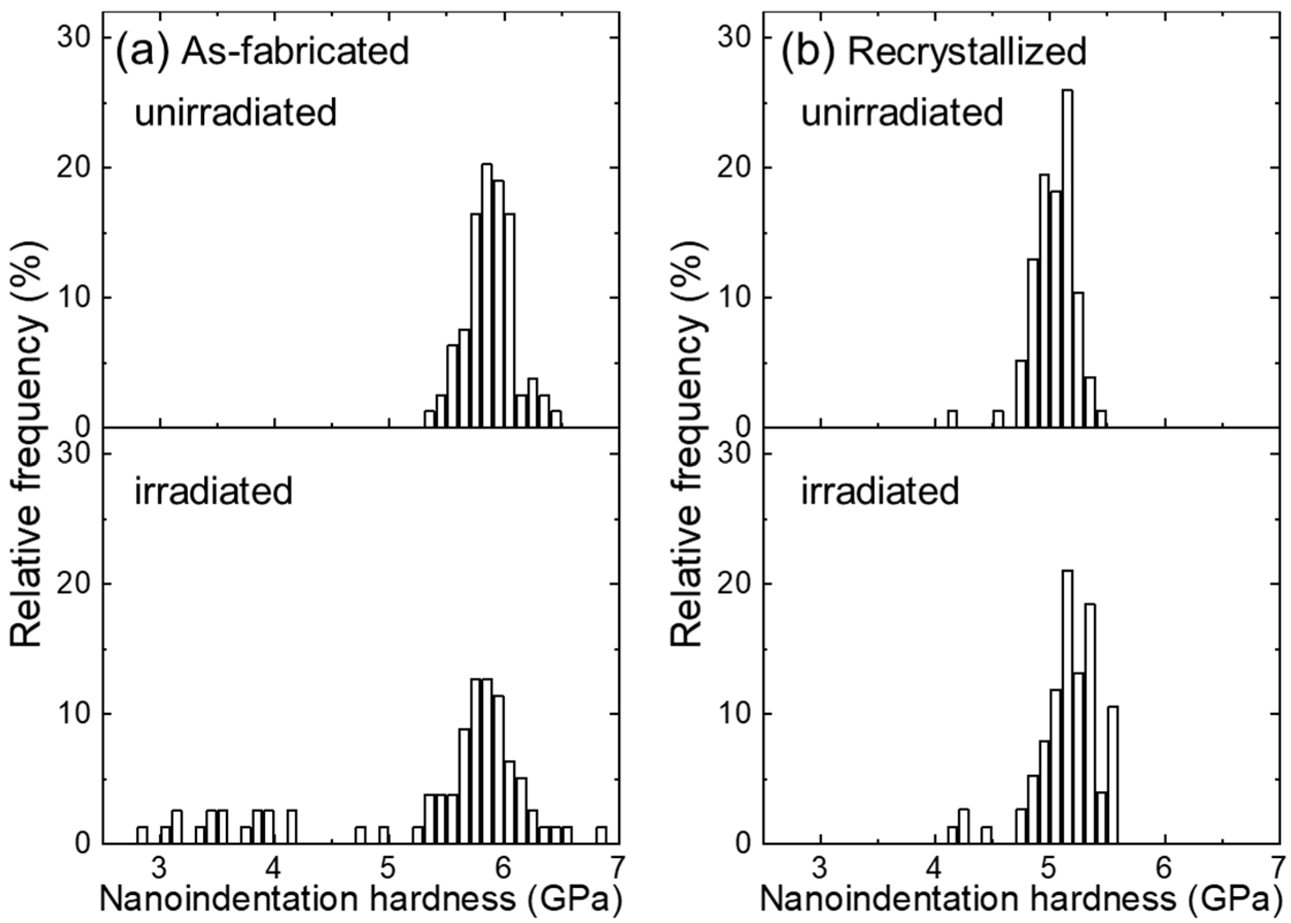
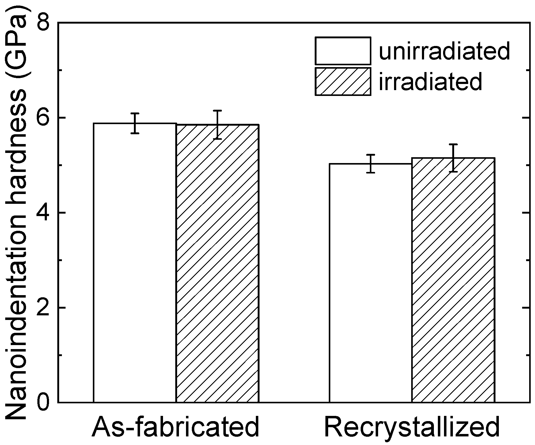
Figure 6 shows the nanoindentation hardness distribution in unirradiated and irradiated as-fabricated and recrystallized specimens. As shown in Figure 6a, there are no hardness values lower than 5.0 GPa in the unirradiated as-fabricated specimen, whereas they appear in the irradiated condition. The lower hardness is not due to the irradiation, but non-uniform microstructures. The specimen is fixed on the stage of a nanoindentation hardness tester, and hardness is automatically measured after choosing the test areas. The microstructure cannot be distinguished under the optical microscope of the nanoindentation hardness tester, and the tested area is randomly selected. The lower hardness areas are not tested, whilst they were measured after irradiation experiments. Microstructure is not uniform in as-fabricated specimens. As mentioned in Figure 2, it consists of coarse recrystallized grains and fine grains containing high density dislocations. During thermal annealing for the cold rolled 12Cr ODS steel, recrystallization preferentially occurs at sites containing no/less oxide particles [24]. The lower hardness comes from the recrystallized grains that have no/lower density of oxide particles and already exist in the as-fabricated condition (see Figure 2a). As shown in Figure 6b, the hardness values of recrystallized specimens are around 5.0 GPa. Hence, the values around 3.0~4.0 GPa are measured on the grains containing no/less oxide particles. To examine the hardening effects upon irradiation, these lower hardness values are not considered in comparison with the unirradiated data, and the corresponding average values are shown in Figure 7. In contrast, with respect to the recrystallized specimen, Figure 6b indicates a similar hardness value distribution for unirradiated and irradiated specimens. There are fewer scattered values in the recrystallized condition, suggesting the homogeneous microstructure after recrystallization.
Figure 7 shows the average nanoindentation hardness of unirradiated and irradiated as-fabricated and recrystallized specimens. No obvious hardening is characterized in the as-fabricated specimen, while the hardness slightly increases for the recrystallized specimen after dual-beam irradiation at 973 K. Ha et al. [10] reported that the hardness was increased by 1.079 GPa after Fe3+ and He+ irradiation up to 90 dpa and 450 appm He at 743 K for recrystallized 15Cr ODS steel, where the oxide particle diameter and number density were 23 nm and 1.9 × 10²¹ m−3, respectively. Relatively larger oxide particles with a lower number density may give rise to lower amounts of sinks for the irradiation-induced defects and thus result in more significant irradiation hardening.
The barrier model [34] is usually applied to explain the irradiation hardening by irradiation-induced defects, which is expressed by the following equation:
σ = M α μ b N d
where M is the Taylor factor (3.06), α is the barrier strength, µ is the shear modulus (81.6 GPa), b is the Burgers vector (0.248 nm), and N and d are number density and diameter of the defects. The obtained nanoindentation hardness should be converted into yield strength according to the equations [35,36] σ = 3.06 H V ,   H V = 94.495 H N , where HV is Vickers hardness, and HN is nanoindentation hardness. Assuming the hardening is caused by the helium bubbles after irradiation, the average diameter (1.7 nm) and number density (3.1 × 10²² m−3) are substituted in the above equations to estimate the barrier strength of helium bubbles. The barrier strength factor αHe can be determined as 0.077, which is much smaller than other defects, such as precipitates, dislocation loops, and clusters. Therefore, helium bubbles seem not to significantly induce hardening after the dual-beam irradiation at 973 K in this study. Similarly, the helium bubbles are not the predominant contributor to the irradiation hardening after dual-beam irradiation at 743 K [10].

4. Conclusions

The microstructure evolution and hardening of as-fabricated and recrystallized 12Cr ODS steel upon dual-ion irradiation at 973 K with 10.6 displacements per atom (dpa) at peak damage and 8900 appm He are studied. The following conclusions can be drawn:
(1)
The size of oxide particles slightly decreases in the as-fabricated specimen, while they are stable in the recrystallized specimen, indicating that smaller oxide particles with a higher density after recrystallization supply more sink areas for the irradiation-induced defects.
(2)
Similar helium bubbles located at grain interiors are characterized in both as-fabricated (average diameter: 1.6 ± 0.3 nm, number density: ~3.8 × 10²² m−3) and recrystallized specimens (average diameter: 1.7 ± 0.3 nm, number density: ~3.1 × 10²² m−3). Larger helium bubbles are observed at the grain boundaries in the as-fabricated specimen, indicating that the grain boundaries are the preferential nucleation sites, and that reduction of grain boundaries would decrease the potential nucleation sites and suppress the helium segregation at the grain boundaries.
(3)
Evident hardening is not observed in the as-fabricated specimen, whereas a little hardness increase occurs in the recrystallized specimen. According to the barrier model, the barrier strength factor of helium bubbles is estimated at 0.077, which is much smaller and suggests that helium bubbles do not significantly cause irradiation hardening in this study.

Author Contributions

J.S.: Conceptualization, Methodology, Formal analysis, Investigation, Data curation, Writing—original draft preparation, Writing—review and editing, Project administration, Funding acquisition. K.Y.: Methodology, Data curation, Project administration. All authors have read and agreed to the published version of the manuscript.

Funding

This study was supported by JSPS KAKENHI JP20K14445 and JP23K13087. It was also performed under the collaborative research project at Nuclear Professional School, School of Engineering, The University of Tokyo, and Joint Usage/Research Program on Zero-Emission Energy Research, Institute of Advanced Energy, Kyoto University.

Institutional Review Board Statement

Not applicable.

Informed Consent Statement

Not applicable.

Data Availability Statement

The original contributions presented in this study are included in the article. Further inquiries can be directed to the corresponding author.

Conflicts of Interest

The authors declare no conflict of interest.

References

  1. Ukai, S.; Nishita, T.; Okuda, T.; Yoshitake, T. R&D of oxide dispersion strengthened ferritic martensitic steels for FBR. J. Nucl. Mater. 1998, 258–263, 1745–1749. [Google Scholar]
  2. Kimura, A.; Han, W.; Je, H.; Yabuuchi, K.; Kasada, R. Oxide Dispersion Strengthened Steels for Advanced Blanket Systems. Plasma Fusion Res. 2016, 11, 2505090. [Google Scholar] [CrossRef]
  3. Zinkle, S.J.; Boutard, J.L.; Hoelzer, D.T.; Kimura, A.; Lindau, R.; Odette, G.R.; Rieth, M.; Tan, L.; Tanigawa, H. Development of next generation tempered and ODS reduced activation ferritic/martensitic steels for fusion energy applications. Nucl. Fusion 2017, 57, 092005. [Google Scholar] [CrossRef]
  4. Jean-Louis, B.; Ana, A.; Rainer, L.; Michael, R. Fissile core and Tritium-Breeding Blanket: Structural materials and their requirements. Comptes Rendus. Physique 2008, 9, 287–302. [Google Scholar]
  5. Edmondson, P.D.; Parish, C.M.; Zhang, Y.; Hallén, A.; Miller, M.K. Helium bubble distributions in a nanostructured ferritic alloy. J. Nucl. Mater. 2013, 434, 210–216. [Google Scholar] [CrossRef]
  6. Edmondson, P.D.; Parish, C.M.; Li, Q.; Miller, M.K. Thermal stability of nanoscale helium bubbles in a 14YWT nanostructured ferritic alloy. J. Nucl. Mater. 2014, 445, 84–90. [Google Scholar] [CrossRef]
  7. Lu, C.Y.; Lu, Z.; Xie, R.; Liu, C.M.; Wang, L.M. Microstructure of a 14Cr-ODS ferritic steel before and after helium ion implantation. J. Nucl. Mater. 2014, 455, 366–370. [Google Scholar] [CrossRef]
  8. Fave, L.; Pouchon, M.A.; Döbeli, M.; Schulte-Borchers, M.; Kimura, A. Helium ion irradiation induced swelling and hardening in commercial and experimental ODS steels. J. Nucl. Mater. 2014, 445, 235–240. [Google Scholar] [CrossRef]
  9. Zhang, H.; Zhang, C.; Yang, Y.; Meng, Y.; Jang, J.; Kimura, A. Irradiation hardening of ODS ferritic steels under helium implantation and heavy-ion irradiation. J. Nucl. Mater. 2014, 455, 349–353. [Google Scholar] [CrossRef]
  10. Ha, Y.; Kimura, A. Effect of recrystallization on ion-irradiation hardening and microstructural changes in 15Cr-ODS steel. Nucl. Instrum. Methods Phys. Res. Sect. B: Beam Interact. Mater. At. 2015, 365, 313–318. [Google Scholar] [CrossRef]
  11. Song, P.; Zhang, Z.; Yabuuchi, K.; Kimura, A. Helium bubble formation behavior in ODS ferritic steels with and without simultaneous addition of Al and Zr. Fusion Eng. Des. 2017, 125, 396–401. [Google Scholar] [CrossRef]
  12. Krsjak, V.; Degmova, J.; Veternikova, J.S.; Yao, C.F.; Dai, Y. Experimental comparison of the (ODS)EUROFER steels implanted by helium ions and irradiated in spallation neutron target. J. Nucl. Mater. 2019, 523, 51–55. [Google Scholar] [CrossRef]
  13. Stan, T.; Wu, Y.; Ciston, J.; Yamamoto, T.; Odette, G.R. Characterization of polyhedral nano-oxides and helium bubbles in an annealed nanostructured ferritic alloy. Acta Mater. 2020, 183, 484–492. [Google Scholar] [CrossRef]
  14. Yan, X.; Su, Z.; Liang, J.; Shi, T.; Sun, B.; Lei, P.; Yun, D.; Shen, T.; Ran, G.; Lu, C. Highly stable nanocrystalline oxide dispersion strengthened alloys with outstanding helium bubble suppression. J. Nucl. Mater. 2021, 557, 153283. [Google Scholar] [CrossRef]
  15. Wu, Z.F.; Xu, L.D.; Chen, H.Q.; Liang, Y.X.; Du, J.L.; Wang, Y.F.; Zhang, S.L.; Cai, X.C.; Sun, B.R.; Zhang, J.; et al. Significant suppression of void swelling and irradiation hardening in a nanograined/nanoprecipitated 14YWT-ODS steel irradiated by helium ions. J. Nucl. Mater. 2022, 559, 153418. [Google Scholar] [CrossRef]
  16. Diao, S.Z.; Zhao, Q.; Wang, S.L.; Han, W.T.; Wang, Z.Q.; Liu, P.P.; Chen, Y.H.; Wan, F.R.; Zhan, Q. The microstructure evolution and irradiation hardening in 15Cr-ODS steel irradiated by helium ions. Mater. Charact. 2022, 184, 111699. [Google Scholar] [CrossRef]
  17. Ukai, S.; Harada, M.; Okada, H.; Inoue, M.; Nomura, S.; Shikakura, S.; Asabe, K.; Nishida, T.; Fujiwara, M. Alloying design of oxide dispersion strengthened ferritic steel for long life FBRs core materials. J. Nucl. Mater. 1993, 204, 65–73. [Google Scholar] [CrossRef]
  18. Okada, H.; Ukai, S.; Inoue, M. Effects of Grain Morphology and Texture on High Temperature Deformation in Oxide Dispersion Strengthened Ferritic Steels. J. Nucl. Sci. Technol. 1996, 33, 936–943. [Google Scholar] [CrossRef]
  19. Hadraba, H.; Fournier, B.; Stratil, L.; Malaplate, J.; Rouffié, A.L.; Wident, P.; Ziolek, L.; Béchade, J.L. Influence of microstructure on impact properties of 9–18%Cr ODS steels for fusion/fission applications. J. Nucl. Mater. 2011, 411, 112–118. [Google Scholar] [CrossRef]
  20. Kasada, R.; Lee, S.G.; Isselin, J.; Lee, J.H.; Omura, T.; Kimura, A.; Okuda, T.; Inoue, M.; Ukai, S.; Ohnuki, S.; et al. Anisotropy in tensile and ductile–brittle transition behavior of ODS ferritic steels. J. Nucl. Mater. 2011, 417, 180–184. [Google Scholar] [CrossRef]
  21. Shen, J.; Nagasaka, T.; Muroga, T.; Li, Y.; Yang, H.; Kano, S.; Abe, H. Study on anisotropy in microstructure and tensile properties of the 12Cr oxide dispersion strengthened (ODS) steel. Fusion Eng. Des. 2019, 146, 1082–1085. [Google Scholar] [CrossRef]
  22. Li, Y.; Zhang, J.; Shan, Y.; Yan, W.; Shi, Q.; Yang, K.; Shen, J.; Nagasasa, T.; Muroga, T.; Yang, H.; et al. Anisotropy in creep properties and its microstructural origins of 12Cr oxide dispersion strengthened ferrite steels. J. Nucl. Mater. 2019, 517, 307–314. [Google Scholar] [CrossRef]
  23. Ukai, S.; Nishida, T.; Okada, H.; Okuda, T.; Fujiwara, M.; Asabe, K. Development of oxide dispersion strengthened ferritic steels for FBR core application. (I) Improvement of mechanical properties by recrystallization processing. J. Nucl. Sci. Technol. 1997, 34, 256–263. [Google Scholar] [CrossRef]
  24. Shen, J.; Yang, H.; Li, Y.; Kano, S.; Matsukawa, Y.; Satoh, Y.; Abe, H. Recrystallization behavior of a two-way cold rolled 12Cr ODS steel. Fusion Eng. Des. 2019, 143, 99–105. [Google Scholar] [CrossRef]
  25. Dolph, C.K.; da Silva, D.J.; Swenson, M.J.; Wharry, J.P. Plastic zone size for nanoindentation of irradiated Fe–9%Cr ODS. J. Nucl. Mater. 2016, 481, 33–45. [Google Scholar] [CrossRef]
  26. Yamamoto, N.; Chuto, T.; Murase, Y.; Nagakawa, J. Correlation between embrittlement and bubble microstructure in helium-implanted materials. J. Nucl. Mater. 2004, 329–333, 993–997. [Google Scholar] [CrossRef]
  27. Suzudo, T.; Yamaguchi, M. Simulation of He embrittlement at grain boundaries in bcc transition metals. J. Nucl. Mater. 2015, 465, 695–701. [Google Scholar] [CrossRef]
  28. Qin, W.; Chauhan, A.K.; Song, M.; Gu, D.; Li, T.L.; Zhu, W.L.; Szpunar, J.A. Microstructure criterion for the preferential locations of helium bubble precipitation in a polycrystal. J. Nucl. Mater. 2023, 577, 154332. [Google Scholar] [CrossRef]
  29. Allen, T.R.; Gan, J.; Cole, J.I.; Miller, M.K.; Busby, J.T.; Shutthanandan, S.; Thevuthasan, S. Radiation response of a 9 chromium oxide dispersion strengthened steel to heavy ion irradiation. J. Nucl. Mater. 2008, 375, 26–37. [Google Scholar] [CrossRef]
  30. Song, P.; Morrall, D.; Zhang, Z.; Yabuuchi, K.; Kimura, A. Radiation response of ODS ferritic steels with different oxide particles under ion-irradiation at 550 °C. J. Nucl. Mater. 2018, 502, 76–85. [Google Scholar] [CrossRef]
  31. Lescoat, M.L.; Ribis, J.; Gentils, A.; Kaïtasov, O.; de Carlan, Y.; Legris, A. In situ TEM study of the stability of nano-oxides in ODS steels under ion-irradiation. J. Nucl. Mater. 2012, 428, 176–182. [Google Scholar] [CrossRef]
  32. Yamashita, S.; Akasaka, N.; Ukai, S.; Ohnuki, S. Microstructural development of a heavily neutron-irradiated ODS ferritic steel (MA957) at elevated temperature. J. Nucl. Mater. 2007, 367–370, 202–207. [Google Scholar] [CrossRef]
  33. Wharry, J.P.; Swenson, M.J.; Yano, K.H. A review of the irradiation evolution of dispersed oxide nanoparticles in the b.c.c. Fe-Cr system: Current understanding and future directions. J. Nucl. Mater. 2017, 486, 11–20. [Google Scholar] [CrossRef]
  34. Zinkle, S.J.; Matsukawa, Y. Observation and analysis of defect cluster production and interactions with dislocations. J. Nucl. Mater. 2004, 329, 88–96. [Google Scholar] [CrossRef]
  35. Busby, J.T.; Hash, M.C.; Was, G.S. The relationship between hardness and yield stress in irradiated austenitic and ferritic steels. J. Nucl. Mater. 2005, 336, 267–278. [Google Scholar] [CrossRef]
  36. Swenson, M.J.; Dolph, C.K.; Wharry, J.P. The effects of oxide evolution on mechanical properties in proton- and neutron-irradiated Fe-9%Cr ODS steel. J. Nucl. Mater. 2016, 479, 426–435. [Google Scholar] [CrossRef]
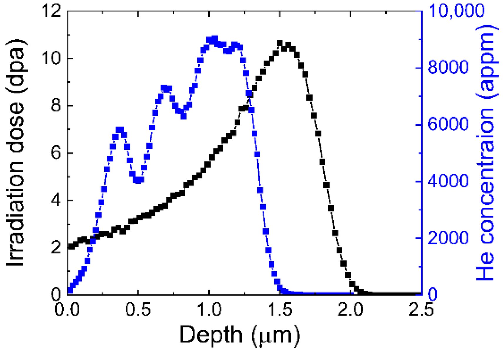
Figure 1. SRIM results show irradiation damage dose and helium concentration as a function of depth.
Materials 18 03246 g001
Figure 2. Bright-field TEM images of (a) as-fabricated and (b) recrystallized 12Cr ODS steel.
Materials 18 03246 g002
Figure 3. Size distribution of oxide particles in (a) as-fabricated and (b) recrystallized 12Cr ODS steel.
Materials 18 03246 g003
Figure 4. Bright-field TEM images of (ac) as-fabricated and (df) recrystallized 12Cr ODS steel after dual-beam irradiation at 973 K. (a) Low magnification, (b) high magnification showing the bubbles at the grain boundaries and (c) in the grains; (d) Low magnification, (e) high magnification showing the bubbles trapped at the large oxide particles and (f) the small oxide particles.
Materials 18 03246 g004
Figure 5. Size distribution of oxide particles in (a) as-fabricated and (b) recrystallized 12Cr ODS steel after dual-beam irradiation at 973 K.
Materials 18 03246 g005
Figure 6. Nanoindentation hardness distribution of (a) as-fabricated and (b) recrystallized 12Cr ODS steel before and after dual-beam irradiation at 973 K.
Materials 18 03246 g006
Figure 7. The average nanoindentation hardness of as-fabricated and recrystallized 12Cr ODS steel before and after dual-beam irradiation at 973 K.
Materials 18 03246 g007
Disclaimer/Publisher’s Note: The statements, opinions and data contained in all publications are solely those of the individual author(s) and contributor(s) and not of MDPI and/or the editor(s). MDPI and/or the editor(s) disclaim responsibility for any injury to people or property resulting from any ideas, methods, instructions or products referred to in the content.