Communication

Dual Effects of Ag Doping and S Vacancies on H2 Detection Using SnS2-Based Photo-Induced Gas Sensor at Room Temperature

1
School of Energy and Chemical Engineering, Puyang Vocational and Technical College, Puyang 457000, China
2
Puyang Institute of Technology, Henan University, Puyang 457000, China
3
College of Chemistry and Chemical Engineering, Henan Institute of Science and Technology, Xinxiang 453003, China
*
Authors to whom correspondence should be addressed.
Materials 2025, 18(12), 2687; https://doi.org/10.3390/ma18122687
Submission received: 25 April 2025 / Revised: 22 May 2025 / Accepted: 4 June 2025 / Published: 6 June 2025
(This article belongs to the Section Catalytic Materials)

Abstract

Hydrogen (H2) monitoring demonstrates significant practical importance for safety assurance in industrial production and daily life, driving the demand for gas-sensing devices with enhanced performance and reduced power consumption. This study developed a room-temperature (RT) hydrogen-sensing platform utilizing two-dimensional (2D) Ag-doped SnS2 nanomaterials activated by light illumination. The Ag-SnS2 nanosheets, synthesized through hydrothermal methods, exhibited exceptional H2 detection capabilities under blue LED light activation. The synergistic interaction between silver dopants and photo-activation enabled remarkable gas sensitivity across a broad concentration range (5.0–2500 ppm), achieving rapid response/recovery times (4 s/18 s) at 2500 ppm under RT. Material characterization revealed that Ag doping induced S vacancies, enhancing oxygen adsorption, while simultaneously facilitating photo-induced hole transfer for surface hydrogen activation. The optimized sensor maintained good response stability after five-week ambient storage, demonstrating excellent operational durability. Experimental results further demonstrated that Ag dopants enhanced hydrogen adsorption–activation, while S vacancies improved the surface oxygen affinity. This work provides fundamental insights into defect engineering strategies for the development of optically modulated gas sensors, proposing a viable pathway for the construction of energy-efficient environmental monitoring systems.

Graphical Abstract

1. Introduction

As a carbon-neutral energy carrier with a high gravimetric energy density, hydrogen has emerged as a pivotal renewable energy source for applications ranging from chemical synthesis to fuel cell technologies and industrial combustion systems [1,2]. However, the widespread adoption of hydrogen energy requires the urgent resolution of its inherent safety concerns, particularly given its hazardous nature, including its wide explosive concentration range (4.0–75.0 vol%), high diffusion coefficient (0.61 cm2/s), and substantial combustion enthalpy (285.5 kJ/mol) [3,4,5]. Thus, it is of great significance to selectively detect trace hydrogen [6]. These intrinsic properties underscore the critical need to develop reliable detection systems capable of selective trace-level hydrogen monitoring (particularly < 1000 ppm) in operational environments. The current hydrogen detection methodologies primarily employ analytical techniques such as gas chromatography (GC), semiconductor gas sensors, and optical spectroscopy approaches [1,6] In contrast, semiconductor-based chemiresistive sensors have garnered significant research interest due to their inherent advantages of low-cost fabrication, scalable manufacturing processes, and balanced performance metrics in terms of sensitivity, stability, and selectivity.
SnS2, as a 2D layered transition metal disulfide, is regarded as a promising gas-sensing material due to its large surface area and great electrical properties [7]. A series of SnS2-based materials have exhibited good gas sensitivity for NO2, NH3, xylene, C2H5OH, and H2S molecules [8,9,10,11,12]. W. Gao et al. demonstrated that a Pd/SnS2/SnO2 sensor exhibited a strong and rapid response to 500 ppm H2 at 300 °C [5]. However, to activate the gas detection abilities of semiconductors, a high operating temperature range of 100–450 °C is typically required, with an additional heater [13,14]. Considering its high resistance and weak adsorption to H2 molecules at room temperature (RT), the structural design of a SnS2 surface under external non-thermal field conditions would play a crucial role in solving the above issues [14,15]. Due to the excellent light absorption capacity of SnS2, light irradiation could be an effective alternative to thermal driving to activate its adsorption behavior toward gas molecules [15]. Under illumination, abundant electron–hole pairs could promote electron transfer, further improving the gas-sensing properties. While UV illumination (λ < 400 nm) has demonstrated effective photo-activation, its practical application is limited by photon-induced material degradation and potential health hazards. Recent advances in LED technology offer a safer alternative through visible light irradiation, combining sufficient photon energy for electron excitation with low power consumption [13]. Therefore, the synergistic combination of defect-engineered SnS2 nanostructures and LED photo-activation is of significance in developing an energy-efficient H2 sensor operating at RT.
In addition, the strategic introduction of sulfur vacancies (Vs) in SnS2 lattices enables selective oxygen chemisorption through isovalent orbital interactions, capitalizing on the chalcogen-group electronic affinity that facilitates superoxide radical (O2) formation at defect sites. Simultaneously, noble metal dopants with high work functions have demonstrated superior hydrogen spillover effects, with their d-orbital electron configurations enabling optimized hydrogen binding energies through charge-transfer interactions [16,17]. In the intrinsic 2D SnS2 lattice, the Sn atom was sandwiched between two layers of S atoms maintained by van der Waals forces [18]. Doped Ag(I) impurities not only led to an adsorbed activated site for hydrogen but also facilitated the formation of S vacancies due to the charge and size differences between Ag(I) and Sn(IV) in SnS2 [18]. Thus, it would be of great interest to investigate the synergistic effects of S vacancies and Ag impurities on the sensing performance of Ag-doped SnS2 sensors.
In this regard, a series of Ag-doped SnS2 (Ag-SnS2) sheets was synthesized and used as sensing materials to sense H2 with the help of LED light illumination. The photo-activated Ag-SnS2 sheet showed significantly enhanced H2-sensing performance with a wide serviceable range from 5.0 ppm to 2500 ppm at RT. Such excellent gas sensitivity was attributed to the effects of Ag doping and light illumination. The influence of Ag doping on the surface structure was investigated by ESR and TEM. Additionally, through O2 absorption measurements and in situ XPS, the possible mechanism was proven.

2. Materials and Methods
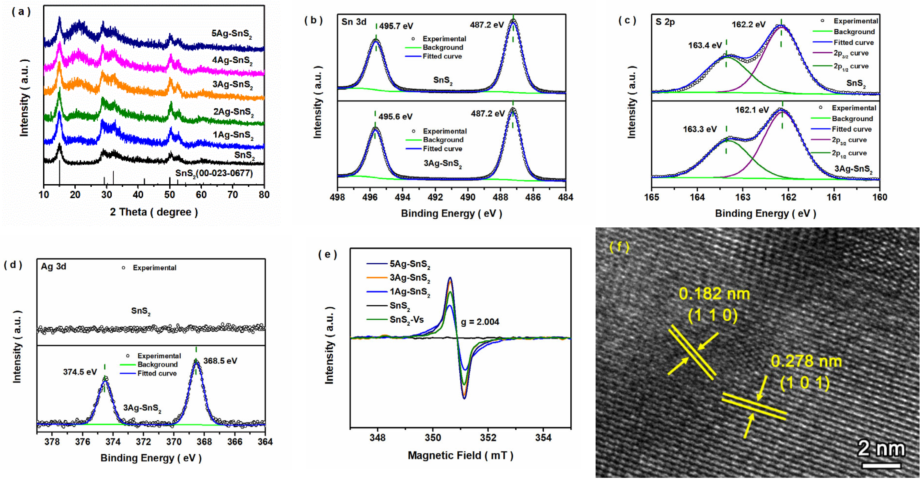
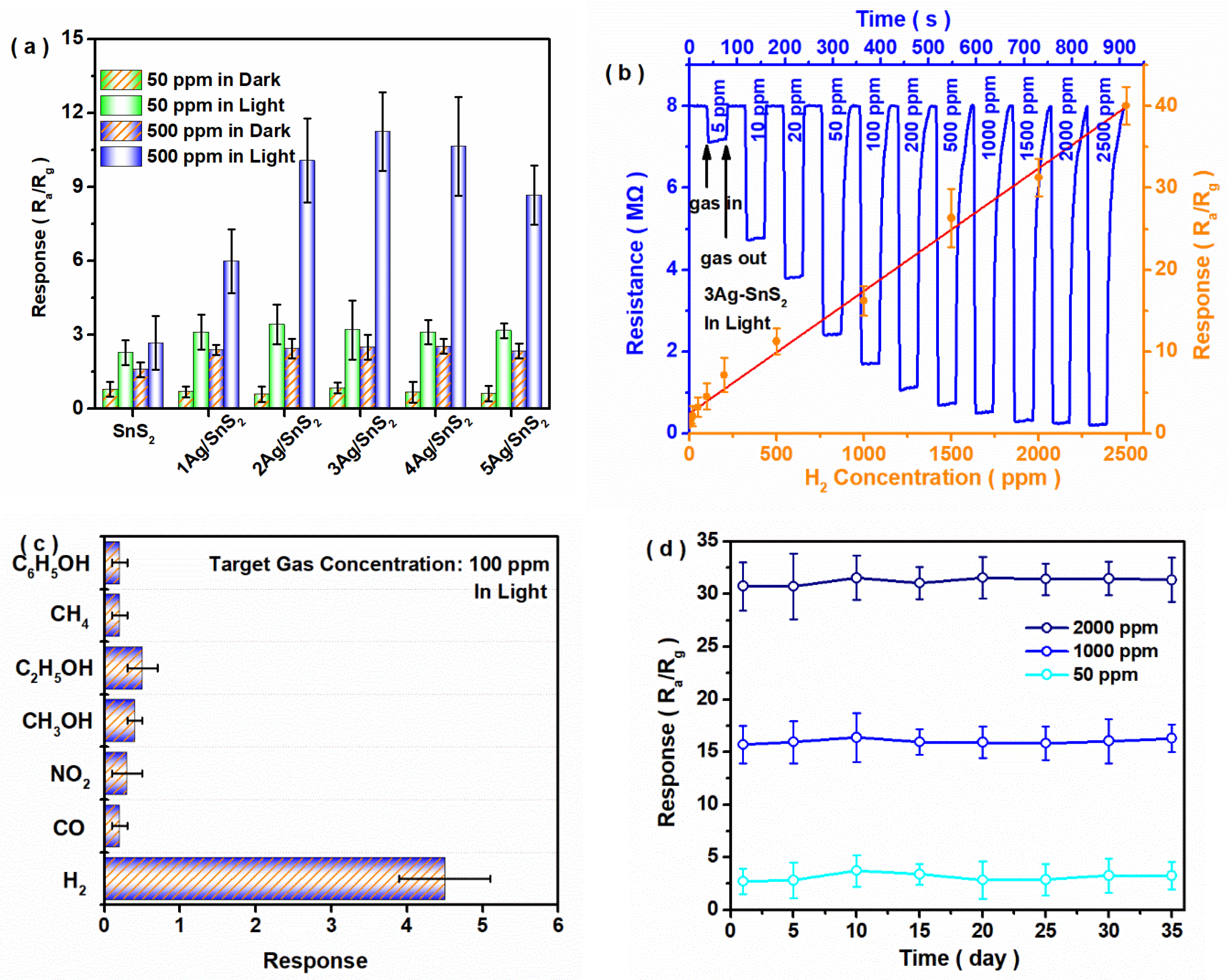
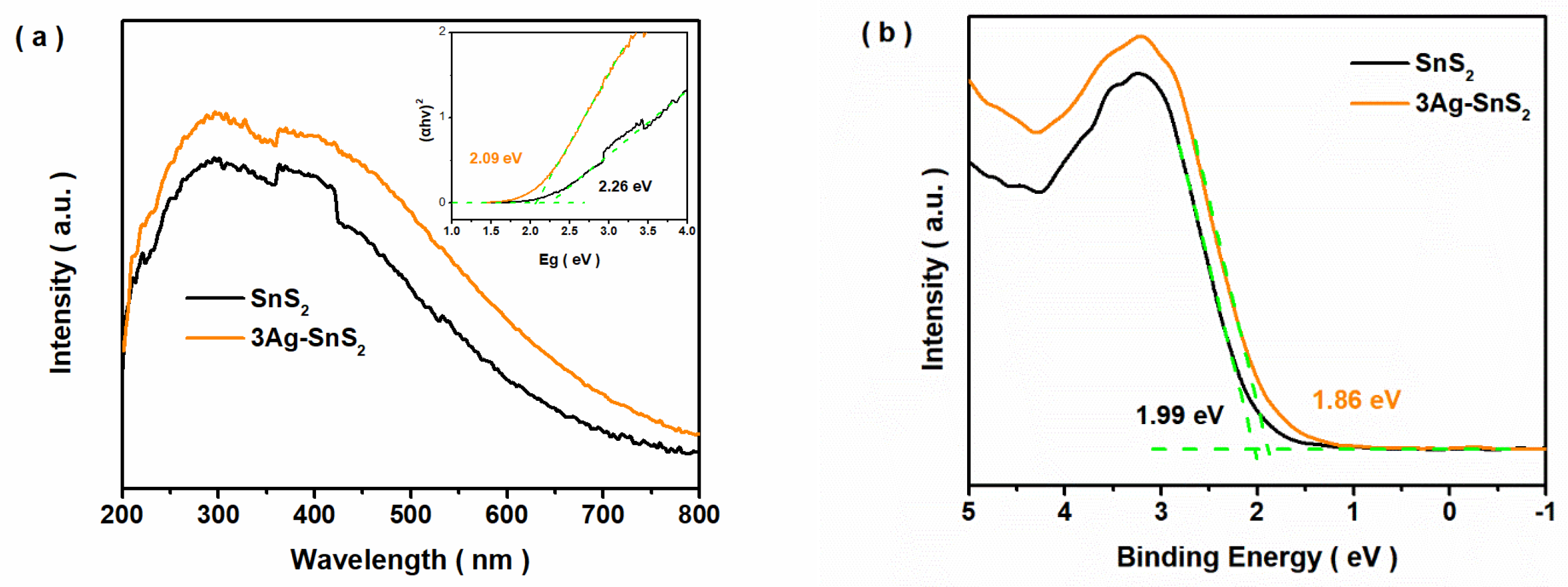
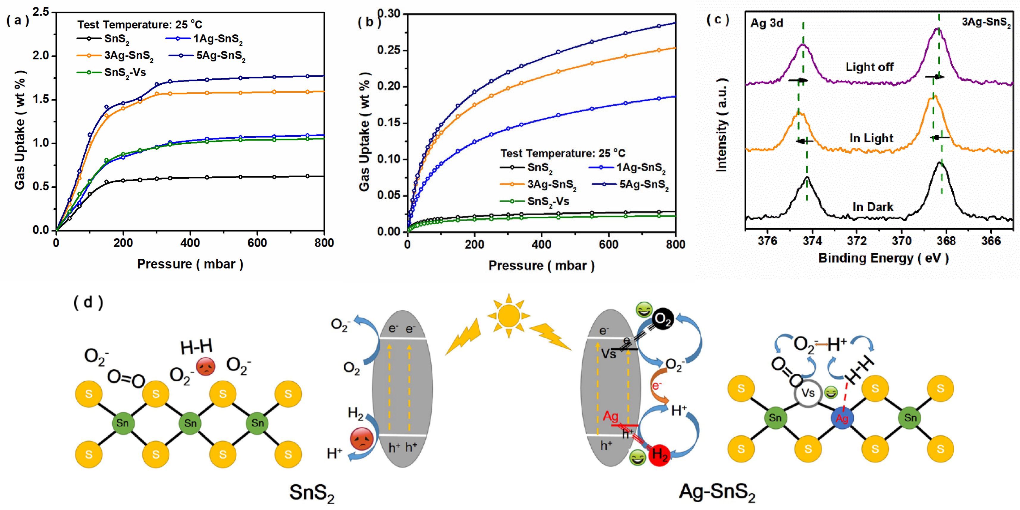
The SnS2-based sample was synthesized through the one-step solvothermal method. All chemicals were directly obtained from Shanghai Macklin Biochemical Technology Co., Ltd. Initially, 2.5 mL Triton X-100, 5.0 mmol citric acid, and 5.0 mmol SnCl4·5H2O were dissolved in 30 mL of deionized water and then added to 0.15 mmol of AgNO3 under strong stirring conditions. Then, 5 mL thioacetamide solution (2.0 mmol/mL) was slowly added to the above colloidal solution under stirring. After ultrasonic processing for 30 min, the resulting mixture was transferred to a Teflon-lined autoclave and heated at 150 °C for 12 h. The Ag-SnS2 composite was finally obtained by centrifugation and washed with water/ethanol (three times), with subsequent drying at 70 °C. This sample was named xAg-SnS2, in which x indicates the ideal ratio (mol/mol %) between Ag and Sn elements. The pristine SnS2 sample was prepared using a similar method but without the addition of AgNO3. The SnS2-Vs sample, as a contrasting sample, was obtained from pristine SnS2 using 300 °C calcination under a 5%H2/95%N2 mixed atmosphere. The gas sensitivity was measured using reconstructive W30-A gas-sensitive equipment (Zhengzhou WeiSheng Electronics Technology Co., Ltd. Figures S1 and S2) [19,20]. The selectivity (Q) was defined as the ratio of SA/ST, where SA and ST denote the average sensing responses in the testing gas and the total testing gas, respectively [21,22]. The theoretical detection limit (LOD) of the 3Ag-SnS2 sensor was also calculated [23,24]. Thus, SnS2-based gas sensors were formed, and the synthesis process is detailed in Supplementary Materials S1. The morphological and structural characterization of the obtained SnS2 samples was performed through XRD, TEM, and XPS measurements. The S vacancies of doped SnS2 were explored using the ESR test(Bruker EMXplus). The above measurements are detailed in Supplementary Materials S2.

3. Results

As shown in Figure 1, five obvious peaks at 15.0°, 28.2°, 32.1°, 50.0°, and 52.5° were found, corresponding to the characteristic peaks of the (0 0 1), (1 0 0), (1 0 1), (1 1 0), and (1 1 1) planes of hexagonal 2T SnS2 materials. With Ag addition, the intensity of one broad peak at about 22.0° gradually increased, while no new peak appeared. A broad peak between 20° and 30° appeared with Ag doping, and its intensity increased with increasing doped Ag content, indicating that the disordered atom part of Ag-SnS2 gradually increased [25,26]. Meanwhile, as shown in Figure S3, the characteristic peak of the (0 0 1) crystal facet moved toward a lower angle, which was due to the doped Ag impurities in the SnS2 structure [27,28]. Based on the ICP-OES results (Table S1), Ag impurities existed in the Ag-SnS2 sample, and its content increased with increasing Ag feedstock input. In the high-resolution Sn 3d spectra (Figure 1b), two stronger peaks at binding energies of 487.2 and 495.7 eV were associated with Sn 3d5/2 and 3d3/2 of Sn(IV) [25,29]. As displayed in Figure 1c, two peaks at 161.9 eV and 163.1 eV were found, corresponding to S 2p3/2 and 2p1/2, respectively (Figure 1c) [30,31]. In Figure 1d, two peaks at 368.0 and 374.1 eV are shown, which were ascribed to 3d5/2 and 3d3/2 of Ag(I) [18,25]. The peaks of both Sn and S shifted negatively after Ag doping, indicating a change in the surrounding electronegativity around the atoms. In the ESR spectra (Figure 1e), a clear sign of S vacancies (g = 2.004) appeared in the sample of Ag-doped SnS2, and its intensity was enhanced with increasing Ag input [18]. Combined with the XRD results, it was found that the ordered arrangement of Sn-S was destroyed by the doped Ag impurities, further forming S vacancies. As shown in Figure S4, a sheet morphology appeared in the SnS2 and 3Ag-SnS2 samples. In Figure 1f, the lattice fringes with spacing of 0.278 and 0.182 nm correspond to the (1 0 1) and (1 1 0) crystal facets of hexagonal 2T SnS2. Therefore, a series of Ag-doped SnS2 sheets with S vacancies were obtained through a one-step hydrothermal process.
The gas sensitivity of the SnS2-based materials for H2 detection was investigated. As shown in Figure S6, the SnS2-based sensor at a 150 °C operating temperature in the dark exhibited higher gas sensitivity than that at a lower temperature. As shown in Figure 2a, all SnS2 materials exhibited sensitivity for H2 gas at room temperature, and the response under illumination was apparently higher than that in the dark. With increasing Ag addition, the response value of the Ag-SnS2 sensor was gradually increased, and the 3Ag-SnS2 sample showed the highest response value for H2 detection. Beyond the optimal value, its sign exhibited a marked decline, which was caused by the excess defects on the SnS2 surface, further decreasing the concentration of active electrons. As shown in Figure S6a, when faced with higher humidity, the gas sensitivity observably declined, especially for that under light. As shown in Figure S6b, smaller liquid contact angles appeared in all samples with S vacancies and/or Ag impurities, proving their good hydrophilicity at RT. This provides a possible reason for the weak water resistance: superfluous H2O molecules were readily adsorbed on the sensor’s surface, inhibiting the capture of H2 molecules. We further explored the relationship between the gas concentration and sensor sign. The results (Figure 2b) showed that, although a fast response rate for the 3Ag-SnS2 photo-induced sensor was found, its recovery time was gradually lengthened with increasing concentrations of H2 gas. Nonetheless, its response and recovery times under 2500 ppm H2 conditions were maintained at ~4 s and ~18 s, respectively. Moreover, a linear correlation (y = 0.01498x + 2.428, R2 0.9923) could be determined at a H2 concentration between 5 ppm and 2500 ppm. The theoretical detection limit was calculated to be ∼923 ppb. Such a low LOD value satisfies the permissible exposure limits of H2 specified by various organizations. As shown in Figure 2c, when faced with multiple different target gases, the 3Ag-SnS2 sensor exhibited good selectivity for H2 gas. As summarized in Table S2, the Q value of the 3Ag-SnS2 sensor for H2 gas could reach 71.4%. The cycling stability of the gas sensor was also considered. As shown in Figure 2d, the photo-induced 3Ag-SnS2 sensor exhibited a stable response value for H2 detection at room temperature over 35 days. Compared with the previous SnS2- or SnO2-based gas sensors, as summarized in Table S3, the 3Ag-SnS2 photo-induced sensor exhibited good sensitivity, selectivity, and stability for H2 detection, with a wide concentration range (5.0 to 2500 ppm) when operating at room temperature [32,33,34,35,36,37,38,39,40,41].
To explore the reasons for the enhanced photo-induced gas sensitivity of Ag-SnS2, the band structures and surface states of the obtained samples were further investigated. Firstly, the influence of Ag impurities on the band structure of SnS2 was studied. As shown in Figure 3a, the light response range of the 3Ag-SnS2 sample was wider than that of pristine SnS2, and their Eg values were calculated and are summarized in Table S4. Then, the VB band position could be measured through VB-XPS measurements. With Ag doping, the VB value modestly decreased, as shown in Figure 3b. Finally, the CB position could be calculated, and the information about their band structures is summarized in Table S4. Additionally, the photo-induced current curve is shown in Figure S7. Although the photo-induced current density of Ag-SnS2 was higher than that of SnS2, excess Ag impurities did not increase the abundance of photo-induced electrons. This may explain why S vacancies were easily formed around the doped Ag site due to the effects of charge balance; however, superfluous vacancies created a recombination center for photo-induced electron–holes, leading to a weak current density. This also explains why the 3Ag-SnS2 sample exhibited better gas sensitivity compared with other samples. As shown in Figure S8, the lift time of photo-induced electrons was calculated, and the average lift time of 3Ag-SnS2 (2.89 ns) was obviously longer than that of the pristine SnS2 sample (2.27 ns). Consistent with existing results about the photo-response current, doped Ag impurities could promote the separation of photo-induced carriers.
As shown in Figure 4a, all obtained samples showed adsorptive properties toward O2 gas at room temperature. It was found that the existence of Ag impurities enhanced the O2 adsorption capacity of SnS2, and its variation trend was in line with the change in S vacancies (Figure 1e). As shown in Figure 4b, the variation trend of the H2 adsorption property was similar to that of the O2 adsorption property. With the increasing density of Ag sites on the surface, the H2 adsorption capacity of SnS2 was increased, which was due to the superior hydrogen-binding capabilities of the high-work-function metal element. Meanwhile, the H2 adsorption property of SnS2-Vs was not increased through increasing that of O2 due to S vacancies. Hence, more adsorbed H2 molecules appeared around the Ag impurities. Additionally, the in situ XPS results (Figure 4c) showed that the peak positions of the Ag impurities in light shifted in a high-energy direction, and this trend was reversed after the light was switched off. It indicates that the electrons in Ag sites are lost under light, and this change in Ag atoms in light/dark conditions is reversible [42]. Hence, doped Ag sites may play a role in the surface transfer of photo-induced holes. Based on the above results, as shown in Figure 4c, the S vacancies around the Ag sites on the surface could easily adsorb the oxygen molecules. Then, metal Ag sites could capture hydrogen molecules in the air, which formed two neighboring adsorbed O2/H2 molecules. Simultaneously, under illumination, the photo-generated electrons and holes were transferred from SnS2 to the adsorbed O2 and H2 molecules, respectively. Moreover, superoxide radicals (O2) and hydrogen ions (H+) were formed and reacted with each other, leading to electron consumption and a change in the resistance of SnS2. The possible mechanism behind the H2 detection of the photo-induced gas sensor is shown in Figure 4d.

4. Conclusions

In conclusion, a series of sheet-like Ag-doped SnS2 materials were prepared through the one-step hydrothermal method. S vacancies were generated, along with Ag impurity doping, leading to enhanced O2 adsorption properties. Compared to the pristine SnS2, which was unable to sense H2 at RT, the Ag-SnS2 sample exhibited excellent gas sensitivity for H2 detection, with a wide concentration range of 5.0 ppm to 2500 ppm under blue LED illumination. When faced with a high concentration of H2 gas (2500 ppm), its response and recovery times were maintained at ~4 s and ~18 s, respectively. Moreover, the sensor maintained its gas response after five weeks of relaxation. The synergistic effects of S vacancies and Ag impurities in enhancing O2/H2 adsorption have been proven. The Ag impurity sites played a key role in enabling photo-induced hole transfer to the active adsorbed hydrogen on the SnS2 surface under illumination. Given their remarkable gas-sensing performance, the formed Ag-doped SnS2 sheets could contribute to enhancing the potential and fueling the exploitation of next-generation, high-performance, gas-sensing, light-modulated devices at RT.

Supplementary Materials

The following supporting information can be downloaded at: https://www.mdpi.com/article/10.3390/ma18122687/s1, S1. Gas sensing characterization. S2. Sample characterization. Figure S1. Schematic diagram of photo-induced gas sensor. Figure S2. Wavelength range of LED light source. Figure S3. Partial XRD patterns of SnS2 and doped SnS2 samples. Figure S4. TEM images of SnS2 and 3Ag-SnS2 samples. Figure S5. Gas sensitivity of SnS2-based sensor at different operating temperatures in dark. Figure S6. Gas sensitivity of SnS2-based sensor at different humidity levels with 500 ppm H2 gas in light and liquid contact angle of water obtained over SnS2 sample surface. Figure S7. Photo-induced current curves of SnS2 and Ag-SnS2 samples. Figure S8. Transient photo-current curves of SnS2 and 3Ag-SnS2 samples. Table S1. Element content of obtained SnS2 sample according to ICP-OES. Table S2. Quantitative sensitivity values of 3Ag-SnS2 sensor. Table S3. Gas sensitivity of SnS2- or SnO2-based materials for H2 detection in previous studies and research. Table S4. Band structures of Ag-SnS2 and SnS2 materials.

Author Contributions

Conceptualization, J.W. and X.S.; methodology, J.W.; software, N.F. and H.M.; validation, H.M.; formal analysis, N.F.; investigation, H.M.; data curation, S.W. and H.M.; writing—original draft preparation, S.W. and J.W.; writing—review and editing, J.W.; project administration, J.W.; funding acquisition, J.W. and X.S. All authors have read and agreed to the published version of the manuscript.

Funding

This research was funded by the Scientific and Technological Research Project of Henan Province (No. 252102230085 and 252102320178), the Outstanding Talent Innovation Foundation of PuYang (No. 160803), and the Training Program for Young Backbone Teachers in Higher Education Institutions in Henan Province (No. 2024GGJS101).

Data Availability Statement

The original contributions presented in this study are included in the article/Supplementary Material. Further inquiries can be directed to the corresponding authors.

Conflicts of Interest

The authors declare no conflicts of interest.

References

  1. Hjiri, M.; Dhahri, R.; Benmansour, N.; Neri, G. Laser irradiated gas sensors: A review. Micro Nanostruct. 2025, 204, 208157. [Google Scholar] [CrossRef]
  2. Shooshtari, M.; Salehi, A. An electronic nose based on carbon nanotube -titanium dioxide hybrid nanostructures for detection and discrimination of volatile organic compounds. Sens. Actuators B 2022, 357, 131418. [Google Scholar] [CrossRef]
  3. Liu, Y.; Chen, S.; Xiao, B.; Chu, J.; Wang, H.; Chen, Y.; Yao, T.; Yang, A.; Han, X.; Rong, M.; et al. Ultra-large Sn3O4 nanosheets with Sn2+ defect for highly efficient hydrogen sensing. Sens. Actuators B 2024, 401, 135025. [Google Scholar] [CrossRef]
  4. Yin, X.-T.; Wu, S.-S.; Dastan, D.; Nie, S.; Liu, Y.; Li, Z.-G.; Zhou, Y.-W.; Li, J.; Faik, A.; Shan, K.; et al. Sensing selectivity of SnO2-Mn3O4 nanocomposite sensors for the detection of H2 and CO gases. Surf. Interfaces 2021, 25, 101190. [Google Scholar] [CrossRef]
  5. Meng, X.; Bi, M.; Xiao, Q.; Gao, W. Ultrasensitive gas sensor based on Pd/SnS2/SnO2 nanocomposites for rapid detection of H2. Sens. Actuators B 2022, 359, 131612. [Google Scholar] [CrossRef]
  6. Zhu, M.; Zhang, H.; Zhang, S.; Yao, H.; Shi, X.; Xu, S. Chemoresistive gas sensors based on noble-metal-decorated metal oxide semiconductors for H2 detection. Materials 2025, 18, 451. [Google Scholar] [CrossRef]
  7. Mishra, R.K.; Choi, H.J.; Ryu, J.W.; Choi, G.J.; Kumar, V.; Kumar, P.; Singh, J.; Kumar, S.; Gwag, J.S. Recent progress in gas sensing based on 2D SnS2 and its heterostructure platforms: A review. Sens. Actuators A 2024, 365, 114860. [Google Scholar] [CrossRef]
  8. Saggu, I.S.; Singh, S.; Chen, K.; Xuan, Z.; Swihart, M.T.; Sharma, S. Ultrasensitive room-temperature NO2 detection using SnS2/MWCNT composites and accelerated recovery kinetics by UV activation. ACS Sens. 2023, 8, 243–253. [Google Scholar] [CrossRef]
  9. Lee, S.M.; Kim, Y.J.; Park, S.J.; Cheon, W.S.; Kim, J.; Nam, G.B.; Kim, Y.; Jang, H.W. In-situ growth of 2D MOFs as a molecular sieving layer on SnS2 nanoflakes for realizing ultraselective H2S detection. Adv. Funct. Mater. 2025, 35, 2417019. [Google Scholar] [CrossRef]
  10. Qiu, P.; Qin, Y.; Wang, X. S-vacancies and Ag nanoparticles in SnS2 nanoflakes for ethanol sensing: A combined experimental and theoretical investigation. ACS Appl. Nano Mater. 2022, 5, 10839–10847. [Google Scholar] [CrossRef]
  11. Lu, Y.; Zhang, J.; Wang, W.; Fan, Y.; Liu, C.; Zhou, J.; Liu, D.; Ruan, S. Au-Pd modified SnS2 nanosheets for conductometric detection of xylene gas. Sens. Actuators B 2022, 351, 130907. [Google Scholar] [CrossRef]
  12. Maria, K.H.; Sakhuja, N.; Jha, R.K.; Bhat, N. Ultra-sonication assisted synthesis of 2D SnS2 nanoflakes for room-temperature NO gas detection. IEEE Sens. J. 2021, 21, 10420–10427. [Google Scholar] [CrossRef]
  13. Eom, T.H.; Cho, S.H.; Suh, J.M.; Kim, T.; Lee, T.H.; Jun, S.E.; Yang, J.W.; Lee, J.; Hong, S.-H.; Jang, H.W. Substantially improved room temperature NO2 sensing in 2-dimensional SnS2 nanoflowers enabled by visible light illumination. J. Mater. Chem. A 2021, 9, 11168–11178. [Google Scholar] [CrossRef]
  14. Ramakrishnan, K.; Ajitha, B.; Ashok Kumar Reddy, Y. Review on metal sulfide-based nanostructures for photodetectors: From ultraviolet to infrared regions. Sens. Actuators A 2023, 349, 114051. [Google Scholar] [CrossRef]
  15. Li, Q.; Wang, X.; Li, H.; Guo, X. Light-activated gas sensors. Chin. Sci. Bull. 2022, 67, 1837–1850. [Google Scholar] [CrossRef]
  16. Arain, S.; Usman, M.; Saeed, F.; Feng, S.; Rehman, W.; Liu, X.; Dai, H. Microemulsion-based synthesis of highly efficient Ag-doped fibrous SiO2-TiO2 photoanodes for photoelectrochemical water splitting. Catalysts 2025, 15, 66. [Google Scholar] [CrossRef]
  17. An, Q.; Li, J.; Peng, J.; Hu, L. DFT study of small gas molecules (C2H2, CH4, CO and H2) adsorbed on Au, Ag-doped ZnO monolayer. Chem. Phys. Lett. 2025, 869, 142043. [Google Scholar] [CrossRef]
  18. Yang, H.; Du, Z.; Yang, Y.; Li, X.; Wu, Q.; Tang, J.; Wang, X.; Zeng, D. Ag intercalated SnS2 with S vacancy and expanded interlayer for enhancing NO2 sensing. Sens. Actuators B 2023, 393, 134140. [Google Scholar] [CrossRef]
  19. Wang, J.-C.; Ma, H.; Shi, W.; Li, W.; Zhang, Z.; Hou, Y.; Zhang, W.; Chen, J. Designed synthesized step-scheme heterojunction of Bi2WO6 nanosheet supported on CuBi2O4 nanorod with remarkable photo-assisted gas sensing for N-butyl alcohol. J. Environ. Chem. Eng. 2024, 12, 112698. [Google Scholar] [CrossRef]
  20. Wang, J.-C.; Shi, W.; Sun, X.-Q.; Wu, F.-Y.; Li, Y.; Hou, Y. Enhanced photo-assisted acetone gas sensor and efficient photocatalytic degradation ssing Fe-doped hexagonal and monoclinic WO3 phase−junction. Nanomaterials 2020, 10, 398. [Google Scholar] [CrossRef]
  21. Yang, J.; Sun, R.; Bao, X.; Liu, J.; Ng, J.W.; Tang, B.; Liu, Z. Enhancing selectivity of two-dimensional materials-based gas sensors. Adv. Funct. Mater. 2025, 35, 2420393. [Google Scholar] [CrossRef]
  22. Qiao, X.; Xu, Y.; Yang, K.; Ma, J.; Li, C.; Wang, H.; Jia, L. Mo doped BiVO4 gas sensor with high sensitivity and selectivity towards H2S. Chem. Eng. J. 2020, 395, 125144. [Google Scholar] [CrossRef]
  23. Venkatesan, A.; Ryu, H.; Devnath, A.; Yoo, H.; Lee, S. Air-stable, all-dry transferred ReS2/GaSe heterostructure-based NO2 gas sensor. J. Mater. Sci. Technol. 2024, 168, 79–87. [Google Scholar] [CrossRef]
  24. Wu, K.; Chai, H.; Xu, K.; Debliquy, M.; Zhang, C. Effect of {010} crystal facets of Bi2MoO6 and 1D/2D heterostructures for conductometric room temperature NH3 gas sensors. Sens. Actuators B 2023, 376, 132983. [Google Scholar] [CrossRef]
  25. Tian, W.; Han, J.; Wan, L.; Li, N.; Chen, D.; Xu, Q.; Li, H.; Lu, J. Enhanced piezocatalytic activity in ion-doped SnS2 via lattice distortion engineering for BPA degradation and hydrogen production. Nano Energy 2023, 107, 108165. [Google Scholar] [CrossRef]
  26. Ageeva, T.A.; Bush, A.A.; Golubev, D.V.; Gorshkova, A.S.; Mozhchil, R.N.; Koifman, O.I.; Kozlov, V.S.; Matis, M.E.; Rumyantseva, V.D.; Sigov, A.S.; et al. Porphyrin complexes of transition metals with a large dipole moment as active components of new film electret materials. Russ. Chem. Bull. 2023, 72, 2070–2082. [Google Scholar] [CrossRef]
  27. Liu, Y.; Zhou, Y.; Zhou, X.; Jin, X.; Li, B.; Liu, J.; Chen, G. Cu doped SnS2 nanostructure induced sulfur vacancy towards boosted photocatalytic hydrogen evolution. Chem. Eng. J. 2021, 407, 127180. [Google Scholar] [CrossRef]
  28. Park, S.J.; Lee, S.M.; Lee, J.; Choi, S.; Nam, G.B.; Jo, Y.K.; Hwang, I.-S.; Jang, H.W. Pd-W18O49 nanowire MEMS gas sensor for ultraselective dual detection of hydrogen and ammonia. Small 2025, 21, 2405809. [Google Scholar] [CrossRef]
  29. Lv, Y.-R.; Wang, Z.-L.; Yang, Y.-X.; Luo, Y.; Yang, S.-Y.; Xu, Y.-H. Tin bisulfide nanoplates anchored onto flower-like bismuth tungstate nanosheets for enhancement in the photocatalytic degradation of organic pollutant. J. Hazard. Mater. 2022, 432, 128665. [Google Scholar] [CrossRef]
  30. Lei, Z.; Wang, W.; Sun, T.; Liu, E.; Gao, T. Efficient photocatalytic H2 evolution over SnS2/twinned Mn0.5Cd0.5S hetero-homojunction with double S-scheme charge transfer routes. J. Mater. Sci. Technol. 2025, 216, 81–92. [Google Scholar] [CrossRef]
  31. Mamo, T.T.; Qorbani, M.; Hailemariam, A.G.; Putikam, R.; Chu, C.-M.; Ko, T.-R.; Sabbah, A.; Huang, C.-Y.; Kholimatussadiah, S.; Billo, T.; et al. Enhanced CO2 photoreduction to CH4 via *COOH and *CHO intermediates stabilization by synergistic effect of implanted P and S vacancy in thin-film SnS2. Nano Energy 2024, 128, 109863. [Google Scholar] [CrossRef]
  32. Qin, Y.; Zhang, Y.; Qiu, P.; Lei, S. SnO2-Co3O4 nanocomposite sensor: Achieving ultra-selective hydrogen detection in mixed gas environments. Sens. Actuators B 2025, 422, 136521. [Google Scholar] [CrossRef]
  33. Li, A.; Zhao, S.; Bai, J.; Xiao, H.; Gao, S.; Shen, Y.; Yuan, Z.; Meng, F. The role of AuSn alloys in optimizing SnO2 nanospheres for chemoresistive hydrogen sensing. Sens. Actuators B 2025, 427, 137214. [Google Scholar] [CrossRef]
  34. Geethukrishnan; Apte, O.; Tadi, K.K. Geethukrishnan; Apte, O.; Tadi, K.K. Electrochemical sensor for dopamine detection based on multiwalled carbon nanotube/molybdenum disulfide quantum dots with machine learning integration and anti-interference capability. Tungsten 2025, 7, 137–151. [Google Scholar] [CrossRef]
  35. Liu, W.; Zou, J.; Li, S.; Li, J.; Li, F.; Zhan, Z.; Zhang, Y. Pd/In2O3-based bilayer H2 sensor with high resistance to silicone toxicity and ultra-fast response. Int. J. Hydrogen Energy 2023, 48, 5743–5753. [Google Scholar] [CrossRef]
  36. Murali, A.S.; Sreelekshmi; Saraswathyamma, B. The role of engineered MoS2 and MoSe2 transition metal dichalcogenides in electrochemical sensors and batteries: A review. Tungsten 2025, 7, 215–242. [Google Scholar] [CrossRef]
  37. Lu, S.; Zhang, Y.; Liu, J.; Li, H.-Y.; Hu, Z.; Luo, X.; Gao, N.; Zhang, B.; Jiang, J.; Zhong, A.; et al. Sensitive H2 gas sensors based on SnO2 nanowires. Sens. Actuators B 2021, 345, 130334. [Google Scholar] [CrossRef]
  38. Tang, C.; Jin, W.; Xiao, X.; Qi, X.; Ma, Y.; Ma, L. Graphene-based chemiresistive hydrogen sensor for room temperature operation. Sens. Actuators B 2025, 424, 136889. [Google Scholar] [CrossRef]
  39. Kumar, G.; Li, X.; Du, Y.; Geng, Y.; Hong, X. UV-light enhanced high sensitive hydrogen (H2) sensor based on spherical Au nanoparticles on ZnO nano-structured thin films. J. Alloys Compd. 2019, 798, 467–477. [Google Scholar] [CrossRef]
  40. Wang, F.; Hu, K.; Liu, H.; Zhao, Q.; Wang, K.; Zhang, Y. Low temperature and fast response hydrogen gas sensor with Pd coated SnO2 nanofiber rods. Int. J. Hydrogen Energy 2020, 45, 7234–7242. [Google Scholar] [CrossRef]
  41. Pandey, G.; Lawaniya, S.D.; Kumar, S.; Dwivedi, P.K.; Awasthi, K. A highly selective, efficient hydrogen gas sensor based on bimetallic (Pd–Au) alloy nanoparticle (NP)-decorated SnO2 nanorods. J. Mater. Chem. A 2023, 11, 26687–26697. [Google Scholar] [CrossRef]
  42. Wang, Z.; Cheng, B.; Zhang, L.; Yu, J.; Tan, H. BiOBr/NiO S-scheme heterojunction photocatalyst for CO2 photoreduction. Sol. RRL 2022, 6, 2100587. [Google Scholar] [CrossRef]
Figure 1. Structure, composition, defects, and morphology of SnS2 and doped SnS2 samples. (a) XRD pattern, (b) XPS: Sn 3d high-resolution spectra, (c) XPS: S 2p high-resolution spectra, (d) XPS: Ag 3d high-resolution spectra, (e) ESR of SnS2 and Ag-SnS2 samples, and (f) HR-TEM of 3Ag-SnS2 sample.
Materials 18 02687 g001
Figure 2. Performance of SnS2- and Ag-SnS2-based gas sensors. (a) Gas sensitivity for different samples; (b) resistance and response of 3Ag-SnS2 sensor at different H2 concentrations; (c) gas selectivity of 3Ag-SnS2 sensor for different samples; (d) cycling stability of 3Ag-SnS2 sensor over 5 weeks.
Materials 18 02687 g002
Figure 3. (a) DRS and (b) VB-XPS of SnS2 and 3Ag-SnS2 samples.
Materials 18 02687 g003
Figure 4. O2 (a) and H2 (b) absorption properties of SnS2 and Ag-SnS2 samples; in situ XPS results of Ag element in light/dark conditions; and (c) schematic diagram of gas sensing mechanism (d).
Materials 18 02687 g004
Disclaimer/Publisher’s Note: The statements, opinions and data contained in all publications are solely those of the individual author(s) and contributor(s) and not of MDPI and/or the editor(s). MDPI and/or the editor(s) disclaim responsibility for any injury to people or property resulting from any ideas, methods, instructions or products referred to in the content.

