
Degree of Standardisation in Ceramic Gingival Systems


 and
and
Abstract
1. Introduction
2. Materials and Methods
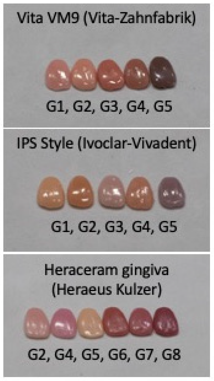
2.1. Sample Preparation and Colour Coordinate Recording
2.2. Statistical Analysis
3. Results
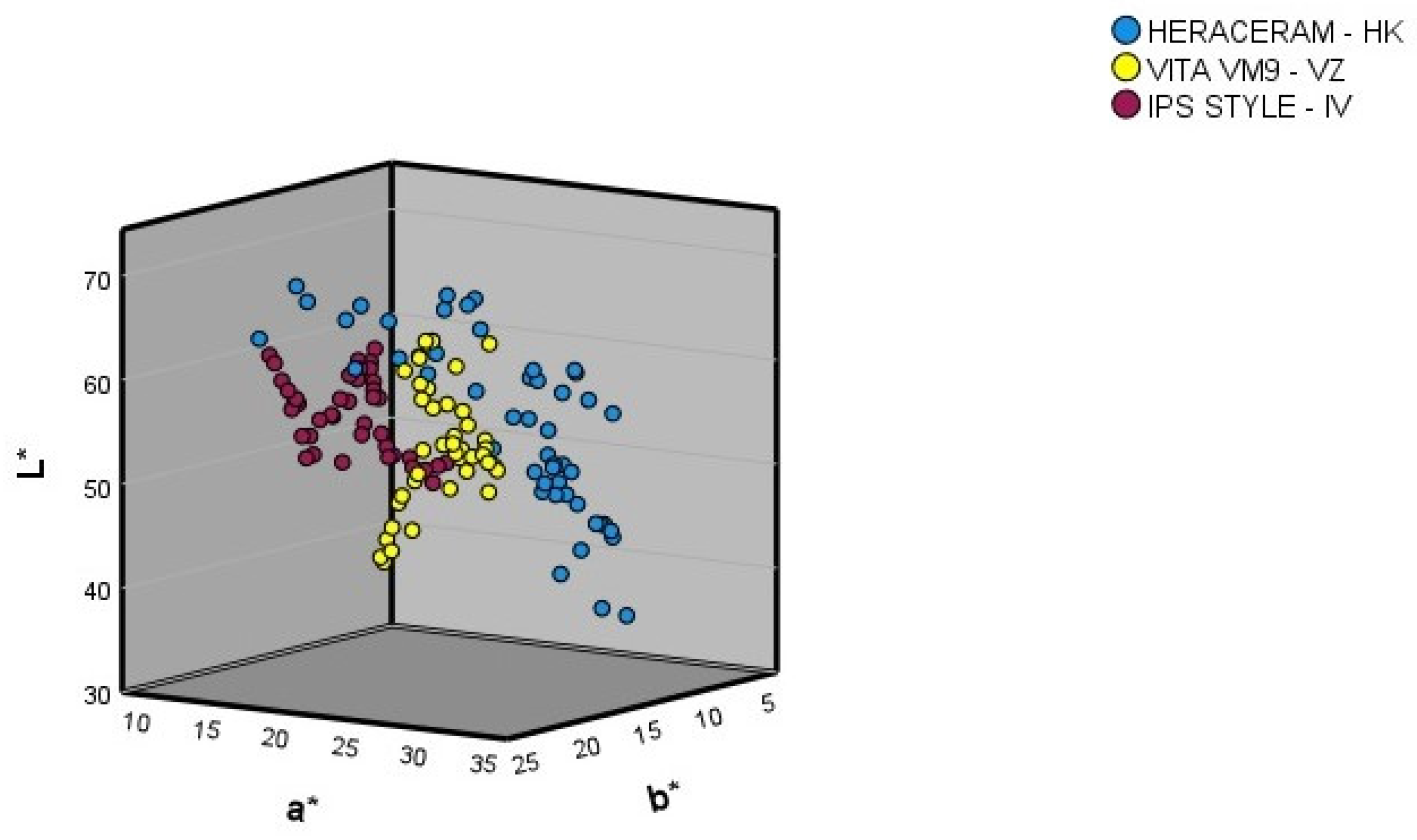
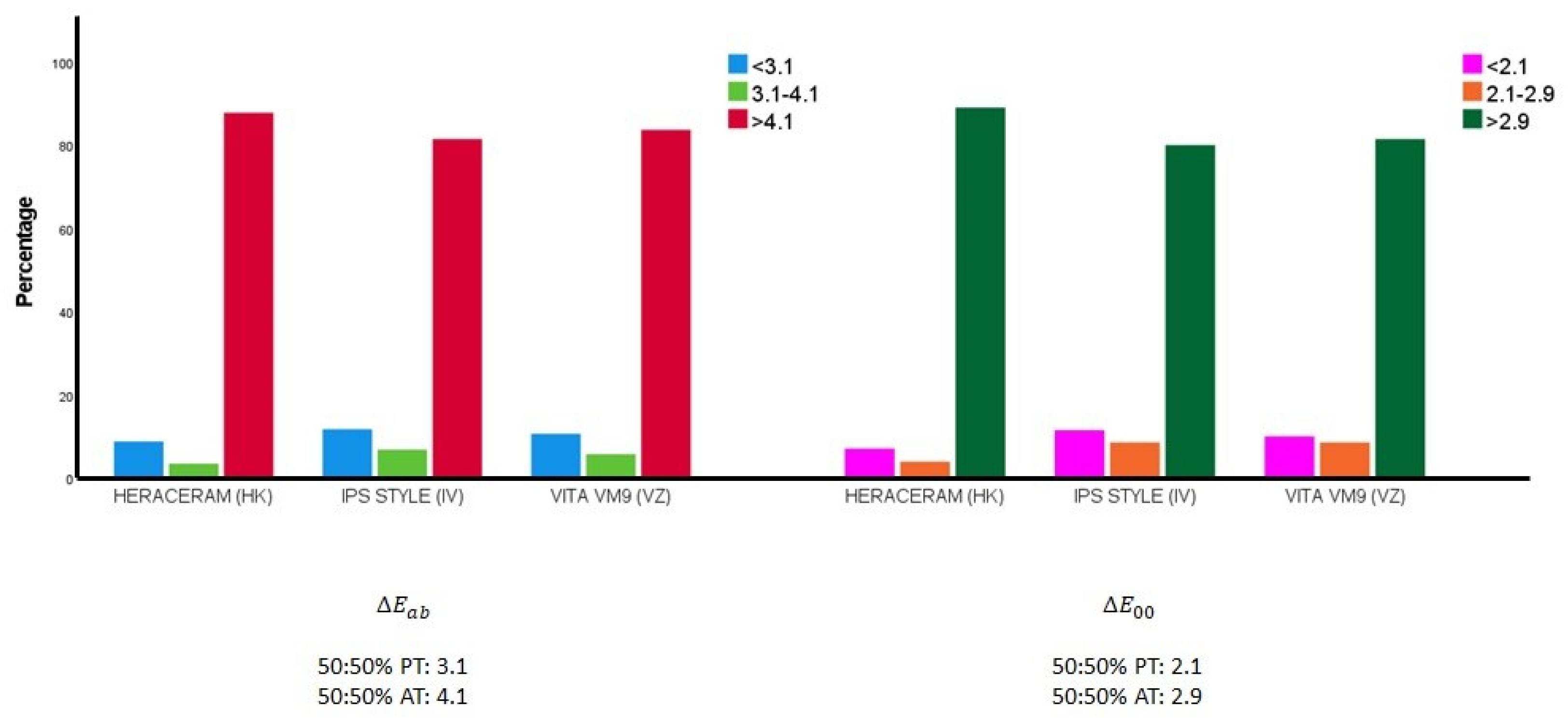
3.1. Description and Comparison of the Basic Colours in the Three Ceramic Gingival Colour Systems
3.2. Description and Comparison of the Three Ceramic Gingival Colour Systems when Expanded with Basic-Colour Mixtures
4. Discussion
4.1. Null Hypotheses
4.2. Strengths and Limitations of the Research
4.3. Potential Clinical Applications
5. Conclusions
Author Contributions
Funding
Institutional Review Board Statement
Informed Consent Statement
Data Availability Statement
Conflicts of Interest
References
- Gómez-Polo, C.; Montero, J.; Santos Marino, J.; Galindo, M.P.; Vicente, M.P. Application of the Social Appeal Scale to a Selected Spanish Population. Int. J. Prosthodont. 2016, 29, 59–62. [Google Scholar] [CrossRef] [PubMed]
- Montero, J.; Gómez-Polo, C.; Santos, J.A.; Portillo, M.; Lorenzo, M.C.; Albaladejo, A. Contributions of dental colour to the physical attractiveness stereotype. J. Oral Rehabil. 2014, 41, 768–782. [Google Scholar] [CrossRef]
- Samorodnitzky-Naveh, G.R.; Geiger, S.B.; Levin, L. Patients’ satisfaction with dental esthetics. J. Am. Dent. Assoc. 2007, 138, 805–808. [Google Scholar] [CrossRef]
- Silva, F.M.F.D.; Magno, M.B.; Neves, A.B.; Coqueiro, R.D.S.; Costa, M.C.; Maia, L.C.; Pithon, M.M. Aesthetic perceptions and social judgments about different enamel opacities. Braz. Oral Res. 2020, 34, e049. [Google Scholar] [CrossRef] [PubMed]
- Montero, J.; Gómez Polo, C.; Rosel, E.; Barrios, R.; Albaladejo, A.; López-Valverde, A. The role of personality traits in self-rated oral health and preferences for different types of flawed smiles. J. Oral Rehabil. 2016, 43, 39–50. [Google Scholar] [CrossRef] [PubMed]
- Sabbah, A. Smile Analysis: Diagnosis and Treatment Planning. Dent. Clin. N. Am. 2022, 66, 307–341. [Google Scholar] [CrossRef]
- Chen, J.; Chiang, C.; Zhang, Y. Esthetic evaluation of natural teeth in anterior maxilla using the pink and white esthetic scores. Clin. Implant Dent. Relat. Res. 2018, 20, 770–777. [Google Scholar] [CrossRef]
- Singh, V.P.; Uppoor, A.S.; Nayak, D.G.; Shah, D. Black triangle dilemma and its management in esthetic dentistry. Dent. Res. J. 2013, 10, 296–301. [Google Scholar]
- Fürhauser, R.; Florescu, D.; Benesch, T.; Haas, R.; Mailath, G.; Watzek, G. Evaluation of soft tissue around single-tooth implant crowns: The pink esthetic score. Clin. Oral Implant. Res. 2005, 16, 639–644. [Google Scholar] [CrossRef]
- Tjan, A.H.; Miller, G.D.; The, J.G. Some esthetic factors in a smile. J. Prosthet. Dent. 1984, 51, 24–28. [Google Scholar] [CrossRef]
- Marini, L.; Tomasi, C.; Gianserra, R.; Graziani, F.; Landi, L.; Merli, M.; Nibali, L.; Roccuzzo, M.; Sforza, N.M.; Tonetti, M.S.; et al. Reliability assessment of the 2018 classification case definitions of peri-implant health, peri-implant mucositis, and peri-implantitis. J. Periodontol. 2023. [Google Scholar] [CrossRef]
- Chisci, G.; Gabriele, G.; Gennaro, P. Periodontal disease before and after fractures of the mandible. Br. J. Oral Maxillofac. Surg. 2023, 61, 116. [Google Scholar] [CrossRef] [PubMed]
- Van der Burgt, T.P.; ten Bosch, J.J.; Borsboom, P.C.F.; Kortsmit, W.J. A comparision of new and conventional methods for quantification of tooth color. J. Prosthet. Dent. 1990, 63, 155–162. [Google Scholar] [CrossRef]
- Gómez-Polo, C.; Gómez-Polo, M.; Celemín Viñuela, A.; Martínez Vázquez de Parga, J.A. A clinical study relating CIELCH coordinates to the color dimensions of the 3D-Master System in a Spanish population. J. Prosthet. Dent. 2015, 113, 185–190. [Google Scholar] [CrossRef]
- Johnston, W.M. Color measurement in dentistry. J. Dent. 2009, 37, e2–e6. [Google Scholar] [CrossRef]
- Mayekar, S.M. Shades of a color illusion or reality? Dent. Clin. N. Am. 2001, 45, 155–172. [Google Scholar] [CrossRef]
- Sproull, R.C. Color matching in dentistry. Part II. Practical applications for the organization of color. J. Prosthet. Dent. 1973, 29, 556–566. [Google Scholar] [CrossRef]
- O’Brien, W.J. A new small-color difference equation for dental shades. J. Dent. Res. 1990, 11, 1762–1990. [Google Scholar] [CrossRef] [PubMed]
- Gómez-Polo, C.; Gómez-Polo, M.; Martínez Vázquez De Parga, J.A.; Celemín-Viñuela, A. Study of the shade tabs of the toothguide 3D master through cluster analysis. Color Res. Appl. 2015, 40, 194–200. [Google Scholar] [CrossRef]
- Van der Burgt, T.P.; ten Bosch, J.J.; Borsboom, P.C.F. A new method for matching tooth colors with color standard. J. Dent. Res. 1985, 64, 837–841. [Google Scholar] [CrossRef]
- Lee, Y.K.; Yu, B.; Lim, H.N. Lightness, chroma, and hue distributions of a shade guide as measured by a spectroradiometer. J. Prosthet. Dent. 2010, 104, 173–181. [Google Scholar] [CrossRef]
- Paul, S.; Peter, A.; Pietrobon, N.; Hämmerle, C.H. Visual and spectrophotometric shade analisys of human teeth. J. Dent. Res. 2002, 81, 578–582. [Google Scholar] [CrossRef] [PubMed]
- Miller, L.L. Organizing color in dentistry. J. Am. Dent. Assoc. 1987, 115, 26E–40E. [Google Scholar] [CrossRef] [PubMed]
- Cal, E.; Güneri, P.; Kose, T. Comparison of digital and spectro- photometric measurements of colour shade guides. J. Oral Rehabil. 2006, 33, 221–228. [Google Scholar] [CrossRef]
- Yap, A.U. Color attributes and accuracy of Vita-based manufacturers’ shade guides. Oper. Dent. 1998, 23, 266–271. [Google Scholar] [PubMed]
- Paravina, R.D.; Majkic, G.; Imai, F.H.; Powers, J.M. Optimization of tooth color and shade guide design. J. Prosthodont. 2007, 16, 269–276. [Google Scholar] [CrossRef]
- Okubo, S.R.; Kanawati, A.; Richards, M.W.; Childress, S. Evaluation of visual and instrument shade matching. J. Prosthet. Dent. 1998, 80, 642–648. [Google Scholar] [CrossRef]
- Hammad, I.A. Intrarater repeatability of shade selections with two shade guides. J. Prosthet. Dent. 2003, 89, 50–53. [Google Scholar] [CrossRef]
- Klemetti, E.; Matela, A.M.; Haag, P.; Kononen, M. Shade selection performed by novice dental professionals and colorimeter. J. Oral Rehabil. 2006, 33, 31–35. [Google Scholar] [CrossRef]
- Dagg, H.; O’Connell, B.; Claffey, N.; Byrne, D.; Gorman, C. The influence of some different factors on the accuracy of shade selection. J. Oral Rehabil. 2004, 31, 900–904. [Google Scholar] [CrossRef]
- Culpepper, W.D. A comparative study of shade matching procedure. J. Prosthet. Dent. 1970, 24, 166–173. [Google Scholar] [CrossRef]
- Terry, D.A. Natural Aesthetics with Composite Resin; Montage Media Corp: Mahwah, NJ, USA, 2004. [Google Scholar]
- Mokhlis, G.R.; Matis, B.A.; Cochran, M.A.; Eckert, G.J. A clinical evaluation of carbamide peroxide and hydrogen peroxide whitening agents during daytime use. J. Am. Dent. Assoc. 2000, 131, 1269–1277. [Google Scholar] [CrossRef] [PubMed]
- Shirani, M.; Emami, M.; Mosharraf, R.; Savabi, O.; Akhavankhaleghi, M.; Azadbakht, K. Comparing the color match of monolithic CAD-CAM dental ceramics with the VITA Classical shade guide. J. Prosthet. Dent. 2022; in press. [Google Scholar] [CrossRef] [PubMed]
- Ferreira Dias, S.B.; Lourenço Silveira, J.M.; Nunes Pereira, R.M.; Cardoso, A.B.; Duarte Sola Pereira da Mata, A.; da Silva Marques, D.N. CIEL*a*b* values in vita classical and vita 3d master by two dental spectrophotometers. Int. J. Prosthodont. 2021, 18, 33750999. [Google Scholar] [CrossRef]
- Kihn, P.W.; Barnes, D.M.; Romberg, E.; Peterson, K. A clinical evaluation of 10 percent vs. 15 percent carbamide peroxide tooth-whitening agents. J. Am. Dent. Assoc. 2000, 131, 1478–1484. [Google Scholar] [CrossRef]
- Heymann, H.O.; Swift, E.J., Jr.; Bayne, S.C.; May, K.N., Jr.; Wilder, A.D., Jr.; Mann, G.B.; Peterson, C.A. Clinical evaluation of two carbamide peroxide tooth-whitening agents. Compend. Contin. Educ. Dent. 1998, 19, 359–362. [Google Scholar]
- Floriani, F.; Brandfon, B.A.; Sawczuk, N.J.; Lopes, G.C.; Rocha, M.G.; Oliveira, D. Color difference between the vita classical shade guide and composite veneers using the dual-layer technique. J. Clin. Exp. Dent. 2022, 1, e615–e620. [Google Scholar] [CrossRef]
- Zenthöfer, A.; Wiesberg, S.; Hildenbrandt, A.; Reinelt, G.; Rammelsberg, P.; Hassel, A.J. Selecting VITA classical shades with the VITA 3D-master shade guide. Int. J. Prosthodont. 2014, 27, 376–382. [Google Scholar] [CrossRef]
- Borse, S.; Chaware, S.H. Tooth shade analysis and selection in prosthodontics: A systematic review and meta-analysis. J. Indian Prosthodont. Soc. 2020, 20, 131–140. [Google Scholar]
- Igiel, C.; Lehmann, K.M.; Ghinea, R.; Weyhrauch, M.; Hangx, Y.; Scheller, H.; Paravina, R.D. Reliability of visual and instrumental color matching. J. Esthet. Restor. Dent. 2017, 29, 303–308. [Google Scholar] [CrossRef]
- Pecho, O.E.; Ghinea, R.; Alessandretti, R.; Pérez, M.M.; Della Bona, A. Visual and instrumental shade matching using CIELAB and CIEDE2000 color difference formulas. Dent. Mater. 2016, 32, 82–92. [Google Scholar] [CrossRef]
- Yılmaz, B.; Yuzugullu, B.; Çınar, D.; Berksun, S. Effects of shade tab arrangement on the repeatability and accuracy of shade selection. J. Prosthet. Dent. 2011, 105, 383–386. [Google Scholar] [CrossRef]
- Rahbani Nobar, B.; Tabatabaian, F.; Namdari, M. Can identical dental shade guides be used interchangeably? J. Esthet. Restor. Dent. 2021, 33, 1150–1159. [Google Scholar] [CrossRef] [PubMed]
- Malament, K.A.; Neeser, S. Prosthodontic management of ridge deficiencies. Dent. Clin. N. Am. 2004, 48, 735–744. [Google Scholar] [CrossRef] [PubMed]
- Coachman, C.; Salama, M.; Garber, D.; Calamita, M.; Salama, H.; Cabral, G. Prosthetic gingival reconstruction in a fixed partial restoration. Part 1: Introduction to artificial gingival as an alternative therapy. Int. J. Periodontics Restor. Dent. 2009, 29, 471–477. [Google Scholar]
- Coachman, C.; Salama, M.; Garber, D.; Calamita, M.; Salama, H.; Cabral, G. Prosthetic gingival reconstruction in fixed partial restorations. Part 3: Laboratory procedures and maintenance. Int. J. Periodontics Restor. Dent. 2010, 30, 19–29. [Google Scholar]
- Priest, G.F.; Lindke, L. Gingival-colored porcelain for implant-supported prostheses in the aesthetic zone. Pract. Periodontics Aesthet. Dent. 1998, 10, 1231–1240. [Google Scholar]
- Hannon, S.M.; Colvin, C.J.; Zurek, D.J. Selective use of gingival-toned ceramics: Case reports. Quintessence Int. 1994, 25, 233–238. [Google Scholar] [PubMed]
- Gomez Polo, C.; Montero, J.; Martin Casado, A.M. Proposal for a gingival shade guide based on in vivo spectrophotometric measurements. J. Adv. Prosthodont. 2019, 11, 239–246. [Google Scholar] [CrossRef]
- Sala, L.; Carrillo-de-Albornoz, A.; Martín, C.; Bascones-Martínez, A. Factors involved in the spectrophotometric measurement of soft tissue: A clinical study of interrater and intrarater reliability. J. Prosthet. Dent. 2015, 113, 558–564. [Google Scholar] [CrossRef]
- Gómez-Polo, C.; Montero, J.; Gómez-Polo, M.; Martín Casado, A.M. Clinical study on natural gingival color. Odontology 2019, 107, 80–89. [Google Scholar] [CrossRef] [PubMed]
- Ghinea, R.; Herrera, L.J.; Pérez, M.M.; Ionescu, A.M.; Paravina, R.D. Gingival shade guides: Colorimetric and spectral modeling. J. Esthet. Restor. Dent. 2018, 30, E31–E38. [Google Scholar] [CrossRef] [PubMed]
- Gómez Polo, C.; Montero, J.; Gómez Polo, M.; Casado, A.M.M. Chromatic compatibility of two gingival shade guides with human keratinized gingiva. Int. J. Prosthodont. 2021, 16, 20–29. [Google Scholar] [CrossRef]
- Gómez-Polo, C.; Martín Casado, A.M.; Gómez-Polo, M.; Montero, J. Colour thresholds of the gingival chromatic space. J. Dent. 2020, 103, 103502. [Google Scholar] [CrossRef] [PubMed]
- Commission Internationale de l’Eclairage (CIE). Annuaire, Roster, Register, Annexe au Bulletin CIE; Bureau Central de la CIE: Paris, France, 1976. [Google Scholar]
- Commission Internationale de l’Eclairage. CIE Technical Report: Colorimetry. CIE Publication No. 15.3; Commission Internationale de l’Eclairage: Vienna, Austria, 2004. [Google Scholar]
- Paravina, R.D.; Ghinea, R.; Herrera, L.J.; Bona, A.D.; Igiel, C.; Linninger, M.; Sakai, M.; Takahashi, H.; Tashkandi, E.; Perez Mdel, M. Color difference thresholds in dentistry. J. Esthet. Restor. Dent. 2015, 27, S1–S9. [Google Scholar] [CrossRef] [PubMed]
- Pérez, M.M.; Ghinea, R.; Herrera, L.J.; Carrillo, F.; Ionescu, A.M.; Paravina, R.D. Color difference thresholds for computer-simulated human Gingiva. J. Esthet. Restor. Dent. 2018, 30, E24–E30. [Google Scholar] [CrossRef]
- Gómez-Polo, C.; Muñoz, M.P.; Luengo, M.C.L.; Vicente, P.; Galindo, P.; Casado, A.M.M. Comparison of the CIELab and CIEDE2000 color difference formulas. J. Prosthet. Dent. 2016, 115, 65–70. [Google Scholar] [CrossRef]
- Gómez-Polo, C.; Montero, J.; Gómez-Polo, M.; Martin Casado, A. Comparison of the CIELab and CIEDE 2000 Color Difference Formulas on Gingival Color Space. J. Prosthodont. 2020, 29, 401–408. [Google Scholar] [CrossRef]
- Gómez-Polo, C.; Montero, J.; Martín Casado, A.M. Dental student, dentist, dental assistant, and layperson perception of pink gingival porcelain color. J. Prosthet. Dent. 2022, 127, 134–140. [Google Scholar] [CrossRef]
- Jung, R.E.; Sailer, I.; Hammerle, C.H.; Attin, T.; Schmidlin, P. In vitro color changes of soft tissues caused by restorative materials. Int. J. Periodontics Restor. Dent. 2007, 27, 251–257. [Google Scholar]
- Jung, R.E.; Holderegger, C.; Sailer, I.; Khraisat, A.; Suter, A.; Hammerle, C.H. The effect of all-ceramic and porcelain-fused-to-metal restorations on marginal peri-implant soft tissue color: A randomized controlled clinical trial. Int. J. Periodontics Restor. Dent. 2008, 28, 357–365. [Google Scholar]
- Sailer, I.; Zembic, A.; Jung, R.E.; Siegenthaler, D.; Holderegger, C.; Hämmerle, C.H. Randomized controlled clinical trial of customized zirconia and titanium implant abutments for canine and posterior single-tooth implant reconstructions at 1 year function. Clin. Oral Implants Res. 2009, 20, 219–225. [Google Scholar] [CrossRef] [PubMed]
- Zembic, A.; Sailer, I.; Jung, R.E.; Hammerle, C.H. Randomized-controlled clinical trial of customized zirconia and titanium implant abutments for single-tooth implants in canine and posterior regions: 3-year results. Clin. Oral Implants Res. 2009, 20, 802–808. [Google Scholar] [CrossRef] [PubMed]
- Bressan, E.; Paniz, G.; Lops, D.; Corazza, B.; Romeo, E.; Favero, G. Influence of abutment material on the gingival color of implant-supported all-ceramic restorations: A prospective multicenter study. Clin. Oral Implants Res. 2011, 22, 631–637. [Google Scholar] [CrossRef]
- Happe, A.; Schulte-Mattler, V.; Fickl, S.; Naumann, M.; Zöller, J.E.; Rothamel, D. Spectrophotometric assessment of peri-implant mucosa after restoration with zirconia abutments veneered with fluorescent ceramic: A controlled, retrospective clinical study. Clin. Oral Implants Res. 2013, 24, 28–33. [Google Scholar] [CrossRef]
- Paravina, R.D. Evaluation of a newly developed visual shade-matching apparatus. Int. J. Prosthodont. 2002, 15, 528–534. [Google Scholar]
- Alani, A.; Maglad, A.; Nohl, F. The prosthetic management of gingival aesthetics. Br. Dent. J. 2011, 22, 63–69. [Google Scholar] [CrossRef]
- Chou, Y.H.; Tsai, C.C.; Wang, J.C.; Ho, Y.P.; Ho, K.Y.; Tseng, C. New classification of crown forms and gingival characteristics in Taiwanese. Open Dent. J. 2008, 2, 114–119. [Google Scholar] [CrossRef]
- De Rouck, T.; Eghbali, R.; Collys, K.; De Bruyn, H.; Cosyn, J. The gingival biotype revisited: Transparency of the periodontal probe through the gingival margin as a method to discriminate thin from thick gingiva. J. Clin. Periodontol. 2009, 36, 428–433. [Google Scholar] [CrossRef]
- Olsson, M.; Lindhe, J. Periodontal characteristics in individual with varying form of the upper central incisors. J. Clin. Periodontol. 1991, 18, 78–92. [Google Scholar] [CrossRef]
- Kleinheinz, J.; Büchter, A.; Fillies, T.; Joos, U. Vascular basis of mucosal color. Head Face Med. 2005, 1, 4. [Google Scholar] [CrossRef]
- Bayindir, F.; Bayindir, Y.Z.; Gozalo-Diaz, D.J.; Wee, A.G. Coverage error of gingival shade guide systems in measuring color of attached anterior gingiva. J. Prosthet. Dent. 2009, 101, 46–53. [Google Scholar] [CrossRef]
- Ito, M.; Marx, D.B.; Cheng, A.C.; Wee, A.G. Proposed shade guide for attached gingiva—A pilot study. J. Prosthodont. 2015, 24, 182–187. [Google Scholar] [CrossRef]
- Schnitzer, S.; Turp, J.C.; Heydecke, G. Color distribution and visual color assessment of human gingiva and mucosa: A systematic review of the literature. Int. J. Prosthodont. 2004, 17, 327–332. [Google Scholar]
- Heydecke, G.; Schnitzer, S.; Türp, J.C. The color of human gingiva and mucosa: Visual measurement and description of distribution. Clin. Oral Investig. 2005, 9, 49–57. [Google Scholar] [CrossRef]
- Pérez, M.M.; Carrillo-Perez, F.; Tejada-Casado, M.; Ruiz-López, J.; Benavides-Reyes, C.; Herrera, L.J. CIEDE2000 lightness, chroma and hue human gingiva thresholds. J. Dent. 2022, 124, 104213. [Google Scholar] [CrossRef]
- Sailer, I.; Fehmer, V.; Ioannidis, A.; Thoma, D.; Hammerle, C. Threshold Value for the Perception of Color Changes of Human Gingiva. Int. J. Periodontics Restor. Dent. 2014, 34, 757–762. [Google Scholar] [CrossRef][Green Version]
- Ho, D.K.; Ghinea, R.; Herrera, L.J.; Angelov, N.; Paravina, R.D. Color Range and Color Distribution of Healthy Human Gingiva: A Prospective Clinical Study. Sci. Rep. 2015, 22, 18498. [Google Scholar] [CrossRef]
- Hyun, H.K.; Kim, S.; Lee, C.; Jeon Shin, T.; Kim, Y.J. Colorimetric distribution of human attached gingiva and alveolar mucosa. J. Prosthet. Dent. 2017, 117, 294–302. [Google Scholar] [CrossRef]
- Grieco, P.C.; Da Silva, J.D.; Ishida, Y.; Ishikawa-Nagai, S. An In Vivo Spectrophotometric Analysis of Gingival Acrylic Shade Guide. Materials 2021, 3, 1768. [Google Scholar] [CrossRef]
- Valente, N.A.; Sailer, I.; Fehmer, V.; Thoma, D.S. Color Differences Between Pink Veneering Ceramics and the Human Gingiva. Int. J. Periodontics Restorative Dent. 2018, 38, s59–s65. [Google Scholar] [CrossRef] [PubMed]
- Wang, J.; Lin, J.; Gil, M.; Da Silva, J.D.; Wright, R.; Ishikawa-Nagai, S. Optical effects of different colors of artificial gingiva on ceramic crowns. J. Dent. 2013, 41, e11–e17. [Google Scholar] [CrossRef] [PubMed]
- Sarmast, N.D.; Angelov, N.; Ghinea, R.; Powers, J.M.; Paravina, R.D. Color Compatibility of Gingival Shade Guides and Gingiva-Colored Dental Materials with Healthy Human Gingiva. Int. J. Periodontics Restorative Dent. 2018, 38, 397–403. [Google Scholar] [CrossRef]
- Della Bona, A.; Barrett, A.A.; Rosa, V.; Pinzetta, C. Visual and instrumental agreement in dental shade selection: Three distinct observer populations and shade matching protocols. Dent. Mater. 2009, 25, 276–281. [Google Scholar] [CrossRef]
- Oh, W.S.; Koh, I.W.; O’Brien, W.J. Estimation of visual shade matching errors with 2 shade guides. Quintessence Int. 2009, 40, 833–836. [Google Scholar] [PubMed]
- Gómez-Polo, C.; Gómez-Polo, M.; Martínez Vázquez de Parga, J.A.; Celemín-Viñuela, A. Clinical Study of the 3D-Master Color System among the Spanish Population. J. Prosthodont. 2018, 27, 708–715. [Google Scholar] [CrossRef]
- Bayindir, F.; Kuo, S.; Johnston, W.M.; Wee, A.G. Coverage error of three conceptually different shade guide systems to vital unrestored dentition. J. Prosthet. Dent. 2007, 98, 175–185. [Google Scholar] [CrossRef]
- Liu, M.; Chen, L.; Liu, X.; Yang, Y.; Zheng, M.; Tan, J. Online colour training system for dental students: A comprehensive as- sessment of different training protocols. J. Oral Rehabil. 2015, 42, 282–290. [Google Scholar] [CrossRef]
- Öngül, D.; Şermet, B.; Balkaya, M.C. Visual and instrumental evaluation of color match ability of 2 shade guides on a ceramic system. J. Prosthet. Dent. 2012, 108, 9–14. [Google Scholar] [CrossRef] [PubMed]


| HERACERAM (HK) | VITA VM9 (VZ) | IPS STYLE (IV) | |||||||||
|---|---|---|---|---|---|---|---|---|---|---|---|
| L* | a* | b* | L* | a* | b* | L* | a* | b* | |||
| G2 | 60.07 | 18.77 | 11.83 | G1 | 60.40 | 22.93 | 14.93 | G1 | 62.30 | 17.37 | 23.55 |
| 90G2+10G4 | 60.20 | 16.37 | 9.43 | 90G1+10G2 | 62.60 | 21.33 | 15.07 | 90G1+10G2 | 63.00 | 17.03 | 23.57 |
| 80G2+20G4 | 65.03 | 18.03 | 10.07 | 80G1+20G2 | 62.83 | 21.50 | 15.80 | 80G1+20G2 | 60.87 | 18.23 | 23.93 |
| 70G2+30G4 | 64.27 | 19.37 | 9.90 | 70G1+30G2 | 57.77 | 24.53 | 17.50 | 70G1+30G2 | 59.43 | 19.57 | 24.27 |
| 60G2+40G4 | 64.60 | 19.20 | 9.13 | 60G1+40G2 | 61.90 | 23.20 | 12.50 | 60G1+40G2 | 60.27 | 19.27 | 24.60 |
| 50G2+50G4 | 57.37 | 22.57 | 8.43 | 50G1+50G2 | 62.23 | 23.23 | 18.30 | 50G1+50G2 | 59.10 | 19.30 | 24.10 |
| 40G2+60G4 | 57.17 | 23.13 | 8.43 | 40G1+60G2 | 59.83 | 24.77 | 19.43 | 40G1+60G2 | 58.43 | 19.33 | 24.37 |
| 30G2+70G4 | 55.87 | 24.03 | 7.43 | 30G1+70G2 | 58.23 | 25.53 | 19.80 | 30G1+70G2 | 58.83 | 19.37 | 23.87 |
| 20G2+80G4 | 55.03 | 25.00 | 6.37 | 20G1+80G2 | 59.17 | 25.20 | 20.33 | 20G1+80G2 | 56.17 | 20.53 | 24.87 |
| 10G2+90G4 | 57.43 | 23.97 | 6.23 | 10G1+90G2 | 61.47 | 23.47 | 19.80 | 10G1+90G2 | 54.47 | 21.53 | 25.67 |
| G4 | 57.70 | 23.90 | 6.27 | G2 | 60.50 | 24.87 | 20.10 | G2 | 54.53 | 21.33 | 24.87 |
| 90G4+10G5 | 53.57 | 25.83 | 5.33 | 90G2+10G3 | 60.03 | 25.13 | 19.77 | 90G2+10G3 | 56.00 | 20.53 | 24.23 |
| 80G4+20G5 | 58.20 | 22.93 | 8.57 | 80G2+20G3 | 55.50 | 27.93 | 20.90 | 80G2+20G3 | 56.27 | 23.30 | 23.13 |
| 70G4+30G5 | 61.80 | 19.73 | 9.27 | 70G2+30G3 | 56.17 | 27.87 | 20.73 | 70G2+30G3 | 56.70 | 19.27 | 22.00 |
| 60G4+20G5 | 63.90 | 18.40 | 10.73 | 60G2+40G3 | 58.20 | 27.53 | 19.63 | 60G2+40G3 | 56.73 | 18.87 | 20.57 |
| 50G4+50G5 | 58.40 | 19.00 | 12.77 | 50G2+50G3 | 57.00 | 28.07 | 19.83 | 50G2+50G3 | 57.57 | 17.93 | 18.77 |
| 40G4+60G5 | 63.50 | 17.13 | 13.87 | 40G2+60G3 | 54.53 | 29.57 | 20.30 | 40G2+60G3 | 56.97 | 17.57 | 17.67 |
| 30G4+70G5 | 65.17 | 16.17 | 15.07 | 30G2+70G3 | 54.00 | 30.27 | 20.67 | 30G2+70G3 | 58.67 | 16.00 | 15.80 |
| 20G4+80G5 | 59.73 | 17.13 | 16.63 | 20G2+80G3 | 55.57 | 28.93 | 19.40 | 20G2+80G3 | 58.20 | 16.23 | 15.40 |
| 10G4+90G5 | 67.33 | 13.63 | 17.47 | 10G2+90G3 | 53.07 | 30.37 | 20.03 | 10G2+90G3 | 59.53 | 15.23 | 14.20 |
| G5 | 63.27 | 13.87 | 20.80 | G3 | 53.33 | 29.90 | 19.73 | G3 | 60.00 | 14.77 | 12.30 |
| 90G5+10G6 | 66.33 | 15.17 | 18.33 | 90G3+10G4 | 54.80 | 29.07 | 19.50 | 90G3+10G4 | 59.13 | 15.30 | 13.27 |
| 80G5+20G6 | 64.20 | 16.27 | 16.40 | 80G3+20G4 | 50.83 | 29.60 | 19.87 | 80G3+20G4 | 58.97 | 16.33 | 14.47 |
| 70G5+30G6 | 60.80 | 19.43 | 15.63 | 70G3+30G4 | 53.37 | 27.07 | 18.33 | 70G3+30G4 | 58.13 | 17.30 | 15.30 |
| 60G5+40G6 | 61.00 | 20.40 | 15.10 | 60G3+40G4 | 53.50 | 25.33 | 17.23 | 60G3+40G4 | 57.63 | 17.87 | 15.97 |
| 50G5+50G6 | 52.67 | 25.07 | 14.33 | 50G3+50G4 | 51.57 | 25.97 | 17.53 | 50G3+50G4 | 57.17 | 18.80 | 16.57 |
| 40G5+60G6 | 55.10 | 25.93 | 12.37 | 40G3+60G4 | 52.53 | 25.00 | 17.00 | 40G3+60G4 | 57.37 | 18.80 | 17.03 |
| 30G5+70G6 | 54.00 | 26.87 | 11.83 | 30G3+70G4 | 53.27 | 25.40 | 17.77 | 30G3+70G4 | 55.47 | 16.70 | 17.97 |
| 20G5+80G6 | 57.83 | 23.53 | 13.97 | 20G3+80G4 | 53.40 | 23.37 | 16.50 | 20G3+80G4 | 54.73 | 20.93 | 18.80 |
| 10G5+90G6 | 55.37 | 25.40 | 13.03 | 10G3+90G4 | 49.47 | 24.30 | 17.00 | 10G3+90G4 | 58.97 | 15.90 | 14.83 |
| G6 | 45.03 | 31.30 | 11.73 | G4 | 52.10 | 20.80 | 15.23 | G4 | 56.20 | 21.17 | 20.50 |
| 90G6+10G7 | 50.50 | 29.00 | 12.37 | 90G4+10G5 | 49.17 | 19.25 | 13.87 | 90G4+10G5 | 52.97 | 21.03 | 22.13 |
| 80G6+20G7 | 51.13 | 28.40 | 12.40 | 80G4+20G5 | 48.20 | 18.50 | 13.27 | 80G4+20G5 | 52.13 | 20.53 | 17.77 |
| 70G6+30G7 | 52.00 | 27.60 | 12.70 | 70G4+30G5 | 42.87 | 17.17 | 11.97 | 70G4+30G5 | 51.83 | 17.67 | 14.67 |
| 60G6+40G7 | 43.67 | 30.87 | 13.70 | 60G4+40G5 | 45.70 | 15.70 | 11.13 | 60G4+40G5 | 50.77 | 17.67 | 14.13 |
| 50G6+50G7 | 51.33 | 28.93 | 13.80 | 50G4+50G5 | 44.67 | 14.83 | 10.43 | 50G4+50G5 | 49.03 | 17.43 | 12.23 |
| 40G6+60G7 | 51.35 | 28.40 | 13.10 | 40G4+60G5 | 41.80 | 13.47 | 9.40 | 40G4+60G5 | 49.57 | 16.50 | 11.40 |
| 30G6+70G7 | 50.83 | 27.90 | 14.13 | 30G4+70G5 | 39.70 | 13.77 | 9.80 | 30G4+70G5 | 47.63 | 15.70 | 9.23 |
| 20G6+80G7 | 50.33 | 29.60 | 15.23 | 20G4+80G5 | 40.47 | 12.83 | 9.10 | 20G4+80G5 | 46.15 | 15.57 | 8.43 |
| 10G6+90G7 | 42.87 | 32.90 | 17.70 | 10G4+90G5 | 38.50 | 12.03 | 8.70 | 10G4+90G5 | 46.93 | 14.13 | 6.37 |
| G7 | 49.47 | 29.37 | 15.17 | G5 | 37.83 | 11.77 | 8.13 | G5 | 46.57 | 13.17 | 4.57 |
| 90G7+10G8 | 49.07 | 29.80 | 14.57 | ||||||||
| 80G7+20G8 | 49.77 | 28.97 | 13.37 | ||||||||
| 70G7+30G8 | 47.97 | 30.43 | 13.50 | ||||||||
| 60G7+40G8 | 38.37 | 32.47 | 13.80 | ||||||||
| 50G7+50G8 | 45.77 | 30.67 | 12.23 | ||||||||
| 40G7+60G8 | 45.57 | 30.60 | 11.80 | ||||||||
| 30G7+70G8 | 45.30 | 30.23 | 11.07 | ||||||||
| 20G7+80G8 | 44.07 | 30.67 | 10.87 | ||||||||
| 10G7+90G8 | 36.90 | 32.07 | 11.30 | ||||||||
| G8 | 48.90 | 29.90 | 13.80 | ||||||||
| (a) | ΔEab | VZ G1 | VZ G2 | VZ G3 | VZ G4 | VZ G5 | (b) | ΔE00 | VZ G1 | VZ G2 | VZ G3 | VZG4 | VZG5 |
| HK G2 | 5.20 | 10.29 | 15.22 | 8.90 | 23.61 | HK G2 | 2.58 | 5.05 | 8.67 | 7.73 | 22.72 | ||
| HK G4 | 9.12 | 14.14 | 15.37 | 11.01 | 23.35 | HK G4 | 6.57 | 9.44 | 9.22 | 8.76 | 21.33 | ||
| HK G5 | 11.17 | 11.36 | 18.89 | 14.28 | 28.50 | HK G5 | 9.10 | 7.99 | 13.74 | 12.58 | 26.70 | ||
| HK G6 | 17.79 | 18.73 | 11.61 | 13.13 | 21.12 | HK G6 | 16.01 | 16.89 | 9.91 | 9.77 | 12.68 | ||
| HK G7 | 12.69 | 12.89 | 6.00 | 8.96 | 22.24 | HK G7 | 10.89 | 11.50 | 4.75 | 5.39 | 14.76 | ||
| HK G8 | 13.49 | 14.13 | 7.40 | 9.75 | 21.99 | HK G8 | 11.66 | 12.47 | 5.82 | 6.27 | 14.49 | ||
| ΔEab | IV G1 | IV G2 | IV G3 | IV G4 | IV G5 | ΔE00 | IV G1 | IV G2 | IV G3 | IV G4 | IV G5 | ||
| HK G2 | 12.01 | 14.40 | 4.03 | 9.79 | 16.32 | HK G2 | 8.43 | 9.30 | 3.01 | 6.33 | 14.28 | ||
| HK G4 | 19.04 | 19.04 | 11.18 | 14.57 | 15.55 | HK G4 | 14.44 | 13.41 | 8.43 | 10.47 | 12.88 | ||
| HK G5 | 4.56 | 12.19 | 9.15 | 10.17 | 23.30 | HK G5 | 2.42 | 9.01 | 6.67 | 8.17 | 19.47 | ||
| HK G6 | 25.14 | 19.03 | 22.31 | 17.44 | 19.55 | HK G6 | 21.43 | 15.32 | 17.53 | 14.68 | 10.20 | ||
| HK G7 | 19.46 | 13.58 | 18.23 | 11.87 | 19.58 | HK G7 | 16.19 | 10.58 | 12.84 | 9.58 | 10.44 | ||
| HK G8 | 20.78 | 15.09 | 18.82 | 13.21 | 19.25 | HK G8 | 17.25 | 11.73 | 13.59 | 10.66 | 10.19 | ||
| ΔEab | IV G1 | IV G2 | IV G3 | IV G4 | IV G5 | ΔE00 | IV G1 | IV G2 | IV G3 | IV G4 | IV G5 | ||
| VZ G1 | 10.43 | 11.65 | 8.58 | 7.19 | 19.85 | VZ G1 | 8.15 | 8.71 | 4.94 | 5.72 | 15.66 | ||
| VZ G2 | 8.45 | 8.42 | 12.77 | 5.69 | 23.92 | VZ G2 | 6.39 | 7.04 | 6.18 | 4.55 | 17.14 | ||
| VZ G3 | 15.88 | 10.06 | 18.13 | 9.22 | 23.57 | VZ G3 | 12.16 | 7.24 | 10.13 | 5.89 | 13.23 | ||
| VZ G4 | 13.60 | 9.96 | 10.36 | 6.69 | 14.23 | VZ G4 | 11.54 | 6.39 | 8.27 | 5.16 | 9.50 | ||
| VZ G5 | 29.46 | 25.51 | 22.76 | 24.06 | 9.54 | VZ G5 | 26.19 | 18.99 | 22.31 | 19.75 | 8.55 |
| Heraceram-Kulzer (HK) | Vita VM9-Vita Zahnfabrik (VZ) | IPS Syle-Ivoclar AG (IV) | ||||||
|---|---|---|---|---|---|---|---|---|
| Pair | ΔEab | ΔE00 | Pair | ΔEab | ΔE00 | Pair | ΔEab | ΔE00 |
| G2-G4 | 7.93 | 5.93 | G1-G2 | 5.52 | 3.00 | G1-G2 | 8.82 | 7.37 |
| G2-G5 | 10.71 | 8.77 | G1-G3 | 11.03 | 7.41 | G1-G3 | 11.77 | 6.93 |
| G2-G6 | 19.57 | 16.20 | G1-G4 | 8.57 | 7.84 | G1-G4 | 7.81 | 6.67 |
| G2-G7 | 15.36 | 11.41 | G1-G5 | 26.08 | 23.70 | G1-G5 | 25.01 | 19.36 |
| G2-G8 | 15.89 | 12.08 | G2-G3 | 8.77 | 7.18 | G2-G3 | 15.20 | 8.67 |
| G4-G5 | 18.52 | 14.85 | G2-G4 | 10.53 | 8.32 | G2-G4 | 4.68 | 3.03 |
| G4-G6 | 15.66 | 13.32 | G2-G5 | 28.79 | 24.39 | G2-G5 | 23.28 | 14.88 |
| G4-G7 | 13.30 | 9.78 | G3-G4 | 10.23 | 4.59 | G3-G4 | 11.07 | 6.22 |
| G4-G8 | 13.05 | 9.90 | G3-G5 | 26.52 | 18.22 | G3-G5 | 15.58 | 14.15 |
| G5-G6 | 26.81 | 22.56 | G4-G5 | 18.32 | 15.02 | G4-G5 | 20.26 | 14.11 |
| G5-G7 | 21.50 | 17.47 | ||||||
| G5-G8 | 22.64 | 18.46 | ||||||
| G6-G7 | 5.93 | 5.12 | ||||||
| G6-G8 | 4.60 | 4.13 | ||||||
| G7-G8 | 1.57 | 1.17 | ||||||
| Heraceram Kulzer (HK) | Vita VM9 Vita-Zahnfabrik (VZ) | IPS Style Ivoclar-AG (IV) | ||||
|---|---|---|---|---|---|---|
| ΔEab | ΔE00 | ΔEab | ΔE00 | ΔEab | ΔE00 | |
| Min | 0.28 | 0.25 | 0.53 | 0.27 | 0.36 | 0.29 |
| Max | 36.11 | 32.79 | 28.84 | 25.95 | 25.42 | 19.86 |
| Mean | 12.22 | 9.95 | 11.50 | 8.74 | 9.10 | 6.47 |
| SD | 7.14 | 6.19 | 7.62 | 6.28 | 5.31 | 4.12 |
Disclaimer/Publisher’s Note: The statements, opinions and data contained in all publications are solely those of the individual author(s) and contributor(s) and not of MDPI and/or the editor(s). MDPI and/or the editor(s) disclaim responsibility for any injury to people or property resulting from any ideas, methods, instructions or products referred to in the content. |
© 2023 by the authors. Licensee MDPI, Basel, Switzerland. This article is an open access article distributed under the terms and conditions of the Creative Commons Attribution (CC BY) license (https://creativecommons.org/licenses/by/4.0/).
Share and Cite
Hernández, A.D.; Martín Casado, A.M.; Gómez-Polo, M.; Viñuela, A.C.; Gómez-Polo, C. Degree of Standardisation in Ceramic Gingival Systems. Materials 2023, 16, 6710. https://doi.org/10.3390/ma16206710
Hernández AD, Martín Casado AM, Gómez-Polo M, Viñuela AC, Gómez-Polo C. Degree of Standardisation in Ceramic Gingival Systems. Materials. 2023; 16(20):6710. https://doi.org/10.3390/ma16206710
Chicago/Turabian StyleHernández, Alejandra Díaz, Ana María Martín Casado, Miguel Gómez-Polo, Alicia Celemín Viñuela, and Cristina Gómez-Polo. 2023. "Degree of Standardisation in Ceramic Gingival Systems" Materials 16, no. 20: 6710. https://doi.org/10.3390/ma16206710
APA StyleHernández, A. D., Martín Casado, A. M., Gómez-Polo, M., Viñuela, A. C., & Gómez-Polo, C. (2023). Degree of Standardisation in Ceramic Gingival Systems. Materials, 16(20), 6710. https://doi.org/10.3390/ma16206710