Article

Photoinhibitive Properties of α-MoO3 on Its Composites with TiO2, ZnO, BiOI, AgBr, and Cu2O

1
Faculty of Physics, Babeş-Bolyai University, M. Kogălniceanu 1, RO-400084 Cluj-Napoca, Romania
2
Centre of Nanostructured Materials and Bio-Nano Interfaces, Institute for Interdisciplinary Research on Bio-Nano-Sciences, Treboniu Laurian 42, RO-400271 Cluj-Napoca, Romania
3
Department of Applied and Environmental Chemistry, University of Szeged, Rerrich tér 1, HU-6720 Szeged, Hungary
4
Laboratory for Advanced Materials and Applied Technologies, Institute for Research, Development and Innovation in Applied Natural Sciences, Fântânele 30, RO-400294 Cluj-Napoca, Romania
*
Authors to whom correspondence should be addressed.
Materials 2023, 16(10), 3621; https://doi.org/10.3390/ma16103621
Submission received: 10 February 2023 / Revised: 13 April 2023 / Accepted: 20 April 2023 / Published: 9 May 2023
(This article belongs to the Section Catalytic Materials)

Abstract

:
Orthorhombic molybdenum trioxide (α-MoO3) is well known as a photocatalyst, adsorbent, and inhibitor during methyl orange photocatalytic degradation via TiO2. Therefore, besides the latter, other active photocatalysts, such as AgBr, ZnO, BiOI, and Cu2O, were assessed via the degradation of methyl orange and phenol in the presence of α-MoO3 using UV-A- and visible-light irradiation. Even though α-MoO3 could be used as a visible-light-driven photocatalyst, our results demonstrated that its presence in the reaction medium strongly inhibits the photocatalytic activity of TiO2, BiOI, Cu2O, and ZnO, while only the activity AgBr is not affected. Therefore, α-MoO3 might be an effective and stable inhibitor for photocatalytic processes to evaluate the newly explored photocatalysts. Quenching the photocatalytic reactions can offer information about the reaction mechanism. Moreover, the absence of photocatalytic inhibition suggests that besides photocatalytic processes, parallel reactions take place.

1. Introduction

Many types of semiconductors have been investigated as photocatalysts; however, combining them may be an effective approach to enhance the photocatalytic degradation yield. The photocatalyst can be activated by electromagnetic irradiation if the photon energy is higher or equal to the band gap of the exposed semiconductor. Due to excitation, charge separation occurs, resulting in active radicals in an aqueous medium, such as ·OH, ·O2. Numerous semiconductors (TiO2 [1], WO3 [2], BiOI [3], AgBr [4], ZnO [5], Cu2O [6]) with various structural properties were recognized as individual photocatalysts (depending on the irradiation type: UV, visible, or NIR light) [7]. However, further efficiency improvement can be achieved by making heterostructures where the lifetime of the photogenerated charge carriers is extended. Coupling the appropriate semiconductors can induce electron and hole sinks hindering the recombination process [8,9]. Moreover, an overall band gap reduction can also be achieved so the light absorbance range of the photocatalyst will be extended in the visible light range [10].
MoO3 is attracting great interest because of its small band gap energy (2.7 eV), which offers visible-light-driven photocatalytic activity [11]. It was also studied in many composites to extend the band gap value or to enhance the photocatalytic performance of the base catalyst. Such an example is the well-known MoO3-TiO2 composite (47 wt.% MoO3), which shows better photocatalytic conversion (based on literature data) than pristine TiO2 or MoO3 [12]. ZnO, another well-known photocatalyst, presented photocatalytic enhancement under visible-light irradiation due to the presence of MoO3 [13]. MoO3 itself was also decorated with AgBr quantum dots to achieve an ultrafast dye-sensitized-assisted electron transfer process, which resulted in excellent photocatalytic activity towards rhodamine B [14]. Interestingly, papers that deal with the composites of MoO3 with BiOI (or any other BiOX) or Cu2O are scarce.
Overall, MoO3 is presented as a co-catalyst in photocatalytic investigations or as a visible light active photocatalyst. The actual functioning mechanism as a photocatalyst of MoO3 is still a debate, as it can also be an adsorbent [11,15]. Various structural characteristics (primary crystallite size, specific surface area, lattice defects, grafted functional groups, etc.) can influence whether MoO3 exhibits stronger adsorptive proprieties or catalytic activity. Ultimately, the interaction in suspension with the other semiconductor components in the composite could hinder or enhance photocatalytic efficiency. In our paper, we demonstrated that besides being a prominent cationic dye, adsorbents strongly inhibit the photocatalytic activity of TiO2 (P25). It was demonstrated that the adsorption of cationic dyes depends on the crystallographic plane ratio of α-MoO3 [11]. α-MoO3 is slightly soluble in water (pKa1 = 3.61–4.0 and pKa2 = 3.89–4.37 [16]). Hence different molybdate anions might be generated in aqueous mediums. It was suggested that dissolved MoO42− anions might be the cause of the photocatalytic activity decrease. It is known that the presence of anions (such as HPO42− CH3COO) can inhibit photocatalytic reactions [17]. Therefore, we were interested if α-MoO3 can act as an inhibitor in photocatalytic processes besides other photocatalysts as well.
To demonstrate this feature, α-MoO3 was mixed with various active photocatalysts (such as AgBr, TiO2, ZnO, BiOI, and Cu2O). Their photocatalytic activity was assessed using two distinct organic pollutants in visible and UV irradiation. The present work aims to draw the attention of the photocatalysis community to be critical in terms of reproducibility and applicability when working with composite photocatalysts.

2. Materials and Methods

2.1. Synthesis of α-MoO3 and Composites

α-MoO3 fibers were synthesized via hydrothermal crystallization. A total of 1 g of Ammonium heptamolybdate tetrahydrate (AHM) (99.5%, NH4)6Mo7O244H2O, Molar Chemicals, Halásztelek, Hungary) was dissolved in 90 mL of 1 M HNO3 (65%, Molar Chemicals, Halásztelek, Hungary), and stirred for 15 min. The transparent solution was poured into a Teflon®-lined stainless-steel autoclave (Toption Instrument CO., LTD., Xi’an, China) and heated up to 180 °C for 20 h. After hydrothermal crystallization, the obtained precipitate was washed with distilled water several times and dried at 40 °C for 24 h. All photocatalysts were synthesized in accordance with our previous works (TiO2 [18], ZnO [5], BiOI [19], AgBr [4], and Cu2O [6]).
The composites were mixed mechanically in an agate mortar at a ratio of 90:10 (wt.%) photocatalyst: α-MoO3. During the MO degradation, it was observed that 10% and 20% of α-MoO3 almost totally inactivated the TiO2, and 4% of α-MoO3 halved TiO2 photocatalytic conversion (Figure S1). As titania is one of the best photocatalysts, it means that MoO3 will probably affect other semiconductors in the same way. Thus, all composites comprised 10% α-MoO3 and 90% photocatalyst. This concentration value was chosen to ensure a difference between the pristine and MoO3-containing material in photoactivity.

2.2. Characterization Methods

X-ray diffraction (XRD) measurements were carried out with a Rigaku Miniflex II diffractometer (Cu Kα λ = 0.15406 nm, 40 kV, 30 mA, scan step size 0.02°, 20–80 (2θ°), Rigaku, Neu-Isenburg, Germany). The primary crystallite size of the identified crystal phases was estimated using the Scherrer equation.
The morphology of the composites and α-MoO3 was investigated with scanning electron microscopy (SEM) using a Hitachi S-4700 device (Hitachi, Tokyo, Japan). During SEM measurements, the electron beam was produced using a cold-field emission gun applying 10 kV acceleration voltage. The samples were fixed on an aluminum sample holder using conductive carbon tape.
The diffuse reflectance spectra of the samples were recorded with a JASCO-V650 spectrophotometer (JASCO, Vienna, Austria) with an integration sphere (ILV-724) between 250 and 800 nm; as a reference, BaSO4 was used. The band gap of the samples was calculated via Kubelka–Munk and the first-derivative method from their reflectance spectra [20].

2.3. The Assessment of the Photocatalytic Activity

The photocatalytic efficiency of the pristine and composite samples was evaluated by the decomposition of phenol (C0,phenol = 0.5 mM) and methyl orange (MO, C0,MO = 0.5 mM, C14H14N3NaO3S, 85%, NORDIC, Romania) in aqueous solutions under UV (Vilber-Lourmat T-6L UV-A, 6 × 6 W fluorescent lamps, λmax ≈ 365 nm) and visible-light irradiation (6 × 6 W fluorescent lamps, λ > 400 nm) (Düwi 25920/R7S-24W), the irradiation time was 2 h. For a typical experiment, 100 mL of the model compound solution was prepared, to which the catalyst was added to set the concentration to 1 g × L−1. This was followed by the sonication of the mixture in the dark for 20 min to reach adsorption–desorption equilibrium. During the photocatalytic experiments, the temperature was kept at 25 °C, the homogeneity was assured by constant magnetic stirring at 400 rpm, and the oxygen concentration was maintained by providing constant air supply (30 L × h−1) during the measurements. Samples were taken each 10 min in the first hour and 20 min in the second hour.
The phenol concentration was monitored by high-performance liquid chromatography (HPLC) with a device consisting of a Merck Hitachi L-7100 low-pressure gradient pump and a Merck-Hitachi L-4250 UV–Vis detector (λdetection = 210 nm) (HPLC, L-7100, Merck-Hitachi, Darmstadt, Germany), using a 50%–50% methanol/water mixture as the eluent. The MO concentration was monitored using the Jasco UV-Vis spectrophotometer (λdetection = 464 nm). It should be mentioned that the pristine α-MoO3 was inactive in all the photocatalytic experiments. Moreover, no photolysis was detected for MO and phenol under UV or under visible light.

3. Results and Discussion

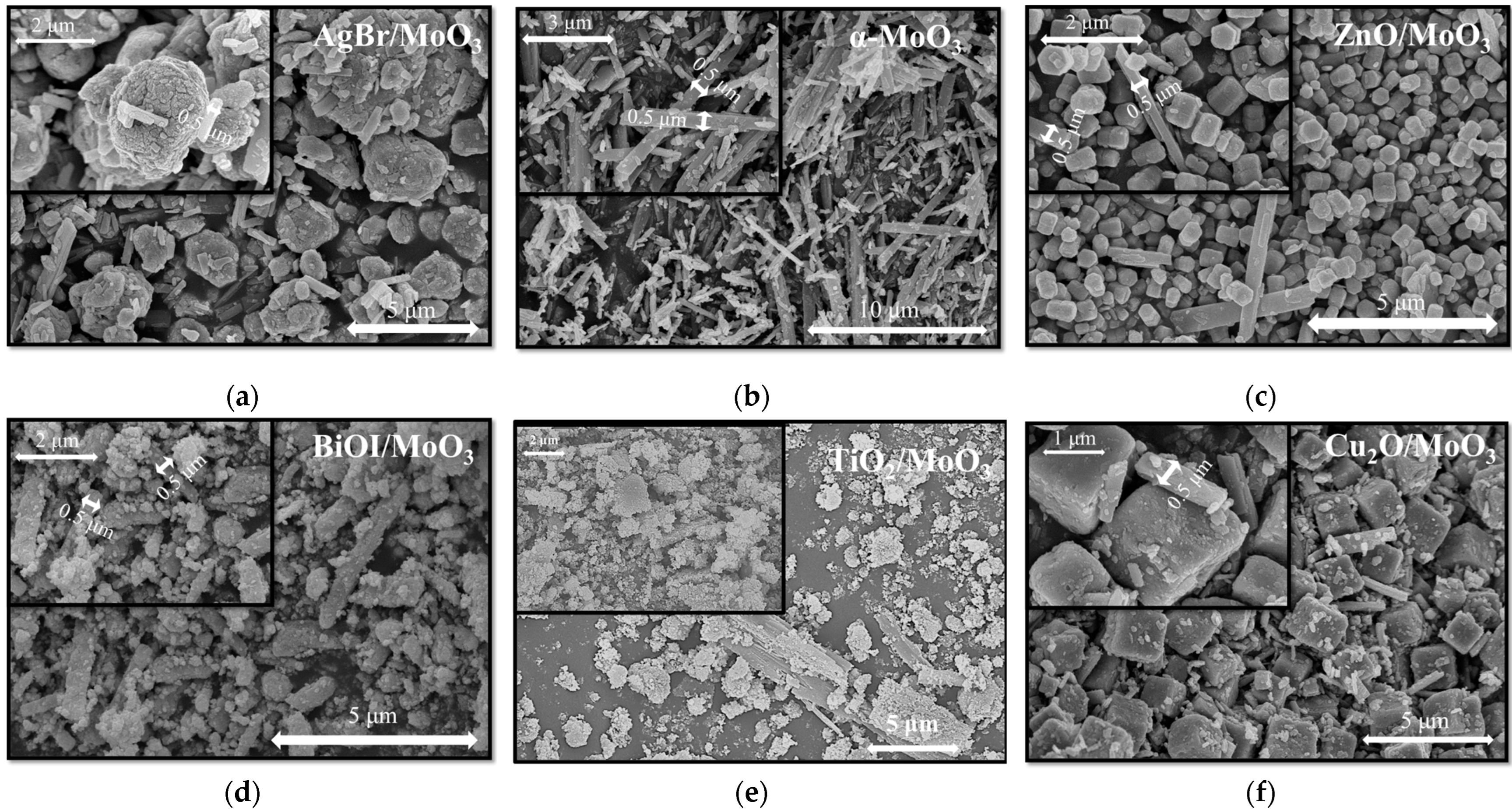
The as-prepared semiconductors and their composites were analyzed via XRD and SEM measurements to ascertain the reproducibility of photocatalysts and the presence of MoO3 in the composites. The structure of the as-prepared pristine photocatalysts was reproducible: TiO2 was obtained in the form of anatase [18], BiOI in tetragonal matlockite [19], ZnO in hexagonal wurtzite [5], AgBr [4], and Cu2O in cubic form [6] (Figure 1). The as-prepared MoO3 presented an orthorhombic phase with (021) a dominant crystallographic plane (Figure 1b). Despite the mechanical mixing, the reflections of α-MoO3 were faintly visible on the XRD patterns of the composites (it was the case of AgBr/MoO3, Cu2O/MoO3, and ZnO/MoO3, while in the case of BiOI/MoO3, no MoO3 reflections were noticed). However, the SEM micrographs showed that the characteristic α-MoO3 fibers were present in all of the composites (Figure 2). Based on these two structural characteristics, it was considered that the active photocatalysts were successfully reproduced and the fibrous α-MoO3 was present in these composites (the synthesis of α-MoO3 was carried out in such a way that the MoO3 particles will be crystalline—that is why 20 h was considered as the minimal crystallization time, which influences the quality of the final product of course [21]). Using the Scherrer equation, the crystallite sizes of the samples were estimated. Unfortunately, in the composites, the peaks corresponding to the MoO3 phase were not suitable for the calculation, but the other photocatalysts showed strong, intensive peaks. The crystallite size of the other component semiconductors showed no significant difference compared to the pristine phase. Hence the calculation results are as follows; TiO2—14.2 nm, AgBr—36.8 nm, ZnO—38.0 nm, and BiOI—14.3 nm, while for Cu2O, the calculation was not possible due to its microcrystalline structure. This indicates that the mechanical mixing was successful and induced no deviation in the crystal structure.
The SEM micrographs (Figure 2) also revealed the size and arrangement of the crystals. In each of the composite samples, the diameter of the MoO3 rods was preserved (~0.5 µm, Figure 2b shows the pristine MoO3). The AgBr particles appeared (Figure 2a) as hierarchical structures with variable sizes (from 1 up to 5 µm), while the shape of the particles was random. In the case of the ZnO-base composite, the MoO3 particles were larger than ZnO (which were smaller than 400 nm, Figure 2c). In the case of BiOI and TiO2, the particles were randomly aggregated and covered, in most cases, the available MoO3 nanorods (Figure 2d,e). No clear size evaluation or particle size distribution can be carried out. The situation is quite different in the case of Cu2O. The crystals were cubes (1–2.5 µm), which were alongside the MoO3 rods (Figure 2f).
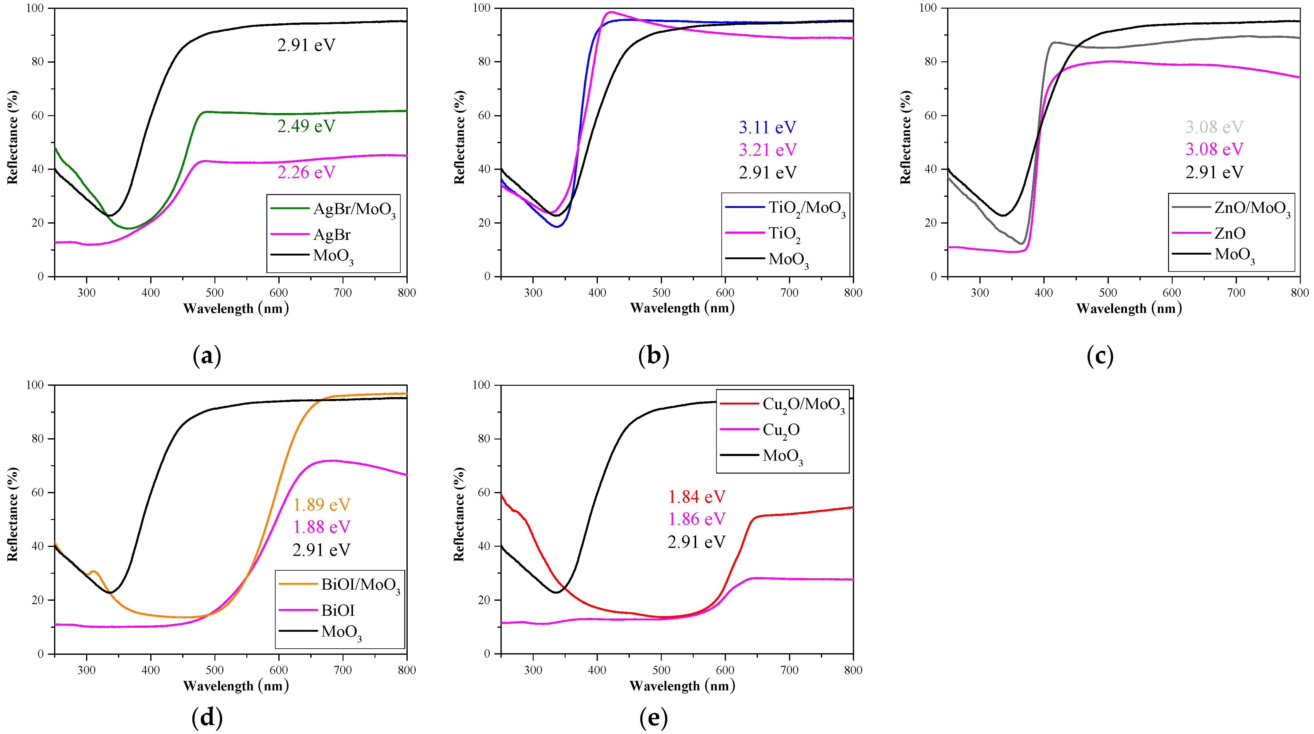
Based on the measured optical properties (Figure 3) and our previous work, TiO2 and ZnO should exhibit photocatalytic activity in the UV range and Cu2O, AgBr, and BiOI in the visible range. The calculated band gap value for α-MoO3 (2.91 eV) suggested that it is a promising visible-light active photocatalyst. In composites, the presence of α-MoO3 did not or only slightly affected the band gap values of the pristine photocatalysts (Figure 3). The band gap value of TiO2 was shifted by 0.1 eV toward the visible range, while in the case of ZnO, it did not change at all. The other three photocatalysts presented an increased reflectance in the visible region; however, the calculated band gap for AgBr increased by 0.23 eV, and for BiOI and Cu2O, the change was irrelevantly small (<0.05 eV) (Figure 3). The first derivative method is more advised to determine the band-gap energies of composite materials because it does not generalize the spectral values and could reveal the band-gap values of the constituent phases. However, in the present case, the values showed no significant difference compared to the Kubelka–Munk method, and the peak corresponding to MoO3 was not observable in the derivative spectra. This could also be explained by the small percentage of the MoO3 phase in the composites.
Photocatalysis is based on the utilization of charge carriers: the organic molecule can be oxidized, whether directly via holes, or by different types of radicals (⋅O2, ·OH). Depending on the MO concentration its photocatalytic decomposition is a very common example of both hole (>1.6 × 10⁻⁴ M) and hydroxyl oxidation (<1.6 × 10⁻⁴ M) [22], while phenol degradation is driven by hydroxyl radicals to form intermediate hydroxylated compounds—such as catechol, benzoquinone, and other compounds [23].
The photocatalytic efficiency values are presented in Figure 4. Pristine photocatalysts reached higher activity in the degradation of methyl orange compared to phenol, except in the case of TiO2 (Figure 4). Higher photocatalytic conversion for MO could be achieved because its degradation mechanism consists of both radicals (hydroxyl and superoxide radicals) and photogenerated holes. Anatase TiO2 and ZnO were active only in UV due to their optical properties. Cu2O presented outstanding activity in the degradation of MO in visible light (92% conversion), while in UV, adsorption was predominant (Figure S2). Cu2O was not active in the photodegradation or adsorption of phenol. The literature also presents that Cu2O is both a visible-light-active photocatalyst and an adsorbent for MO [24,25]. BiOI and AgBr, independently from the light source, presented stable activity in MO decomposition. However, phenol degradation reached higher conversion under UV illumination (Figure 4).
In the present work, the motivation was to assess the effect of α-MoO3 upon the above-presented pristine photocatalysts and their activity. We demonstrated before that α-MoO3 reduces the photocatalytic activity of TiO2, although band gap narrowing or the electron–hole recombination suppression might occur [1]. Our current results confirm that not only TiO2 activity but the activity of photocatalysts (ZnO, BiOI, AgBr, and Cu2O) can be hindered in the presence of α-MoO3. As it was mentioned before, inhibition might occur due to the formation of HMoO4 and MoO42− anions, and those may function as hole scavengers, such as HPO42− and CH3COO [16]. The presence of MoO42− anions should induce a pH drop. However, an acidic medium does not affect the degradation rate of MO via TiO2. Its apparent rate constant is unchanged between pH 3 and 8 [17].
The mentioned Mo-species (HMoO4 and MoO42− anions) may be pretty challenging to follow under the current circumstances as the equilibrium constant (or the pKa values: pKa1 = 3.61–4.0 and pKa2 = 3.89–4.37, [11]) for the hydrolysis of MoO3 suggests that the solubilization is not the favored reaction. This means that a smaller (nM or a few µM) concentration of Mo species can always be found in the solution. However, if hydrolysis is the dominant process, MoO3 would be dissolved entirely. Furthermore, during the hole-scavenging process, after the electron transfer, MoO3 may be re-deposited. Hence the presence of HMoO4 and MoO42− is transitory and challenging to follow. Moreover, the activity decrease can occur due to the OH radical scavenging effect of MoO3 [26].
Light shielding via MoO3 might be another reason for inactivity. However, this scenario is less likely, because the amount of MoO3 used in the experiment was 10 wt.%, which is not much, considering that in the literature, sometimes nearly 50 wt.% of MoO3 (33 molar%) was used. Still, photoactivity was registered for these samples [12]. Further, when the influence of the MoO3 content was investigated on titania photocatalysts, it was found that the activity decrease was not linear with the MoO3 content increase [11]. Hence, the presence of α-MoO3 or its anions should be the cause that inactivates the photocatalysts.
In MO photodegradation, all the composite materials presented lower conversion than without α-MoO3 (Figure 4a, Figure S2). TiO2 and ZnO presented a ~50% conversion drop, which is particularly high as only 10% photocatalyst was substituted with α-MoO3. The photocatalytic activity of Cu2O completely disappeared, and the tendency to adsorb MO has ceased. Finally, BiOI conversion was least affected by α-MoO3; only 15–30% conversion drop was observed. The inhibition varied depending on the form of illumination. A more intense inhibition was observed during visible-light irradiation. On the contrary, the activity of AgBr in MO decomposition was hindered only in UV, though after 2 h illumination, the AgBr/MoO3 composites reached a similar conversion to pristine AgBr. An opposite behavior was observed when ZnO was analyzed. Under visible-light irradiation, the composite achieved ~10% conversion, while the bare ZnO was inactive. This suggests that, to some extent, MoO3 can be activated as well.
Phenol photocatalytic decomposition was also inhibited by α-MoO3 as the methyl orange decomposition (Figure 4b, Figure S3). ZnO and Cu2O photocatalytic activity or adsorption were negligible during phenol degradation. Hence their composite activity was not counted. TiO2 presented a ~40% conversion drop, similar to MO degradation. In the case of BiOI, the inhibition was higher (40–60% conversion drop) compared to MO photocatalytic tests. In both irradiation cases, BiOI photocatalytic activity was decreased in the presence of α-MoO3. The pollutant conversion was below ~10%, although, without MoO3, BiOI phenol decomposition reached 65.7% in UV and 43.4% in vis. Since all the other photocatalysts were inhibited by α-MoO3 regardless of the model pollutant or irradiation, as a surprising fact, phenol decomposition by AgBr slightly increased under UV or visible irradiation. Compared to the other photocatalysts, AgBr is known to be unstable during photocatalytic treatment. AgBrO3, Ag, and Ag2O can be formed on AgBr surfaces during the photocatalytic process [4]. The formation of these species might be the first cause of the MO and phenol degradation and not solely the photocatalysis, which can be hindered by α-MoO3. It should be noted that in the case of ZnO-based composites, a small amount of phenol degradation occurs under visible-light irradiation (~7%), emphasizing the issue raised for this composite in the case of MO degradation.
Light screening could be another issue for catalyst inactivation. However, this was not the case. The phenomenon requires a material that absorbs light intensively instead of the main catalyst. The simultaneous prerequisite conditions are a low band-gap energy, a significant difference (above 1.0 eV) between the band-gap values, and low (below 30%) reflectivity of the minor composite component (MoO3). Neither of these conditions is valid at the same time.

4. Conclusions

Our results suggested that α-MoO3, next to an active photocatalyst, predominantly acts as a photocatalytic inhibitor rather than an enhancement factor, as stated frequently in the literature. α-MoO3 inhibition was manifested by a decrease in the activity of the photocatalysts, whether MO or phenol was decomposed under visible or UV light. Only the activity of AgBr was unaffected, yet it is known that AgBr is unstable during photocatalysis. Therefore, not only photocatalysis occurs, but the formation of Ag compounds, which might oxidize the organic pollutants, and that cannot be inhibited by α-MoO3. We deduced that α-MoO3, or its anions, can inhibit photocatalytic processes, and its inhibition efficiency varies depending on the type of irradiation, the pollutant, and the photocatalyst. Moreover, these results point out that the current scientific community does not present those very important results in which a negative aspect is presented regarding a specific material. It should be noted that charge transfer inhibition and inactivation may influence other research areas besides photocatalysis (e.g., supercapacitive materials, sensor applications, energy storage devices). Hence appropriate documentation of negative phenomena should be considered. As α-MoO3 tends to transform in aqueous media, environmental aspects should also be considered, as Mo is a heavy metal, after all.

Supplementary Materials

The following supporting information can be downloaded at: https://www.mdpi.com/article/10.3390/ma16103621/s1, Figure S1. Photocatalytic degradation of MO with TiO2/MoO3 composites in different weight percentages under UV irradiation: (a) TiO2, (b) 96/4, (c) 90/10, and (d) 80/20; Figure S2. Methyl orange degradation curves under UV and visible irradiation: (a) AgBr, AgBr/MoO3, (b) TiO2, TiO2/MoO3, (c) ZnO, ZnO/MoO3, (d) BiOI, BiOI/MoO3, and (e) Cu2O, Cu2O/MoO3; Figure S3. Phenol degradation curves under UV and visible irradiation: (a) AgBr, AgBr/MoO3, (b) TiO2, TiO2/MoO3, (c) ZnO, ZnO/MoO3, (d) BiOI, BiOI/MoO3, and (e) Cu2O, Cu2O/MoO3.

Author Contributions

Conceptualization, E.-Z.K. and Z.P.; methodology, E.B., A.R., Z.-R.T., S.M. and Z.K.; software, E.-Z.K. and A.R.; investigation, A.R. and E.-Z.K.; data curation, E.-Z.K.; writing—original draft preparation, E.-Z.K. and Z.P.; writing—review and editing, E.-Z.K. and L.B.; supervision, L.B. and Z.P.; funding acquisition, Z.P and L.B. All authors have read and agreed to the published version of the manuscript.

Funding

This research was funded by the PN-III-P1-1.1-TE-2019-1318 project, provided by UEFISCDI (Executive Agency for Higher Education, Research, Development and Innovation Funding).

Institutional Review Board Statement

Not applicable.

Informed Consent Statement

Not applicable.

Data Availability Statement

The data presented in this study are available on request from the corresponding author. The data are not publicly available due to their implication in future industrial research.

Acknowledgments

Zsolt Pap expresses his gratitude towards the Hungarian Academy of Sciences, Bolyai János research scholarship.

Conflicts of Interest

The authors declare no conflict of interest.

References

  1. Kedves, E.-Z.; Pap, Z.; Hernadi, K.; Baia, L. Significance of the surface and bulk features of hierarchical TiO2 in their photocatalytic properties. Ceram. Int. 2021, 47, 7088–7100. [Google Scholar] [CrossRef]
  2. Yao, S.; Zhang, X.; Qu, F.; Umar, A.; Wu, X. Hierarchical WO3 nanostructures assembled by nanosheets and their applications in wastewater purification. J. Alloys Compd. 2016, 689, 570–574. [Google Scholar] [CrossRef]
  3. Bárdos, E.; Király, A.K.; Pap, Z.; Baia, L.; Garg, S.; Hernádi, K. The effect of the synthesis temperature and duration on the morphology and photocatalytic activity of BiOX (X = Cl, Br, I) materials. Appl. Surf. Sci. 2019, 479, 745–756. [Google Scholar] [CrossRef]
  4. Tóth, Z.-R.; Pap, Z.; Kiss, J.; Baia, L.; Gyulavári, T.; Czekes, Z.; Todea, M.; Magyari, K.; Kovács, G.; Hernadi, K. Shape tailoring of AgBr microstructures: Effect of the cations of different bromide sources and applied surfactants. RSC Adv. 2021, 11, 9709–9720. [Google Scholar] [CrossRef] [PubMed]
  5. Kovács, Z.; Molnár, C.; Štangar, U.L.; Cristea, V.-M.; Pap, Z.; Hernadi, K.; Baia, L. Optimization Method of the Solvothermal Parameters Using Box–Behnken Experimental Design—The Case Study of ZnO Structural and Catalytic Tailoring. Nanomaterials 2021, 11, 1334. [Google Scholar] [CrossRef]
  6. Fodor, S.; Baia, L.; Baán, K.; Kovács, G.; Pap, Z.; Hernadi, K. The Effect of the Reducing Sugars in the Synthesis of Visible-Light-Active Copper(I) Oxide Photocatalyst. Molecules 2021, 26, 1149. [Google Scholar] [CrossRef]
  7. Mavridi-Printezi, A.; Menichetti, A.; Guernelli, M.; Montalti, M. Extending photocatalysis to the visible and NIR: The molecular strategy. Nanoscale 2021, 13, 9147–9159. [Google Scholar] [CrossRef]
  8. Muhmood, T.; Uddin, A. Fabrication of spherical-graphitic carbon nitride via hydrothermal method for enhanced photo-degradation ability towards antibiotic. Chem. Phys. Lett. 2020, 753, 137604. [Google Scholar] [CrossRef]
  9. Muhmood, T.; Cai, Z.; Lin, S.; Xiao, J.; Hu, X. Dimensions controllable synthesis of silver Nano-morphologies via moderate one step methodology. Adv. Powder Technol. 2021, 32, 3388–3394. [Google Scholar] [CrossRef]
  10. Lam, S.-M.; Sin, J.-C.; Abdullah, A.Z.; Mohamed, A.R. Investigation on visible-light photocatalytic degradation of 2,4-dichlorophenoxyacetic acid in the presence of MoO3/ZnO nanorod composites. J. Mol. Catal. A Chem. 2013, 370, 123–131. [Google Scholar] [CrossRef]
  11. Kedves, E.-Z.; Bárdos, E.; Gyulavári, T.; Pap, Z.; Hernadi, K.; Baia, L. Dependence of cationic dyes’ adsorption upon α-MoO3 structural properties. Appl. Surf. Sci. 2022, 573, 151584. [Google Scholar] [CrossRef]
  12. Diniz, J.; Nunes, C.D.; Monteiro, O.C. Novel approach to synthesise MoO3-TiO2 nanocomposites for the photo-assisted oxidation of benzyl alcohol to benzaldehyde. Inorg. Chem. Commun. 2020, 119, 108099. [Google Scholar] [CrossRef]
  13. Selvakumar, G.; Palanivel, C. A study on synthesis, characterization and catalytic applications of MoO3-ZnO nanocompositematerial. Mater. Sci. Energy Technol. 2022, 5, 36–44. [Google Scholar] [CrossRef]
  14. Feng, B.; Wu, Z.; Liu, J.; Zhu, K.; Li, Z.; Jin, X.; Hou, Y.; Xi, Q.; Cong, M.; Liu, P.; et al. Combination of ultrafast dye-sensitized-assisted electron transfer process and novel Z-scheme system: AgBr nanoparticles interspersed MoO3 nanobelts for enhancing photocatalytic performance of RhB. Appl. Catal. B Environ. 2017, 206, 242–251. [Google Scholar] [CrossRef]
  15. Huan, C.; Wang, P.; He, B.; Cai, Y.; Ke, Q. Oxygen deficient α-MoO3 with enhanced adsorption and state-quenching of H2O for gas sensing: A DFT study. J. Mater. Chem. C 2022, 10, 1839–1849. [Google Scholar] [CrossRef]
  16. Minubayeva, Z.; Seward, T.M. Molybdic acid ionisation under hydrothermal conditions to 300 °C. Geochim. Cosmochim. Acta 2010, 74, 4365–4374. [Google Scholar] [CrossRef]
  17. Barka, N.; Assabbane, A.; Nounah, A.; Dussaud, J.; Ait Ichou, Y. Photocatalytic degradation of methyl orange with immobilized TiO2 nanoparticles: Effect of pH and some inorganic anions photocatalytic degradation of methyl orange with immobilized tio 2 nanoparticles: Effect of ph and some inorganic anions. Artic. Phys. Chem. News 2008, 41, 85–88. [Google Scholar]
  18. Kedves, E.-Z.; Székely, I.; Baia, L.; Baia, M.; Csavdári, A.; Pap, Z. The Comparison of the Photocatalytic Performance Shown by TiO2 and TiO2/WO3 Composites—A Parametric and Kinetic Study. J. Nanosci. Nanotechnol. 2018, 19, 356–365. [Google Scholar] [CrossRef]
  19. Bárdos, E.; Márta, V.; Baia, L.; Todea, M.; Kovács, G.; Baán, K.; Garg, S.; Pap, Z.; Hernadi, K. Hydrothermal crystallization of bismuth oxybromide (BiOBr) in the presence of different shape controlling agents. Appl. Surf. Sci. 2020, 518, 146184. [Google Scholar] [CrossRef]
  20. Roy Choudhury, A.K. 4-Instrumental Colourant Formulation; Roy Choudhury, A.K., Ed.; Woodhead Publishing: Oxford, UK, 2015; pp. 117–173. ISBN 978-1-78242-367-6. [Google Scholar]
  21. Muhmood, T.; Xia, M.; Lei, W.; Wang, F. Under vacuum synthesis of type-I heterojunction between red phosphorus and graphene like carbon nitride with enhanced catalytic, electrochemical and charge separation ability for photodegradation of an acute toxicity category-III compound. Appl. Catal. B 2018, 238, 568–575. [Google Scholar] [CrossRef]
  22. Yu, L.; Xi, J.; Li, M.-D.; Chan, H.T.; Su, T.; Phillips, D.L.; Chan, W.K. The degradation mechanism of methyl orange under photo-catalysis of TiO2. Phys. Chem. Chem. Phys. 2012, 14, 3589–3595. [Google Scholar] [CrossRef] [PubMed]
  23. Dang, T.T.T.; Le, S.T.T.; Channei, D.; Khanitchaidecha, W.; Nakaruk, A. Photodegradation mechanisms of phenol in the photocatalytic process. Res. Chem. Intermed. 2016, 42, 5961–5974. [Google Scholar] [CrossRef]
  24. Dan, Z.; Yang, Y.; Qin, F.; Wang, H.; Chang, H. Facile Fabrication of Cu2O Nanobelts in Ethanol on Nanoporous Cu and Their Photodegradation of Methyl Orange. Materials 2018, 11, 446. [Google Scholar] [CrossRef] [PubMed]
  25. Prado-Chay, D.A.; Cortés-Jácome, M.A.; Angeles-Chávez, C.; Oviedo-Roa, R.; Martínez-Magadán, J.M.; Zuriaga-Monroy, C.; Hernández-Hernández, I.J.; Mayoral, P.R.; Gómora-Herrera, D.R.; Toledo-Antonio, J.A. Synthesis and Photocatalytic Activity of Cu2O Microspheres upon Methyl Orange Degradation. Top. Catal. 2020, 63, 586–600. [Google Scholar] [CrossRef]
  26. Kedves, E.-Z.; Fodor, C.; Fazekas, Á.; Székely, I.; Szamosvölgyi, Á.; Sápi, A.; Kónya, Z.; Cristian Pop, L.; Baia, L.; Pap, Z. α-MoO3 with inhibitive properties in Fenton reactions and insights on its general impact on OH radical based advanced oxidation processes. Appl. Surf. Sci. 2023, 624, 156914. [Google Scholar] [CrossRef]
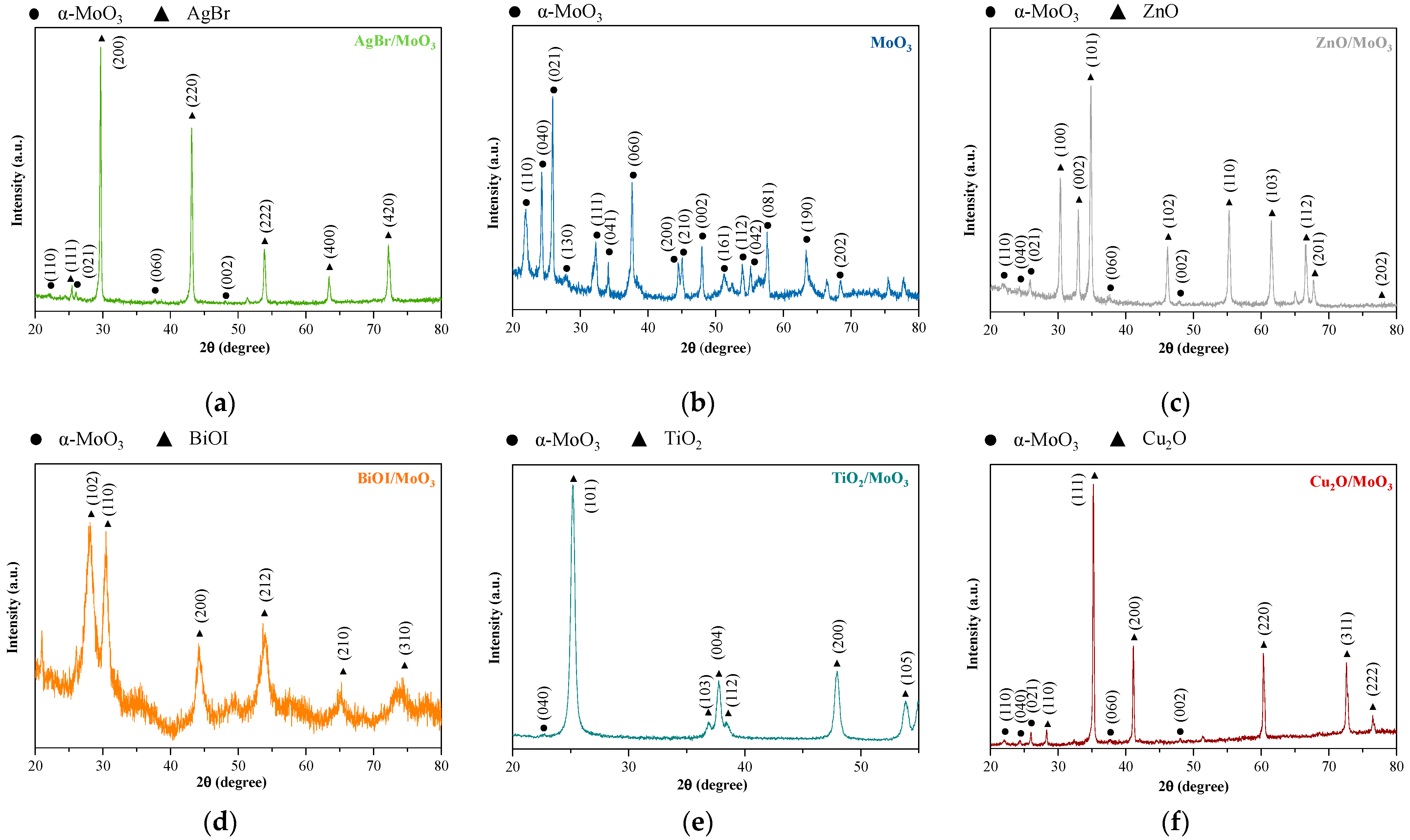
Figure 1. X-ray diffractograms of the as-prepared composites: (a) AgBr/MoO3, (b) α-MoO3, (c) ZnO/MoO3, (d) BiOI/MoO3, (e) TiO2/MoO3, and (f) Cu2O/MoO3 composites’ XRD patterns.
Materials 16 03621 g001
Figure 2. Micrographs of the composites: (a) AgBr/MoO3, (b) α-MoO3, (c) ZnO/MoO3, (d) BiOI/MoO3, (e) TiO2/MoO3, and (f) Cu2O/MoO3.
Materials 16 03621 g002
Figure 3. DRS spectra and their respective band gap values: (a) AgBr/MoO3, (b) TiO2/MoO3, (c) ZnO/MoO3, (d) BiOI/MoO3, and (e) Cu2O/MoO3.
Materials 16 03621 g003
Figure 4. The photocatalytic degradation of (a) methyl orange and (b) phenol via the pristine photocatalysts and the as-prepared α-MoO3-containing composites in UV and visible irradiation.
Materials 16 03621 g004
Disclaimer/Publisher’s Note: The statements, opinions and data contained in all publications are solely those of the individual author(s) and contributor(s) and not of MDPI and/or the editor(s). MDPI and/or the editor(s) disclaim responsibility for any injury to people or property resulting from any ideas, methods, instructions or products referred to in the content.