Article

Structure and Magnetic Properties of SrFe12−xInxO19 Compounds for Magnetic Hyperthermia Applications

by
Polina I. Nikolenko
,
Timur R. Nizamov
,
Igor G. Bordyuzhin
,
Maxim A. Abakumov
,
Yulia A. Baranova
,
Alexander D. Kovalev
and
Igor V. Shchetinin
*
Department of Physical Materials Science, University of Science and Technology MISIS, Leninsky Prospekt 4s1, 119049 Moscow, Russia
*
Author to whom correspondence should be addressed.
Materials 2023, 16(1), 347; https://doi.org/10.3390/ma16010347
Submission received: 25 November 2022 / Revised: 26 December 2022 / Accepted: 27 December 2022 / Published: 30 December 2022

Abstract

:
In this work, complex studies of the structure and magnetic properties of SrFe12−xInxO19 powders obtained by the mechanochemical and citrate methods were carried out. The solubility of In in strontium hexaferrite SrFe₁₂O₁₉ at 1200 °C was determined. The structure and properties of the powders were studied using X-ray diffraction analysis, Mössbauer spectroscopy and scanning electron microscopy. Measurements of magnetic properties in magnetic fields up to 1600 kA/m were also performed. Additionally, the hyperthermia effect was investigated. The possibility of controlling the coercivity of the samples in the range from 188.9 kA/m to 22.3 kA/m and saturation magnetization from 63.5 A·m2/kg to 44.2 A·m2/kg with an increase in the degree of In doping was also demonstrated. Investigation of the magnetic hyperthermia of the samples was carried out by temperature measurement with an IR camera when they were introduced into alternating magnetic fields of various frequencies (144, 261 and 508 kHz) and amplitudes (between 11.96 and 19.94 kA/m). According to the study result, there was detected the heating of the SrFe12−xInxO19 sample (x = 1.7). The highest values of magnetic hyperthermia of the sample were observed in a 19.94 kA/m magnetic field and a frequency of 261 kHz. At a concentration of 56.67 g/L, the sample was heated from 23 to 41 °C within 2 min. The parameters SLP (specific loss power) and ILP (intrinsic loss power) were calculated.

1. Introduction

Hard magnetic ferrites with a magnetoplumbite structure have been known since the middle of the 20th century [1]. From the moment of discovery to the present day, due to the unique combination of properties, hexaferrites have been widely used as materials for permanent magnets [2], microwave technology [3,4], information carriers [5,6], magneto-optics [7,8], absorbing materials [9], electronics and telecommunications [10], etc. Hexagonal ferrites can be obtained by a variety of methods such as ceramic synthesis [2], mechanochemical [11,12,13], co-precipitation [14,15,16], crystallization of oxide glass [17,18], hydrothermal [19,20,21], from salt melt [22], sol-gel [23,24], etc.
Other iron-based oxide materials, such as magnetite, maghemite and hematite, have been well studied in the literature for biomedicine applications as contrast agents for MRI [25,26,27], local hyperthermia [28,29], targeted drug delivery [30], in magnetic sensors and magnetic refrigerants [31]. Hexaferrites also belong to oxide materials and, in comparison with iron oxides, have a number of advantages [32,33,34,35]. At present time, the attention of scientists is attracted to research related to the biomedical applications of hexaferrites for diagnostics and therapy, and, in particular, for hyperthermia. There are studies that testify to the non-toxic nature of hexaferrites. Thus, it was demonstrated in [36] that the implantation of barium hexaferrite in tumors of tumor-bearing mice does not have a toxic effect on most organs.
Most of the research related to the magnetic hyperthermia is associated with stable colloidal suspensions of magnetic nanoparticles. Strontium hexaferrites SrFe12O19 are among the few hard magnetic materials based on nanoparticles of which it was possible to obtain stable colloidal solutions. The possibility of obtaining colloidal solutions of strontium hexaferrite was demonstrated in the studies [18,37,38]. This body of evidence indicates the potential of strontium hexaferrites as agents for magnetic hyperthermia in biomedical applications.
There are the following two main mechanisms for heating magnetic particles: hysteresis losses and relaxation. In multidomain particles, the main heating mechanism is hysteresis losses due to magnetization reversal. In single-domain particles, the main mechanism is relaxation, which can take place via the following two pathways: Neel relaxation is the rotation of magnetic moments inside the domain; Brown’s relaxation is the rotation of the body of the nanoparticle in the liquid.
The efficiency of magnetic particle heating Increases with higher frequency and amplitude of the applied magnetic field. However, there are restrictions on the maximum allowable safe amplitude-frequency product of the field. This is due to eddy currents, which, when using alternating magnetic fields, occur in all tissues, both affected and healthy. The estimated maximum values of the amplitude-frequency product, according to the Brezovich constraint, are given at the level of H0f = 4.85 × 10⁸ [39].
In high-frequency fields with an admissibly low amplitude, hard magnetic materials with a high coercivity experience have almost no hysteresis losses and do not emit heat [40]. In this regard, the study of the doping effect on the formation of the structure and properties of strontium hexaferrites in order to adapt and use for hyperthermia is an urgent task.

2. Materials and Methods

In this work, the mechanochemical method was used to study the solubility limit of In in the SrFe12O19 compound and the compositions and the citrate method was used to synthesize samples to study the functional properties.
Strontium carbonate SrCO3 (99.9%) and Fe2O3 (99.9%), In2O3 (99.9%) oxides were used for the mechanochemical method, where they were mixed in certain quantities to obtain a hexaferrite phase, according to the following chemical reaction:
2 SrCO3 + (12–− x) Fe2O3 + x In2O3 → 2 SrFe12−xInxO19 + 2 CO2, where x = 0, 0.25, 0.5, 1, 3.
The amount of SrCO3 powder was between 1.43 and 1.22 g, the amount of Fe2O3 varied from 18.57 to 11.89 g and the mass of In2O3 was taken from 0 to 6.89 g. Total mass of the powder mixture per vial was 20 g. Then the powder mixtures were subjected to mechanochemical synthesis in an «Activator 2S» (Novosibirsk, Russia) planetary ball mill for 3 h at a rotation speed of 200/400 rpm (disk/vials). The ball-to-powder ratio was 1:10; steel balls and steel vials were used for milling. After mechanochemical synthesis the samples were annealed at 1200 °C for 1 h. The solubility limit was determined by changing the lattice spacings and the unit cell volume of the SrFe12−xInxO19 compound.
Aqueous solution of Sr(NO3)2, Fe(NO3)3 and In(NO3)3 was used SrFe12−xInxO19 for the citrate method synthesis. Then water was evaporated at 95 °C and the annealing was performed at 1200 °C (1 h in air).
The X-ray diffraction (XRD) phase analysis of SrFe12−xInxO19 compounds was carried out on a Rigaku Ultima IV (Tokyo, Japan) diffractometer with CoKα radiation and a graphite monochromator on a diffracted beam. XRD studies were carried out on powders that were placed into round cuvettes, and their surface was aligned with glass. The real degree of alloying of the samples obtained by the citrate method was determined from the change in the lattice spacings of the SrFe12−xInxO19 phase. The particle morphology of the samples was studied by scanning electron microscopy (SEM) in secondary electrons using a JEOL JSM-IT500 (Tokyo, Japan) electron microscope. The magnetic properties of the samples were measured on a VSM-250 (Changchun, China) vibrating sample magnetometer in fields up to 1600 kA/m. The powder samples were placed into copper cylinders, which were filled with paraffin. Then they were attached to a VSM rod vibrating during the measurements in an external magnetic field between the poles of electromagnet (in opened magnetic flux).
The study of magnetic hyperthermia of the samples was carried out in an aqueous medium with a small addition of sodium citrate (100 μL of 5% sodium citrate solution per 2 mL of water). The heating of the samples after applying the alternating magnetic field into the chamber was recorded by an infrared camera. Based on the study results, the SLP (specific loss power) and ILP (intrinsic loss power) parameters were calculated. The calculation was carried out in accordance with expressions (1) and (2) as follows:
S L P = m s o l m p a r t · Δ T Δ t · C
where SLP is the specific power loss in W/g, msol is the mass of the sol in kg, mpart is the mass of particles in g, C is the heat capacity of the medium (for water 4183 J/(kg·K)), ∆T is the change of temperature in °C or K, ∆t—time in s.
I L P = S L P · ( f H 2 )
where ILP is the intrinsic power loss in (nH·m2)/kg, SLP is the specific power loss in W/kg, f is the frequency of the magnetic field in kHz, H is the field strength in kA/m.

3. Results and Discussion

Samples of SrFe12−xInxO19, where x = 0, 0.25, 0.5, 1, 3 (at. In per formula unit), were obtained by mechanochemical synthesis and annealed at 1200 °C for 1 h. The XRD spectra and results of its analysis are presented in Figure 1 and Table 1.
According to Table 1, a graph of changes in hexaferrite lattice spacings was plotted as a function of the In doping degree (Figure 2). The phase lattice spacings change in the single-phase area, and upon transition to the two-phase/multiphase region, they become constant. Since in the two-phase/multiphase region a mixture of phases of the limiting composition is in equilibrium. Indium alloying leads to an increase in the lattice spacings and cell volume (Figure 2). The lattice spacings of the solid solution increase up to x ≈ 2.1 and reach a constant level above x ≈ 2.1. The cell volume of the phase behaves similarly with an increase in the atomic percentage of indium per formula unit. Since the In3+ ion radius (94 pm) is greater than the Fe3+ radius (69 pm) [41], the dissolution of In in the SrFe12−xInxO19 phase should lead to an increase in the lattice spacing and cell volume, which is observed in Figure 2. The point of intersection of the rising line and the constant value from the two-phase area corresponds to the maximum solubility of In in the SrFe12−xInxO19 phase at a temperature of 1200 °C. As shown in Figure 2 the maximum solubility of In in the SrFe12−xInxO19 phase at 1200 °C is x ≈ 2.1.
According to Mössbauer spectroscopy (Figure 3, Table 2), the spectrum of non-alloyed strontium hexaferrite shows five sextets corresponding to five nonequivalent positions of iron in the structure. In the hexaferrite structure, iron ions occupy the following five non-equivalent sites: three of them are octahedral (12k, 2a and 4f2), one is tetrahedral (4f1) and one is trigonal bipyramidal (2b). Alloying of SrFe12−xInxO19 phase with indium leads to a change in the superfine parameters of the sextets (Table 2) and the appearance of the sixth sextet 12k’ (yellow), the share of which in the total spectrum increases with increasing degree of doping of the phase, and the share of the 12k (red) sextet in the total spectrum decreases, and their sum remains at the 50% level. The appearance of the 12k’ (yellow) sextet can be associated with a change in the immediate environment in the 12k (red) position due to the occupation of this position by indium. Moreover, when doping with indium, a decrease in the parameters of the hyperfine magnetic field of the sextets is observed, which may be associated with a decrease in the magnetocrystalline anisotropy constant [42].
Estimating the Mössbauer parameters in Table 2, it can be noted that there are the changes in the isomeric shift (IS) of the Fe3+ ions for all positions (sextets 1–6). The results of the analysis show high IS values for the Fe3+ ions in the octahedral positions 12k, 4f2 and 2a (IS = 0.33–0.46 mm/s) compared to the tetrahedral 4f1 (IS = 0.15–0.26 mm/s). This can be explained by the greater covalence of bonds between iron ions due to the smaller volume of 4f1 polyhedra. In this case the quadrupole splitting QS of the Fe3+ ions in the 12k’ and 2b positions increases due to the larger deviation of the symmetry from the ideal state.
The obtained data are in good agreement with the results of the works [41,43]. Since chemical synthesis can lead to heterogeneity, and the activity of the components is different, the resulting linear dependence (Figure 2) was used to determine the real In content in the SrFe12−xInxO19 phase obtained by the citrate method. The citrate method was used to synthesize samples in a wide range of indium concentrations (x = 0–2), samples C1–C4 (Table 3). Samples C1–C3 with the degree of Indium alloying x ≤ 1 corresponded to the composition of the charge, and in sample C4 an underestimated content of indium was observed, which is associated with the formation of the In2O3 phase. The results of the phase analysis of SrFe12−xInxO19 samples obtained by the citrate method and annealed at 1200 °C for 1 h are presented in Table 3 and Figure 4. The presented data confirm that the synthesized samples contain the SrFe12−xInxO19 phase.
By the XRD analysis data, the samples obtained by the citrate method with x = 0–1.0 were in a single-phase state. Additionally, there were small XRD peaks observed in the sample with x = 1.7, coinciding with the position of the In2O3 peaks. The estimated amount of In2O3 was 2%.
The obtained photographs of the particles of SrFe12−xInxO19 samples C2 (x = 0.5), C3 (x = 1.0) and C4 (x = 1.7) are shown in Figure 5. According to the SEM data, it can be seen that the particles have a size of 0.5–5 µm. Figure 5 shows that at low doping levels C2 and C3 (x = 0.5 and x = 1.0), the average particle size does not generally exceed 2.5 µm, while in the photo of the C4 (x = 1.7) sample, the particle size is at the level of 5 µm, which is due to influence of In and is consistent with the results of the work [43]. Moreover, it can be suggested by the XRD spectra analysis that with an increase in the degree of doping by indium, an increase in lattice microstrain occurs. This fact can be associated with the appearance of defects in the crystal structure and also with the effective increase in the diffusion coefficient.
The magnetic hysteresis loops and the magnetic parameters of the SrFe12−xInxO19 samples obtained by the citrate method are shown in Figure 6 and Table 4. Hysteresis loops show that alloying with In leads to a fast decrease in the coercivity to 22.3 kA/m and magnetization to 42.95 A·m2/kg. This behavior of the magnetic properties is due to the fact that In generally occupies the 12k position, which, along with the 2b position, is responsible for the magnetocrystalline anisotropy constant [43,44], which leads to a decrease in the magnetocrystalline anisotropy constant and of the coercivity, respectively. Moreover, the coercivity decrease can be due to the grain growth of the SrFe12−xInxO19 phase, which is observed in a highly alloyed sample C4 (x = 1.7). In the structure of strontium hexaferrite, the magnetic moments of the Fe3+ ions in positions 2a, 2b and 12k are directed ferromagnetically, and in positions 4f1 and 4f2—antiferromagnetically [2]. When the nonmagnetic In3+ ion substitutes the Fe3+ in the 12k position, the total ferromagnetic moment in the cell decreases as well as the magnetization of the whole sample [43]. It should be noted that In doping makes it possible to control the magnetic properties of the samples over a wide range. For use materials in hyperthermia, the coercivity must be less than the amplitude of the high-frequency field to emit the remagnetization losses in the form of heat.
SrFe12−xInxO19 samples C2 (x = 0.5) and C3 (1.0), did not experience visible heating when introduced into a low-amplitude alternating magnetic field. For the sample with the lowest coercivity, SrFe12−xInxO19 C4 (x = 1.7), when introduced into an alternating magnetic field evident heating was observed. Sample temperature changes depending on the experiment time are shown in Figure 7. Video of these sample temperature changes vs time can be accessible in Supplementary materials to this paper. The results of measured and calculated SLP and ILP parameters for this sample are shown in Table 5. Indium alloying leads to a decrease in the coercivity to the level of high-frequency field amplitude used for medical purposes. This causes a reversal magnetization of the particles by these fields and heat generation. The obtained values correspond to the level of properties of the materials researched at the present time [45,46].

4. Conclusions

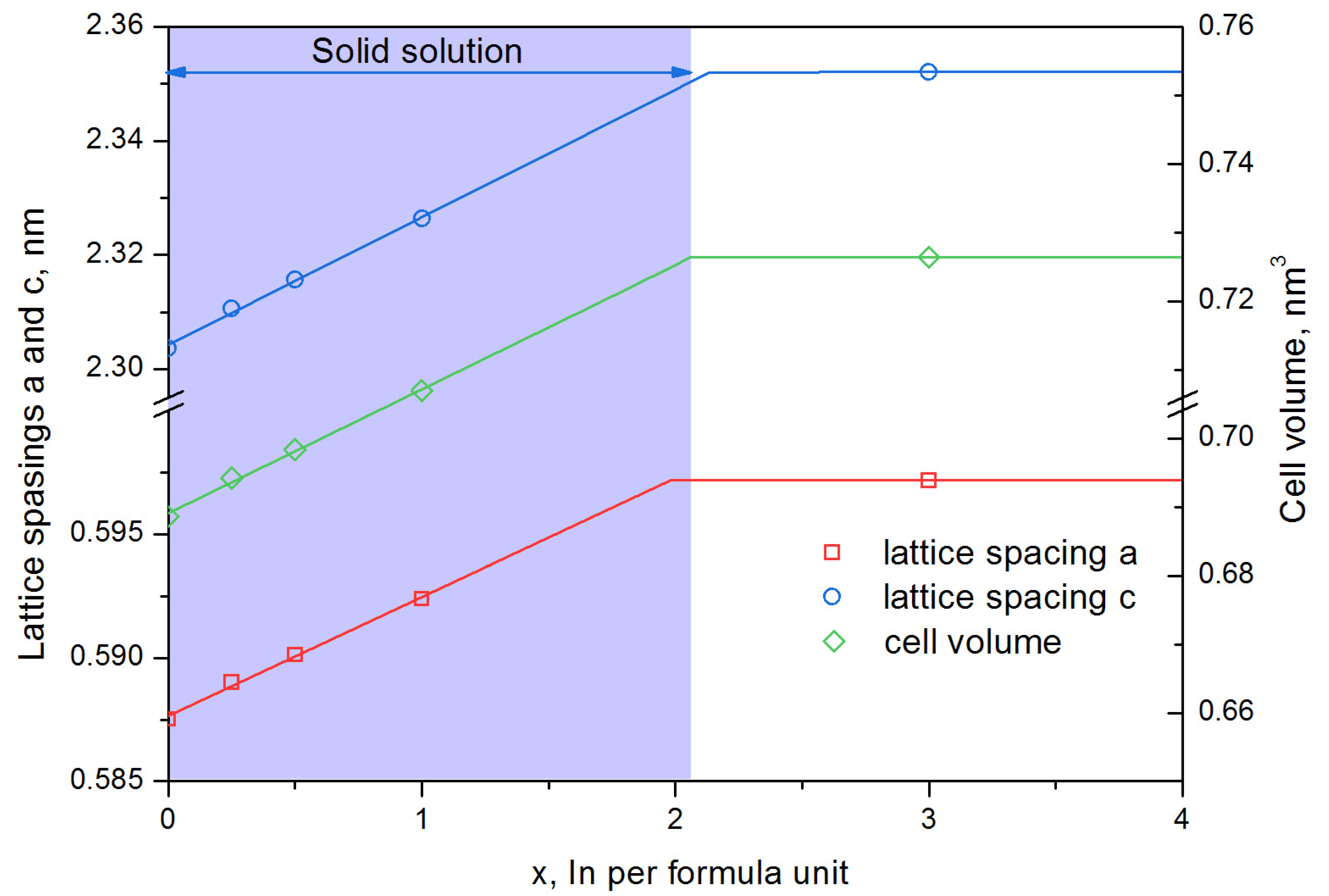
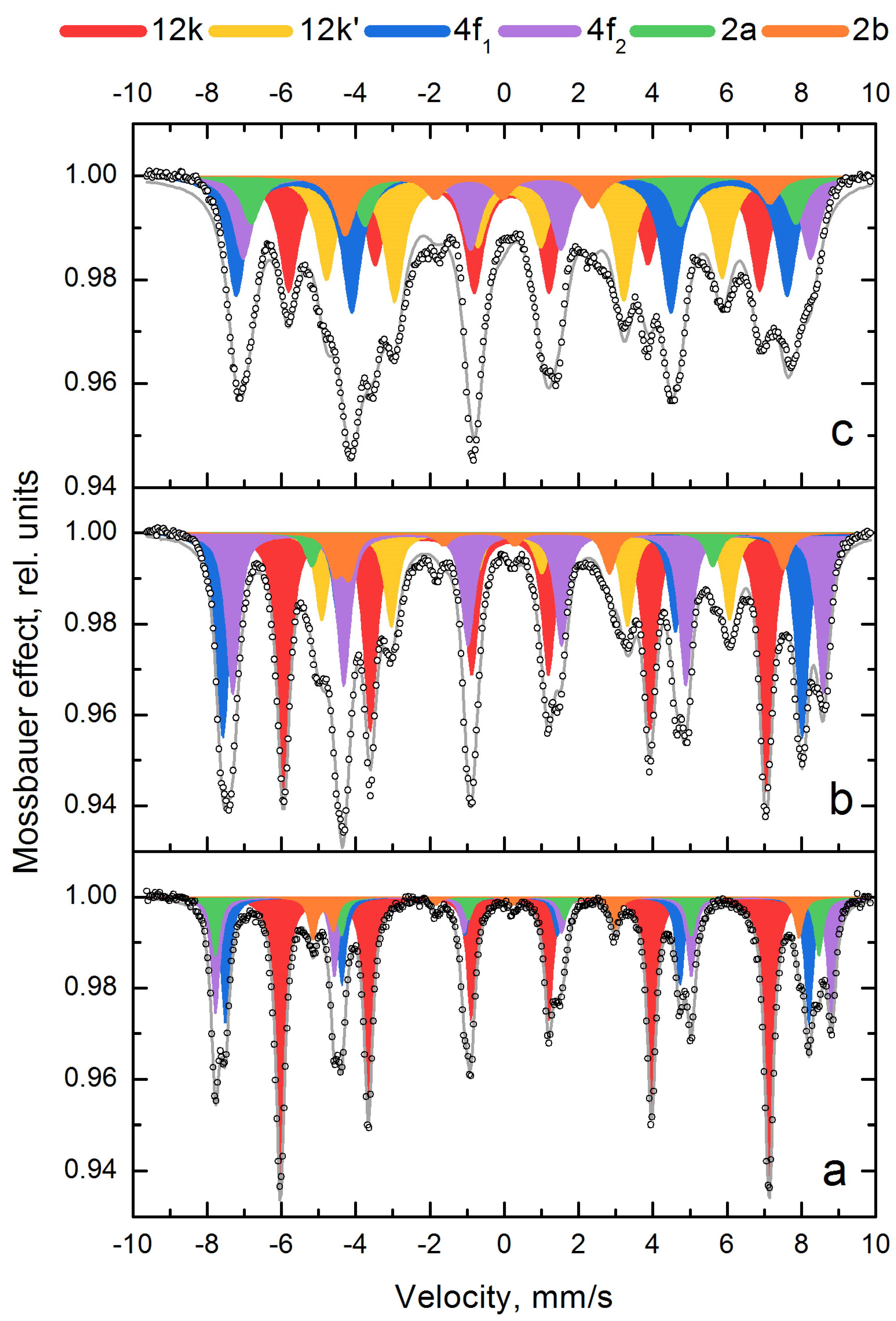
  • The solubility limit of In in hexaferrite SrFe12O19 at 1200 °C was determined—it reaches to x = 2.1 In atoms per hexaferrite formula unit. According to Mössbauer spectroscopy data, In predominantly occupies the 12k positions;
  • Using the citrate method and subsequent annealing at 1200 °C, the SrFe12−xInxO19 powders with x = 0.5, x = 1.0 and x = 1.7 were obtained. All synthesized samples contained at least 97% of the SrFe12−xInxO19 phase. Sample particles had sizes in the range of 0.5–5 µm and were collected in agglomerates. As the degree of doping increases, an increase in the average particle size is observed;
  • As the degree of substitution of iron by indium increases from x = 0.5 to x = 1.7, the coercivity of the samples strongly decreases from 188.9 kA/m to 22.3 kA/m. The specific magnetization decreases from 63.5 A·m2/kg to 44.2 A·m2/kg;
  • SrFe12−xInxO19 sample with x = 1.7 experienced evident heating when introduced into an alternating magnetic field. The best results of magnetic hyperthermia of the sample were observed at a field strength of 19.94 kA/m and a frequency of 261 kHz (at a high sample concentration (56.67 g/L), heating from 23 to 41 °C was achieved in 2 min).

Supplementary Materials

The following supporting information can be downloaded at: https://www.mdpi.com/article/10.3390/ma16010347/s1. Video S1: Sample temperature changes depending on the experiment time.

Author Contributions

P.I.N. investigation and synthesis; T.R.N. visualization and Mössbauer spectroscopy; I.G.B. synthesis and magnetic properties investigations; M.A.A. investigation of the magnetic hyperthermia; I.V.S. writing—original draft preparation and XRD investigations; Y.A.B. investigation and validation; A.D.K. Formal analysis, Investigation. All authors have read and agreed to the published version of the manuscript.

Funding

This work was supported by the Ministry of Science and Higher Education of the Russian Federation within the framework of the strategic academic leadership program “Priority 2030” of the University of Science and Technology MISIS, project № K1-2022-035.

Institutional Review Board Statement

Not applicable.

Informed Consent Statement

Not applicable.

Data Availability Statement

Not applicable.

Conflicts of Interest

The authors declare no conflict of interest.

References

  1. Went, J.J.; Rathenau, G.W.; Gorter, E.W.; van Oosterhout, G.W. Ferroxdure, a class of new permanent magnet materials. Philips Tech. Rew. 1951, 13, 194–208. [Google Scholar]
  2. Pullar, R.C. Hexagonal ferrites: A review of the synthesis, properties and applications of hexaferrite ceramics. Prog. Mater. Sci. 2012, 57, 1191–1334. [Google Scholar] [CrossRef]
  3. Asghar, G.; Anis-ur-Rehman, M. Structural, dielectric and magnetic properties of Cr–Zn doped strontium hexa-ferrites for high frequency applications. J. Alloys Compd. 2012, 526, 85–90. [Google Scholar] [CrossRef]
  4. Rehman, A.; Shaukat, S.F.; Haidyrah, A.S.; Akhtar, M.N.; Ahmad, M. Synthesis and investigations of structural, magnetic and dielectric properties of Cr-substituted W-type Hexaferrites for high frequency applications. J. Electroceram. 2021, 46, 93–106. [Google Scholar] [CrossRef]
  5. Ashiq, M.N.; Iqbal, M.J.; Najam-ul-Haqa, M.; Gomez, P.H.; Qureshi, A.M. Synthesis, magnetic and dielectric properties of Er–Ni doped Sr-hexaferrite nanomaterials for applications in High density recording media and microwave devices. J. Magn. Magn. Mater. 2012, 324, 15–19. [Google Scholar] [CrossRef]
  6. Nielsch, K.; Wehrspohn, R.B.; Barthel, J.; Kirschner, J.; Fischer, S.F.; Kronmüller, H.; Schweinböck, T.; Weiss, D.; Gösele, U. High density hexagonal nickel nanowire array. J. Magn. Magn. Mater. 2002, 249, 234–240. [Google Scholar] [CrossRef]
  7. Ferk, G.; Krajnc, P.; Hamler, A.; Mertelj, A.; Cebollada, F.; Drofenik, M.; Lisjak, D. Monolithic Magneto-Optical Nanocomposites of Barium Hexaferrite Platelets in PMMA. Sci. Rep. 2015, 5, 11395. [Google Scholar] [CrossRef] [PubMed] [Green Version]
  8. Auwal, I.A.; Baykal, A.; Güner, S.; Sertkol, M.; Sözeri, H. Magneto-optical properties BaBixLaxFe12−2xO19 (0.0≤x≤0.5) hexaferrites. J. Magn. Magn. Mater. 2016, 409, 92–98. [Google Scholar] [CrossRef]
  9. Charles, A.D.M.; Rider, A.N.; Brown, S.A.; Wang, C.H. Multifunctional magneto-polymer matrix composites for electromagnetic interference suppression, sensors and actuators. Prog. Mater. Sci. 2021, 115, 100705. [Google Scholar] [CrossRef]
  10. Darwish, M.A.; Afifi, A.I.; Abd El-Hameed, A.S.; Abosheiasha, H.F.; Henaish, A.M.A.; Salogub, D.; Morchenko, A.T.; Kostishyn, V.G.; Turchenko, V.A.; Trukhanov, A.V. Can hexaferrite composites be used as a new artificial material for antenna applications? Ceram. Int. 2021, 47, 2615–2623. [Google Scholar] [CrossRef]
  11. Icin, K.; Ozturk, S.; Cakil, D.D.; Sunbul, S.E. Effect of the stoichiometric ratio on phase evolution and magnetic properties of SrFe12O19 produced with mechanochemical process using mill scale. Ceram. Int. 2020, 46, 14150–14160. [Google Scholar] [CrossRef]
  12. Ketov, S.V.; Yagodkun, Y.D.; Lebed, A.L.; Chernopyatova, Y.V.; Khlopkov, K. Structure and magnetic properties of nanocrystalline SrFe12O19 alloy produced by high-energy ball milling and annealing. J. Magn. Magn. Mater. 2006, 300, e479–e481. [Google Scholar] [CrossRef]
  13. Chen, Y.; Wang, Y.; Liu, Y.; Liu, Q.; Wu, C.; Qi, B.; Zhang, H. Microstructure and magnetic properties of Y substituted M-type hexaferrite BaYxFe12−xO19. J. Mater. Sci. Mater. Electron. 2019, 30, 5911–5916. [Google Scholar] [CrossRef]
  14. Zi, Z.F.; Sun, Y.P.; Zhu, X.B.; Yang, Z.R.; Dai, J.M.; Song, W.H. Structural and magnetic properties of SrFe12O19 hexaferrite synthesized by a modified chemical co-precipitation method. J. Magn. Magn. Mater. 2008, 320, 2746–2751. [Google Scholar] [CrossRef]
  15. Hessien, M.M.; Rashad, M.M.; El-Barawy, K. Controlling the composition and magnetic properties of strontium hexaferrite synthesized by co-precipitation method. J. Magn. Magn. Mater. 2008, 320, 336–343. [Google Scholar] [CrossRef]
  16. Matutes-Aquino, J.; Díaz-Castanon, S.; Mirabal-Garcıa, M.; Palomares-Sanchez, S.A. Synthesis by coprecipitation and study of barium hexaferrite powders. Scr. Mater. 2000, 42, 295–299. [Google Scholar] [CrossRef]
  17. Zaitsev, D.D.; Kushnir, S.E.; Kazin, P.E.; Tretyakov, Y.D.; Jansen, M. Preparation of the SrFe12O19-based magnetic composites via boron oxide glass devitrification. J. Magn. Magn. Mater. 2006, 301, 489–494. [Google Scholar] [CrossRef]
  18. Anokhin, E.O.; Trusov, L.A.; Kozlov, D.A.; Chumakov, R.G.; Sleptsova, A.E.; Uvarov, O.V.; Kozlov, M.I.; Petukhov, D.I.; Eliseev, A.A.; Kazin, P.E. Silica coated hard-magnetic strontium hexaferrite nanoparticles. Adv. Powder Technol. 2019, 30, 1976–1984. [Google Scholar] [CrossRef]
  19. Jean, M.; Nachbaur, V.; Bran, J.; Le Breton, J.-M. Synthesis and characterization of SrFe12O19 powder obtained by hydrothermal process. J. Alloys Compd. 2010, 496, 306–312. [Google Scholar] [CrossRef]
  20. Eikeland, A.Z.; Holscher, J.; Christensen, M. Hydrothermal synthesis of SrFe12O19 nanoparticles: Effect of the choice of base and base concentration. J. Phys. D Appl. Phys. 2021, 54, 134004. [Google Scholar] [CrossRef]
  21. Jing, Y.; Jia, L.; Zheng, Y.; Zhang, H. Hydrothermal synthesis and competitive growth of flake-like M-type strontium hexaferrite. RSC Adv. 2019, 9, 33388–33394. [Google Scholar] [CrossRef] [PubMed]
  22. Chin, T.-S.; Hsu, S.L.; Deng, M.C. Barium ferrite particulates prepared by a salt-melt method. J. Magn. Magn. Mater. 1993, 120, 64–68. [Google Scholar] [CrossRef]
  23. Eikeland, A.Z.; Stingaciu, M.; Mamakhel, A.H.; Saura-Muzquiz, M.; Christensen, M. Enhancement of magnetic properties through morphology control of SrFe12O19 nanocrystallites. Sci. Rep. 2018, 8, 7325. [Google Scholar] [CrossRef] [PubMed] [Green Version]
  24. Mirkazemi, S.; Alamolhoda, S.; Ghiami, Z. Microstructure and Magnetic Properties of SrFe12O19 Nano-sized Powders Prepared by Sol-Gel Auto-combustion Method with CTAB Surfactant. J. Supercond. Nov. Magn. 2015, 28, 1543–1549. [Google Scholar] [CrossRef]
  25. Nikitin, A.; Fedorova, M.; Naumenko, V.; Schetinin, I.; Abakumov, M.; Erofeev, A.; Gorelkin, P.; Meshkov, G.; Beloglazkina, E.; Ivanenkov, Y. Synthesis, characterization and MRI application of magnetite water-soluble cubic nanoparticles. J. Magn. Magn. Mater. 2017, 441, 6–13. [Google Scholar] [CrossRef]
  26. Nikitin, A.A.; Fedorova, N.D.; Tabachkova, N.Y.; Schetinin, I.V.; Abakumov, M.A.; Soldatov, M.V.; Soldatov, A.V.; Beloglazkina, E.K.; Sviridenkova, N.V. Synthesis of Iron Oxide Nanoclusters by Thermal Decomposition. Langmuir 2018, 34, 4640–4650. [Google Scholar] [CrossRef]
  27. Metelkina, O.N.; Lodge, R.W.; Rudakovskaya, P.G.; Gerasimov, V.M.; Lucas, C.H.; Grebennikov, I.S.; Shchetinin, I.V.; Savchenko, A.G.; Pavlovskaya, G.E.; Rance, G.A. Nanoscale engineering of hybrid magnetite–carbon nanofibre materials for magnetic resonance imaging contrast agents. J. Mater. Chem. C 2017, 5, 2167–2174. [Google Scholar] [CrossRef] [Green Version]
  28. Nikitin, A.; Khramtsov, M.; Garanina, A.; Mogilnikov, P.; Sviridenkova, N.; Shchetinin, I.; Savchenko, A.; Abakumov, A.; Majouga, A. Synthesis of iron oxide nanorods for enhanced magnetic hyperthermia. J. Magn. Magn. Mater. 2019, 469, 443–449. [Google Scholar] [CrossRef]
  29. Levy, M.; Wilhelm, C.; Siaugue, J.M. Magnetically induced hyperthermia: Size-dependent heating power of γ -Fe2O3 nanoparticles. J. Phys. Condens. Matter 2008, 20, 204133. [Google Scholar] [CrossRef]
  30. Vangijzegem, T.; Stanicki, D.; Laurent, S. Magnetic iron oxide nanoparticles for drug delivery: Applications and characteristics. Expert Opin. Drug Deliv. 2019, 16, 69–78. [Google Scholar] [CrossRef]
  31. Zhang, H.; Wang, Y.; Wang, H.; Huo, D.; Tan, W. Room-temperature magnetoresistive and magnetocaloric effect in La1−xBaxMnO3 compounds: Role of Griffiths phase with ferromagnetic metal cluster above Curie temperature. J. Appl. Phys. 2022, 131, 043901. [Google Scholar] [CrossRef]
  32. Ertug, B. Investigation of Ferrimagnetic Sr-hexaferrite Nanocrystals for Clinical Hyperthermia. Mater. Sci. Medzg. 2021, 27, 341–346. [Google Scholar] [CrossRef]
  33. Wu, K.; Yu, B.; Yangyang, T.; Di, L.; Liu, Y.; Jiang, J. Recent Advances in Nanoplatforms for the Treatment of Osteosarcoma. Front. Oncol. 2022, 12, 805978. [Google Scholar] [CrossRef] [PubMed]
  34. Hu, J.; Gorsak, T.; Rodriguez, E.M.; Calle, D.; Munoz-Ortiz, T. Magnetic Nanoplatelets for High Contrast Cardiovascular Imaging by Magnetically Modulated Optical Coherence Tomography. ChemPhotoChem 2019, 3, 529–539. [Google Scholar] [CrossRef]
  35. Gorsak, T.; Drab, M.; Krizaj, D.; Jeran, M.; Genova, J.; Kralj, S.; Lisjak, D. Magneto-mechanical actuation of barium-hexaferrite nanoplatelets for the disruption of phospholipid membranes. J. Colloid Interface Sci. 2020, 579, 508–519. [Google Scholar] [CrossRef]
  36. Vershinina, S.F.; Evtushenko, V.I. The effect of intratumor implantation of barium hexaferrite, magnetite, hematite, aluminum oxide and silica on the growth dynamics of Ehrlich’s tumor and the survival rate of tumor-bearing mice. Med. Acad. J. 2020, 20, 75–82. [Google Scholar] [CrossRef]
  37. Trusov, L.; Vasiliev, A.; Lukatskaya, M. Stable colloidal solutions of strontium hexaferrite hard magnetic nanoparticles. Chem. Commun. 2014, 50, 14581–14584. [Google Scholar] [CrossRef]
  38. Kushnir, S.E.; Gavrilov, A.I.; Kazin, P.E. Synthesis of colloidal solutions of SrFe12O19 plate-like nanoparticles featuring extraordinary magnetic-field-dependent optical transmission. J. Mater. Chem. 2012, 22, 18893–18901. [Google Scholar] [CrossRef]
  39. Atkinson, W.J.; Brezovich, I.A.; Chakraborty, D.P. Usable frequencies in hyperthermia with thermal seeds. IEEE Trans. Biomed. Eng. 1984, 1, 70–75. [Google Scholar] [CrossRef]
  40. Najafinezhad, A.; Abdellahi, M.; Saber-Samandari, S. Hydroxyapatite—M-type strontium hexaferrite: A new composite for hyperthermia applications. J. Alloys Compd. 2018, 734, 290–300. [Google Scholar] [CrossRef]
  41. Trukhanov, S.V.; Trukhanov, A.V.; Turchenko, V.A.; Trukhanov, A.V.; Trukhanova, E.L.; Tishkevich, D.I.; Panina, L.V. Polarization origin and iron positions in indium doped barium hexaferrites. Ceram. Int. 2018, 44, 290–300. [Google Scholar] [CrossRef]
  42. Turchenko, V.A.; Trukhanov, S.V.; Kostishin, V.G.; Damay, F.; Porcher, F.; Klygach, D.S.; Vakhitov, M.G.; Lyakhov, D.; Michels, D.; Bozzo, B.; et al. Features of structure, magnetic state and electrodynamic performance of SrFe12−xInxO19. Sci. Rep. 2021, 11, 18342. [Google Scholar] [CrossRef] [PubMed]
  43. Zhou, S.; Yang, Y.; Lei, R.-Y.; Zhou, J.-P.; Chen, X.-M. The effects of indium doping on the electrical, magnetic, and magnetodielectric properties of M-type strontium hexaferrites. J. Magn. Magn. Mater. 2021, 539, 168333. [Google Scholar] [CrossRef]
  44. Trusov, L.A.; Gorbachev, E.A.; Lebedev, V.A.; Sleptsova, A.E.; Roslyakov, I.V.; Jansen, M.; Kazin, P.E. Ca–Al double-substituted strontium hexaferrites with giant coercivity. Chem. Commun. 2018, 54, 479–482. [Google Scholar] [CrossRef] [PubMed] [Green Version]
  45. Lemine, O.M.; Madkhali, M.; Hjiri, M.; Aida, M.S. Comparative heating efficiency of hematite (α-Fe2O3) and nickel ferrite nanoparticles for magnetic hyperthermia application. Ceram. Int. 2020, 46, 28821–28827. [Google Scholar] [CrossRef]
  46. Al Nasir, M.H.; Siddique, S.; Aisida, S.O.; Altowairqi, Y.; Fadhali, M.M.; Shariq, M.; Khan, M.S.; Qamar, M.A.; Shahid, T.; Shahzad, M.I.; et al. Structural, Magnetic, and Magnetothermal Properties of Co100−xNix Nanoparticles for Self-Controlled Hyperthermia. Coatings 2022, 12, 1272. [Google Scholar] [CrossRef]
Figure 1. XRD patterns of samples SrFe12−xInxO19 after mechanochemical synthesis and annealing at 1200 °C, 1 h: x = 0 (a); x = 0.5 (b); x = 1.0 (c); x = 3.0 (d).
Materials 16 00347 g001
Figure 2. Dependence of lattice spacings and cell volume of the SrFe12−xInxO19 phase on the degree of alloying by In, after annealing at 1200 °C for 1 h.
Materials 16 00347 g002
Figure 3. Mössbauer spectra of SrFe12−xInxO19 samples after mechanochemical synthesis and annealing at 1200 °C, 1 h with different degrees of Indium alloying: x = 0 (a); x = 0.5 (b); x = 1.0 (c). Circle – experimental data, grey line – calculated data.
Materials 16 00347 g003
Figure 4. XRD patterns of SrFe12−xInxO19 samples obtained by the citrate method and annealed at 1200 °C for 1 h.
Materials 16 00347 g004
Figure 5. SEM images of SrFe12−xInxO19 particles, where x = 0.5 (a), x = 1.0 (b), x = 1.7 (c), obtained by the citrate method and annealed at 1200 °C for 1 h.
Materials 16 00347 g005
Figure 6. Hysteresis loops and magnetic properties of SrFe12−xInxO19 samples obtained by the citrate method and annealed at 1200 °C for 1 h.
Materials 16 00347 g006
Figure 7. Temperature of sample C4 as time function: (a)—0 min.; (b)—1 min.; (c)—2 min.; (d)—3 min.
Materials 16 00347 g007
Table 1. The XRD results of SrFe12−xInxO19 samples obtained by mechanochemical synthesis followed by annealing at 1200 °C for 1 h.
x, at. in per Formula UnitPhase Composition, % (±3)Phase SrFe12−xInxO19 Parameters
a, nm (±0.0001)c, nm (±0.0003)Cell Volume, nm3 (±0.003)c/a Ratio (±0.001)
0SrFe12O19—100%0.58752.30360.68863.921
0.25SrFe12−xO19—100%0.5892.31060.69423.923
0.5SrFe12−xO19—100%0.59012.31570.69843.924
1.0SrFe12−xO19—100%0.59242.32630.70703.927
3SrFe12−xO19—83%
In2O3—17%
0.59722.3520.72643.939
Table 2. Mössbauer spectra parameters of samples SrFe12−xInxO19 after mechanochemical synthesis and annealing at 1200 °C, 1 h.
Hyperfine Parameters of Mössbauer SpectraSampleFe Site
12k12k’4f14f22a2b
Hyperfine magnetic field Hhf, kOex = 0409-488515505406
x = 0.5403341484494488378
x = 1.0394330461474455355
Isomer shift (IS), mm/sx = 00.35 0.260.370.340.29
x = 0.50.350.310.150.460.420.35
x = 1.00.370.330.190.460.510.24
Quadrupole splitting (QS), mm/sx = 00.40-0.170.290.022.2
x = 0.50.390.370.110.390.082.2
x = 1.00.340.400.000.300.022.4
Relative intensity, %x = 048-191896
x = 0.53514182076
x = 1.02624211586
Table 3. Phase analysis of SrFe12−xInxO19 samples obtained by the citrate method and annealed at 1200 °C for 1 h.
Samplex, at. in per Formula Unit (Estimated)Phase Composition, %Phase SrFe12−xInxO19 Parameters
a, nm (±0.0001)c, nm (±0.0003)Cell Volume, nm3 (±0.03)c/a Ratio (±0.001)
C10SrFe12O19—100%0.58762.30340.68883.920
C20.5SrFe12O19—100%0.59032.31440.69843.921
C31.0SrFe12O19—99.5%
Fe2O3—0.5%
0.59262.32640.70753.926
C41.7SrFe12O19—98%
In2O3—2%
0.59552.3430.71963.935
Table 4. Magnetic properties of SrFe12−xInxO19 samples obtained by the citrate method and annealed at 1200 °C for 1 h.
Samplex, at. in per Formula Unit (Estimated Quantity)Coercitivity Force Hc, kA/mRemanence Magnetization σr, A·m2/kgSaturation Magnetization σs, A·m2/kg
C10254.165.333.5
C20.5188.963.528.6
C31.0135.657.726.2
C41.722.344.27.7
Table 5. Magnetic hyperthermia of the SrFe12−xInxO19 sample C4 (x = 1.7), obtained by the citrate method and annealed at 1200 °C for 1 h.
Concentration, g/LTime, sf, kHzH, kA/mTstart, °CTfinish, °CSLP, W/gILP, (nH·m2)/kg
10.0012026119.9420226.970.067
56.6712026119.94234111.070.107
56.6712050811.9622315.540.076
56.6712014419.9421273.690.064
Disclaimer/Publisher’s Note: The statements, opinions and data contained in all publications are solely those of the individual author(s) and contributor(s) and not of MDPI and/or the editor(s). MDPI and/or the editor(s) disclaim responsibility for any injury to people or property resulting from any ideas, methods, instructions or products referred to in the content.

