
Evaluation of Antimicrobial Efficacy of UVC Radiation, Gaseous Ozone, and Liquid Chemicals Used for Disinfection of Silicone Dental Impression Materials

 , ,
, ,  ,
, 
Abstract
1. Introduction
2. Materials and Methods
2.1. Sample Preparation
2.2. Inoculation of Samples
2.3. Disinfection of Samples
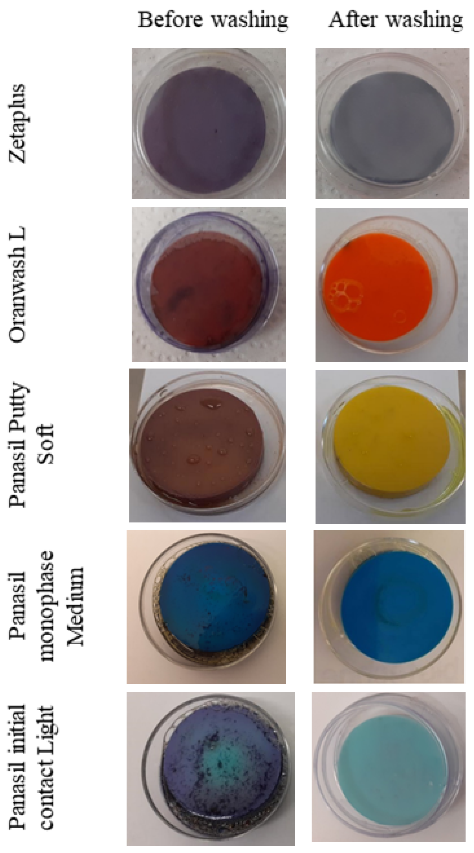
2.4. Validation of the Method of Washing Away of Adhered Microorganism Cultures—MTT Assay
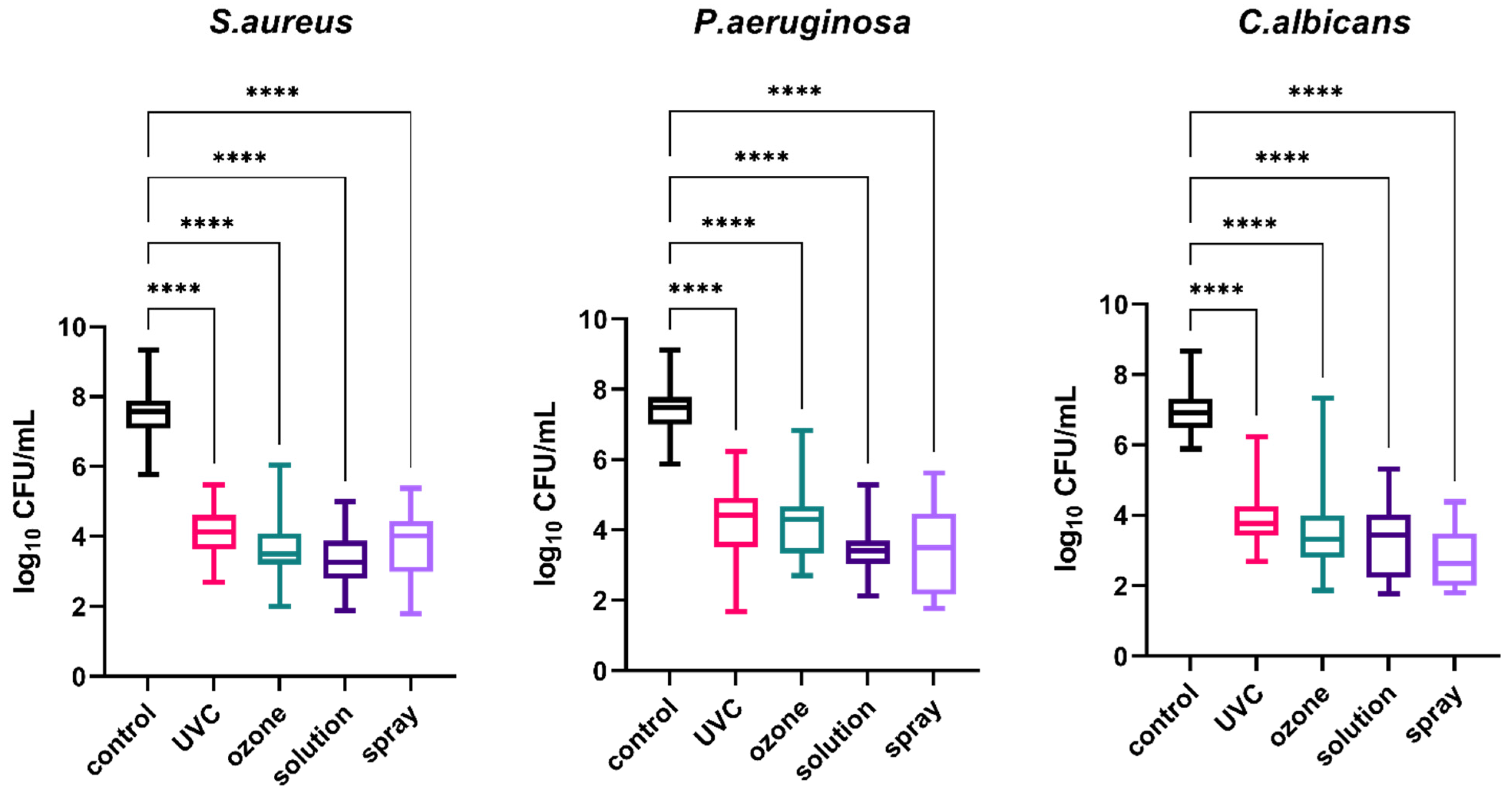
2.5. Evaluation of the Efficacy of Disinfection—Microbial Cell Counting
2.6. Statistical Analysis
3. Results
4. Discussion
5. Conclusions
Author Contributions
Funding
Institutional Review Board Statement
Informed Consent Statement
Data Availability Statement
Conflicts of Interest
Appendix A

| Material | Pathogen | Method of Disinfection | Log10 CFU/mL | ||
|---|---|---|---|---|---|
| Mean | Median | SD | |||
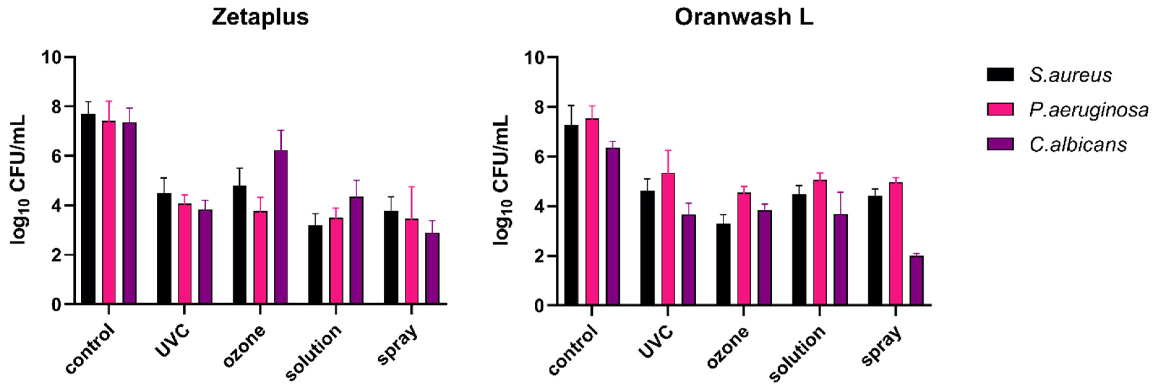
| Zetaplus | S. aureus | Control | 7.685 | 7.890 | 0.5063 |
| UVC | 4.500 | 4.455 | 0.6063 | ||
| Ozone | 4.808 | 4.780 | 0.6936 | ||
| Solution | 3.207 | 3.000 | 0.4593 | ||
| Spray | 3.782 | 3.890 | 0.5684 | ||
| P. aeruginosa | Control | 7.412 | 7.120 | 0.8025 | |
| UVC | 4.068 | 4.085 | 0.3617 | ||
| Ozone | 3.766 | 3.840 | 0.5546 | ||
| Solution | 3.312 | 3.365 | 0.4501 | ||
| Spray | 3.466 | 3.950 | 1.287 | ||
| C. albicans | Control | 7.360 | 7.240 | 0.5651 | |
| UVC | 3.825 | 3.825 | 0.3826 | ||
| Ozone | 6.240 | 6.105 | 0.7975 | ||
| Solution | 4.343 | 4.420 | 0.6690 | ||
| Spray | 2.905 | 2.980 | 0.4716 | ||
| Oranwash L | S. aureus | Control | 7.272 | 7.560 | 0.7835 |
| UVC | 4.637 | 4.490 | 0.4694 | ||
| Ozone | 3.295 | 3.300 | 0.3552 | ||
| Solution | 4.487 | 4.480 | 0.3435 | ||
| Spray | 4.428 | 4.430 | 0.2576 | ||
| P. aeruginosa | Control | 7.535 | 7.650 | 0.5001 | |
| UVC | 5.342 | 5.760 | 0.9061 | ||
| Ozone | 4.539 | 4.560 | 0.2567 | ||
| Solution | 5.063 | 5.180 | 0.2687 | ||
| Spray | 4.963 | 4.960 | 0.1899 | ||
| C. albicans | Control | 6.356 | 6.380 | 0.2550 | |
| UVC | 3.658 | 3.560 | 0.4657 | ||
| Ozone | 3.835 | 3.890 | 0.2446 | ||
| Solution | 3.676 | 4.040 | 0.8783 | ||
| Spray | 2.000 | 2.000 | 0.09126 | ||
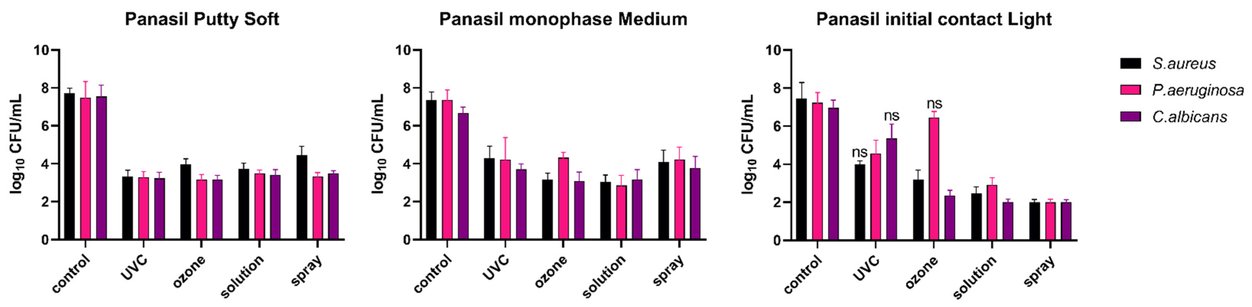
| Panasil Putty Soft | S. aureus | Control | 7.724 | 7.710 | 0.2628 |
| UVC | 3.329 | 3.330 | 0.3251 | ||
| Ozone | 3.960 | 4.000 | 0.2952 | ||
| Solution | 3.734 | 3.780 | 0.3058 | ||
| Spray | 4.462 | 4.330 | 0.4559 | ||
| P. aeruginosa | Control | 7.480 | 7.780 | 0.8451 | |
| UVC | 3.280 | 3.300 | 0.3065 | ||
| Ozone | 3.156 | 3.220 | 0.2819 | ||
| Solution | 3.496 | 3.500 | 0.1778 | ||
| Spray | 3.312 | 3.360 | 0.2140 | ||
| C. albicans | Control | 7.540 | 7.600 | 0.6089 | |
| UVC | 3.233 | 3.260 | 0.3088 | ||
| Ozone | 3.156 | 3.130 | 0.2294 | ||
| Solution | 3.416 | 3.430 | 0.2754 | ||
| Spray | 3.492 | 3.480 | 0.1476 | ||
| Panasil monophase Medium | S. aureus | Control | 7.366 | 7.450 | 0.4230 |
| UVC | 4.291 | 4.390 | 0.6401 | ||
| Ozone | 3.162 | 3.190 | 0.3354 | ||
| Solution | 3.048 | 3.180 | 0.3561 | ||
| Spray | 4.075 | 3.945 | 0.6491 | ||
| P. aeruginosa | Control | 7.363 | 7.480 | 0.5281 | |
| UVC | 4.207 | 4.670 | 1.170 | ||
| Ozone | 4.318 | 4.355 | 0.2876 | ||
| Solution | 2.865 | 2.850 | 0.5252 | ||
| Spray | 4.229 | 4.000 | 0.6533 | ||
| C. albicans | Control | 6.663 | 6.670 | 0.3241 | |
| UVC | 3.702 | 3.700 | 0.2846 | ||
| Ozone | 3.096 | 3.060 | 0.4659 | ||
| Solution | 3.183 | 3.300 | 0.5017 | ||
| Spray | 3.775 | 3.880 | 0.6012 | ||
| Panasil initial contact Light | S. aureus | Control | 7.449 | 7.120 | 0.8477 |
| UVC | 4.013 | 4.000 | 0.1653 | ||
| Ozone | 3.189 | 3.350 | 0.4944 | ||
| Solution | 2.460 | 2.360 | 0.3540 | ||
| Spray | 2.000 | 2.000 | 0.1452 | ||
| P. aeruginosa | Control | 7.227 | 7.340 | 0.5471 | |
| UVC | 4.574 | 4.840 | 0.6885 | ||
| Ozone | 6.450 | 6.530 | 0.3190 | ||
| Solution | 2.913 | 2.950 | 0.3807 | ||
| Spray | 2.007 | 2.000 | 0.1448 | ||
| C. albicans | Control | 6.966 | 7.000 | 0.4021 | |
| UVC | 5.356 | 5.530 | 0.7530 | ||
| Ozone | 2.360 | 2.390 | 0.2690 | ||
| Solution | 2.000 | 2.000 | 0.1594 | ||
| Spray | 2.000 | 2.000 | 0.1355 | ||
References
- Punj, A.; Bompolaki, D.; Garaicoa, J. Dental Impression Materials and Techniques. Dent. Clin. N. Am. 2017, 61, 779–796. [Google Scholar] [CrossRef] [PubMed]
- Winkler, J.; Gkantidis, N. Trueness and Precision of Intraoral Scanners in the Maxillary Dental Arch: An In Vivo Analysis. Sci. Rep. 2020, 10, 1172. [Google Scholar] [CrossRef] [PubMed]
- Mantena, S.R.; Mohd, I.; Sajjan, S.; Ramaraju, A. Disinfection of Impression Materials: A Comprehensive Review of Disinfection. Int. J. Dent. Mater. 2019, 1, 7–16. [Google Scholar] [CrossRef]
- Amin, W. The Effects of Disinfectants on Dimensional Accuracy and Surface Quality of Impression Materials and Gypsum Casts. J. Clin. Med. Res. 2009, 1, 81–89. [Google Scholar] [CrossRef] [PubMed][Green Version]
- Powell, G.L.; Runnells, R.D.; Saxon, B.A.; Whisenant, B.K. The presence and identification of organisms transmitted to dental laboratories. J. Prosthet. Dent. 1990, 64, 235–237. [Google Scholar] [CrossRef]
- Egusa, H.; Abe, K. Clinical Evaluation of the Efficacy of Removing Microorganisms to Disinfect Patient-Derived Dental Impressions. J. Prosthet. Dent. 2009, 102, 56. [Google Scholar] [CrossRef]
- Pang, S.; Millar, B. Cross Infection Control of Impressions: A Questionnaire Survey of Practice among Private Dentists in Hong Kong. Hong Kong Dent. J. 2006, 3, 89–93. [Google Scholar]
- Ait-Ou-amar, S.; Berrazzouk, S.; Ennibi, O. Handwashing Revisited in Dental Practice during the COVID-19 Outbreak. Dent. Med. Probl. 2021, 58, 243–252. [Google Scholar] [CrossRef]
- Peng, X.; Xu, X.; Li, Y.; Cheng, L.; Zhou, X.; Ren, B. Transmission Routes of 2019-NCoV and Controls in Dental Practice. Int. J. Oral Sci. 2020, 12, 9. [Google Scholar] [CrossRef]
- Torul, D.; Omezli, M.M. Is saliva a reliable biofluid for the detection of COVID-19? Dent. Med. Probl. 2021, 58, 229–235. [Google Scholar] [CrossRef]
- Gupta, M.; George, V.T.; Balakrishnan, D. A Comparative Evaluation of Tear Strength and Tensile Strength of Autoclavable and Non-Autoclavable Vinylpolysiloxane Impression Material: An In Vitro Study. J. Int. Oral Health 2020, 12, 153–157. [Google Scholar] [CrossRef]
- Badrian, H.; Ghasemi, E.; Khalighinejad, N.; Hosseini, N. The Effect of Three Different Disinfection Materials on Alginate Impression by Spray Method. ISRN Dent. 2012, 2012, 695151. [Google Scholar] [CrossRef] [PubMed]
- Celebi, H.; Büyükerkmen, E.B.; Torlak, E. Disinfection of Polyvinyl Siloxane Impression Material by Gaseous Ozone. J. Prosthet. Dent. 2018, 120, 138–143. [Google Scholar] [CrossRef] [PubMed]
- Bhasin, A.; Vinod, V.; Bhasin, V.; Mathew, X.; Sajjan, S.; Ahmed, S.T. Evaluation of Effectiveness of Microwave Irradiation for Disinfection of Silicone Elastomeric Impression Material. J. Indian Prosthodont. Soc. 2013, 13, 89–94. [Google Scholar] [CrossRef]
- Choi, Y.R.; Kim, K.N.; Kim, K.M. The Disinfection of Impression Materials by Using Microwave Irradiation and Hydrogen Peroxide. J. Prosthet. Dent. 2014, 112, 981–987. [Google Scholar] [CrossRef]
- Aeran, H.; Sharma, S.; Kumar, V.; Gupta, N. Use of Clinical UV Chamber to Disinfect Dental Impressions: A Comparative Study. J. Clin. Diagn. Res. 2015, 9, ZC67–ZC70. [Google Scholar] [CrossRef]
- Kotwal, M.; Singh, V.P.; Mushtaq, H.; Ahmed, R.; Rai, G.; Kumar, A. Disinfection of impression materials with glutaraldehyde, ultraviolet radiation, and autoclave: A comparative study. J. Pharm. Bioall. Sci. 2021, 13, S289. [Google Scholar] [CrossRef]
- Godbole, S.R.; Dahane, T.M.; Patidar, N.A.; Nimonkar, S.V. “Evaluation of the Effect of Ultraviolet Disinfection on Dimensional Stability of the Polyvinyl Silioxane Impressions.” An In-Vitro Study. J. Clin. Diagn. Res. 2014, 8, ZC73–ZC76. [Google Scholar] [CrossRef]
- AlZain, S. Effect of Chemical, Microwave Irradiation, Steam Autoclave, Ultraviolet Light Radiation, Ozone and Electrolyzed Oxidizing Water Disinfection on Properties of Impression Materials: A Systematic Review and Meta-Analysis Study. Saudi Dent. J. 2020, 32, 161–170. [Google Scholar] [CrossRef]
- Nagamatsu, Y.; Chen, K.K.; Nagamatsu, H.; Kozono, Y.; Shimizu, H. Application of Neutral Electrolyzed Water to Disinfection of Alginate Impression. Dent. Mater. J. 2016, 35, 270–277. [Google Scholar] [CrossRef][Green Version]
- Rentzia, A.; Coleman, D.C.; O’Donnell, M.J.; Dowling, A.H.; O’Sullivan, M. Disinfection Procedures: Their Efficacy and Effect on Dimensional Accuracy and Surface Quality of an Irreversible Hydrocolloid Impression Material. J. Dent. 2011, 39, 133–140. [Google Scholar] [CrossRef] [PubMed]
- Gothwal, G.; Meena, S.; Padiyar, U.N.; Sharma, H.K.; Kaurani, P.; Singh, D.P. Comparative evaluation of elastic recovery of three different elastomeric impression materials on chemical disinfection and autoclaving: An in vitro study. J. Indian Prosthodont. Soc. 2019, 19, 345–352. [Google Scholar] [CrossRef] [PubMed]
- Demajo, J.; Cassar, V.; Farrugia, C.; Millan-Sango, D.; Sammut, C.; Valdramidis, V.; Camilleri, J. Effectiveness of Disinfectants on Antimicrobial and Physical Properties of Dental Impression Materials. Int. J. Prosthodont. 2016, 29, 63–67. [Google Scholar] [CrossRef] [PubMed]
- Azevedo, M.J.; Correia, I.; Portela, A.; Sampaio-Maia, B. A Simple and Effective Method for Addition Silicone Impression Disinfection. J. Adv. Prosthodont. 2019, 11, 155–161. [Google Scholar] [CrossRef]
- Khatri, M.; Mantri, S.S.; Deogade, S.C.; Bhasin, A.; Mantri, S.; Khatri, N.; Jain, P.; Chauhan, D. Effect of Chemical Disinfection on Surface Detail Reproduction and Dimensional Stability of a New Vinyl Polyether Silicone Elastomeric Impression Material. Contemp. Clin. Dent. 2020, 11, 10–14. [Google Scholar] [CrossRef]
- Lepe, X.; Johnson, G.H. Accuracy of polyether and addition silicone after long-term immersion disinfection. J. Prosthet. Dent. 1997, 78, 245–249. [Google Scholar] [CrossRef]
- Morio., K.A.; Sternowski, R.H.; Brogden, K.A. Dataset of endodontic microorganisms killed at 265 nm wavelength by an ultraviolet C light emitting diode in root canals of extracted, instrumented teeth. Data Brief. 2021, 40, 107750. [Google Scholar] [CrossRef]
- Lubojanski, A.; Dobrzynski, M.; Nowak, N.; Rewak-Soroczynska, J.; Sztyler, K.; Zakrzewski, W.; Dobrzynski, W.; Szymonowicz, M.; Rybak, Z.; Wiglusz, K.; et al. Application of Selected Nanomaterials and Ozone in Modern Clinical Dentistry. Nanomaterials 2021, 11, 259. [Google Scholar] [CrossRef]
- PN-EN ISO 4823:2015; Dentistry—Elastomeric Impression Materials. Available online: https://www.iso.org/standard/60586.html (accessed on 18 October 2020).
- PN-EN 13727+A2:2015-12; Chemical Disinfectants and Antiseptics—Quantitative Suspension Test for the Evaluation of Bactericidal Activity in the Medical Area—Test Method and Requirements (Phase 2, Step 1). Available online: https://sklep.pkn.pl/pn-en-13727-a2-2015-12e.html (accessed on 11 January 2021).
- PN-EN 13624:2013-12; Chemical Disinfectants and Antiseptics—Quantitative Suspension Test for the Evaluation of Fungicidal or Yeasticidal Activity in the Medical area—Test Method and Requirements (Phase 2, Step 1). Available online: https://sklep.pkn.pl/pn-en-13624-2013-12p.html (accessed on 11 January 2021).
- Giammanco, G.M.; Melilli, D.; Rallo, A.; Pecorella, S.; Mammina, C.; Pizzo, G. Resistance to Disinfection of a Polymicrobial Association Contaminating the Surface of Elastomeric Dental Impressions. New Microbiol. 2009, 32, 167–172. [Google Scholar]
- Mosmann, T. Rapid colorimetric assay for cellular growth and survival: Application for proliferation and cytotoxicity assays. J. Immunol. Meth. 1983, 65, 55–63. [Google Scholar] [CrossRef]
- Al Mortadi, N.; Al-Khatib, A.; Alzoubi, K.H.; Khabour, O.F. Disinfection of Dental Impressions: Knowledge and Practice among Dental Technicians. Clin. Cosmet. Investig. Dent. 2019, 11, 103–108. [Google Scholar] [CrossRef] [PubMed]
- Casemiro, L.A.; Pires-de-Souza, F.D.C.P.; Panzeri, H.; Martins, C.H.G.; Ito, I.Y. In Vitro Antimicrobial Activity of Irreversible Hydrocolloid Impressions against 12 Oral Microorganisms. Braz. Oral Res. 2007, 21, 323–329. [Google Scholar] [CrossRef] [PubMed]
- Ginjupalli, K.; Shaw, T.; Tellapragada, C.; Alla, R.; Gupta, L.; Perampalli, N.U. Does the Size Matter? Evaluation of Effect of Incorporation of Silver Nanoparticles of Varying Particle Size on the Antimicrobial Activity and Properties of Irreversible Hydrocolloid Impression Material. Dent. Mater. 2018, 34, e158–e165. [Google Scholar] [CrossRef] [PubMed]
- Mahajan, T. Comparative Evaluation of Disinfecting Alginate Using Aloe Vera and 0.2% Chlorhexidine Digluconate by Internal Disinfection Method—An In Vivo Study. J. Evol. Med. Dent. Sci. 2020, 9, 1944–1947. [Google Scholar] [CrossRef]
- Rajendran, V.; Suma, K.; Ali, S.A.; Karthigeyan, R.; Kalarani, G. Antimicrobial efficacy of irreversible hydrocolloid impression impregnated with silver nanoparticles compared to surface disinfected impressions—An in vivo study. J. Pharm. Bioall. Sci. 2021, 13, S532. [Google Scholar] [CrossRef] [PubMed]
- Tetteh, S.; Bibb, R.J.; Martin, S.J. Mechanical and Morphological Effect of Plant Based Antimicrobial Solutions on Maxillofacial Silicone Elastomer. Materials 2018, 11, 925. [Google Scholar] [CrossRef]
- Dai, T.; Vrahas, M.S.; Murray, C.K.; Hamblin, M.R. Ultraviolet C irradiation: An alternative antimicrobial approach to localized infections? Expert Rev. Anti-Infect. Ther. 2012, 10, 185–195. [Google Scholar] [CrossRef]
- Smith, N.; Wilson, A.; Gandhi, J.; Vatsia, S.; Khan, S. Ozone Therapy: An Overview of Pharmacodynamics, Current Research, and Clinical Utility. Med. Gas Res. 2017, 7, 212–219. [Google Scholar] [CrossRef]
- Irie, M.S.; Dietrich, L.; Souza, G.L.D.; Soares, P.B.F.; Mours, C.C.G.; Silva, G.R.D.; Paranhos, L.R. Ozone Disinfection for Viruses with Applications in Healthcare Environments: A Scoping Review. Braz. Oral Res. 2022, 36, 1–16. [Google Scholar] [CrossRef]
- Savabi, O.; Nejatidanesh, F.; Bagheri, K.P.; Karimi, L.; Savabi, G. Prevention of cross-contamination risk by disinfection of irreversible hydrocolloid impression material with ozonated water. Int. J. Prev. Med. 2018, 9, 37. [Google Scholar] [CrossRef]
- Ganji, K.K.; Alshammari, S.M.; Rushdallah, M.A.; Ghazy, A.A.; Taher, I.; Taha, A.E.; Issrani, R.; Alhazmi, M.A.N. Activity of Ozonated Water in Sterilising and Disinfecting Dental Unit Water Pipelines System: A Comparative Study. Oral Health Prev. Dent. 2022, 20, 61–68. [Google Scholar] [CrossRef] [PubMed]
- Moreira Fonseca, P.M.; Buendía Palacios, D.A.; de Sá Júnior, P.L.; Miyakawa, W.; Damião, Á.J.; Fernandes, A.B.; José de Lima, C. Preliminary Study: Comparative Analysis of the Effects of Ozone and Ultrasound on Streptococcus Mutans. Ozone Sci. Eng. 2021, 43, 263–275. [Google Scholar] [CrossRef]
- Al Shikh, A.; Milosevic, A. Effectiveness of Alcohol and Aldehyde Spray Disinfectants on Dental Impressions. Clin. Cosmet. Investig. Dent. 2020, 12, 25–30. [Google Scholar] [CrossRef] [PubMed]


| Type of Material | Consistency | Name | Manufacturer |
|---|---|---|---|
| C-silicone | Putty | Zetaplus | Zhermack (Badia Polesine, Italy) |
| Light | Oranwash L | Zhermack (Badia Polesine, Italy) | |
| A-silicone | Putty | Panasil Putty Soft | Kettenbach (Eschenburg, Germany) |
| Medium | Panasil monophase Medium | Kettenbach (Eschenburg, Germany) | |
| Light | Panasil initial contact Light | Kettenbach (Eschenburg, Germany) |
| Method | Material or Equipment | Description |
|---|---|---|
| UVC | UV-C Blue (Activeshop, Wroclaw, Poland) | Irradiation for 40 min at 254 nm; power of 8 W, distance between the lamp and samples: 80 mm |
| Ozone | Ozox Professional G168 (MediaSklep24, Bojszowy, Poland) | Ozonation for 10 min at an ozone flow rate of 800 mg/h |
| Solution | Zeta 7 Solution (Zhermack, Badia Polesine, Italy) | Immersion for 10 min in 100× diluted solution and rinsing with distilled water |
| Spray | Zeta 7 spray (Zhermack, Badia Polesine, Italy) | Spraying and allowing to dry |
| Method of Disinfection | C-Silicones | A-Silicones | |||
|---|---|---|---|---|---|
| Zetaplus | Oranwash L | Panasil Putty Soft | Panasil Monophase Medium | Panasil Initial Contact Light | |
| S. aureus | |||||
| UVC | <0.0001 | <0.0001 | <0.0001 | <0.0001 | 0.9109 |
| Ozone | <0.0001 | <0.0001 | <0.0001 | <0.0001 | 0.0015 |
| Solution | <0.0001 | <0.0001 | <0.0001 | <0.0001 | <0.0001 |
| Spray | <0.0001 | <0.0001 | <0.0001 | <0.0001 | <0.0001 |
| P. aeruginosa | |||||
| UVC | 0.0060 | 0.0357 | <0.0001 | 0.0024 | 0.0048 |
| Ozone | 0.0002 | <0.0001 | <0.0001 | 0.0007 | >0.9999 |
| Solution | <0.0001 | 0.0058 | 0.0149 | <0.0001 | <0.0001 |
| Spray | <0.0001 | 0.0005 | <0.0001 | <0.0001 | <0.0001 |
| C. albicans | |||||
| UVC | <0.0001 | 0.0003 | <0.0001 | 0.0030 | 0.6131 |
| Ozone | <0.0001 | 0.0012 | <0.0001 | <0.0001 | 0.0002 |
| Solution | <0.0001 | 0.0055 | <0.0001 | <0.0001 | <0.0001 |
| Spray | <0.0001 | <0.0001 | <0.0001 | 0.0458 | <0.0001 |
Publisher’s Note: MDPI stays neutral with regard to jurisdictional claims in published maps and institutional affiliations. |
© 2022 by the authors. Licensee MDPI, Basel, Switzerland. This article is an open access article distributed under the terms and conditions of the Creative Commons Attribution (CC BY) license (https://creativecommons.org/licenses/by/4.0/).
Share and Cite
Wezgowiec, J.; Wieczynska, A.; Wieckiewicz, M.; Czarny, A.; Malysa, A.; Seweryn, P.; Zietek, M.; Paradowska-Stolarz, A. Evaluation of Antimicrobial Efficacy of UVC Radiation, Gaseous Ozone, and Liquid Chemicals Used for Disinfection of Silicone Dental Impression Materials. Materials 2022, 15, 2553. https://doi.org/10.3390/ma15072553
Wezgowiec J, Wieczynska A, Wieckiewicz M, Czarny A, Malysa A, Seweryn P, Zietek M, Paradowska-Stolarz A. Evaluation of Antimicrobial Efficacy of UVC Radiation, Gaseous Ozone, and Liquid Chemicals Used for Disinfection of Silicone Dental Impression Materials. Materials. 2022; 15(7):2553. https://doi.org/10.3390/ma15072553
Chicago/Turabian StyleWezgowiec, Joanna, Anna Wieczynska, Mieszko Wieckiewicz, Anna Czarny, Andrzej Malysa, Piotr Seweryn, Marek Zietek, and Anna Paradowska-Stolarz. 2022. "Evaluation of Antimicrobial Efficacy of UVC Radiation, Gaseous Ozone, and Liquid Chemicals Used for Disinfection of Silicone Dental Impression Materials" Materials 15, no. 7: 2553. https://doi.org/10.3390/ma15072553
APA StyleWezgowiec, J., Wieczynska, A., Wieckiewicz, M., Czarny, A., Malysa, A., Seweryn, P., Zietek, M., & Paradowska-Stolarz, A. (2022). Evaluation of Antimicrobial Efficacy of UVC Radiation, Gaseous Ozone, and Liquid Chemicals Used for Disinfection of Silicone Dental Impression Materials. Materials, 15(7), 2553. https://doi.org/10.3390/ma15072553