
Corrosive Studies of a Prosthetic Ni-Cr Alloy Coated with Ti(C,N) Type Layers


Abstract
:1. Introduction
- Development of metal alloys with a higher corrosion resistance,
- Enrichment of metal prosthetic restorations through a modification of their surface.
2. Aim of the Studies
3. Materials and Methods
Corrosion Studies
4. Results
5. Discussion
6. Conclusions
Author Contributions
Funding
Institutional Review Board Statement
Informed Consent Statement
Data Availability Statement
Conflicts of Interest
References
- Makuch, K.; Koczorowski, R. Biocompatibility of Titanium and Its Alloys Used in Dentistry. Dent. Med. Probl. 2010, 47, 81–88. [Google Scholar]
- Roberts, H.W.; Berzins, D.W.; Moore, B.K.; Charlton, D.G. Metal-ceramic alloys in dentistry: A review. J. Prosthod. 2009, 18, 188–194. [Google Scholar] [CrossRef]
- Singh, R.; Dahotre, N.B. Corrosion degradation and prevention by surface modification of biometallic materials. J. Mater. Sci. Mater. Med. 2007, 18, 725–751. [Google Scholar] [CrossRef] [PubMed]
- Taher, N.M.; Al Jabab, A.S. Galvanic corrosion behavior of implant suprastructure dental alloys. Dent. Mater. 2003, 19, 54–59. [Google Scholar] [CrossRef]
- Näpänkangas, R.; Raustia, A. An 18-Year Retrospective Analysis of Treatment Outcomes with Metal-Ceramic Fixed Partial Dentures. Int. J. Prosthodont. 2011, 24, 314–319. [Google Scholar]
- Levi, L.; Barak, S.; Katz, J. Allergic reactions associated with metal alloys in porcelain-fused-to-metal fixed prosthodontic devices—Systemayic review. Quintessence Int. 2012, 43, 871–877. [Google Scholar]
- Ristic, L.; Vucevic, D.; Radovic, L.; Djordjevic, S.; Nikacevic, M.; Colic, M. Corrosive and Cytotoxic Properties of Compact Specimens and Microparticles of Ni-Cr Dental Alloy. J. Prosthodont. 2013, 23, 221–226. [Google Scholar] [CrossRef]
- Drago, C.; Howell, K. Concepts for designing and fabricating metal implant frameworks for hybrid implant prostheses. J. Prosthodont. 2012, 21, 413–424. [Google Scholar] [CrossRef]
- Wylie, C.M.; Sheltonb, R.M.; Fleming, G.J.P.; Davenporta, A.J. Corrosion of nickel-based dental casting alloys. Dent. Mater. 2007, 23, 714–723. [Google Scholar] [CrossRef]
- Upadhyaya, D.; Panchal, M.A.; Dubey, R.S.; Srivastava, V.K. Corrosion of alloys used in dentistry: A review. Mater. Sci. Eng. A 2006, 432, 1–11. [Google Scholar] [CrossRef]
- Sheikh, T.; Ghorbani, M.; Tahmasbi, S.; Yaghoubnejad, Y. Galvanic Corrosion of Orthodontic Brackets and Wires in Acidic Artificial Saliva: Part II. J. Dent. Sch. 2015, 33, 88–97. [Google Scholar]
- Bakhtari, A.; Bradley, T.G.; Lobb, W.K.; Berzins, D.W. Galvanic corrosion between various combinations of orthodontic brackets and archwires. Am. J. Orthod. Dentofac. Orthop. 2011, 140, 25–31. [Google Scholar] [CrossRef] [PubMed]
- Saranya, R.; Rajendran, S. Influence of D-Glucose on Corrosion Resistance of Ss316 Lin Presence of Artificial Saliva. Rasayan J. Chem. 2018, 11, 103–110. [Google Scholar] [CrossRef]
- Jelínek, M.; Smetanac, K.; Kocoureka, T.; Dvořánková, B.; Zemeka, J.; Remsaa, J.; Luxbachere, T. Biocompatibility and sp3/sp2 ratio of laser created DLC films. Mater. Sci. Eng. B 2010, 169, 89–93. [Google Scholar] [CrossRef]
- Wu, F.; Chen, T.; Wang, H.; Liu, D. Effect of Mo on Microstructures and Wear Properties of In Situ Synthesized Ti(C,N)/Ni-Based Composite Coatings by Laser Cladding. Materials 2017, 10, 1047. [Google Scholar] [CrossRef] [PubMed] [Green Version]
- Nematia, A.; Saghafia, M.; Khamseh, S.; Alibakhshic, E.; Zarrintajd, P.; Saebe, M.R. Magnetron-sputtered TixNy thin films applied on titanium-based alloys for biomedical applications: Composition-microstructure-property relationships. Surf. Coat. Technol. 2018, 349, 251–259. [Google Scholar] [CrossRef]
- Pawlak, R.; Tomczyk, M.; Walczak, M. The favorable and unfavorable effects of oxide and intermetallic phases in conductive materials using laser micro technologies. Mater. Sci. Eng. B 2012, 177, 1273–1280. [Google Scholar] [CrossRef]
- Krzak-Roś, J.; Filipiak, J.; Pezowicz, C.; Baszczuk, A.; Miller, M.; Kowalski, M.; Będziński, R. The effect of substrate roughness on the surface structure of TiO2, SiO2, and doped thin films prepared by the sol-gel method. Acta Bioeng. Biomech. 2009, 11, 21–29. [Google Scholar]
- Szymanowski, H.; Sobczyk, A.; Gazicki-Lipman, M.; Jakubowski, W.; Klimek, L. Plasma enhanced CVD deposition of titanium oxide for biomedical applications. Surf. Coat. Technol. 2005, 200, 1036–1040. [Google Scholar] [CrossRef]
- Wang, G.; Zreiqat, H. Functional Coatings or Films for Hard-Tissue Applications. Materials 2010, 3, 3994–4050. [Google Scholar] [CrossRef] [PubMed] [Green Version]
- Pietrzyk, B.; Miszczak, S.; Szymanowski, H.; Kucharski, D. Plasma enhanced aerosol-gel deposition of Al2O3 coatings. J. Eur. Ceram. Soc. 2013, 33, 2341–2346. [Google Scholar] [CrossRef]
- Asri, R.I.M.; Harun, W.S.W.; Samykano, M.; Lah, N.A.C.; Ghani, S.A.C.; Tarlochan, F.; Raza, M.R. Corrosion and surface modification on biocompatible metals: A review. Mater. Sci. Eng. C 2017, 77, 1261–1274. [Google Scholar] [CrossRef] [PubMed] [Green Version]
- Banaszek, K.; Januszewicz, B.; Wołowiec, E.; Klimek, L. Complex XRD and XRF Characterization of TiN-TiCN-TiC Surface Coatings for Medical Applications. Solid State Phenom. 2015, 225, 159–168. [Google Scholar] [CrossRef]
- De Viteri, V.S.; Barandika, M.G.; de Gopegui, U.R.; Bayón, R.; Zubizarreta, C.; Fernández, X.; Igartua, A.; Agullo-Rueda, F. Characterization of Ti–C–N coatings deposited on Ti6Al4V for biomedical applications. J. Inorg. Biochem. 2012, 117, 359–366. [Google Scholar] [CrossRef] [PubMed]
- deViteri, V.S.; Barandika, M.G.; Bayon, R.; Fernandeza, X.; Ciarsolo, I.; Igartua, A.; Tanoira, R.P.; Moreno, J.E.; Peremarch, C.P.J. Development of Ti–C–N coatings with improved tribological behaviour and antibacterial properties. J. Mech. Behav. Biomed. Mater. 2016, 55, 75–86. [Google Scholar]
- Banaszek, K.; Pietnicki, K.; Klimek, L. Effect of carbon and nitrogen content in Ti(C,N) coatings on selected mechanical properties. Met. Form. 2015, 26, 33–45. [Google Scholar]
- Banaszek, K.; Klimek, L. Wettability and surface free energy of Ti(C,N) coatings on nickel-based casting prosthetic alloys. Arch. Foundry Eng. 2015, 15, 11–16. [Google Scholar] [CrossRef] [Green Version]
- Banaszek, K.; Klimek, L.; Dąbrowski, J.R.; Jastrzębski, W. Fretting Wear in Orthodontic and Prosthetic Alloys with Ti(C,N) Coatings. Processes 2019, 7, 874. [Google Scholar] [CrossRef] [Green Version]
- Banaszek, K.; Wiktorowska-Owczarek, A.; Kowalczyk, E.; Klimek, L. Possibilities of applying Ti(C,N) coatings on prosthetic elements–research with the use of human endothelial cells. Acta Bioeng. Biomiech. 2016, 18, 119–126. [Google Scholar] [CrossRef]
- Banaszek, K.; Klimek, L.; Zgorzynska, E.; Swarzynska, A.; Walczewska, A. Cytotoxicity of titanium carbonitride coatings for prostodontic alloys with different amounts of carbon and nitro gen. Biomed. Mater. 2018, 13, 045003. [Google Scholar] [CrossRef]
- Shan, L.; Wang, Y.; Li, J.; Li, H.; Wu, X.; Chen, J. Tribological behaviours of PVD TiN and TiCN coatings in artificial seawater. Surf. Coat. Technol. 2013, 226, 40–50. [Google Scholar] [CrossRef]
- El Azhari, I.; Garcia, J.; Zamanzade, M.; Soldera, F.; Pauly, C.; Llanes, L.; Mücklich, F. Investigations on micro-mechanical properties of polycrystalline Ti(C,N) and Zr(C,N) coatings. Acta Mater. 2018, 149, 364–376. [Google Scholar] [CrossRef] [Green Version]
- Azadi, M.; Rouhaghdam, A.S. Mechanical Behavior of TiN/TiC-n Multilayer Coatings and Ti(C,N) Multicomponent Coatings Produced by PACVD. Strength Mater. 2016, 48, 279–289. [Google Scholar] [CrossRef]
- Pochrząst, M.; Marciniak, J.; Wróbel, K.; Bączkowski, B. Electrochemical Properties of Ni-Cr and Co-Cr Alloys Used in Prosthodontics. Solid State Phenom. 2011, 183, 143–148. [Google Scholar] [CrossRef]
- Schmutz, P.; Quach-Vu, N.-C.; Gerber, I. Metallic Medical Implants: Electrochemical Characterization of Corrosion Processes. Electrochem. Soc. Interface 2008, 17, 35–40. [Google Scholar] [CrossRef]
- Nierlich, J.; Papageorgiou, S.N.; Bourauel, C.; Hültenschmidt, R.; Bayer, S.; Stark, H.; Keilig, L. Corrosion behavior of dental alloys used for retention elements in prosthodontics. Eur. J. Oral Sci. 2016, 124, 287–294. [Google Scholar] [CrossRef] [PubMed] [Green Version]
- Mercieca, S.; Conti, C.M.; Buhagiar, J.; Camilleri, J. Assessment of corrosion resistance of cast cobalt- and nickel-chromium dental alloys in acidic environments. J. Appl. Biomater. Funct. Mater. 2018, 16, 47–54. [Google Scholar] [CrossRef] [Green Version]
- PN EN ISO 9626; Stainless Steel Test Needle Tubing for the Manufacture of Medical Devices. International Organization for Standardization: Geneva, Switzerland, 2016.
- PN EN ISO 9227; Corrosion Test in Artificial Atmospheres. Salt Spray Test. International Organization for Standardization: Geneva, Switzerland, 2017.
- ASTM G85-09; Standard Practice for Modified Salt Spray (Fog) Testing. ASTM International: West Conshohocken, PA, USA, 2009.
- Banaszek, K.; Klimek, L. Ti(C, N) as Barrier Coatings. Coatings 2019, 9, 432. [Google Scholar] [CrossRef] [Green Version]
- Lee, J.-J.; Song, K.-Y.; Ahn, S.-G.; Choi, J.-Y.; Seo, J.-M.; Park, J.-M. Evaluation of effect of galvanic corrosion between nickel-chromium metal and titanium on ion release and cell toxicity. Adv. Prosthodont. 2015, 7, 172–177. [Google Scholar] [CrossRef] [Green Version]
- Holm, C.; Morisbak, E.; Kalfoss, T.; Dahl, J.E. In vitro element release and biological aspects of base–metal alloys for metal-ceramic applications. Acta Biomater. Odontol. Scand. 2015, 1, 70–75. [Google Scholar] [CrossRef] [Green Version]
- Baričević, M.; Ratkaj, I.; Mladinić, M.; Želježić, D.; Pavelić Kraljević, S.; Lončar, B.; Mravak Stipetić, M. In vivo assessment of DNA damage induced in oral mucosa cells by fixed and removable metal prosthodontic appliances. Clin. Oral Investig. 2012, 16, 325–331. [Google Scholar] [CrossRef] [PubMed]
- Milheiro, A.; Nozaki, K.; Kleverlaan, C.J.; Muris, J.; Miura, H.; Feilzer, A.J. In vitro cytotoxicity of metallic ions released from dental alloys. Odontology 2016, 104, 136–142. [Google Scholar] [CrossRef] [PubMed]
- Chandar, S.; Kotian, R.; Madhyastha, P.; Kabekkodu, S.P.; Rao, P. In vitro evaluation of cytotoxicity and corrosion behavior of commercially pure titanium and Ti-6Al-4V alloy for dental implants. J. Indian Prosthodont. Soc. 2017, 17, 35–40. [Google Scholar] [CrossRef] [PubMed]
- Mc Ginley, E.L.; Fleming, G.J.P.; Moran, G.P. Development of a discriminatory biocompatibility testing model for non-precious dental casting alloys. Dent. Mater. 2011, 27, 1295–1306. [Google Scholar] [CrossRef] [PubMed]
- Ramírez-Ledesma, A.L.; Roncagliolo, P.; Álvarez-Pérez, M.A.; Lopez, H.F.; Juárez-Islas, J.A. Corrosion Assessment of an Implantable Dental Co-Cr Alloy in Artificial Saliva and Biocompatibility Behavior. J. Mater. Eng. Perform. 2020, 29, 1657–1670. [Google Scholar] [CrossRef]
- Zhao, L.; Hong, Y.; Yang, D.; Lü, X.; Xi, T.; Zhang, D.; Hong, Y.; Yuan, J. The underlying biological mechanisms of biocompatibility differences between bare and TiN-coated NiTi alloys. Biomed. Mater. 2011, 6, 025012. [Google Scholar]


| Element Percentage wt. % | ||||||
|---|---|---|---|---|---|---|
| Cr | Mo | Si | Fe | Co | Mn | Ni |
| 24.79 | 8.89 | 1.57 | 1.33 | 0.17 | 0.12 | residue |
| Coating | Element Percentage at. % | ||
|---|---|---|---|
| Ti | C | N | |
| S1 | 51.50 | 48.50 | 0.00 |
| S2 | 52.91 | 33.91 | 13.18 |
| S3 | 51.94 | 28.22 | 19.84 |
| S4 | 47.78 | 20.05 | 32.17 |
| S5 | 46.79 | 0.00 | 53.21 |
| Element Percentage wt. % | |||
| Ti | C | N | |
| S1 | 80.18 | 19.82 | 0.00 |
| S2 | 79.51 | 13.90 | 6.59 |
| S3 | 78.76 | 11.67 | 9.57 |
| S4 | 75.26 | 8.61 | 16.13 |
| S5 | 79.78 | 0.00 | 20.22 |
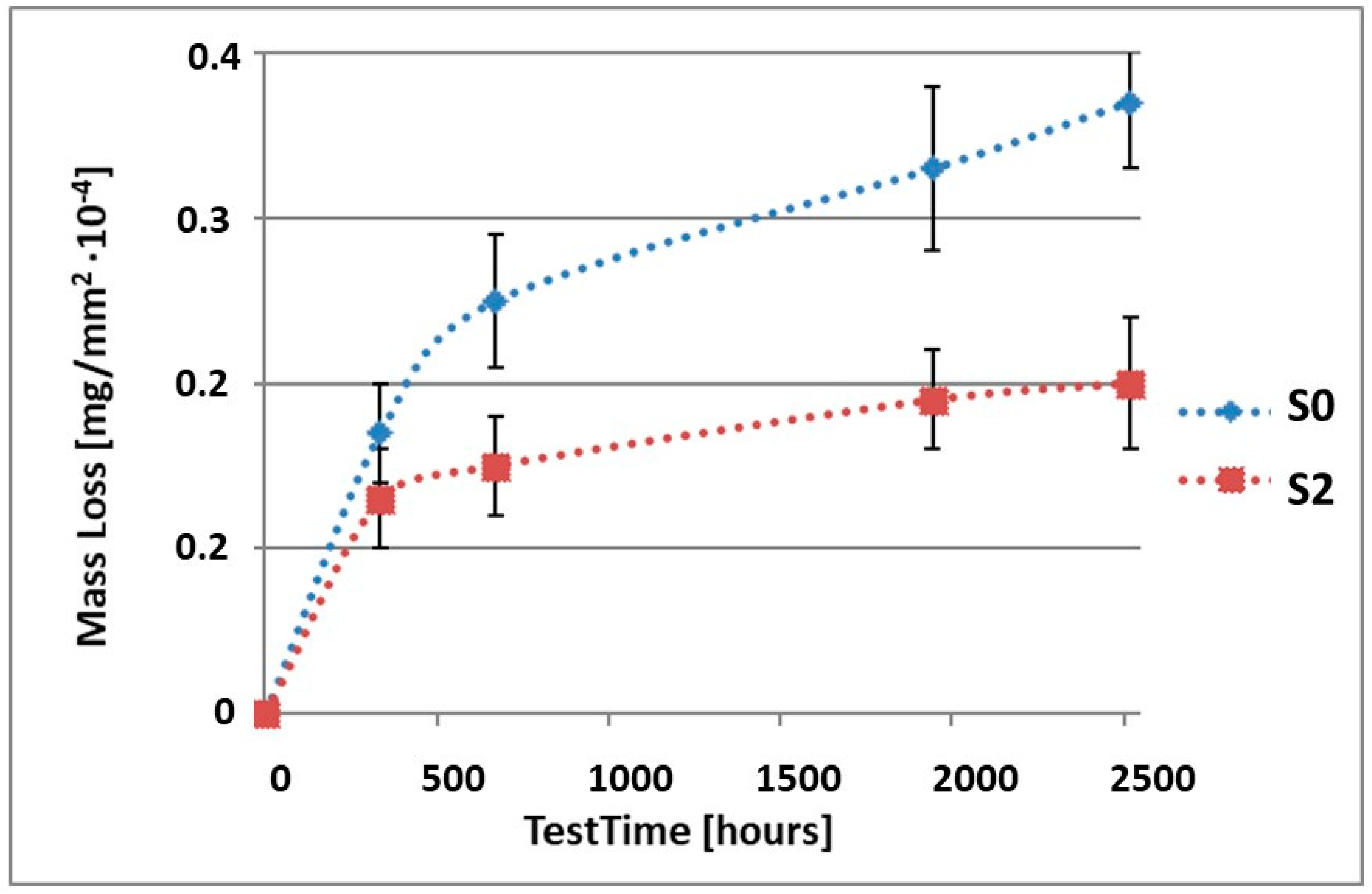
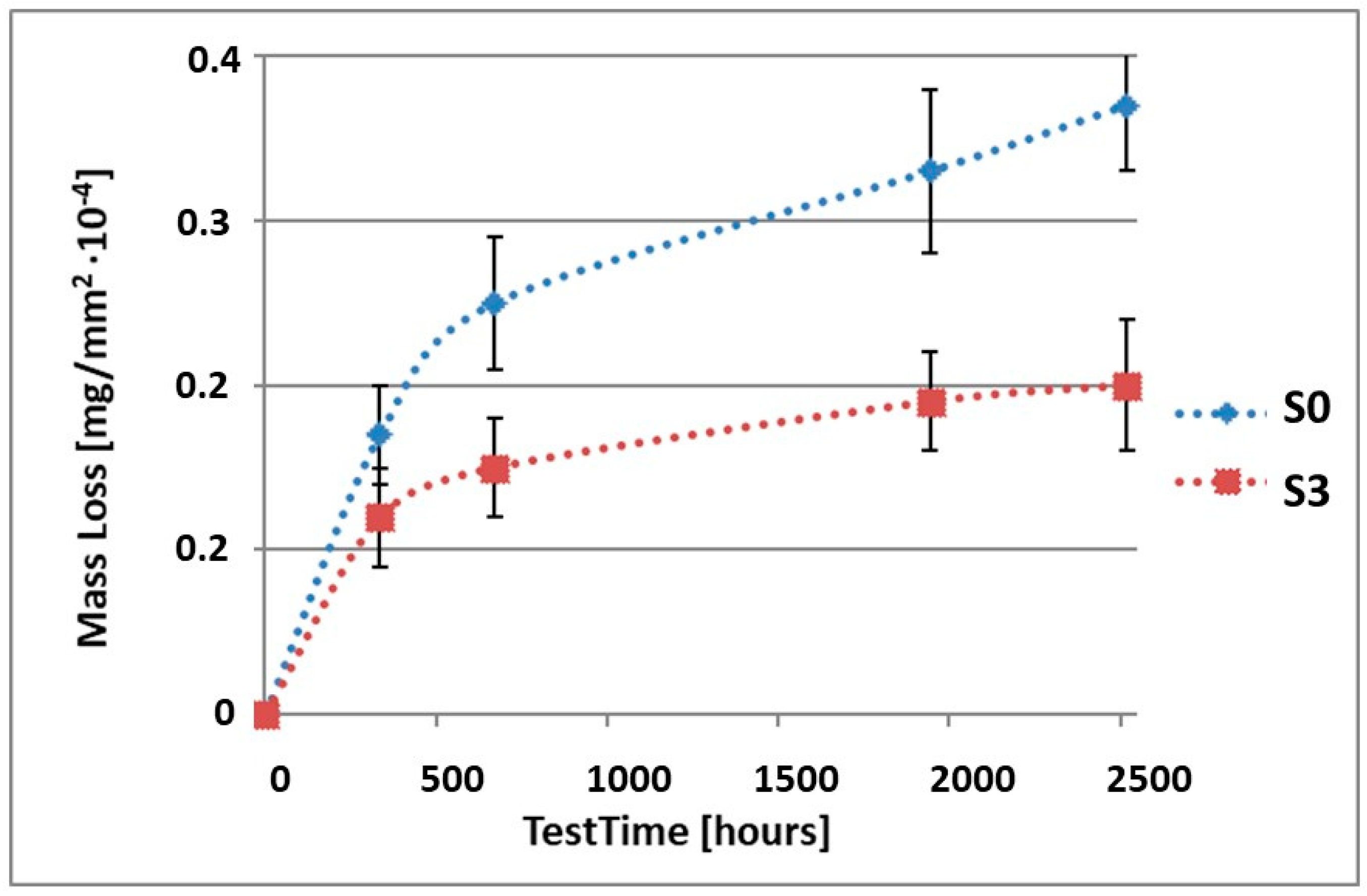
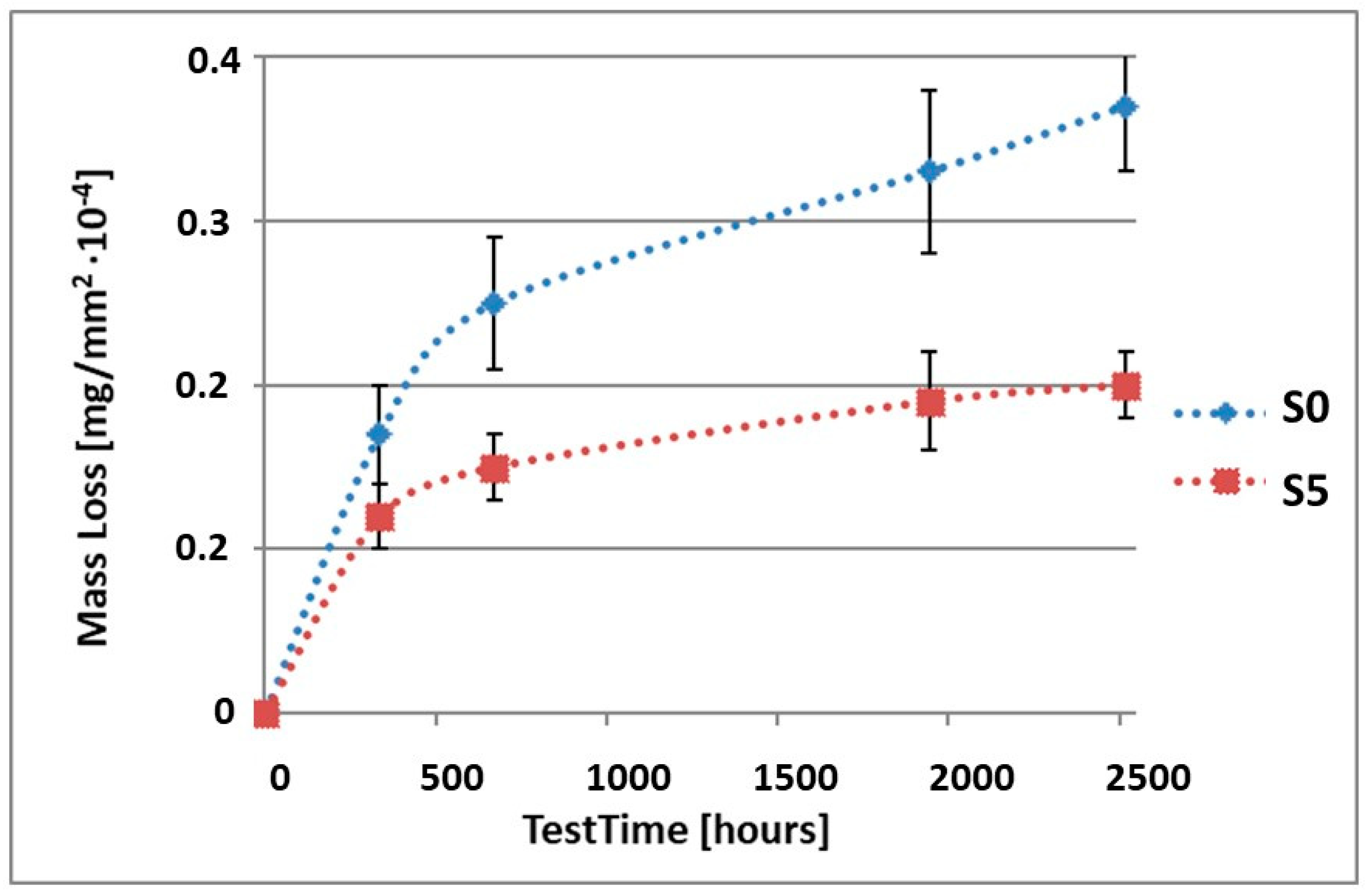
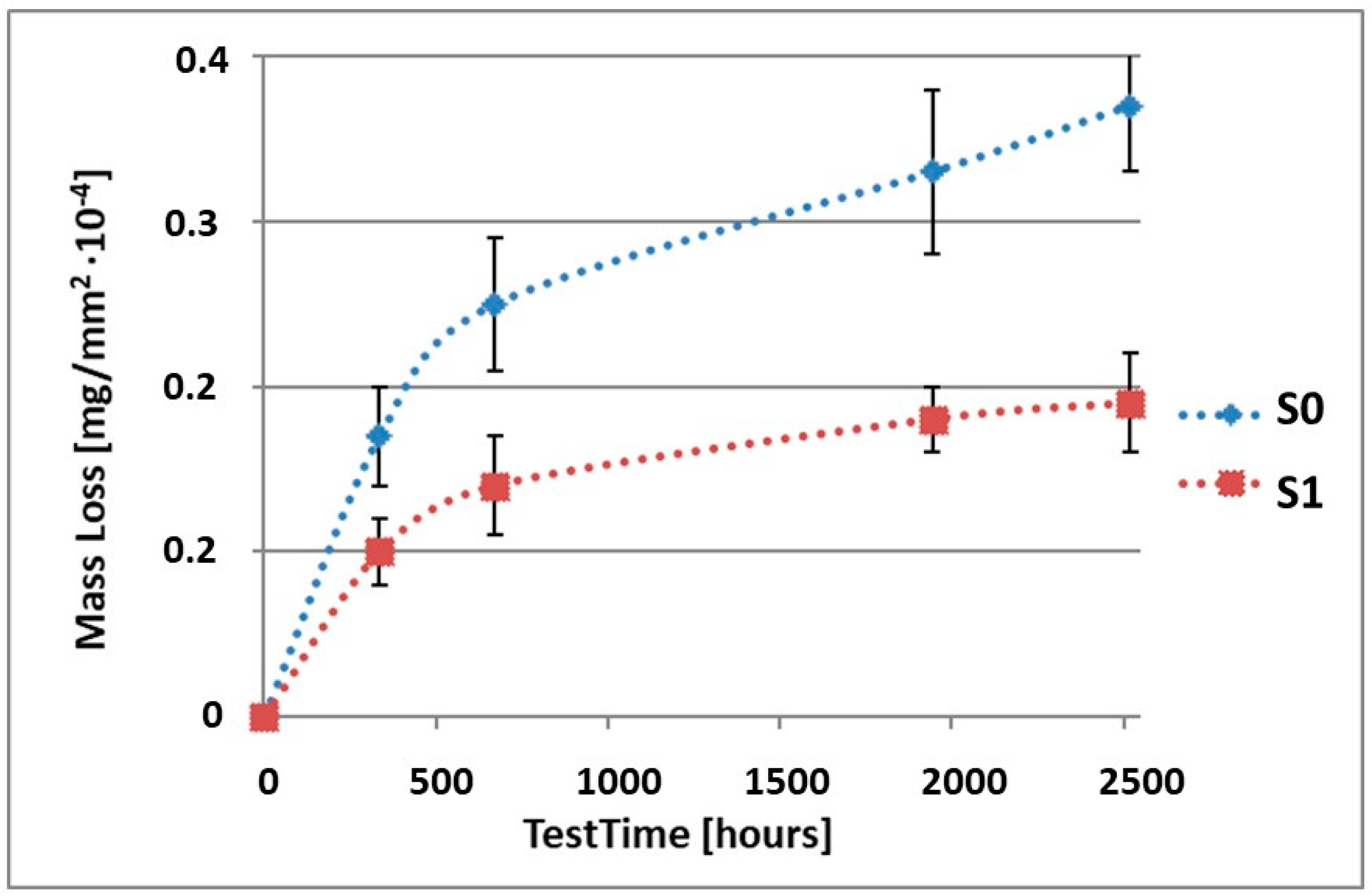
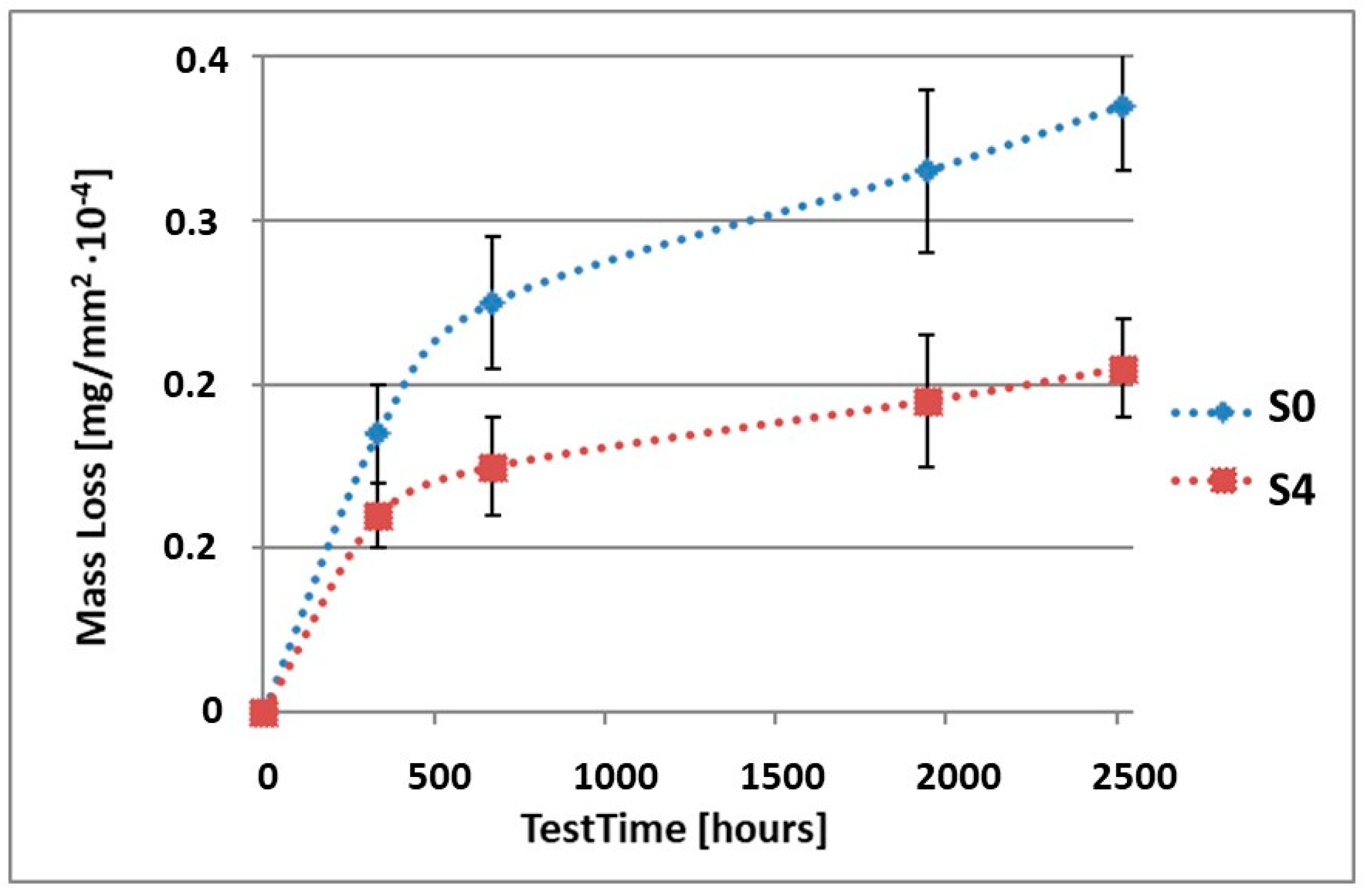
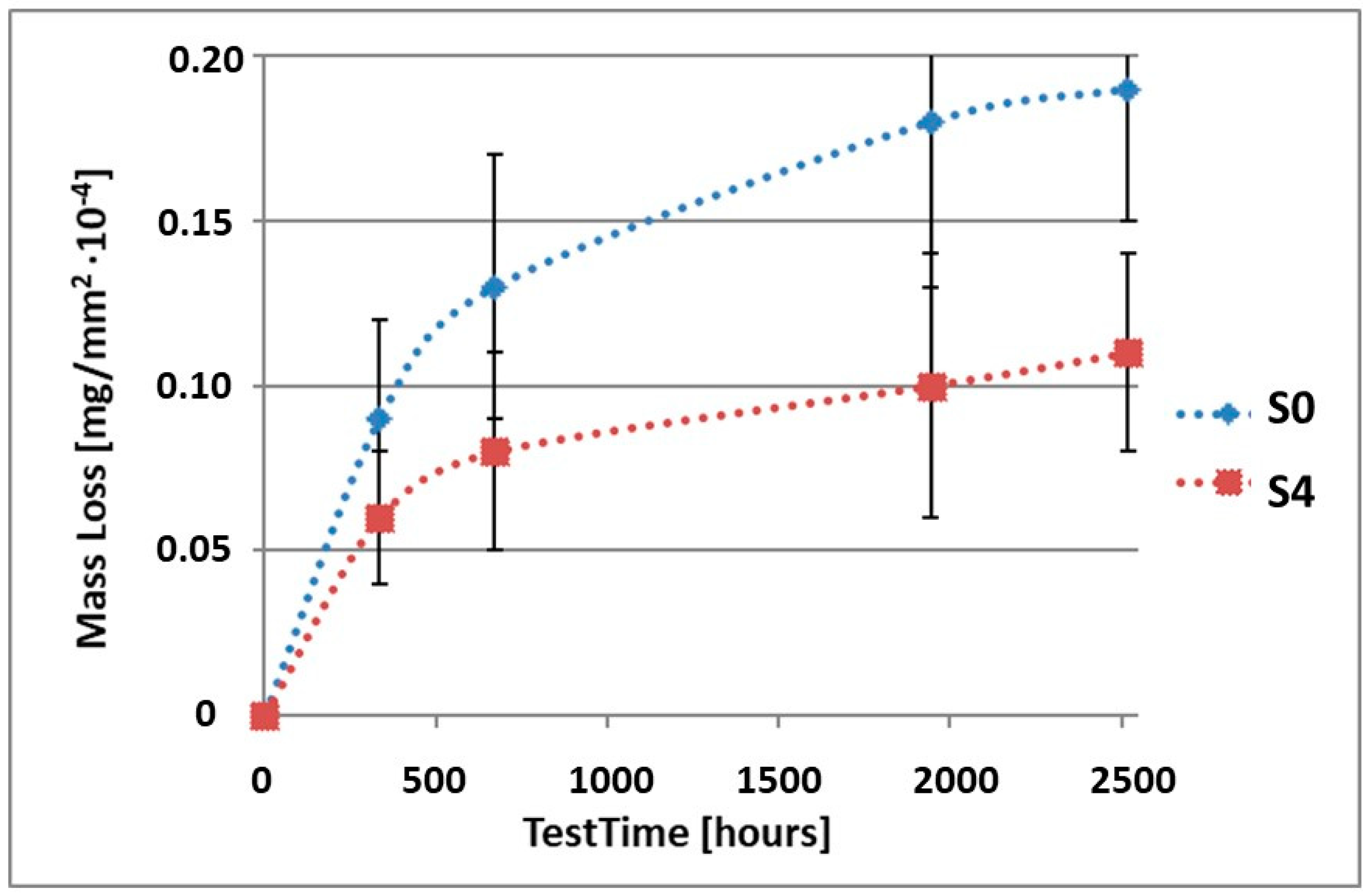
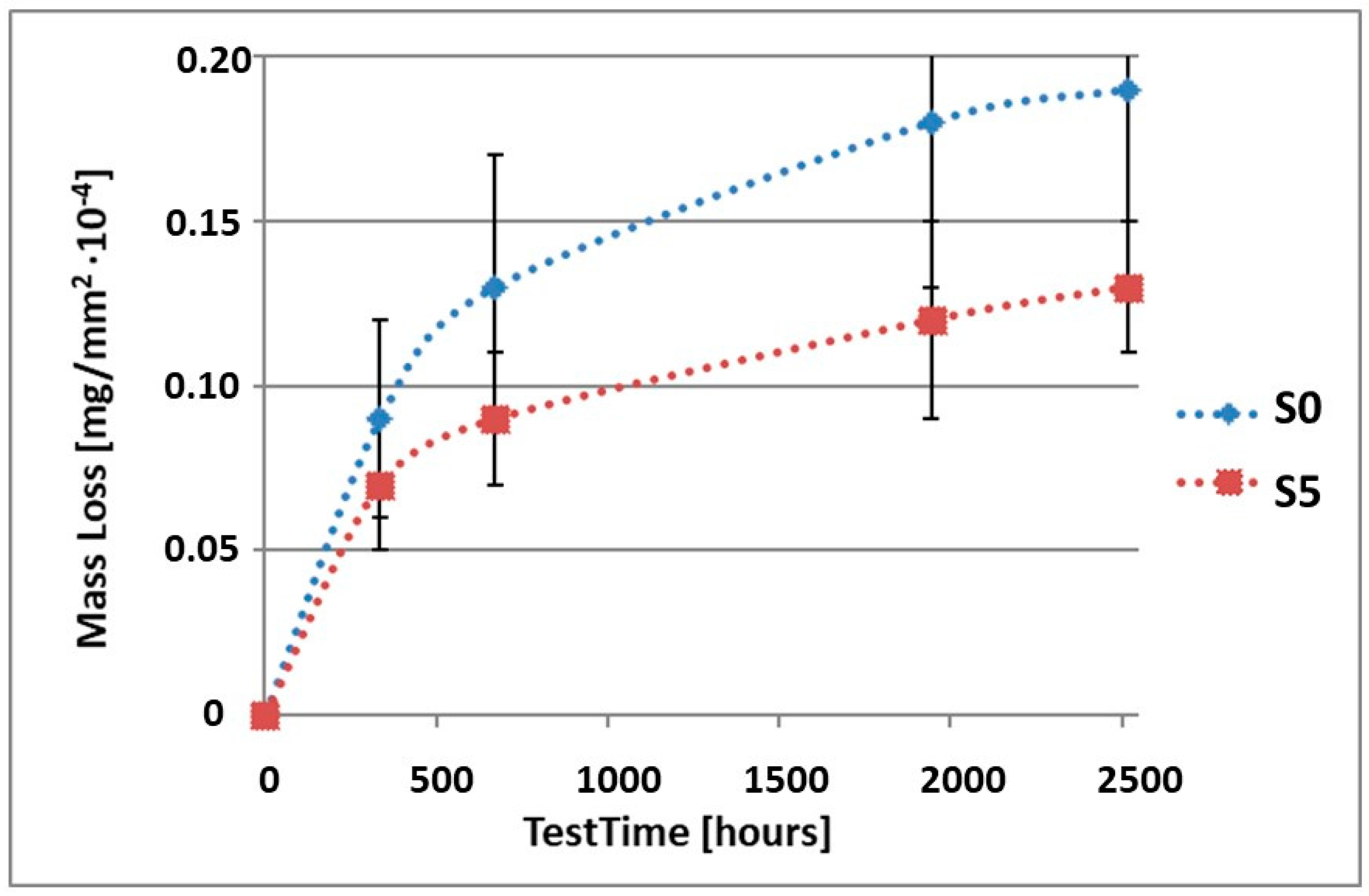
| Mass Loss of Samples after Specific Test Times [mg/mm2 ×10−4} | ||||||
|---|---|---|---|---|---|---|
| Sample | Test Time [Days/Hours] | |||||
| 0 | 14/336 | 28/672 | 81/1994 | 105/2520 | ||
| S0 | Average | 0.17 | 0.25 | 0.33 | 0.37 | |
| Standard deviation | 0.03 | 0.04 | 0.05 | 0.04 | ||
| S1 | Average | 0.10 | 0.14 | 0.18 | 0.19 | |
| Standard deviation | 0.02 | 0.03 | 0.02 | 0.03 | ||
| S2 | Average | 0.13 | 0.15 | 0.19 | 0.20 | |
| Standard deviation | 0.03 | 0.03 | 0.03 | 0.04 | ||
| S3 | Average | 0.12 | 0.15 | 0.19 | 0.20 | |
| Standard deviation | 0.03 | 0.03 | 0.03 | 0.04 | ||
| S4 | Average | 0.12 | 0.15 | 0.19 | 0.21 | |
| Standard deviation | 0.02 | 0.03 | 0.04 | 0.03 | ||
| S5 | Average | 0.12 | 0.15 | 0.19 | 0.20 | |
| Standard deviation | 0.02 | 0.02 | 0.03 | 0.02 | ||
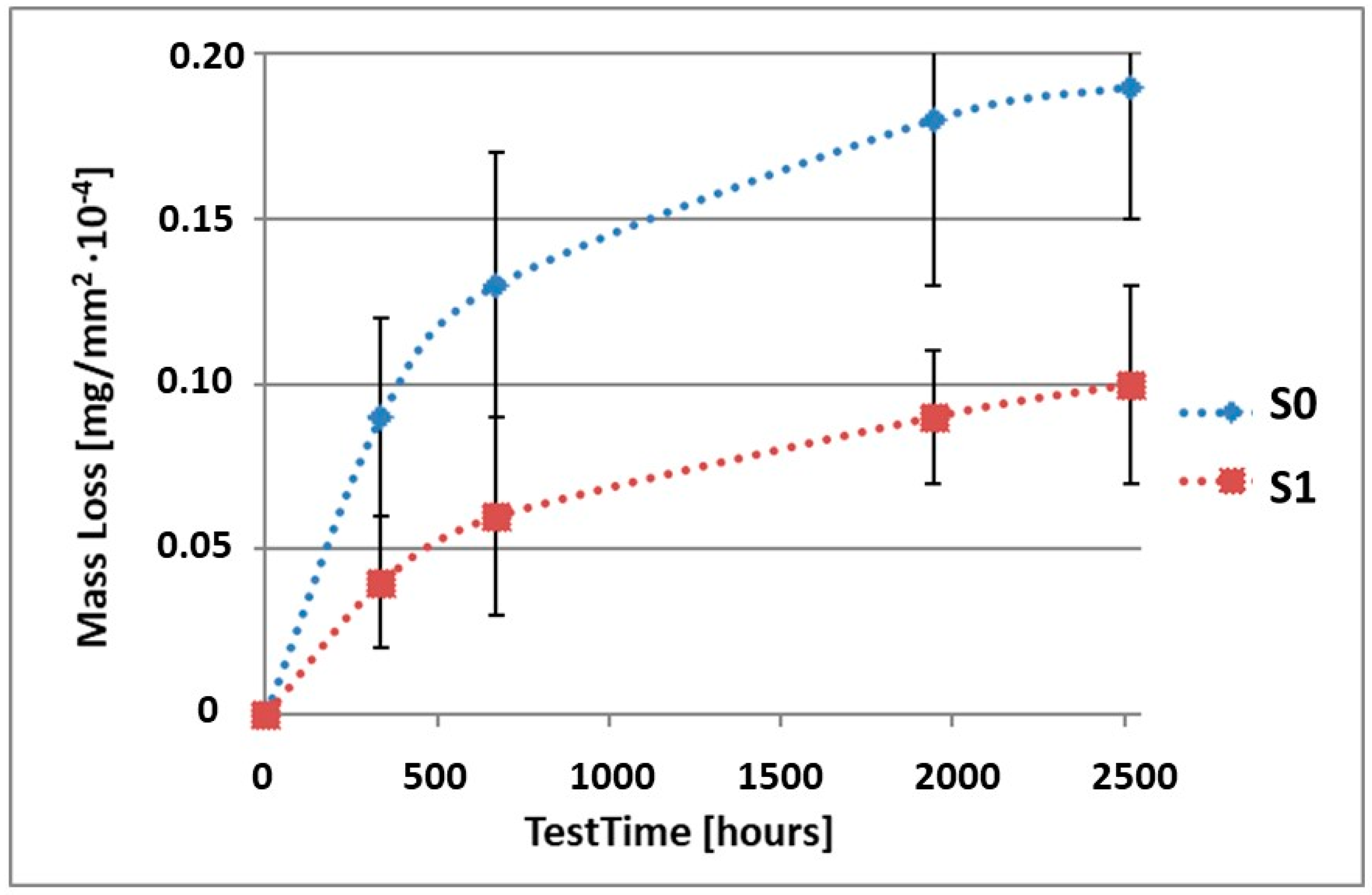
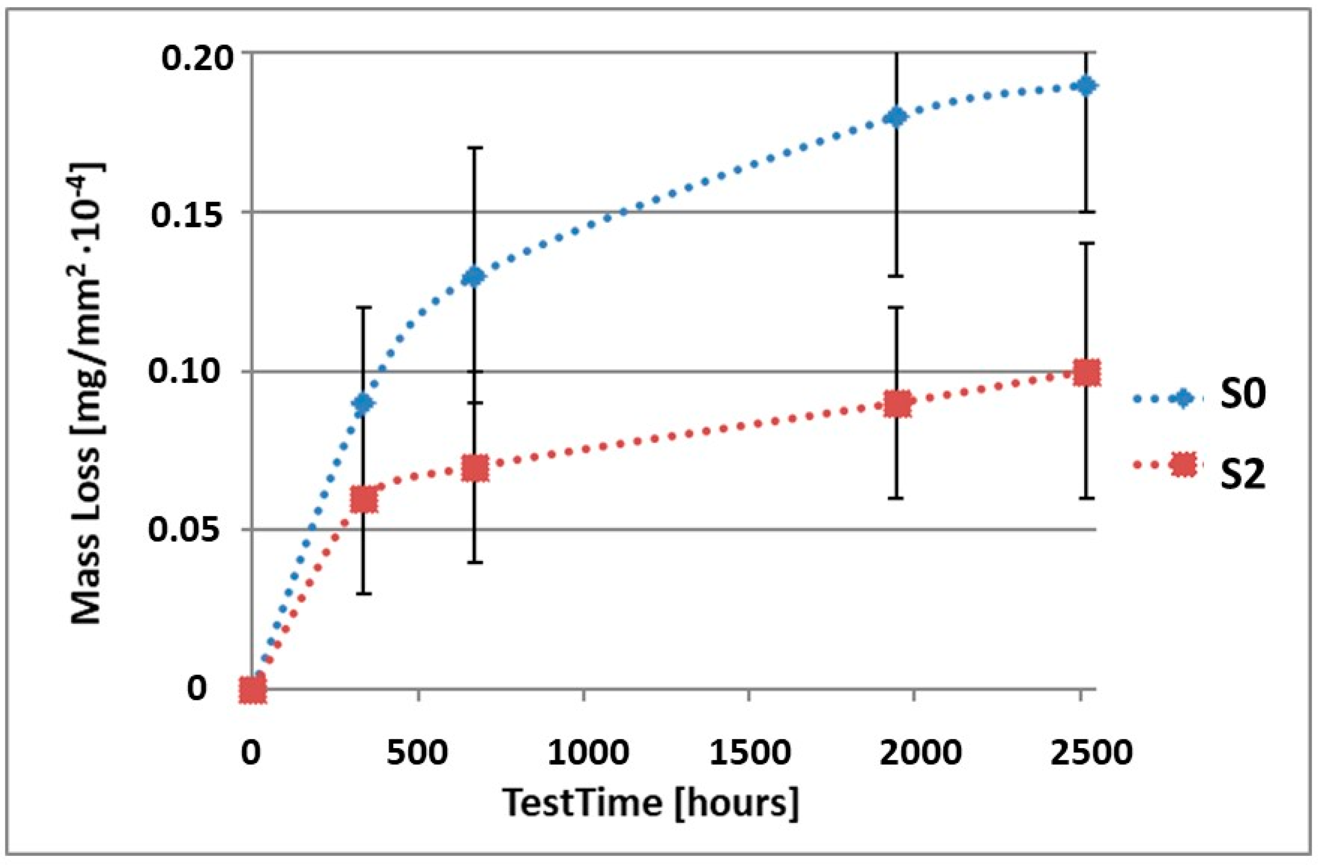
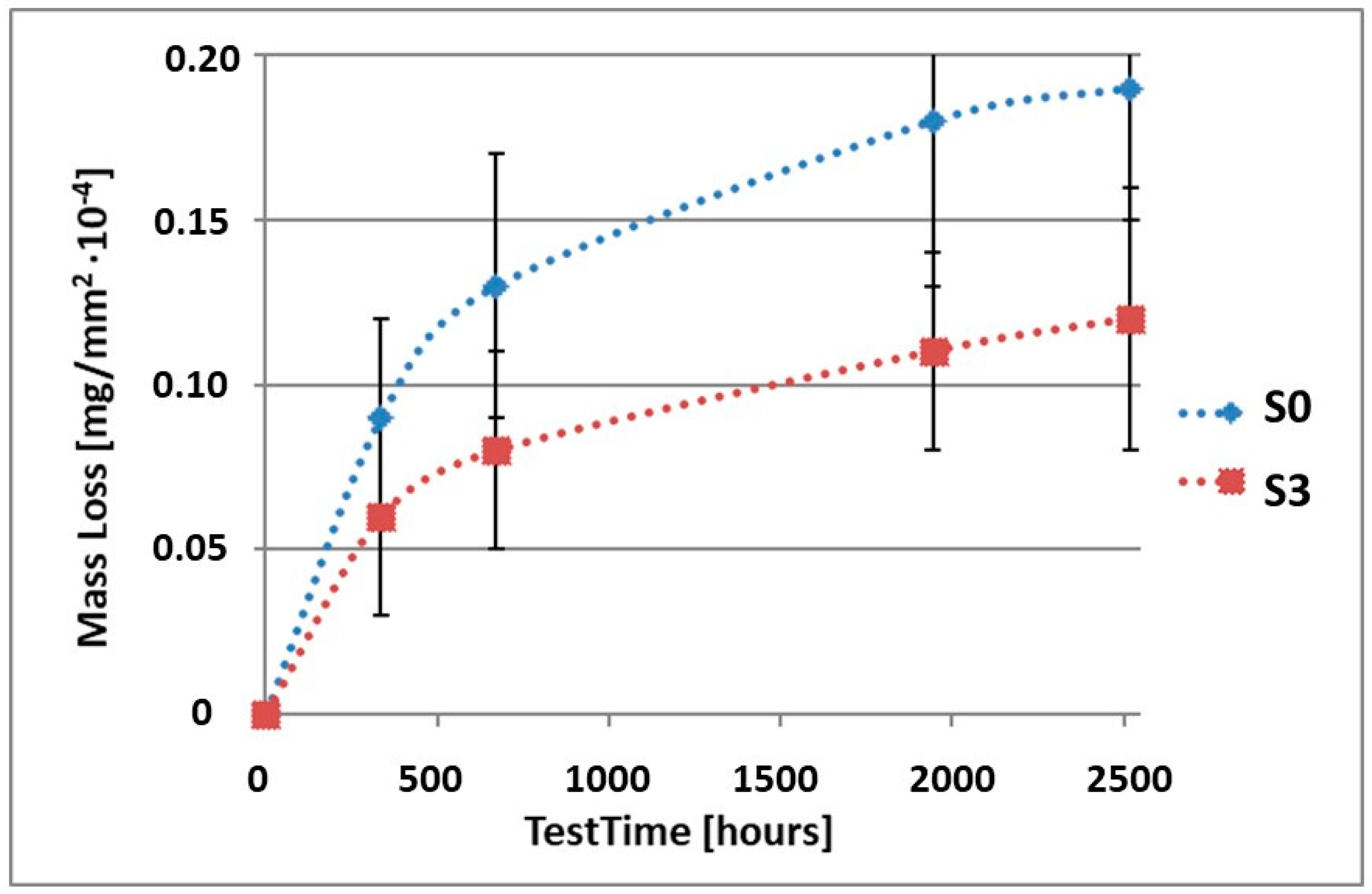
| Mass Loss of Samples after Specific Test Times [mg/mm2 ×10−4} | ||||||
|---|---|---|---|---|---|---|
| Sample | Test Time [Days/Hours] | |||||
| 0 | 14/336 | 28/672 | 81/1994 | 105/2520 | ||
| S0 | Average | 0.09 | 0.13 | 0.18 | 0.19 | |
| Standard deviation | 0.03 | 0.04 | 0.05 | 0.04 | ||
| S1 | Average | 0.04 | 0.06 | 0.09 | 0.10 | |
| Standard deviation | 0.02 | 0.03 | 0.02 | 0.03 | ||
| S2 | Average | 0.06 | 0.07 | 0.09 | 0.10 | |
| Standard deviation | 0.03 | 0.03 | 0.03 | 0.04 | ||
| S3 | Average | 0.06 | 0.08 | 0.11 | 0.12 | |
| Standard deviation | 0.03 | 0.03 | 0.03 | 0.04 | ||
| S4 | average | 0.06 | 0.08 | 0.10 | 0.11 | |
| Standard deviation | 0.02 | 0.03 | 0.04 | 0.03 | ||
| S5 | Average | 0.07 | 0.09 | 0.12 | 0.13 | |
| Standard deviation | 0.02 | 0.02 | 0.03 | 0.02 | ||
Publisher’s Note: MDPI stays neutral with regard to jurisdictional claims in published maps and institutional affiliations. |
© 2022 by the authors. Licensee MDPI, Basel, Switzerland. This article is an open access article distributed under the terms and conditions of the Creative Commons Attribution (CC BY) license (https://creativecommons.org/licenses/by/4.0/).
Share and Cite
Banaszek, K.; Maślanka, M.; Semenov, M.; Klimek, L. Corrosive Studies of a Prosthetic Ni-Cr Alloy Coated with Ti(C,N) Type Layers. Materials 2022, 15, 2471. https://doi.org/10.3390/ma15072471
Banaszek K, Maślanka M, Semenov M, Klimek L. Corrosive Studies of a Prosthetic Ni-Cr Alloy Coated with Ti(C,N) Type Layers. Materials. 2022; 15(7):2471. https://doi.org/10.3390/ma15072471
Chicago/Turabian StyleBanaszek, Katarzyna, Marek Maślanka, Michael Semenov, and Leszek Klimek. 2022. "Corrosive Studies of a Prosthetic Ni-Cr Alloy Coated with Ti(C,N) Type Layers" Materials 15, no. 7: 2471. https://doi.org/10.3390/ma15072471
APA StyleBanaszek, K., Maślanka, M., Semenov, M., & Klimek, L. (2022). Corrosive Studies of a Prosthetic Ni-Cr Alloy Coated with Ti(C,N) Type Layers. Materials, 15(7), 2471. https://doi.org/10.3390/ma15072471