
Raman Spectroscopy as a Novel Method for the Characterization of Polydioxanone Medical Stents Biodegradation

 , ,
, , 
Abstract
:1. Introduction
2. Materials and Methods
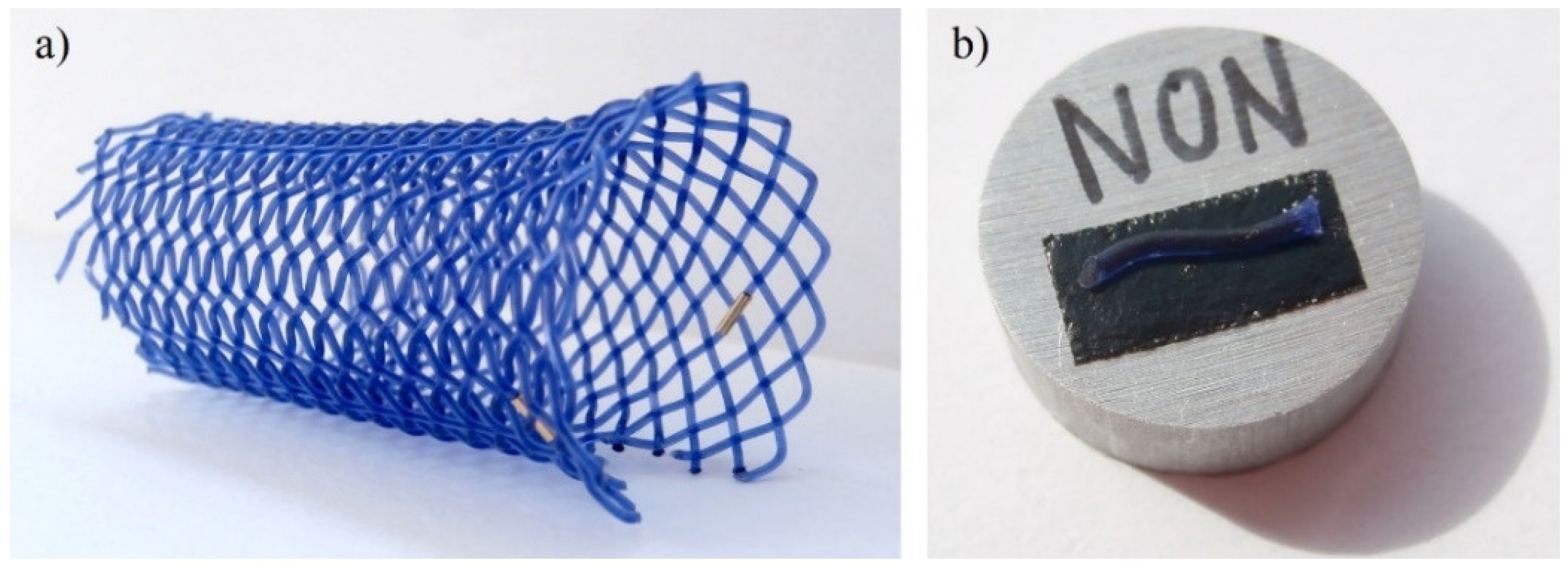
2.1. Degradation Process
2.2. Determination of Crystallinity by Differential Scanning Calorimetry (DSC)
2.3. Surface Morphology
2.4. Infrared Spectroscopy
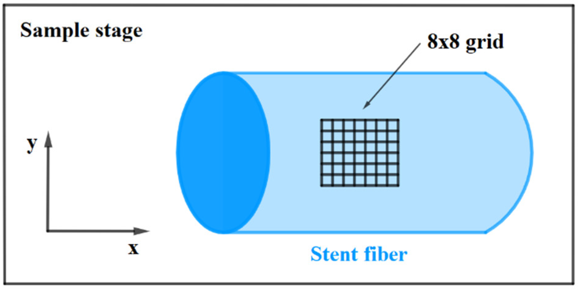
2.5. Raman Spectroscopy
3. Results and Discussion
3.1. Determination of Crystallinity by DSC
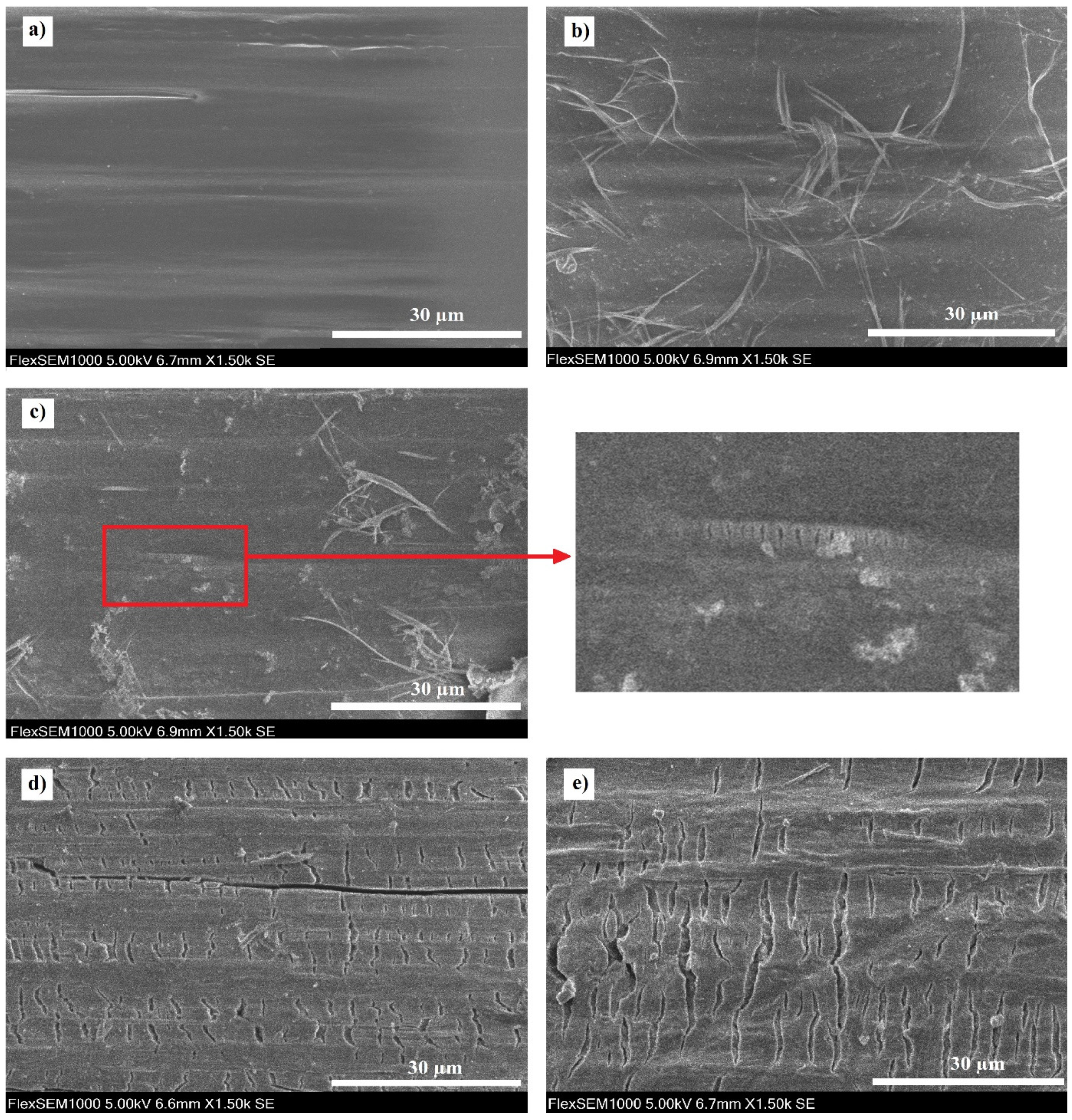
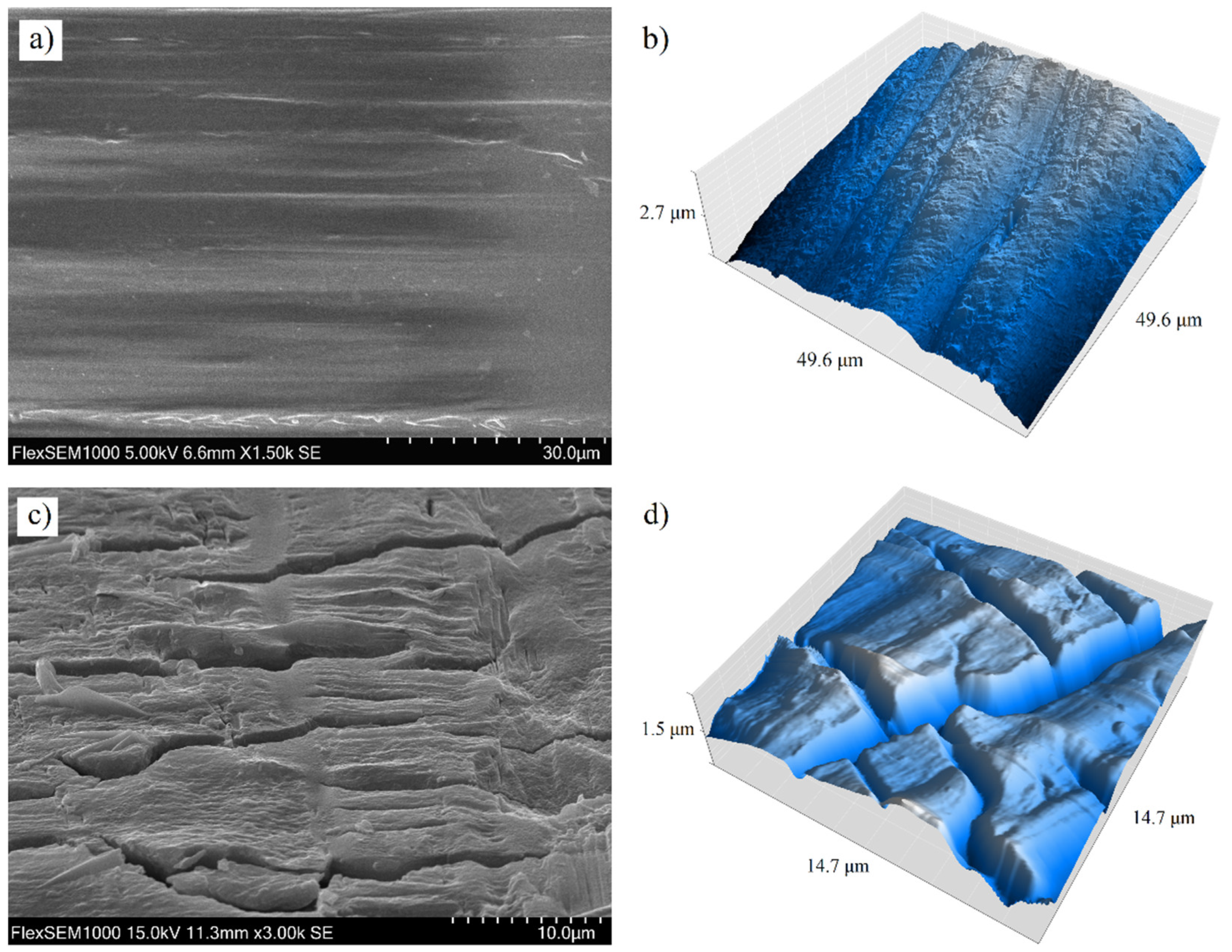
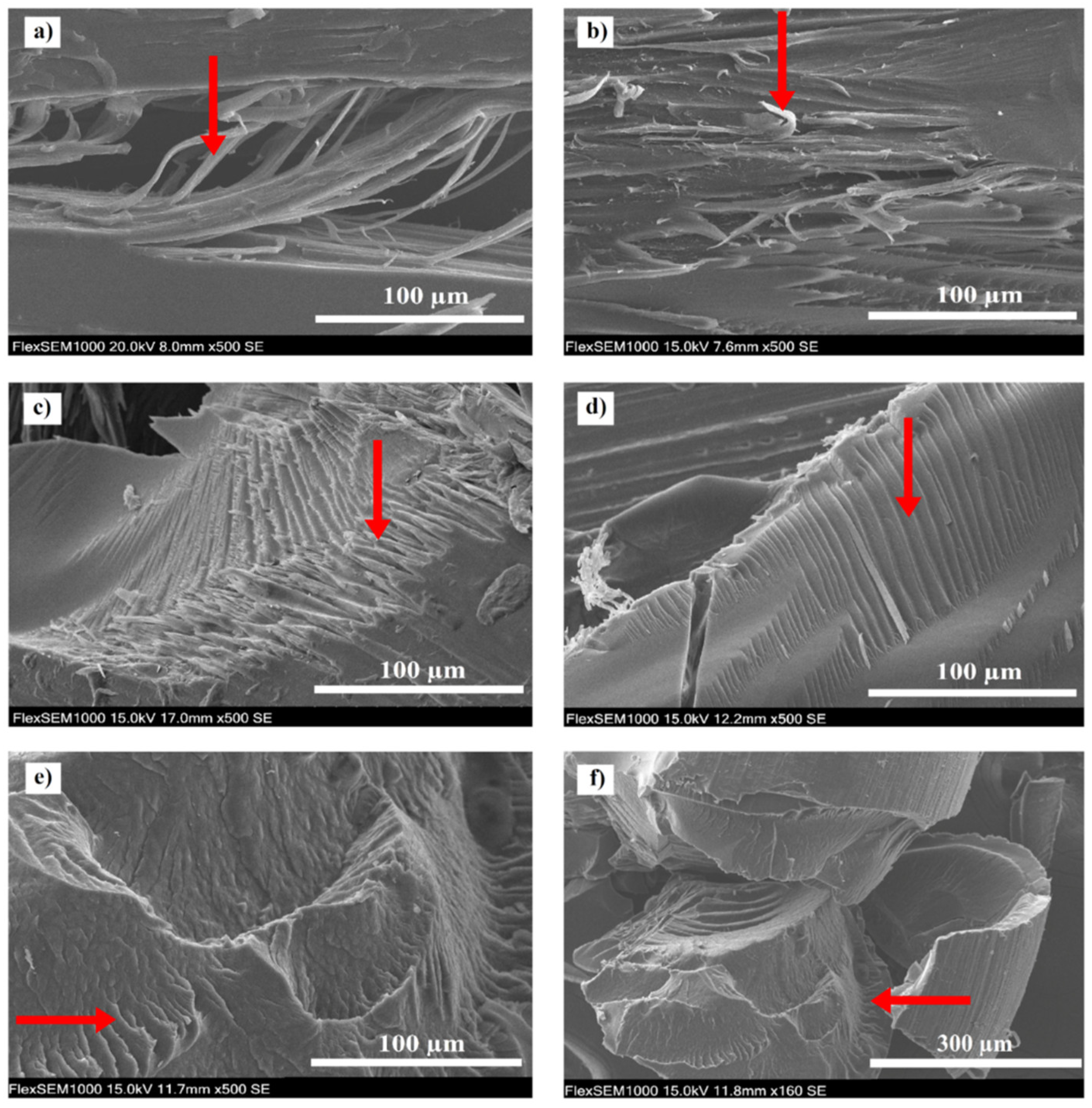
3.2. Surface Morphology
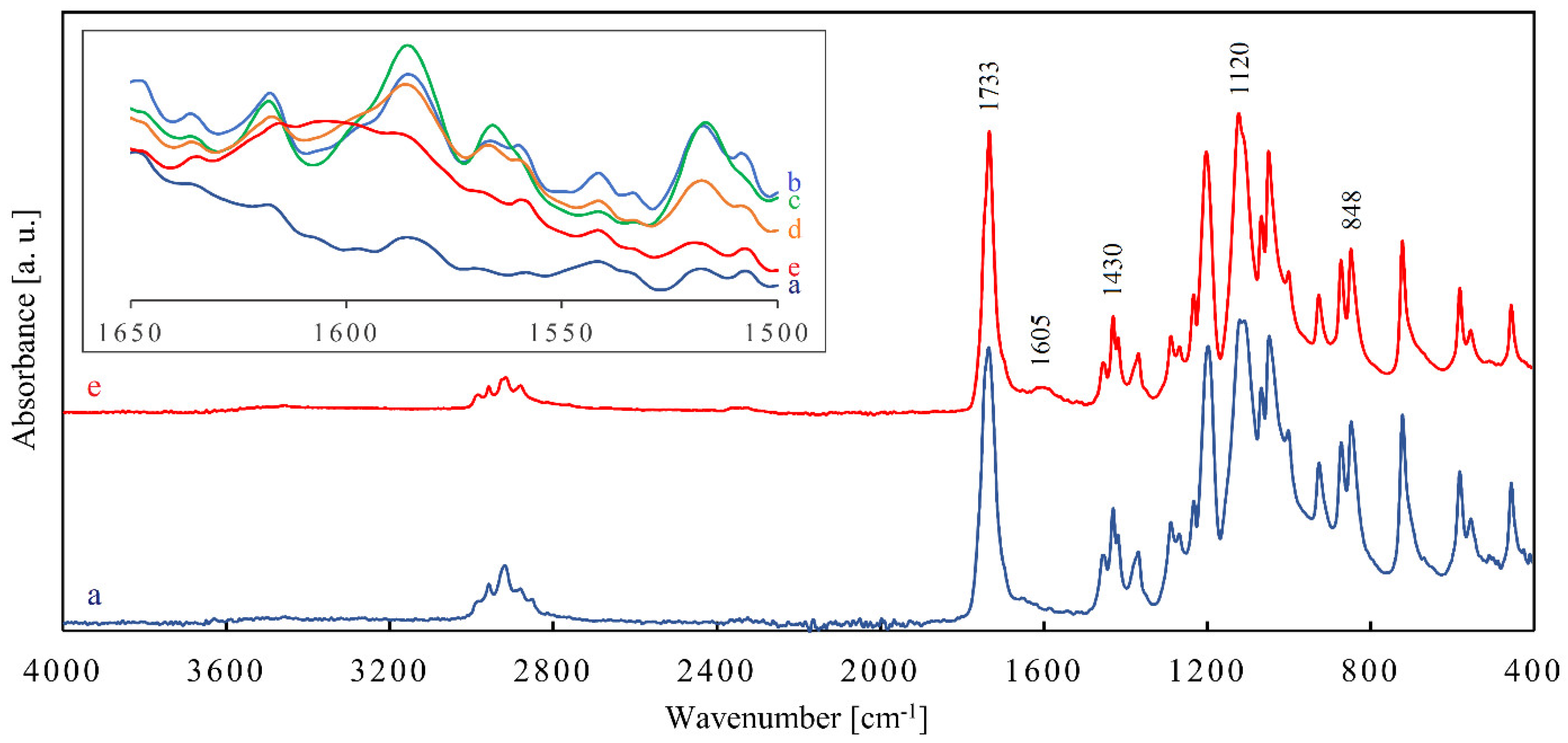
3.3. Infrared Spectroscopy
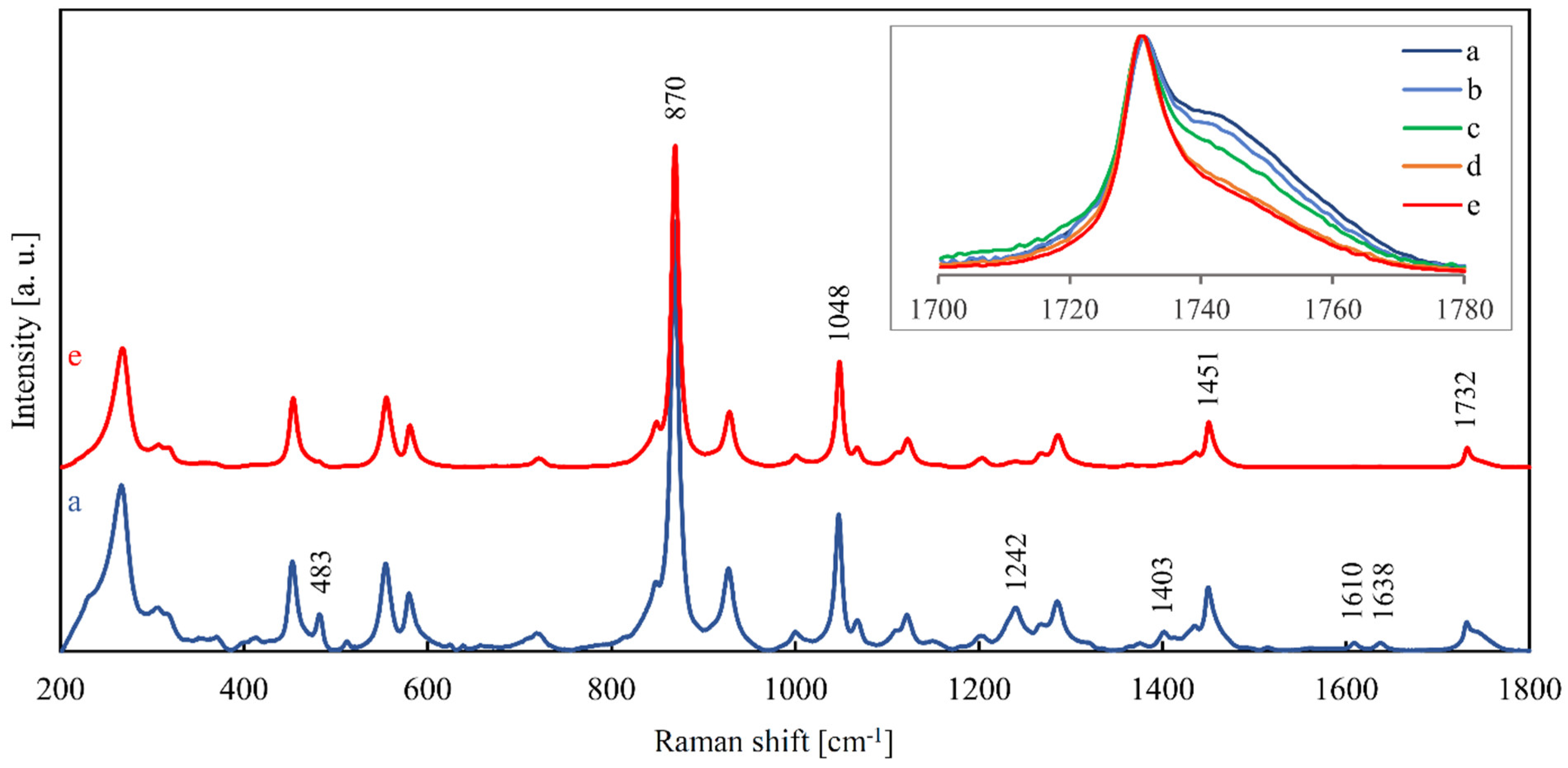
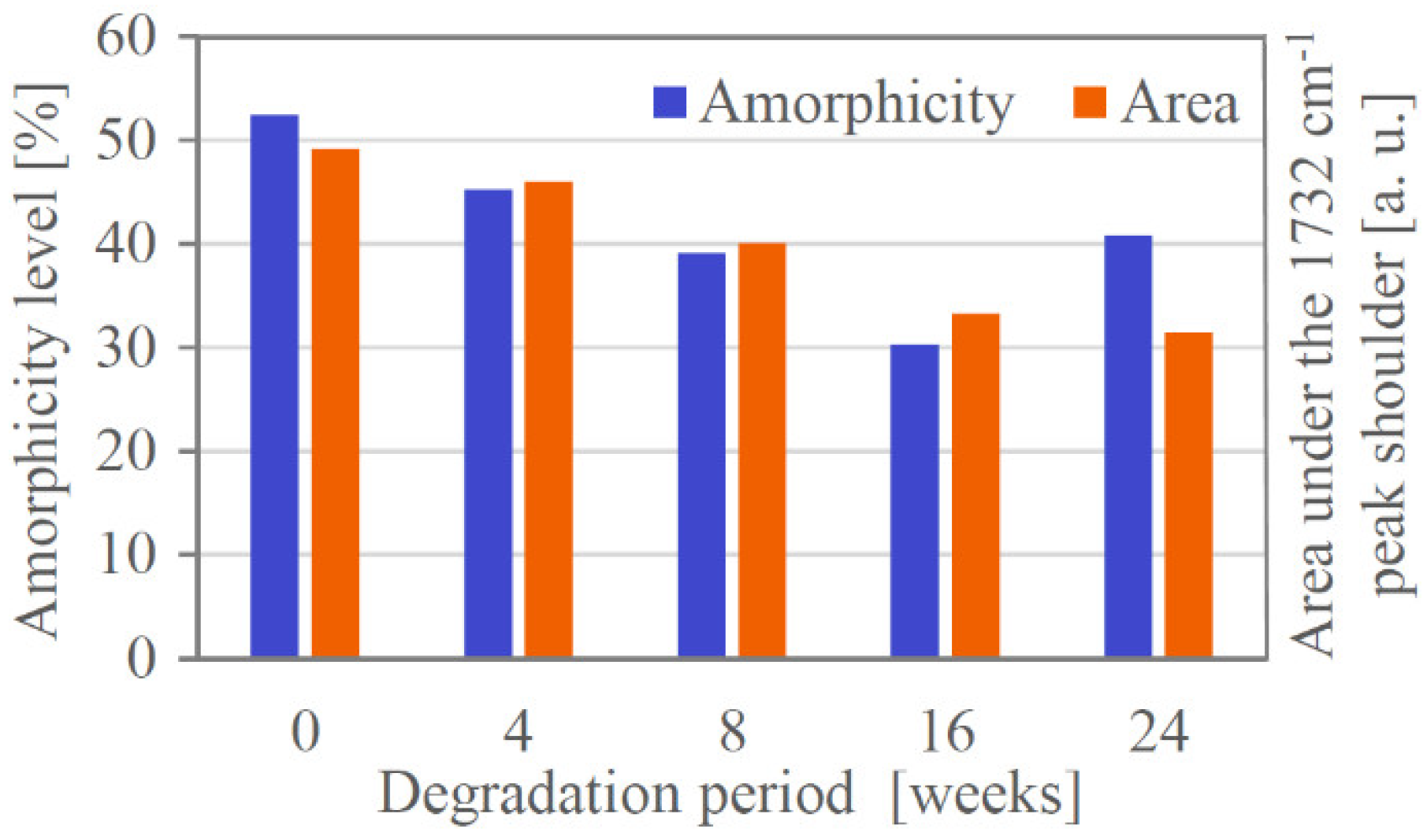
3.4. Raman Spectroscopy
4. Conclusions
Supplementary Materials
Author Contributions
Funding
Institutional Review Board Statement
Informed Consent Statement
Data Availability Statement
Acknowledgments
Conflicts of Interest
References
- Škrlová, K.; Malachová, K.; Muñoz-Bonilla, A.; Měřinská, D.; Rybková, Z.; Fernández-García, M.; Plachá, D. Biocompatible polymer materials with antimicrobial properties for preparation of stents. Nanomaterials 2019, 9, 1548. [Google Scholar] [CrossRef] [PubMed] [Green Version]
- Alexy, R.D.; Levi, D.S. Materials and manufacturing technologies available for production of a pediatric bioabsorbable stent. Biomed Res. Int. 2013, 2013, 137985. [Google Scholar] [CrossRef] [Green Version]
- Pillai, S.K.C.; Sharma, C.P. Review paper: Absorbable polymeric surgical sutures: Chemistry, production, properties, biodegradability, and performance. J. Biomater. Appl. 2010, 25, 291–366. [Google Scholar] [CrossRef]
- Cachia, V.V. Bone Fixation Device. US5893850A, 13 April 1999. [Google Scholar]
- Gertzman, A.; Thompson, D.R. Annealed Polydioxanone Surgical Device and Method for Producing the Same. US4591630A, 27 May 1986. [Google Scholar]
- Zhang, T.; Zhou, S.; Gao, X.; Yang, Z.; Sun, L.; Zhang, D. A multi-scale method for modeling degradation of bioresorbable polyesters. Acta Biomater. 2017, 50, 462–475. [Google Scholar] [CrossRef]
- Goonoo, N.; Jeetah, R.; Bhaw-Luximon, A.; Jhurry, D. Polydioxanone-based materials for tissue engineering and cell/drug delivery applications. Eur. J. Pharm. Biopharm. 2015, 97, 371–391. [Google Scholar] [CrossRef] [PubMed]
- Saska, S.; Pilatti, L.; Silva, E.S.d.S.; Nagasawa, M.A.; Câmara, D.; Lizier, N.; Finger, E.; Konwińska, M.D.; Kempisty, B.; Tunchel, S.; et al. Polydioxanone-based membranes for bone regeneration. Polymers 2021, 13, 1685. [Google Scholar] [CrossRef] [PubMed]
- McManus, M.C.; Sell, S.A.; Bowen, W.C.; Koo, H.P.; Simpson, D.G.; Bowlin, G.L. Electrospun fibrinogen-polydioxanone composite matrix: Potential for in situ urologic tissue engineering. J. Eng. Fiber. Fabr. 2008, 3, 12–21. [Google Scholar] [CrossRef] [Green Version]
- Adhikari, K.R.; Stanishevskaya, I.; Caracciolo, P.C.; Abraham, G.A.; Thomas, V. Novel poly(ester urethane urea)/polydioxanone blends: Electrospun fibrous meshes and films. Molecules 2021, 26, 3847. [Google Scholar] [CrossRef] [PubMed]
- Przybysz-Romatowska, M.; Haponiuk, J.; Formela, K. Reactive extrusion of biodegradable aliphatic polyesters in the presence of free-radical-initiators: A review. Polym. Degrad. Stab. 2020, 182, 109383. [Google Scholar] [CrossRef]
- Zahir, L.; Kida, T.; Ryo, T.; Nakayama, Y.; Shiono, T.; Kawasaki, N.; Yamano, N.; Nakayuma, A. Synthesis of thermoplastic elastomers with high biodegradability in seawater. Polym. Degrad. Stab. 2021, 184, 109467. [Google Scholar] [CrossRef]
- Yang, K.-K.; Wang, X.-L.; Wang, Y.-Z. Poly(p-dioxanone) and its copolymers. J. Macromol. Sci. Part C Polym. Rev. 2007, 42, 373–398. [Google Scholar] [CrossRef]
- Berg, M.; Walter, D.; Vries, E.; Vleggaar, F.P.; van Berge Henegouwen, M.; van Hillegersberg, R.; Siersema, P.; Fockens, P.; Hooft, J. Biodegradable stent placement before neoadjuvant chemoradiotherapy as a bridge to surgery in patients with locally advanced esophageal cancer. Gastrointest. Endosc. 2014, 80, 908–913. [Google Scholar] [CrossRef]
- Del-Pozo-García, A.J.; Piedracoba-Cadahia, C.; Sánchez-Gómez, F.; Marín-Gabriel, J.C.; Rodríguez-Muñoz, S. Complete resolution of dysphagia after sequential polyflexTM stenting in a case of recurrent anastomotic stenosis in an adult with congenital esophageal atresia. Rev. Esp. Enfermedades Dig. 2018, 110, 826–829. [Google Scholar] [CrossRef] [Green Version]
- Rejchrt, S.; Kopacova, M.; Brozik, J.; Bures, J. Biodegradable stents for the treatment of benign stenoses of the small and large intestines. Endoscopy 2011, 43, 911–917. [Google Scholar] [CrossRef]
- Griffiths, B.; James, P.; Morgan, G.; Diamantopoulos, A.; Durward, A.; Nyman, A. Biodegradable stents for the relief of vascular bronchial compression in children with left atrial enlargement. J. Bronchol. Interv. Pulmonol. 2020, 27, 200–204. [Google Scholar] [CrossRef] [PubMed]
- Antón-Pacheco, J.L.; Luna, C.; García, E.; López, M.; Morante, R.; Tordable, C.; Palacios, A.; de Miguel, M.; Benavent, I.; Gómez, A. Initial experience with a new biodegradable airway stent in children: Is this the stent we were waiting for? Pediatr. Pulmonol. 2016, 51, 607–612. [Google Scholar] [CrossRef]
- Siiki, A.; Rinta-Kiikka, I.; Sand, J.; Laukkarinen, J. Endoscopic biodegradable biliary stents in the treatment of benign biliary strictures: First report of clinical use in patients. Dig. Endosc. 2017, 29, 118–121. [Google Scholar] [CrossRef] [PubMed]
- Grolich, T.; Crha, M.; Novotný, L.; Kala, Z.; Hep, A.; Nečas, A.; Hlavsa, J.; Mitas, L.; Misik, J. Self-expandable biodegradable biliary stents in porcine model. J. Surg. Res. 2015, 193, 606–612. [Google Scholar] [CrossRef]
- Kwon, C.-I.; Son, J.S.; Kim, K.S.; Moon, J.P.; Park, S.; Jeon, J.; Kim, G.; Choi, S.H.; Ko, K.H.; Jeong, S.; et al. Mechanical properties and degradation process of biliary self-expandable biodegradable stents. Dig. Endosc. 2020, in press. [Google Scholar]
- Zhu, Y.; Yang, K.; Cheng, R.; Xiang, Y.; Yuan, T.; Cheng, Y.; Sarmento, B.; Cui, W. The current status of biodegradable stent to treat benign luminal disease. Mater. Today 2017, 20, 516–529. [Google Scholar] [CrossRef]
- Bezrouk, A.; Hosszu, T.; Hromadko, L.; Zmrhalova, Z.O.; Kopecek, M.; Smutny, M.; Krulichova, I.S.; Macak, J.M.; Kremlacek, J. Mechanical properties of a biodegradable self-expandable polydioxanone monofilament stent: In vitro force relaxation and its clinical relevance. PLoS ONE 2020, 15, e0235842. [Google Scholar] [CrossRef] [PubMed]
- Tian, Y.; Zhang, J.; Cheng, J.; Wu, G.; Zhang, Y.; Ni, Z.; Zhao, G. A poly(L-lactic acid) monofilament with high mechanical properties for application in biodegradable biliary stents. J. Appl. Polym. Sci. 2021, 138, 49656. [Google Scholar] [CrossRef]
- Han, C.-M.; Lih, E.; Choi, S.-K.; Bedair, T.M.; Lee, Y.-J.; Park, W.; Han, D.K.; Son, J.S.; Joung, Y.K. Biodegradable sheath-core biphasic monofilament braided stent for bio-functional treatment of esophageal strictures. J. Ind. Eng. Chem. 2018, 67, 396–406. [Google Scholar] [CrossRef]
- Wang, C.; Zhang, P. Design and characterization of PDO biodegradable intravascular stents. Text. Res. J. 2017, 87, 1968–1976. [Google Scholar] [CrossRef]
- Li, G.; Li, Y.; Lan, P.; Li, J.; Zhao, Z.; He, X.; Zhang, J.; Hu, H. Biodegradable weft-knitted intestinal stents: Fabrication and Physical changes investigation in vitro degradation. J. Biomed. Mater. Res. Part A 2014, 102, 982–990. [Google Scholar] [CrossRef]
- Wang, P.-J.; Ferralis, N.; Conway, C.; Grossman, J.C.; Edelman, E.R. Strain-induced accelerated asymmetric spatial degradation of polymeric vascular scaffolds. Proc. Natl. Acad. Sci. USA 2018, 115, 2640–2645. [Google Scholar] [CrossRef] [PubMed] [Green Version]
- Vano-Herrera, K.; Vogt, C. Degradation of poly(l-lactic acid) coating on permanent coronary metal stent investigated ex vivo by micro raman spectroscopy. J. Raman Spectrosc. 2017, 48, 711–719. [Google Scholar] [CrossRef]
- Doddi, N.; Versfelt, C.C.; Wasserman, D. Synthetic Absorbable Surgical Devices of Poly-Dioxanone. US4052988A, 11 October 1977. [Google Scholar]
- Gestí, S.; Lotz, B.; Casas, M.T.; Alemán, C.; Puiggali, J. Morphology and structure of poly(p-dioxanone). Eur. Polym. J. 2007, 43, 4662–4674. [Google Scholar] [CrossRef]
- Qu, L.; Cao, J.; Huang, X.-M. Clinical application of biodegradable polydioxanone. J. Clin. Rehabil. Tissue Eng. Res. 2011, 15, 527–530. [Google Scholar] [CrossRef]
- Sahmel, O.; Arbeiter, D.; Siewert, S.; Schümann, K.; Schmitz, K.-P.; Grabow, N. Optimization of manufacturing processes for biodegradable polymeric stents regarding improved mechanical properties. Curr. Dir. Biomed. Eng. 2018, 4, 583–585. [Google Scholar] [CrossRef]
- Guerra, A.J.; Cano, P.; Rabionet, M.; Puig, T.; Ciurana, J. 3D-printed PCL/PLA composite stents: Towards a new solution to cardiovascular problems. Materials 2018, 11, 1679. [Google Scholar] [CrossRef] [PubMed] [Green Version]
- Furuhashi, Y.; Nakayama, A.; Monno, T.; Kawahara, Y.; Yamane, H.; Kimura, Y.; Iwata, T. X-ray and electron diffraction study of poly(p-dioxanone). Macromol. Rapid Commun. 2004, 25, 1943–1947. [Google Scholar] [CrossRef]
- Ooi, C.P.; Cameron, R.E. The hydrolytic degradation of polydioxanone (PDSII) sutures. Part I: Morphological aspects. J. Biomed. Mater. Res. 2002, 63, 280–290. [Google Scholar] [CrossRef]
- Jaidann, M.; Brisson, J. Conformation Study of poly(p-dioxanone) fibers by polarized raman spectroscopy, X-ray diffraction, and conformation analysis. J. Polym. Sci. Part B Polym. Phys. 2008, 46, 406–417. [Google Scholar] [CrossRef]
- Farah, S.; Anderson, D.G.; Langer, R. Physical and mechanical properties of PLA, and their functions in widespread applications—A comprehensive review. Adv. Drug Deliv. Rev. 2016, 107, 367–392. [Google Scholar] [CrossRef] [PubMed] [Green Version]
- Chu, C.C. Hydrolytic degradation of polyglycolic acid: Tensile strength and crystallinity study. J. Appl. Polym. Sci. 1981, 26, 1727–1734. [Google Scholar] [CrossRef]
- Bower, D.I. An Introduction to Polymer Physics; Cambridge University Press: Cambridge, UK, 2002. [Google Scholar]
- Rizzarelli, P.; Carroccio, S. Modern mass spectrometry in the characterization and degradation of biodegradable polymers. Anal. Chim. Acta 2014, 808, 18–43. [Google Scholar] [CrossRef] [PubMed]
- Hakkarainen, M.; Adamus, G.; Höglund, A.; Kowalczuk, M.; Albertsson, A.-C. ESI-MS reveals the influence of hydrophilicity and architecture on the water-soluble degradation product patterns of biodegradable homo- and copolyesters of 1,5-dioxepan-2-one and epsilon-caprolactone. Macromolecules 2008, 41, 3547–3554. [Google Scholar] [CrossRef]
- Márquez, Y.; Franco, L.; Turon, P.; Martínez, J.C.; Puiggalí, J. Study of non-isothermal crystallization of polydioxanone and analysis of morphological changes occurring during heating and cooling processes. Polymers 2016, 8, 351. [Google Scholar] [CrossRef] [Green Version]
- Sabino, M.; Feijoo, J.; Müller, A. Crystallisation and morphology of neat and degraded poly( p-dioxanone). Polym. Degrad. Stab. 2001, 73, 541–547. [Google Scholar] [CrossRef]
- Sabino, M.; González, S.; Márquez, L.; Feijoo, J. Study of the hydrolytic degradation of polydioxanone PPDX. Polym. Degrad. Stab. 2000, 69, 209–216. [Google Scholar] [CrossRef]
- Bower, D.I.; Maddams, W.F. The Vibrational Spectroscopy of Polymers; Cambridge Solid State Science Series; Cambridge University Press: Cambridge, UK, 1989. [Google Scholar]
- ELLA-CS. BD Stent. Available online: https://www.ellacs.cz/en/bd-stent (accessed on 15 September 2021).
- Ferreira, T.; Rasband, W. ImageJ User Guide—IJ 1.46r. 2012. Available online: https://imagej.nih.gov/ij/docs/guide/user-guide.pdf (accessed on 15 September 2021).
- Gil-Castell, O.; Badia, J.D.; Bou, J.; Ribes-Greus, A. Performance of polyester-based electrospun scaffolds under in vitro hydrolytic conditions: From short-term to long-term applications. Nanomaterials 2019, 9, 786. [Google Scholar] [CrossRef] [PubMed] [Green Version]
- Callister, W.D.; Rethwisch, D.G. Materials Science and Engineering, 9th ed.; John Wiley & Sons: Hoboken, NJ, USA, 2015. [Google Scholar]
- Kwon, D.; Kim, J., II; Kim, D.; Kang, H.; Lee, B.; Lee, K.; Kim, M. Biodegradable stent. J. Biomed. Sci. Eng. 2012, 5, 208–216. [Google Scholar] [CrossRef] [Green Version]
- Shockley, M.F.; Muliana, A.H. Modeling temporal and spatial changes during hydrolytic degradation and erosion in biodegradable polymers. Polym. Degrad. Stab. 2020, 180, 109298. [Google Scholar] [CrossRef]
- Schrader, B. Infrared and Raman Spectroscopy—Methods and Applications; VCH: Weinheim, Germany, 1995. [Google Scholar]


| Degradation Period | Crystallinity [%] |
| Non-degraded | 47.6 ± 2.3 |
| 4 weeks degraded | 54.8 ± 1.1 |
| 8 weeks degraded | 60.9 ± 0.7 |
| 16 weeks degraded | 69.7 ± 2.2 |
| 24 weeks degraded | 59.2 ± 3.7 |
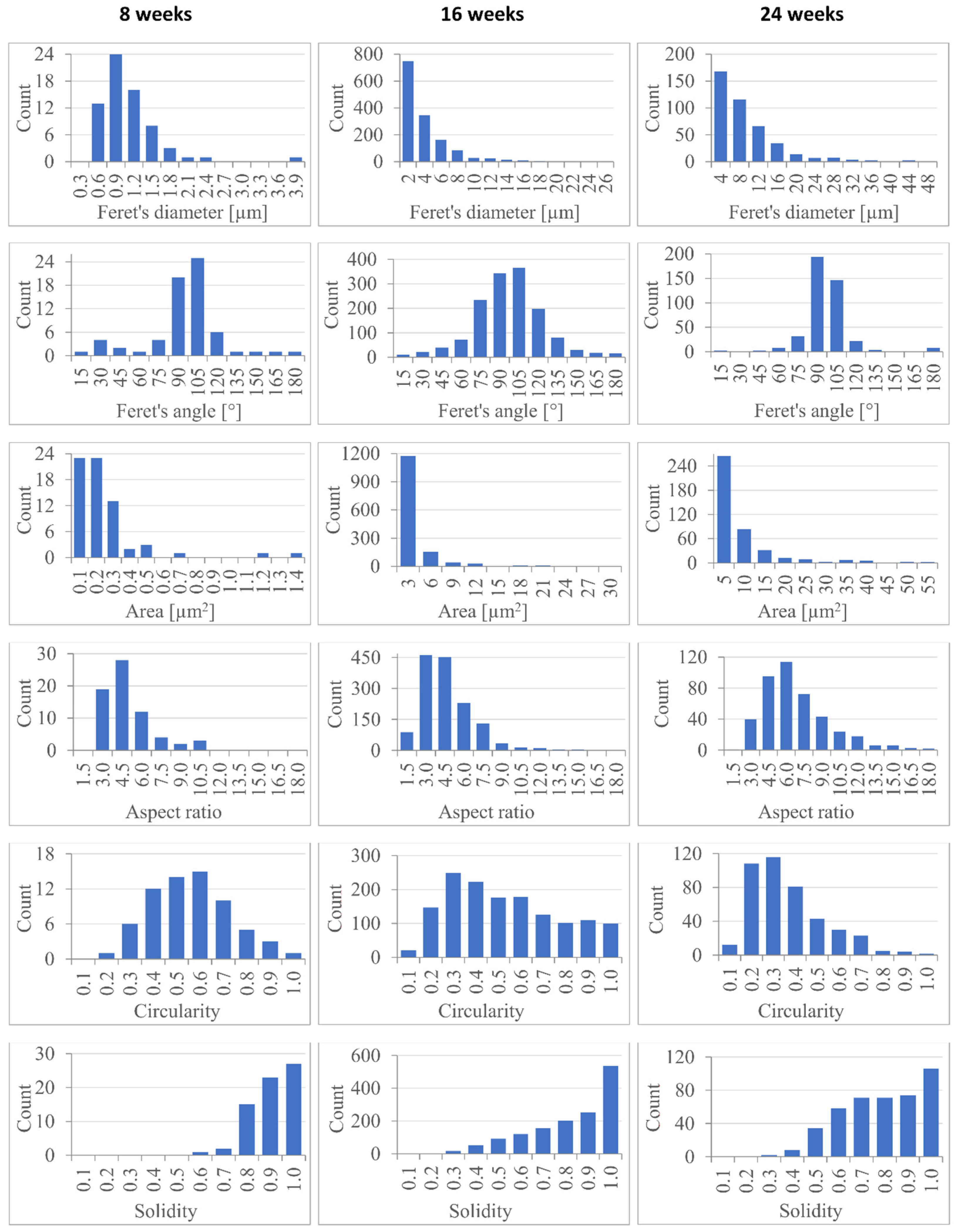
| Size/Shape Descriptor | 8 Weeks Degraded (Mean) | 16 Weeks Degraded (Mean) | 24 Weeks Degraded (Mean) | 8 Weeks Degraded (Median) | 16 Weeks Degraded (Median) | 24 Weeks Degraded (Median) |
| Feret’s diameter [µm] | 0.96 ± 0.51 | 3.04 ± 3.39 | 7.36 ± 6.96 | 0.85 | 1.87 | 5.21 |
| Feret’s angle [°] | 87.3 ± 28.5 | 89.6 ± 27.0 | 89.0 ± 19.1 | 94.5 | 89.0 | 87.8 |
| Area [µm2] | 0.20 ± 0.22 | 1.97 ± 3.92 | 6.85 ± 11.86 | 0.14 | 0.62 | 3.00 |
| Aspect ratio | 4.00 ± 1.82 | 3.90 ± 2.24 | 6.05 ± 2.81 | 3.61 | 3.45 | 5.48 |
| Circularity | 0.51 ± 0.17 | 0.48 ± 0.24 | 0.32 ± 0.17 | 0.51 | 0.45 | 0.27 |
| Solidity | 0.85 ± 0.08 | 0.77 ± 0.19 | 0.74 ± 0.18 | 0.89 | 0.84 | 0.76 |
| Degradation Period | Normalized Area [cm−1] |
| Non-degraded | 9.83 ± 0.22 |
| 4 weeks | 9.24 (8.98, 9.37) |
| 8 weeks | 7.97 (7.80, 8.18) |
| 16 weeks | 6.65 ± 0.24 |
| 24 weeks | 6.30 ± 0.22 |
Publisher’s Note: MDPI stays neutral with regard to jurisdictional claims in published maps and institutional affiliations. |
© 2021 by the authors. Licensee MDPI, Basel, Switzerland. This article is an open access article distributed under the terms and conditions of the Creative Commons Attribution (CC BY) license (https://creativecommons.org/licenses/by/4.0/).
Share and Cite
Loskot, J.; Jezbera, D.; Bezrouk, A.; Doležal, R.; Andrýs, R.; Francová, V.; Miškář, D.; Myslivcová Fučíková, A. Raman Spectroscopy as a Novel Method for the Characterization of Polydioxanone Medical Stents Biodegradation. Materials 2021, 14, 5462. https://doi.org/10.3390/ma14185462
Loskot J, Jezbera D, Bezrouk A, Doležal R, Andrýs R, Francová V, Miškář D, Myslivcová Fučíková A. Raman Spectroscopy as a Novel Method for the Characterization of Polydioxanone Medical Stents Biodegradation. Materials. 2021; 14(18):5462. https://doi.org/10.3390/ma14185462
Chicago/Turabian StyleLoskot, Jan, Daniel Jezbera, Aleš Bezrouk, Rafael Doležal, Rudolf Andrýs, Vendula Francová, Dominik Miškář, and Alena Myslivcová Fučíková. 2021. "Raman Spectroscopy as a Novel Method for the Characterization of Polydioxanone Medical Stents Biodegradation" Materials 14, no. 18: 5462. https://doi.org/10.3390/ma14185462
APA StyleLoskot, J., Jezbera, D., Bezrouk, A., Doležal, R., Andrýs, R., Francová, V., Miškář, D., & Myslivcová Fučíková, A. (2021). Raman Spectroscopy as a Novel Method for the Characterization of Polydioxanone Medical Stents Biodegradation. Materials, 14(18), 5462. https://doi.org/10.3390/ma14185462