FLUKA Simulations of Kβ/Kα Intensity Ratios of Copper in Ag–Cu Alloys

 ,
,  , , , , , , , , and
Abstract
:1. Introduction
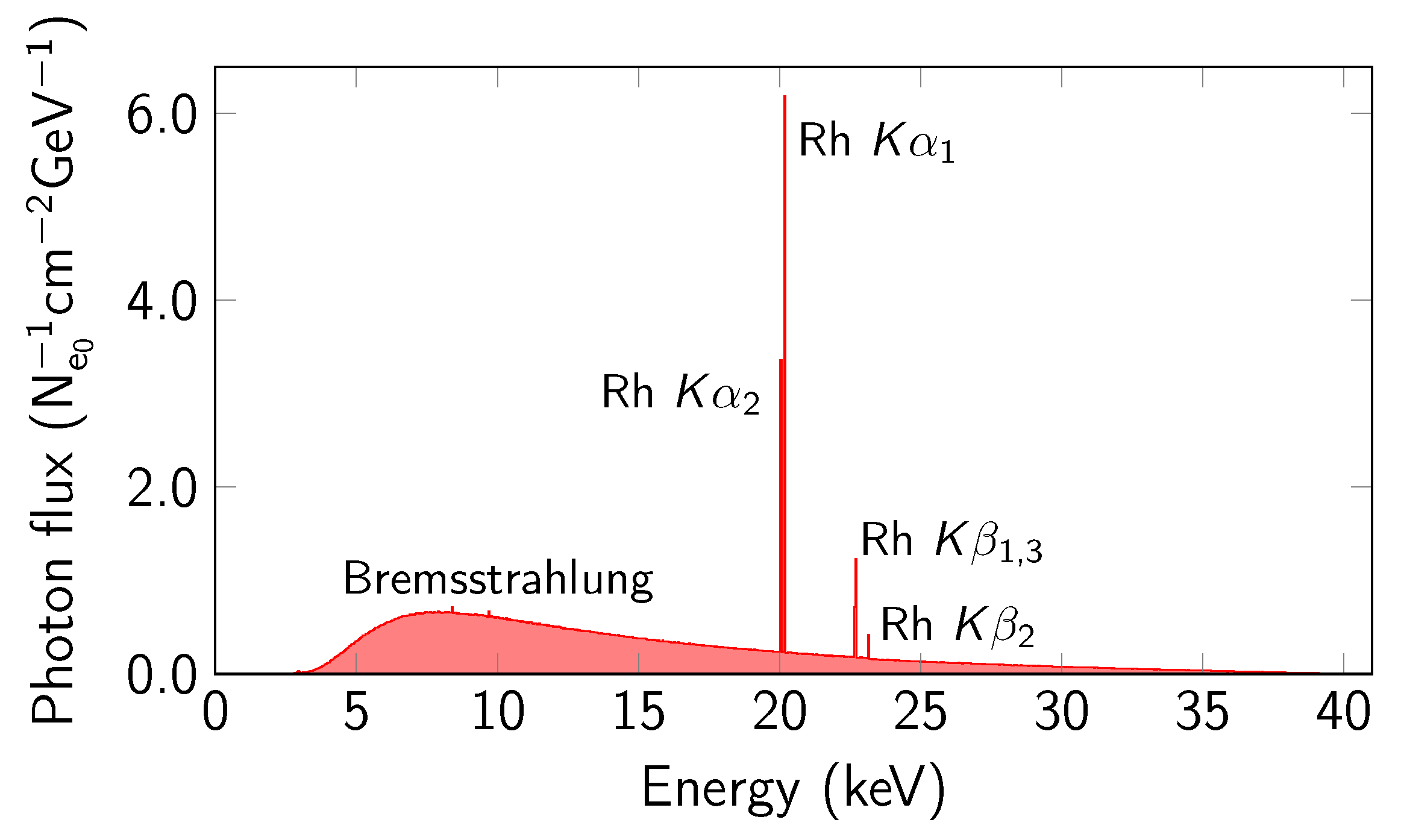
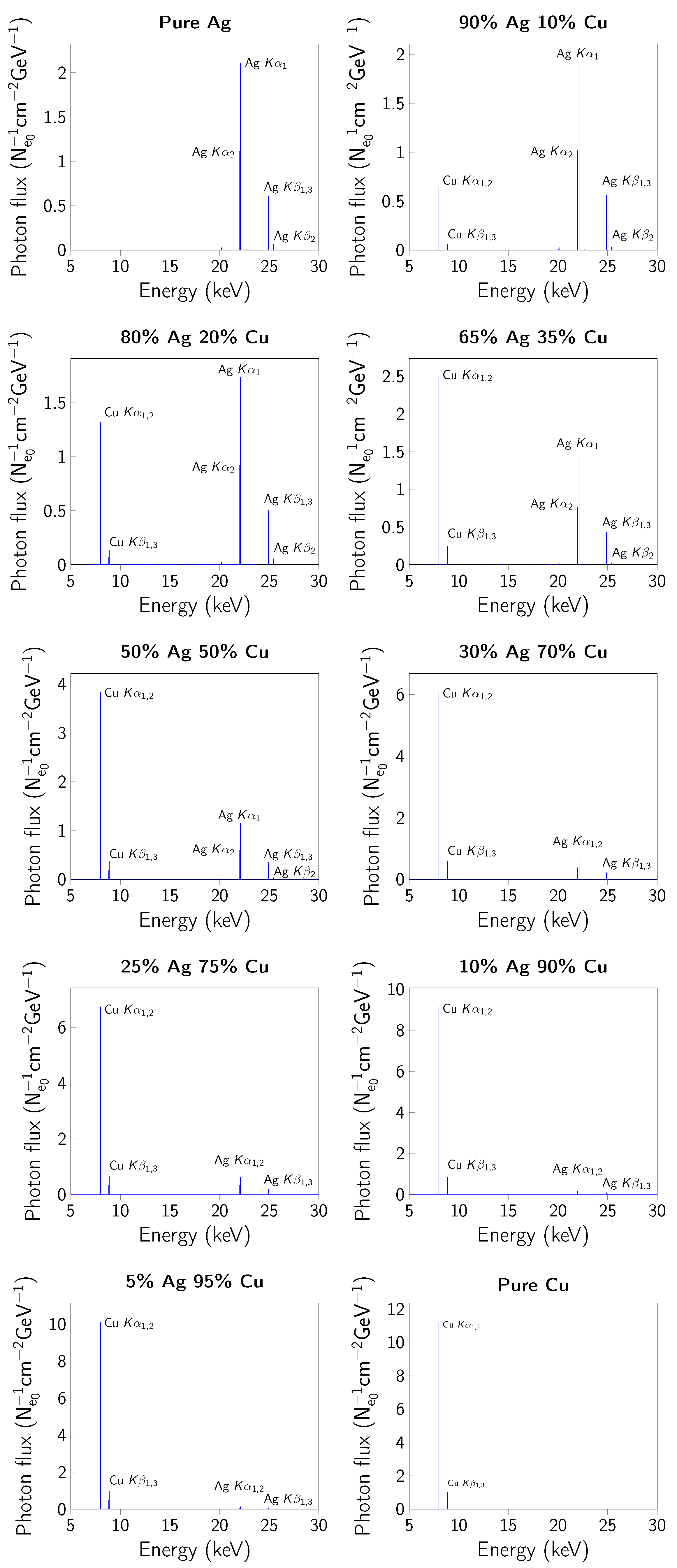
2. Experiment Simulations
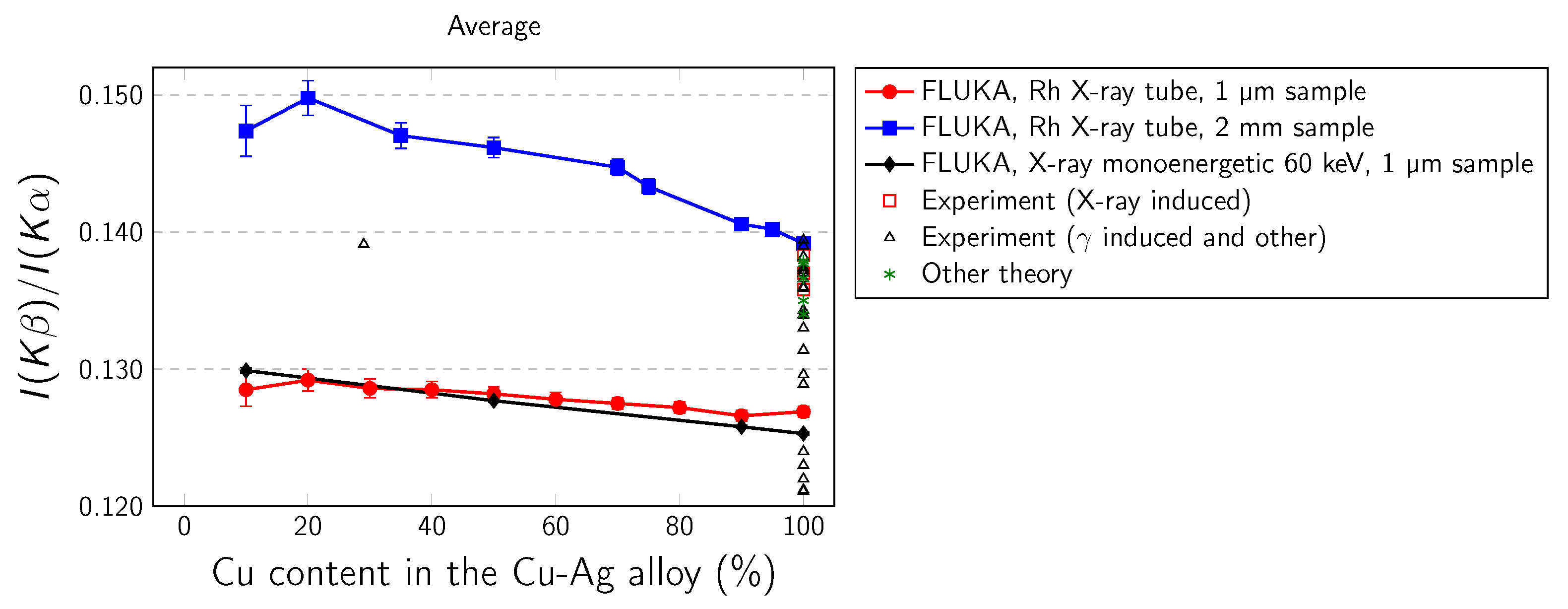
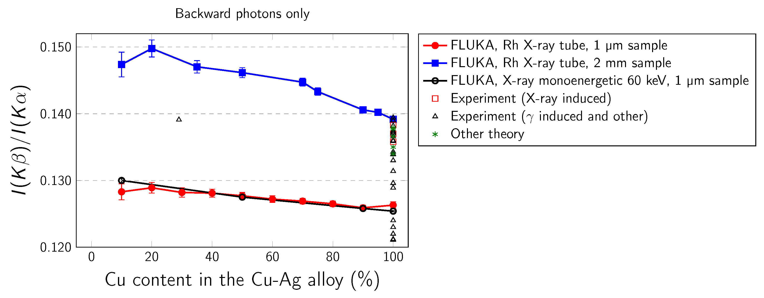
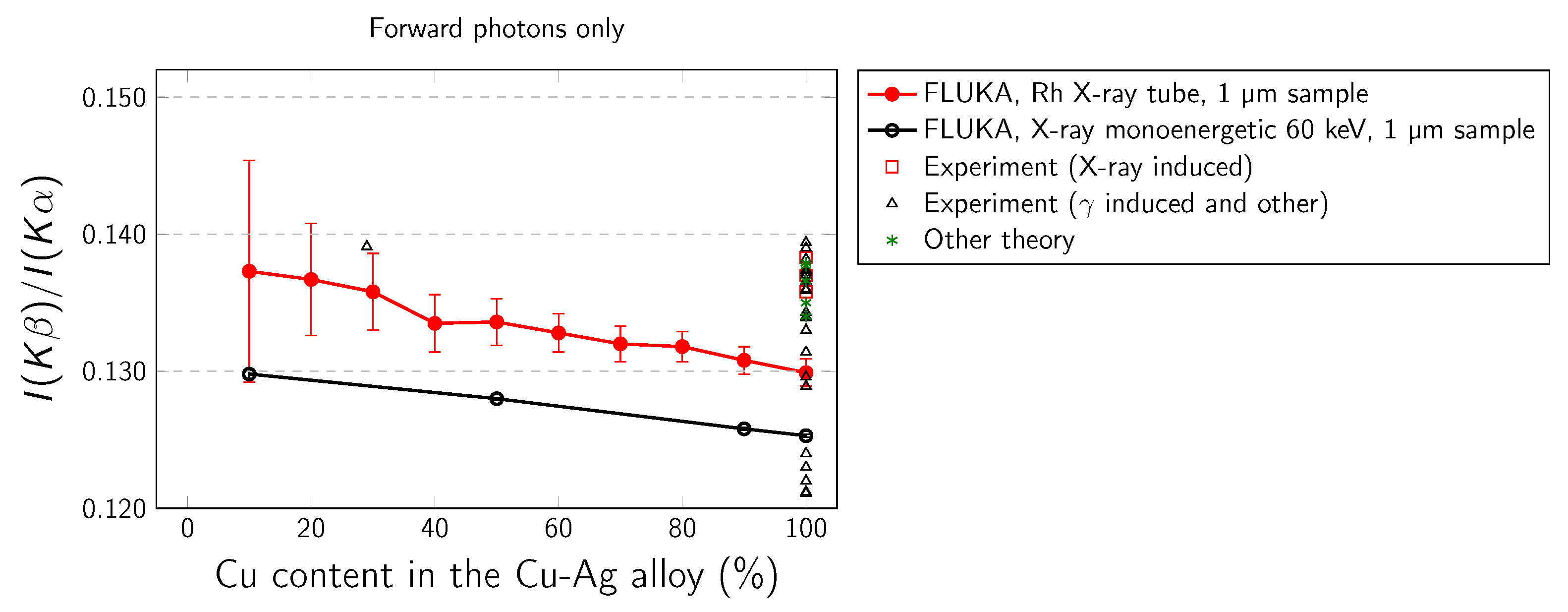
3. Results and Discussion
4. Conclusions
Author Contributions
Funding
Institutional Review Board Statement
Informed Consent Statement
Data Availability Statement
Acknowledgments
Conflicts of Interest
References
- Daoudi, S.; Kahoul, A.; Kup Aylikci, N.; Sampaio, J.; Marques, J.; Aylikci, V.; Sahnoune, Y.; Kasri, Y.; Deghfel, B. Review of experimental photon-induced Kβ/Kα intensity ratios. At. Data Nucl. Data Tables 2020, 132, 101308. [Google Scholar] [CrossRef]
- Scofield, J.H. Exchange corrections of K x-ray emission rates. Phys. Rev. A 1974, 9, 1041–1049. [Google Scholar] [CrossRef]
- Jankowski, K.; Polasik, M. On the calculation of Kβ/Kα X-ray intensity ratios. J. Phys. At. Mol. Opt. Phys. 1989, 22, 2369–2376. [Google Scholar] [CrossRef]
- Polasik, M. Influence of changes in the valence electronic configuration on the Kβ-to-Kα X-ray intensity ratios of the 3d transition metals. Phys. Rev. A 1998, 58, 1840–1845. [Google Scholar] [CrossRef]
- Ertugral, B.; Cevik, U.; Tirasoglu, E.; Kopya, A.; Ertugrul, M.; Dogan, O. Measurement of K to L shell vacancy transfer probabilities for the elements 52≤Z≤68. J. Quant. Spectrosc. Radiat. Transf. 2003, 78, 163–169. [Google Scholar] [CrossRef]
- Ertuğral, B.; Baltaş, H.; Çelik, A.; Kobya, Y. Vacancy Transfer Probabilities from K to L Shell for Low Atomic Number Elements at 5.96 keV. Acta Phys. Pol. A 2010, 117, 900–903. [Google Scholar] [CrossRef]
- Puri, S.; Mehta, D.; Chand, B.; Singh, N.; Trehan, P. Measurements of K to L shell vacancy transfer probability for the elements 37≤Z≤42. Nucl. Instrum. Methods Phys. Res. Sect. B Beam Interact. Mater. Atoms. 1993, 73, 443–446. [Google Scholar] [CrossRef]
- Durak, R.; Özdemır, Y. Measurement of K-shell fluorescence cross-sections and yields of 14 elements in the atomic number range 25≤Z≤47 using photoionization. Radiat. Phys. Chem. 2001, 61, 19–25. [Google Scholar] [CrossRef]
- Şahin, M.; Demir, L.; Budak, G. Measurement of K X-ray fluorescence cross-sections and yields for 5.96 keV photons. Appl. Radiat. Isot. 2005, 63, 141–145. [Google Scholar] [CrossRef] [PubMed]
- Yılmaz, R. Kβ/Kα X-ray intensity ratios for some elements in the atomic number range 28≤Z≤39 at 16.896 keV. J. Radiat. Res. Appl. Sci. 2017, 10, 172–177. [Google Scholar] [CrossRef] [Green Version]
- Küçükönder, A.; Söğüt, Ö.; Büyükkasap, E.; Küçükönder, E.; Çam, H. Kβ/Kα x-ray intensity ratios for bromine and iodine compounds. X-ray Spectrom. 2003, 32, 60–63. [Google Scholar] [CrossRef]
- Casnati, E.; Tartari, A.; Baraldi, C.; Napoli, G. Measurement of the K β /K α yield ratios of Cu, Mo and Cd stimulated by 59.54 keV photons. J. Phys. B At. Mol. Phys. 1985, 18, 2843–2849. [Google Scholar] [CrossRef]
- Çevik, U.; Değirmencioğlu, İ.; Ertuğral, B.; Apaydın, G.; Baltaş, H. Chemical effects on the Kβ/Kα X-ray intensity ratios of Mn, Ni and Cu complexes. Eur. Phys. J. D 2005, 36, 29–32. [Google Scholar] [CrossRef]
- Coelho, L.F.S.; Gaspar, M.B.; Eichler, J. Kβ-to-Kα x-ray intensity ratios after ionization by γ rays. Phys. Rev. A 1989, 40, 4093–4096. [Google Scholar] [CrossRef]
- Küçükönder, A.; Sa̧hín, Y.; Büyükkasap, E. Dependence of the Kβ/Kα intensity ratio on the oxidation state. J. Radioanal. Nucl. Chem. Artic. 1993, 170, 125–132. [Google Scholar] [CrossRef]
- Padhi, H.C.; Dhal, B. X-ray intensity ratios of Fe, Co, Ni, Cu, Mo, Ru, Rh and Pd in equiatomic aluminides. Solid State Commun. 1995, 96, 171–173. [Google Scholar] [CrossRef]
- Raj, S.; Dhal, B.B.; Padhi, H.C.; Polasik, M. Influence of solid-state effects on the Kβ-to-Kα x-ray intensity ratios of Ni and Cu in various silicide compounds. Phys. Rev. B 1998, 58, 9025–9029. [Google Scholar] [CrossRef]
- Ertuğrul, M.; Söğüt, Ö.; Şimşek, Ö.; Büyükkasap, E. Measurement of Kβ/Kα intensity ratios for elements in the range 22≤Z≤69 at 59.5 keV. J. Phys. B At. Mol. Opt. Phys. 2001, 34, 909–914. [Google Scholar] [CrossRef]
- Raj, S.; Padhi, H.C.; Palit, P.; Basa, D.K.; Polasik, M.; Pawłowski, F. Relative K x-ray intensity studies of the valence electronic structure of 3d transition metals. Phys. Rev. B 2002, 65, 193105. [Google Scholar] [CrossRef]
- Söğüt, Ö.; Baydaş, E.; Büyükkasap, E.; Şahin, Y.; Küçükönder, A. Chemical effects on L shell fluorescence yields of Ba, La and Ce compounds. J. Radioanal. Nucl. Chem. 2002, 251, 119–122. [Google Scholar] [CrossRef]
- Öz, E. Determination of ratios of emission probabilities of Auger electrons and K–L-shell radiative vacancy transfer probabilities for 17 elements from Mn to Mo at 59.5 keV. J. Quant. Spectrosc. Radiat. Transf. 2006, 97, 41–50. [Google Scholar] [CrossRef]
- Han, I.; Şahin, M.; Demir, L.; Şahin, Y. Measurement of K X-ray fluorescence cross-sections, fluorescence yields and intensity ratios for some elements in the atomic range 22≤Z≤68. Appl. Radiat. Isot. 2007, 65, 669–675. [Google Scholar] [CrossRef]
- Cevik, U.; Kaya, S.; Ertugral, B.; Baltas, H.; Karabıdak, S. K-shell X-ray fluorescence cross-sections and intensity ratios for some pure metals at 59.5 and 123.6 keV. Nucl. Instrum. Methods Phys. Res. Sect. B Beam Interact. Mater. Atoms. 2007, 262, 165–170. [Google Scholar] [CrossRef]
- Ertuğral, B.; Apaydın, G.; Çevik, U.; Ertuğrul, M.; Kobya, A.I. Kβ/Kα X-ray intensity ratios for elements in the range 16≤Z≤92 excited by 5.9, 59.5 and 123.6 keV photons. Radiat. Phys. Chem. 2007, 76, 15–22. [Google Scholar] [CrossRef]
- Aylikci, N.K.; Tiraşoğlu, E.; Apaydın, G.; Cengiz, E.; Aylikci, V.; Bakkaloğlu, Ö.F. Influence of alloying effect on X-ray fluorescence parameters of Co and Cu in CoCuAg alloy films. Chem. Phys. Lett. 2009, 475, 135–140. [Google Scholar] [CrossRef]
- Akkuş, T.; Şahin, Y.; Yılmaz, D.; Tuzluca, F.N. The K-beta/K-alpha intensity ratios of some elements at different azimuthal scattering angles at 59.54 keV. Can. J. Phys. 2017, 95, 220–224. [Google Scholar] [CrossRef]
- Dogan, M.; Olgar, M.; Cengiz, E.; Tıraşoglu, E. Alloying effect on K shell X-ray fluorescence cross-sections and intensity ratios of Cu and Sn in Cu 1 Sn 1-x alloys using the 59.5 keV gamma rays. Radiat. Phys. Chem. 2016, 126, 111–115. [Google Scholar] [CrossRef]
- Dhal, B.B.; Padhi, H.C. Relative K x-ray intensities in some selected elements between Mn and Sb following ionization by 59.54-keV γ rays. Phys. Rev. A 1994, 50, 1096–1100. [Google Scholar] [CrossRef] [PubMed]
- Porikli, S.; Demir, D.; Kurucu, Y. Chemical Effects on X-ray Emission Spectrum of Some Mn and Fe Compounds. Balk. Phys. Lett. 2008, 431–436. [Google Scholar]
- Kaçal, M.R.; Han, İ.; Akman, F. Determination of K shell absorption jump factors and jump ratios of 3d transition metals by measuring K shell fluorescence parameters. Appl. Radiat. Isot. 2015, 95, 193–199. [Google Scholar] [CrossRef]
- Han, I.; Demir, L. Effect of annealing treatment on Kβ-to-Kα x-ray intensity ratios of 3D transition-metal alloys. Phys. Rev. A 2010, 81, 062514. [Google Scholar] [CrossRef]
- Anand, L.F.M.; Gudennavar, S.B.; Bubbly, S.G.; Kerur, B.R. K β to K α X-ray intensity ratios and K to L shell vacancy transfer probabilities of Co, Ni, Cu, and Zn. J. Exp. Theor. Phys. 2015, 121, 961–965. [Google Scholar] [CrossRef]
- Anand, L.F.M.; Gudennavar, S.B.; Bubbly, S.G.; Kerur, B.R. K-Shell X-ray Fluorescence Parameters of a Few Low Z Elements. J. Exp. Theor. Phys. 2018, 126, 1–7. [Google Scholar] [CrossRef]
- Hansen, J.; Freund, H.; Fink, R. Relative X-ray transition probabilities to the K-shell. Nucl. Phys. A 1970, 142, 604–608. [Google Scholar] [CrossRef]
- Venkateswara Rao, N.; Bhuloka Reddy, S.; Satyanarayana, G.; Sastry, D. Kβ/Kα X-ray intensity ratios in elements with 20≤Z≤50. Physica B+C 1986, 138, 215–218. [Google Scholar] [CrossRef]
- Bé, M.M.; Lépy, M.C.; Plagnard, J.; Duchemin, B. Measurement of relative X-ray intensity ratios for elements in the 22≤Z≤29 Region. Appl. Radiat. Isot. 1998, 49, 1367–1372. [Google Scholar] [CrossRef]
- McCrary, J.H.; Singman, L.V.; Ziegler, L.H.; Looney, L.D.; Edmonds, C.M.; Harris, C.E. K-Fluorescent-X-ray Relative-Intensity Measurements. Phys. Rev. A 1971, 4, 1745–1750. [Google Scholar] [CrossRef]
- Paić, G.; Pečar, V. Study of anomalies in Kβ/Kα ratios observed following K-electron capture. Phys. Rev. A 1976, 14, 2190–2192. [Google Scholar] [CrossRef]
- Gójska, A.M.; Kozioł, K.; Miśta-Jakubowska, E.A.; Diduszko, R. Determination of the Kβ/Kα intensity ratios of silver in Ag-Cu alloys. Nucl. Instrum. Methods Phys. Res. Sect. B Beam Interact. Mater. Atoms. 2020, 468, 65–70. [Google Scholar] [CrossRef]
- Ahlberg, M. Simple depth profile determination by proton-induced x-ray emission. Nucl. Instrum. Methods 1975, 131, 381–384. [Google Scholar] [CrossRef]
- Perujo, A.; Maxwell, J.A.; Teesdale, W.J.; Campbell, J.L. Deviation of the Kβ /Kα intensity ratio from theory observed in proton-induced X-ray spectra in the 22≤Z≤32 region. J. Phys. B At. Mol. Phys. 1987, 20, 4973–4982. [Google Scholar] [CrossRef]
- Brunner, G.; Nagel, M.; Hartmann, E.; Arndt, E. Chemical sensitivity of the Kβ/Kα X-ray intensity ratio for 3d elements. J. Phys. At. Mol. Phys. 1982, 15, 4517–4522. [Google Scholar] [CrossRef]
- Mukoyama, T.; Taniguchi, K.; Adachi, H. Variation of Kβ/Kα x-ray intensity ratios in 3d elements. X-ray Spectrom. 2000, 29, 426–429. [Google Scholar] [CrossRef]
- Greiner, M.T.; Jones, T.E.; Beeg, S.; Zwiener, L.; Scherzer, M.; Girgsdies, F.; Piccinin, S.; Armbrüster, M.; Knop-Gericke, A.; Schlögl, R. Free-atom-like d states in single-atom alloy catalysts. Nat. Chem. 2018, 10, 1008–1015. [Google Scholar] [CrossRef]
- Bhuinya, C.R.; Padhi, H.C. Alloying effect on the K beta /K alpha intensity ratios of Ti, Cr and Ni. J. Phys. B At. Mol. Opt. Phys. 1992, 25, 5283–5287. [Google Scholar] [CrossRef]
- Linke, R.; Schreiner, M. Energy Dispersive X-Ray Fluorescence Analysis and X-Ray Microanalysis of Medieval Silver Coins. Microchim. Acta 2000, 133, 165–170. [Google Scholar] [CrossRef]
- Linke, R.; Schreiner, M.; Demortier, G. The application of photon, electron and proton induced X-ray analysis for the identification and characterisation of medieval silver coins. Nucl. Instrum. Methods Phys. Res. Sect. B Beam Interact. Mater. Atoms. 2004, 226, 172–178. [Google Scholar] [CrossRef]
- Beck, L.; Bosonnet, S.; Réveillon, S.; Eliot, D.; Pilon, F. Silver surface enrichment of silver–copper alloys: A limitation for the analysis of ancient silver coins by surface techniques. Nucl. Instrum. Methods Phys. Res. Sect. B Beam Interact. Mater. Atoms. 2004, 226, 153–162. [Google Scholar] [CrossRef]
- Cesareo, R.; de Assis, J.T.; Roldán, C.; Bustamante, A.D.; Brunetti, A.; Schiavon, N. Multilayered samples reconstructed by measuring Kα/Kβ or Lα/Lβ X-ray intensity ratios by EDXRF. Nucl. Instrum. Methods Phys. Res. Sect. B Beam Interact. Mater. Atoms. 2013, 312, 15–22. [Google Scholar] [CrossRef]
- Khan, M.R.; Karimi, M. Kβ/Kα ratios in energy-dispersive x-ray emission analysis. X-ray Spectrom. 1980, 9, 32–35. [Google Scholar] [CrossRef]
- Böhlen, T.; Cerutti, F.; Chin, M.; Fassò, A.; Ferrari, A.; Ortega, P.; Mairani, A.; Sala, P.; Smirnov, G.; Vlachoudis, V. The FLUKA Code: Developments and Challenges for High Energy and Medical Applications. Nucl. Data Sheets 2014, 120, 211–214. [Google Scholar] [CrossRef] [Green Version]
- Ferrari, A.; Sala, P.; Fasso, A.; Ranft, J. FLUKA: A Multi-Particle Transport Code; Technical Report October; Stanford Linear Accelerator Center (SLAC): Menlo Park, CA, USA, 2005. [Google Scholar] [CrossRef] [Green Version]
- Świerk Computing Centre. Infrastructure and Services for Power Industry. Available online: http://www.cis.gov.pl/ (accessed on 9 August 2021).
- Cullen, D.; Hubbell, J.; Kissel, L. EPDL97: The Evaluated Photo Data Library ‘97 Version; Technical report; Lawrence Livermore National Laboratory (LLNL): Livermore, CA, USA, 1997. [Google Scholar] [CrossRef] [Green Version]
- Uğurlu, M.; Demir, L. Relative K X-ray intensity ratios of the first and second transition elements in the magnetic field. J. Mol. Struct. 2020, 1203, 127458. [Google Scholar] [CrossRef]
- Porikli, S.; Kurucu, Y. Effect of an External Magnetic Field on the K α and K β x-Ray Emission Lines of the 3d Transition Metals. Instrum. Sci. Technol. 2008, 36, 341–354. [Google Scholar] [CrossRef]
- Mirji, S.; Bennal, A.; Krishnananda; Badiger, N.; Tiwari, M.; Lodha, G. Determination of K-L vacancy transfer probabilities of some 3d elements using synchrotron radiation. Can. J. Phys. 2015, 93, 760–764. [Google Scholar] [CrossRef]
- Miśta-Jakubowska, E.A.; Fijał-Kirejczyk, I.; Diduszko, R.; Gójska, A.M.; Kalbarczyk, P.; Milczarek, J.J.; Trela, K.; Żabiński, G. A silvered shield grip from the Roman Period: A technological study of its silver coating. Archaeol. Anthropol. Sci. 2019, 11, 3343–3355. [Google Scholar] [CrossRef] [Green Version]
- Miśta-Jakubowska, E.; Czech Błońska, R.; Duczko, W.; Gójska, A.M.; Kalbarczyk, P.; Żabiński, G.; Trela, K. Archaeometric studies on early medieval silver jewellery from Central and Eastern Europe. Archaeol. Anthropol. Sci. 2019, 11, 6705–6723. [Google Scholar] [CrossRef] [Green Version]
- Gójska, A.; Miśta-Jakubowska, E.; Banaś, D.; Kubala-Kukuś, A.; Stabrawa, I. Archaeological applications of spectroscopic measurements. Compatibility of analytical methods in comparative measurements of historical Polish coins. Measurement 2019, 135, 869–874. [Google Scholar] [CrossRef]
- Abdul Salam, A.; Singaravelan, R.; Vasanthi, P.; Bangarusudarsan Alwar, S. Electrochemical fabrication of Ag–Cu nano alloy and its characterization: An investigation. J. Nanostruct. Chem. 2015, 5, 383–392. [Google Scholar] [CrossRef] [Green Version]
- Paszkiewicz, M.; Goła̧biewska, A.; Rajski, Ł.; Kowal, E.; Sajdak, A.; Zaleska-Medynska, A. The Antibacterial and Antifungal Textile Properties Functionalized by Bimetallic Nanoparticles of Ag/Cu with Different Structures. J. Nanomater. 2016, 2016, 6056980. [Google Scholar] [CrossRef] [Green Version]
- Kim, K.O.; Kim, S. Surface Morphology Control of Cu–Ag Alloy Thin Film on W Diffusion Barrier by Seedless Electrodeposition. J. Nanosci. Nanotechnol. 2016, 16, 11701–11706. [Google Scholar] [CrossRef]


| Cu (%) | |||
|---|---|---|---|
| Average | Backward Only | Forward Only | |
| 10 | 0.1285(12) | 0.1283(12) | 0.1373(81) |
| 20 | 0.1292(8) | 0.1289(8) | 0.1367(41) |
| 30 | 0.1286(7) | 0.1282(7) | 0.1358(28) |
| 40 | 0.1285(6) | 0.1281(6) | 0.1335(21) |
| 50 | 0.1282(5) | 0.1277(5) | 0.1336(17) |
| 60 | 0.1278(5) | 0.1272(5) | 0.1328(14) |
| 70 | 0.1275(4) | 0.1269(4) | 0.1320(13) |
| 80 | 0.1272(4) | 0.1265(4) | 0.1318(11) |
| 90 | 0.1266(4) | 0.1259(4) | 0.1308(10) |
| 100 | 0.1269(4) | 0.1263(5) | 0.1299(10) |
| Cu (%) | |||
|---|---|---|---|
| Average | Backward Only | Forward Only | |
| 10 | 0.1299(2) | 0.1300(3) | 0.1298(3) |
| 50 | 0.1277(1) | 0.1275(1) | 0.1280(1) |
| 90 | 0.1258(1) | 0.1258(1) | 0.1258(1) |
| 100 | 0.1253(1) | 0.1254(1) | 0.1253(1) |
| Cu (%) | |
|---|---|
| 10 | 0.1474(19) |
| 20 | 0.1498(13) |
| 35 | 0.1470(9) |
| 50 | 0.1462(7) |
| 70 | 0.1447(6) |
| 75 | 0.1433(6) |
| 90 | 0.1406(5) |
| 95 | 0.1402(4) |
| 100 | 0.1392(4) |
| Reference | Excitation Source | |
|---|---|---|
| Experiment: | ||
| 0.1382(16) | [12] | 241Am |
| 0.1370(110) | [13] | 241Am |
| 0.1330(33) | [14] | 241Am |
| 0.1212(90) | [10] | 241Am |
| 0.1211(19) | [15] | 241Am |
| 0.1360(6) | [17] | 241Am |
| 0.1340(130) | [18] | 241Am |
| 0.1343(12) | [19] | 241Am |
| 0.1374(113) | [20] | 241Am |
| 0.1390(130) | [21] | 241Am |
| 0.1220(100) | [22] | 241Am |
| 0.1360(60) | [13] | 241Am |
| 0.1359(30) | [24] | 241Am |
| 0.1314(87) | [25] | 241Am |
| 0.1289(86) | [26] | 241Am |
| 0.1296(66) | [27] | 241Am |
| 0.1360(10) | [28] | 241Am |
| 0.1394(70) | [55] | 241Am |
| 0.1360(60) | [35] | 238Pu |
| 0.1366(330) | [56] | 109Cd |
| 0.1370 | [30] | 109Cd |
| 0.1390(56) | [31] | 109Cd |
| 0.1240(30) | [33] | 137Cs |
| 0.1240(90) | [32] | 137Cs |
| 0.1339 | [34] | 57Co |
| 0.1360(20) | [38] | K-capture |
| 0.1372(10) | [41] | 1 MeV protons |
| 0.1358(17) | [36] | 50 kV W X-ray tube |
| 0.1383(55) | [37] | 35 mA W X-ray tube |
| 0.1370(20) | [38] | 30 kV Mo X-ray tube |
| 0.123(7) | [57] | 10 keV synchrotron radiation |
| Theory: | ||
| 0.1379 | [2] | |
| 0.1377 | [3] | |
| 0.1340, 0.1350, 0.1366, 0.1377 | [4] * | |
| Cu Alloy | Relative | Reference | |
|---|---|---|---|
| Cu29Ag71 | 0.1391(7) | - | [28] |
| Cu94Sn6 | 0.1351(6) | - | [28] |
| Cu48.4Sn51.6 | 0.1419(72) | 1.0949 | [27] |
| Cu14Sn86 | 0.1429(73) | 1.1026 | [27] |
| Cu6.1Sn93.9 | 0.1381(70) | 1.0656 | [27] |
| Co25Cu74Ag1 | 0.1388(92) | 1.0563 | [25] |
| Co31Cu68Ag1 | 0.1444(96) | 1.0989 | [25] |
| Co36Cu63.6Ag0.4 | 0.1371(91) | 1.0434 | [25] |
| Co10.7Cu89.1Ag0.2 | 0.1341(89) | 1.0205 | [25] |
| CuAl | 0.1335(6) | - | [16] |
Publisher’s Note: MDPI stays neutral with regard to jurisdictional claims in published maps and institutional affiliations. |
© 2021 by the authors. Licensee MDPI, Basel, Switzerland. This article is an open access article distributed under the terms and conditions of the Creative Commons Attribution (CC BY) license (https://creativecommons.org/licenses/by/4.0/).
Share and Cite
Gójska, A.M.; Kozioł, K.; Kozioł, K.; Wasilewski, A.; Wasilewski, A.; Miśta-Jakubowska, E.A.; Miśta-Jakubowska, E.A.; Mazerewicz, P.; Mazerewicz, P.; Szymanowski, J.; et al. FLUKA Simulations of Kβ/Kα Intensity Ratios of Copper in Ag–Cu Alloys. Materials 2021, 14, 4462. https://doi.org/10.3390/ma14164462
Gójska AM, Kozioł K, Kozioł K, Wasilewski A, Wasilewski A, Miśta-Jakubowska EA, Miśta-Jakubowska EA, Mazerewicz P, Mazerewicz P, Szymanowski J, et al. FLUKA Simulations of Kβ/Kα Intensity Ratios of Copper in Ag–Cu Alloys. Materials. 2021; 14(16):4462. https://doi.org/10.3390/ma14164462
Chicago/Turabian StyleGójska, Aneta Maria, Karol Kozioł, Karol Kozioł, Adam Wasilewski, Adam Wasilewski, Ewelina Agnieszka Miśta-Jakubowska, Ewelina Agnieszka Miśta-Jakubowska, Piotr Mazerewicz, Piotr Mazerewicz, Jakub Szymanowski, and et al. 2021. "FLUKA Simulations of Kβ/Kα Intensity Ratios of Copper in Ag–Cu Alloys" Materials 14, no. 16: 4462. https://doi.org/10.3390/ma14164462
APA StyleGójska, A. M., Kozioł, K., Kozioł, K., Wasilewski, A., Wasilewski, A., Miśta-Jakubowska, E. A., Miśta-Jakubowska, E. A., Mazerewicz, P., Mazerewicz, P., Szymanowski, J., & Szymanowski, J. (2021). FLUKA Simulations of Kβ/Kα Intensity Ratios of Copper in Ag–Cu Alloys. Materials, 14(16), 4462. https://doi.org/10.3390/ma14164462