
Volume Percentage of Filling Voids in Root Canals Prepared by a Novel Nickel-Titanium Rotary System (TruNatomy) Using Two Different Obturation Techniques


Abstract
:1. Introduction
2. Materials and Methods
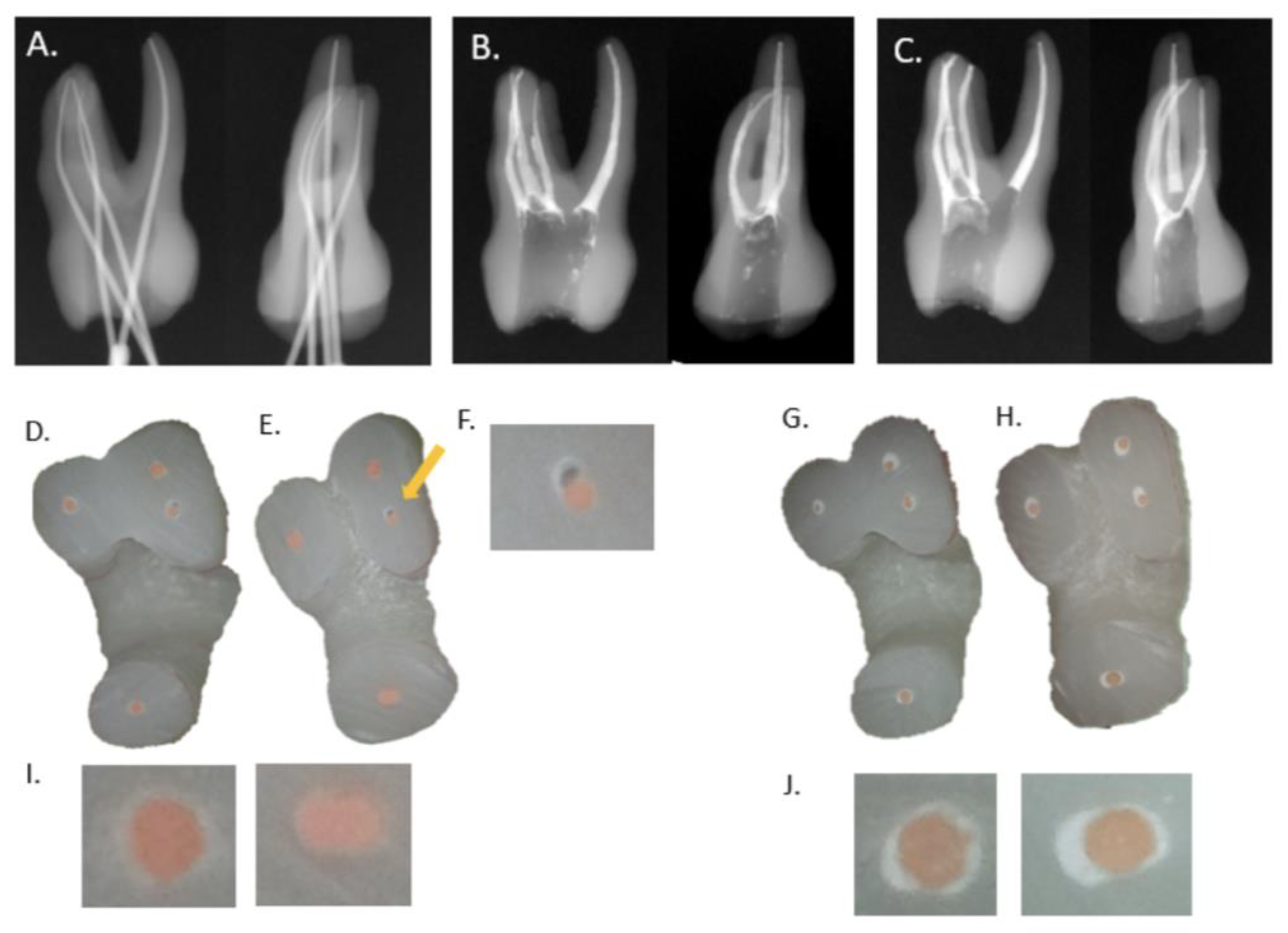
2.1. Preparation of Plastic Tooth Samples
2.2. Obturation of the Plastic Tooth Samples
- Modified continuous wave technique group (CW, n = 12): The GP cone tip was coated with a small amount of AH Plus (Dentsply DeTrey) sealer at the apical 3 to 4 mm part and inserted into the canal. The GP cone was cut at 7 mm from the WL, using a fine sized heat plugger (#30, 0.04 taper) (SybronEndo, Orange, CA, USA) and System B (SybronEndo), packed with BL S-Kondenser (#35) (B & L Biotech, Ansan, Korea) at the 7 mm level and backfilled by using SuperEndo Beta 2 (tip size #25) (B & L Biotech) at a temperature setting of 200 °C.
- Single-cone technique group (SC, n = 11): CeraSeal (Meta Biomed, Cheongju, Korea) was snugly inserted in the canal space by using the provided needle tip; the tip was gently moved towards the orifice from the point from which it was engaged in. A GP cone was moved upward and downward three times to ensure good penetration of the sealer, cut using a fine-sized heat plugger and System B (SybronEndo), and gently packed with BL S-Kondenser (B & L Biotech) at the orifice level.
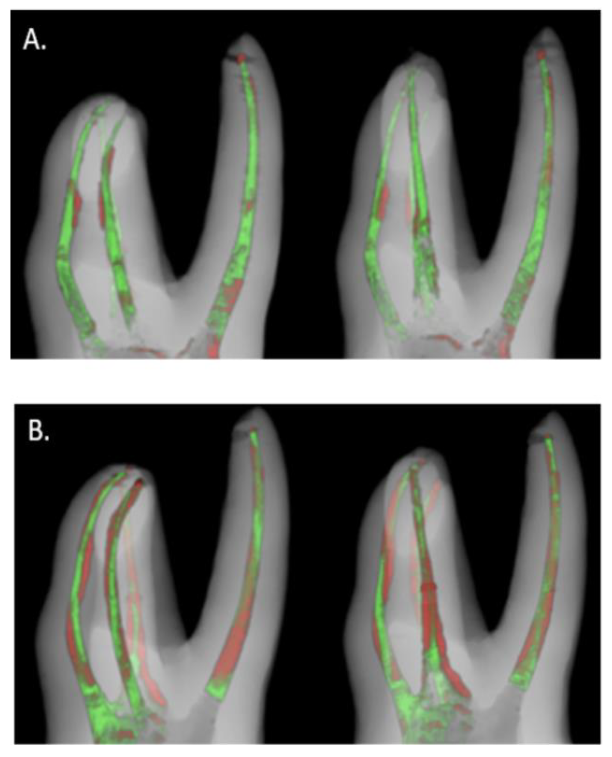
2.3. Micro-CT Imaging and Analysis
2.4. Statistical Analysis
3. Results
4. Discussion
5. Conclusions
Author Contributions
Funding
Institutional Review Board Statement
Informed Consent Statement
Data Availability Statement
Conflicts of Interest
References
- Tang, W.; Wu, Y.; Smales, R.J. Identifying and Reducing Risks for Potential Fractures in Endodontically Treated Teeth. J. Endod. 2010, 36, 609–617. [Google Scholar] [CrossRef] [PubMed]
- TruNatomy. Available online: https://www.dentsplysirona.com/en/explore/endodontics/trunatomy.html (accessed on 4 February 2021).
- Elkholy, M.M.; Nawar, N.N.; Ha, W.N.; Saber, S.M.; Kim, H.-C. Impact of Canal Taper and Access Cavity Design on the Lifespan of an Endodontically Treated Mandibualr Molar: A Finite Element Analysis. J. Endod. 2021. [Google Scholar] [CrossRef] [PubMed]
- Mendes, A.T.; Da Silva, P.B.; Só, B.B.; Hashizume, L.N.; Vivan, R.R.; Da Rosa, R.A.; Duarte, M.A.H.; Só, M.V.R. Evaluation of Physicochemical Properties of New Calcium Silicate-Based Sealer. Braz. Dent. J. 2018, 29, 536–540. [Google Scholar] [CrossRef] [PubMed] [Green Version]
- Al-Haddad, A.Y.; Kutty, M.G.; Abu Kasim, N.H.; Ab Aziz, Z.A.C. The effect of moisture conditions on the constitution of two bioceramic-based root canal sealers. J. Dent. Sci. 2017, 12, 340–346. [Google Scholar] [CrossRef] [PubMed]
- Shakya, V.K.; Gupta, P.; Tikku, A.P.; Pathak, A.K.; Chandra, A.; Yadav, R.K.; Bharti, R.; Singh, R.K. An Invitro Evaluation of Antimicrobial Efficacy and Flow Characteristics for AH Plus, MTA Fillapex, CRCS and Gutta Flow 2 Root Canal Sealer. J. Clin. Diagn. Res. 2016, 10, ZC104–ZC108. [Google Scholar] [CrossRef] [PubMed]
- Peters, O.A.; Arias, A.; Choi, A. Mechanical Properties of a Novel Nickel-titanium Root Canal Instrument: Stationary and Dynamic Tests. J. Endod. 2020, 46, 994–1001. [Google Scholar] [CrossRef]
- Mustafa, R.; Al Omari, T.; Al-Nasrawi, S.; Al Fodeh, R.; Dkmak, A.; Haider, J. Evaluating In Vitro Performance of Novel Nickel-Titanium Rotary System (TruNatomy) Based on Debris Extrusion and Preparation Time from Severely Curved Canals. J. Endod. 2021, 47, 976–981. [Google Scholar] [CrossRef]
- Pérez Morales, M.L.N.; González Sánchez, J.A.; Olivieri, J.G.; Elmsmari, F.; Salmon, P.; Jaramillo, D.E.; Terol, F.D. Micro-computed Tomographic Assessment and Comparative Study of the Shaping Ability of 6 Nickel-Titanium Files: An In Vitro Study. J. Endod. 2021, 47, 812–819. [Google Scholar] [CrossRef]
- Weine, F.S.; Healey, H.J.; Gerstein, H.; Evanson, L. Canal configuration in the mesiobuccal root of the maxillary first molar and its endodontic significance. Oral Surg. Oral Med. Oral Pathol. 1969, 28, 419–425. [Google Scholar] [CrossRef]
- Kim, S.; Kim, S.; Park, J.W.; Jung, I.Y.; Shin, S.J. Comparison of the percentage of voids in the canal filling of a calcium sili-cate-based sealer and gutta percha cones using two obturation techniques. Materials 2017, 10, 1170. [Google Scholar] [CrossRef] [Green Version]
- Jung, J.; Kim, S.; Kim, E.; Shin, S.-J. Volume of Voids in Retrograde Filling: Comparison between Calcium Silicate Cement Alone and Combined with a Calcium Silicate–based Sealer. J. Endod. 2020, 46, 97–102. [Google Scholar] [CrossRef] [PubMed]
- Somma, F.; Cretella, G.; Carotenuto, M.; Pecci, R.; Bedini, R.; De Biasi, M.; Angerame, D. Quality of thermoplasticized and single point root fillings assessed by micro-computed tomography. Int. Endod. J. 2011, 44, 362–369. [Google Scholar] [CrossRef] [PubMed]
- Celikten, B.; Uzuntas, C.F.; Orhan, A.I.; Orhan, K.; Tufenkci, P.; Kursun, S.; Demiralp, K.Ö. Evaluation of root canal sealer filling quality using a single-cone technique in oval shaped canals: An In vitro Micro-CT study. Scanning 2016, 38, 133–140. [Google Scholar] [CrossRef] [PubMed]
- Keleş, A.; Alcin, H.; Kamalak, A.; Versiani, M. Micro-CT evaluation of root filling quality in oval-shaped canals. Int. Endod. J. 2014, 47, 1177–1184. [Google Scholar] [CrossRef]
- Huang, Y.; Orhan, K.; Celikten, B.; Orhan, A.I.; Tufenkci, P.; Sevimay, S. Evaluation of the sealing ability of different root canal sealers: A combined SEM and micro-CT study. J. Appl. Oral Sci. 2018, 26, e20160584. [Google Scholar] [CrossRef] [Green Version]
- Huang, Y.; Celikten, B.; Vasconcelos, K.D.F.; Nicolielo, L.F.P.; Lippiatt, N.; Buyuksungur, A.; Jacobs, R.; Orhan, K. Micro-CT and nano-CT analysis of filling quality of three different endodontic sealers. Dentomaxillofacial Radiol. 2017, 46, 20170223. [Google Scholar] [CrossRef]
- Celikten, B.; Uzuntas, C.F.; Orhan, A.I.; Tufenkci, P.; Misirli, M.; Demiralp, K.O.; Orhan, K. Micro-CT assessment of the sealing ability of three root canal filling techniques. J. Oral Sci. 2015, 57, 361–366. [Google Scholar] [CrossRef] [Green Version]
- Antunes, T.B.M.; Janini, A.C.P.; Pelepenko, L.E.; Abuna, G.F.; Paiva, E.M.; Sinhoreti, M.A.C.; Raimundo, I.M.; Gomes, B.P.F.A.; De-Jesus-Soares, A.; Marciano, M.A.; et al. Heating stability, physical and chemical analysis of calcium silicate-based endodontic sealers. Int. Endod. J. 2021, 54, 1175–1188. [Google Scholar] [CrossRef]
- Yamauchi, S.; Watanabe, S.; Okiji, T. Effects of heating on the physical properties of premixed calcium silicate-based root canal sealers. J. Oral Sci. 2021, 63, 65–69. [Google Scholar] [CrossRef]
- Jung, M.; Lommel, D.; Klimek, J.; Jung, M.; Lommel, D.; Klimek, J. The imaging of root canal obturation using micro-CT. Int. Endod. J. 2005, 38, 617–626. [Google Scholar] [CrossRef]
- Wolf, M.; Küpper, K.; Reimann, S.; Bourauel, C.; Frentzen, M. 3D analyses of interface voids in root canals filled with different sealer materials in combination with warm gutta-percha technique. Clin. Oral Investig. 2014, 18, 155–161. [Google Scholar] [CrossRef] [PubMed]
- Hammad, M.; Qualtrough, A.; Silikas, N. Evaluation of Root Canal Obturation: A Three-dimensional In Vitro Study. J. Endod. 2009, 35, 541–544. [Google Scholar] [CrossRef] [PubMed]
- Saunders, W.P.; Saunders, E.M. Coronal leakage as a cause of failure in root-canal therapy: A review. Dent. Traumatol. 1994, 10, 105–108. [Google Scholar] [CrossRef] [PubMed]
- Zogheib, C.; Naaman, A.; Sigurdsson, A.; Médioni, E.; Bourbouze, G.; Arbab-Chirani, R. Comparative micro-computed tomographic evaluation of two carrier-based obturation systems. Clin. Oral Investig. 2012, 17, 1879–1883. [Google Scholar] [CrossRef] [PubMed]
- Angerame, D.; De Biasi, M.; Pecci, R.; Bedini, R.; Tommasin, E.; Marigo, L.; Somma, F. Analysis of single point and continuous wave of condensation root filling techniques by micro-computed tomography. Annali dell’Istituto Superiore di Sanità 2012, 48, 35–41. [Google Scholar]
- Chybowski, E.A.; Glickman, G.N.; Patel, Y.; Fleury, A.; Solomon, E.; He, J. Clinical Outcome of Non-Surgical Root Canal Treatment Using a Single-cone Technique with Endosequence Bioceramic Sealer: A Retrospective Analysis. J. Endod. 2018, 44, 941–945. [Google Scholar] [CrossRef] [PubMed]


Publisher’s Note: MDPI stays neutral with regard to jurisdictional claims in published maps and institutional affiliations. |
© 2021 by the authors. Licensee MDPI, Basel, Switzerland. This article is an open access article distributed under the terms and conditions of the Creative Commons Attribution (CC BY) license (https://creativecommons.org/licenses/by/4.0/).
Share and Cite
Lee, Y.J.; Kim, S.; Shin, S.-J. Volume Percentage of Filling Voids in Root Canals Prepared by a Novel Nickel-Titanium Rotary System (TruNatomy) Using Two Different Obturation Techniques. Materials 2021, 14, 3846. https://doi.org/10.3390/ma14143846
Lee YJ, Kim S, Shin S-J. Volume Percentage of Filling Voids in Root Canals Prepared by a Novel Nickel-Titanium Rotary System (TruNatomy) Using Two Different Obturation Techniques. Materials. 2021; 14(14):3846. https://doi.org/10.3390/ma14143846
Chicago/Turabian StyleLee, You Jin, Sunil Kim, and Su-Jung Shin. 2021. "Volume Percentage of Filling Voids in Root Canals Prepared by a Novel Nickel-Titanium Rotary System (TruNatomy) Using Two Different Obturation Techniques" Materials 14, no. 14: 3846. https://doi.org/10.3390/ma14143846
APA StyleLee, Y. J., Kim, S., & Shin, S.-J. (2021). Volume Percentage of Filling Voids in Root Canals Prepared by a Novel Nickel-Titanium Rotary System (TruNatomy) Using Two Different Obturation Techniques. Materials, 14(14), 3846. https://doi.org/10.3390/ma14143846