Selected Spectroscopic Techniques for Surface Analysis of Dental Materials: A Narrative Review
Abstract
1. Introduction
2. Search Strategy
3. Fundamentals and Division of Spectroscopy
4. Infrared Spectroscopy (IR) and Fourier Transform Infrared Spectroscopy (FT-IR)
4.1. Principle of the Technique
4.2. Type of Tested Samples
4.3. Sample Characteristics
4.4. Advantages and Limitations
4.5. Applications
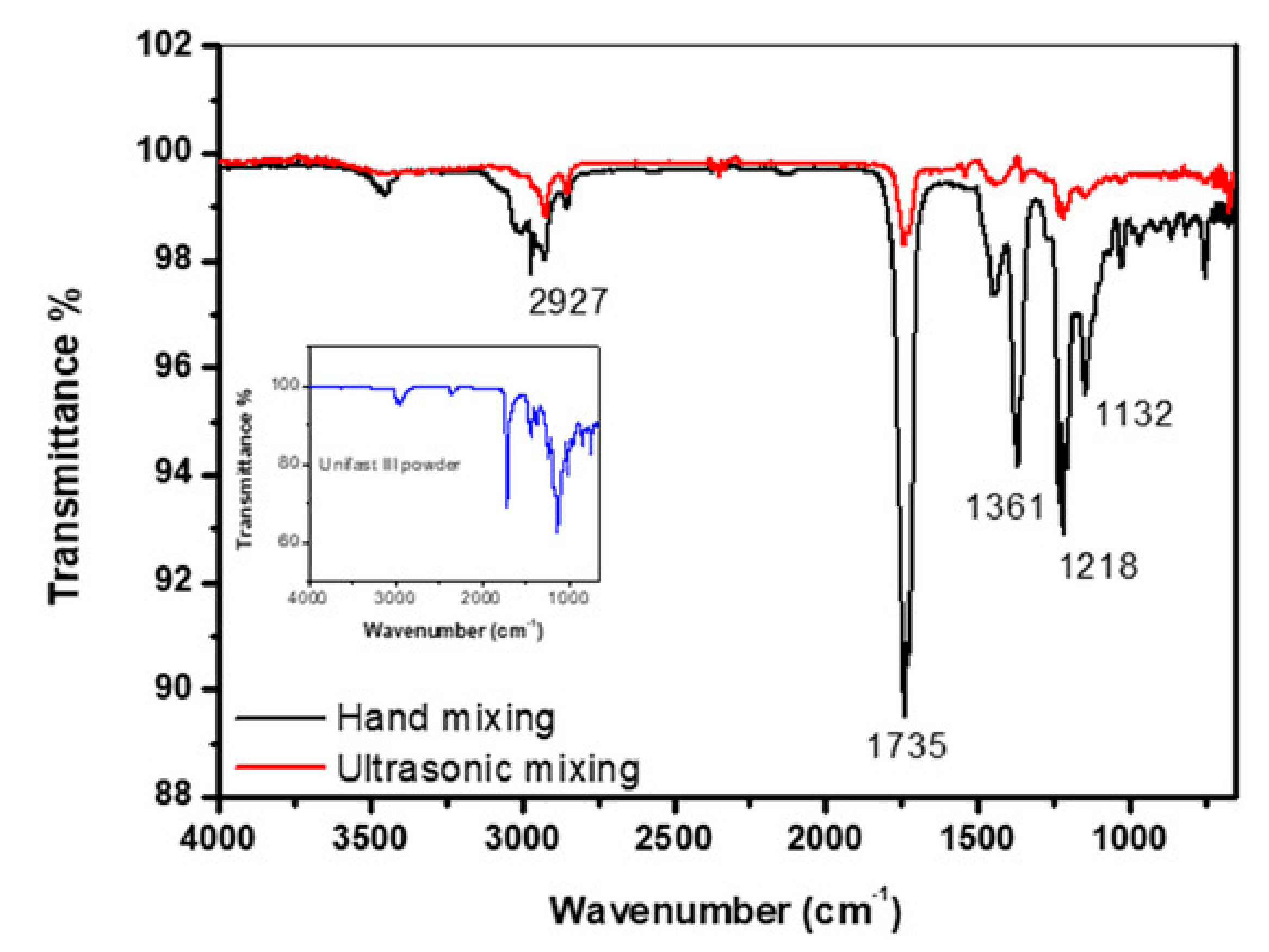
4.6. Spectrum Example
5. Raman Spectroscopy
5.1. Principle of Technique
5.2. Type of Tested Samples
5.3. Sample Characterisctics
5.4. Advantages and Limitations
5.5. Applications
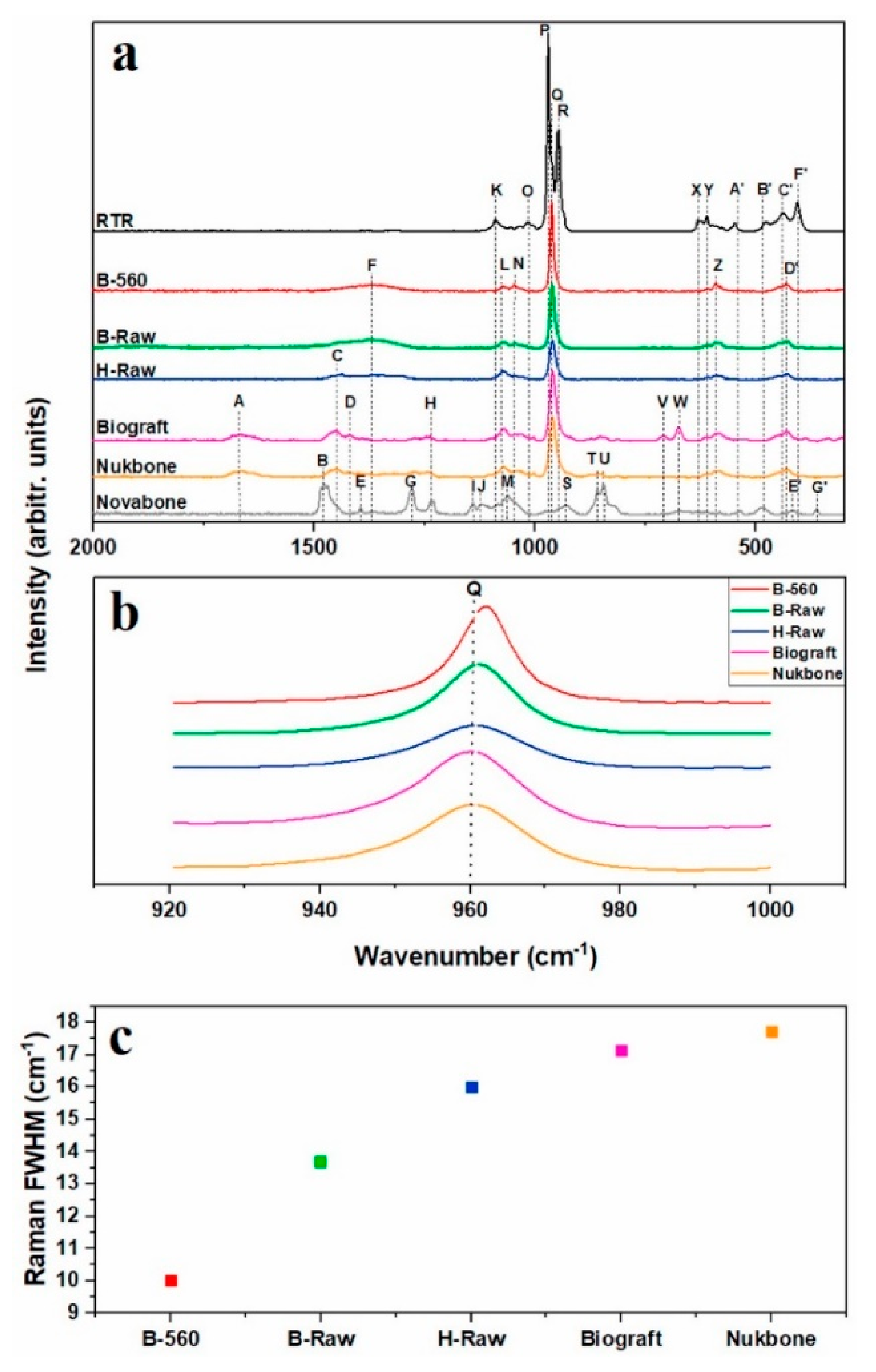
5.6. Spectrum Example
6. Ultraviolet and Visible Spectroscopy (UV-Vis)
6.1. Principle of Technique
6.2. Type of Tested Samples
6.3. Sample Characteristics
6.4. Advantages and Limitations
6.5. Applications
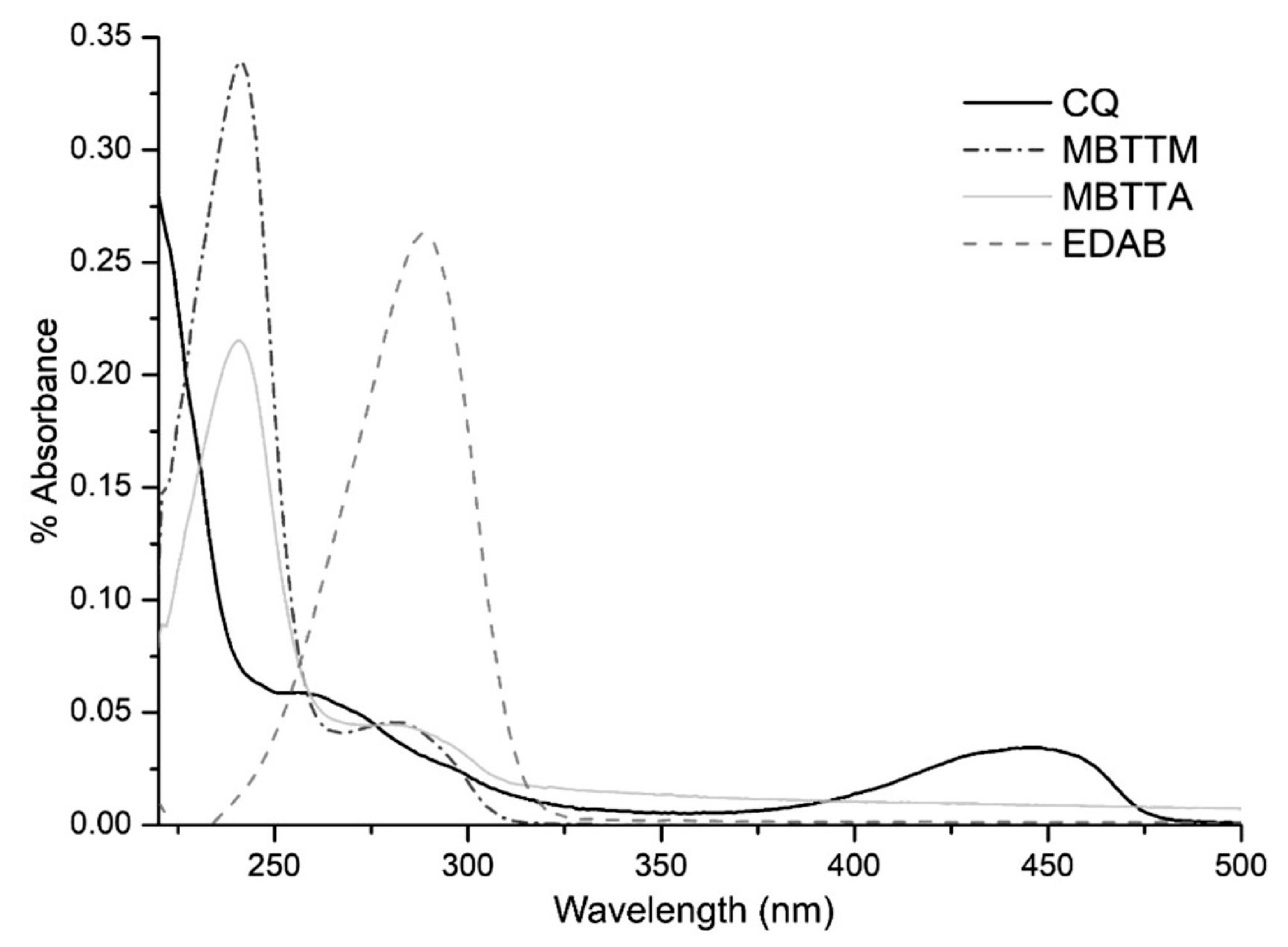
6.6. Spectrum Example
7. X-ray Spectroscopy
7.1. Principle of Technique
7.2. Type of Tested Samples
7.3. Sample Characteristics
7.4. Advantages and Limitations
7.5. Applications
7.6. Spectrum Example
8. Mass Spectrometry (MS)
8.1. Principle of Operation
8.2. Type of Tested Samples
8.3. Sample Characteristics
8.4. Advantages and Limitations
8.5. Applications
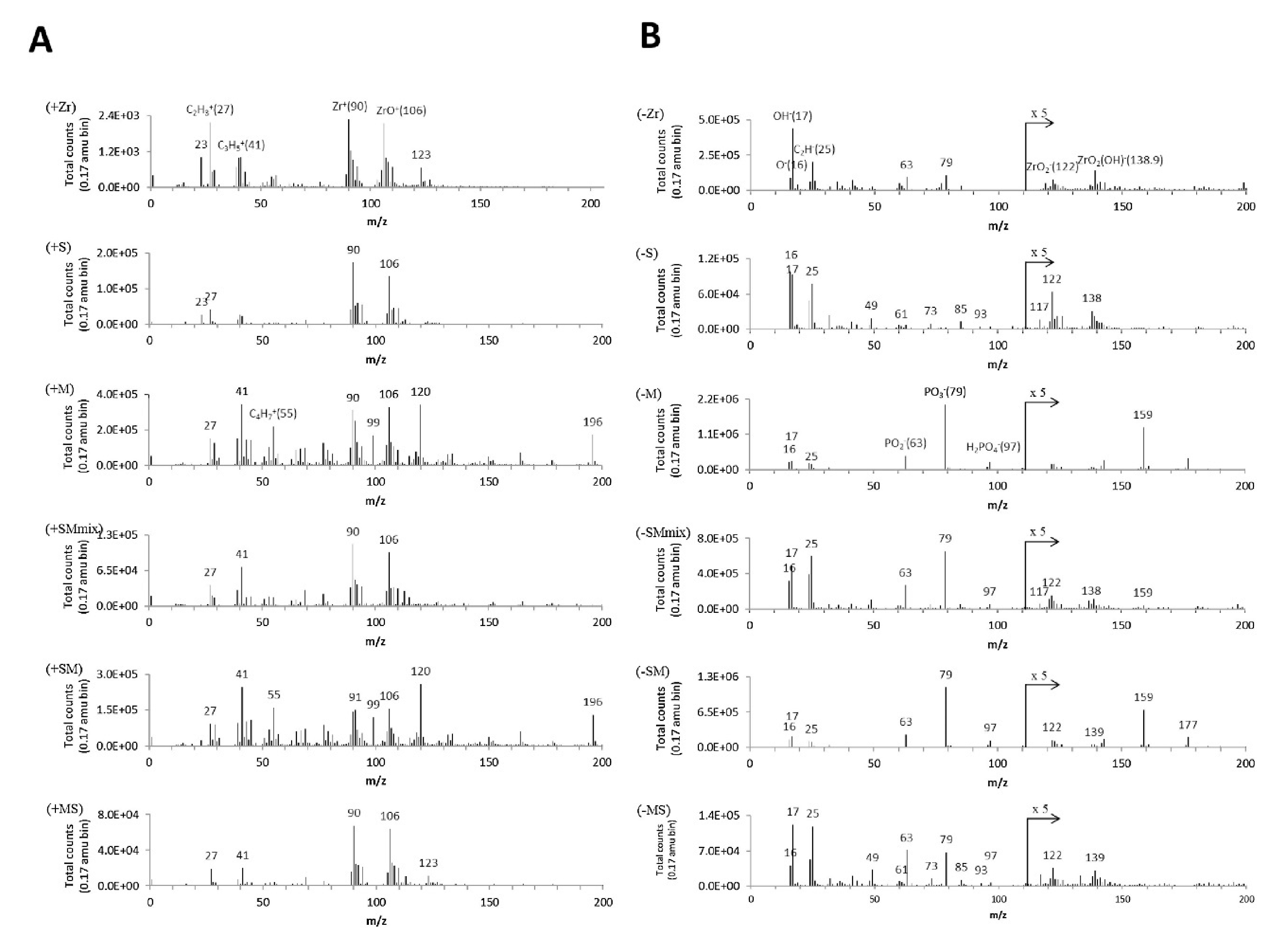
8.6. Spectrum Example
9. Summary and Future Aspect
Author Contributions
Funding
Institutional Review Board Statement
Informed Consent Statement
Data Availability Statement
Acknowledgments
Conflicts of Interest
References
- Alzahrani, K.M. Implant Bio-mechanics for Successful Implant Therapy: A Systematic Review. J. Int. Soc. Prev. Commun. Dent. 2020, 10, 700–714. [Google Scholar] [CrossRef]
- Yu, S.H.; Hao, J.; Fretwurst, T.; Liu, M.; Kostenuik, P.; Giannobile, W.V.; Jin, Q. Sclerostin-Neutralizing Antibody Enhances Bone Regeneration Around Oral Implants. Tissue Eng. Part A 2018, 24, 1672–1679. [Google Scholar] [CrossRef] [PubMed]
- Beltramini, G.; Russillo, A.; Baserga, C.; Pellati, A.; Piva, A.; Candotto, V.; Bolzoni, A.; Beltramini, G.; Rossi, D.; Bolzoni, A.; et al. Collagenated heterologous cortico-cancelleus bone mix stimulated dental pulp derived stem cells. J. Biol. Regul. Homeost. Agents 2020, 34, 1–5. [Google Scholar] [PubMed]
- Jiang, X.; Cao, Z.; Yao, Y.; Zhao, Z.; Liao, W. Aesthetic evaluation of the labiolingual position of maxillary lateral incisors by orthodontists and laypersons. BMC Oral Health 2021, 21, 42. [Google Scholar] [CrossRef]
- Gomez-Meda, R.; Esquivel, J.; Blatz, M.B. The esthetic biological contour concept for implant restoration emergence profile design. J. Esthet. Restor. Dent. 2021, 33, 173–184. [Google Scholar] [CrossRef]
- Furukawa, M.; Wang, J.; Kurosawa, M.; Ogiso, N.; Shikama, Y.; Kanekura, T.; Matsushita, K. Effect of green propolis extracts on experimental aged gingival irritation in vivo and in vitro. J. Oral Biosci. 2021, 63, 58–65. [Google Scholar] [CrossRef]
- Koutouzis, T. Implant-abutment connection as contributing factor to peri-implant diseases. Periodontology 2000 2019, 81, 152–166. [Google Scholar] [CrossRef]
- Birch, S.; Ahern, S.; Brocklehurst, P.; Chikte, U.; Gallagher, J.; Listl, S.; Lalloo, R.; O’Malley, L.; Rigby, J.; Tickle, M.; et al. Planning the oral health workforce: Time for innovation. Commun. Dent. Oral Epidemiol. 2021, 49, 17–22. [Google Scholar] [CrossRef]
- Joda, T.; Yeung, A.W.K.; Hung, K.; Zitzmann, N.U.; Bornstein, M.M. Disruptive Innovation in Dentistry: What It Is and What Could Be Next. J. Dent. Res. 2020, 100, 448–453. [Google Scholar] [CrossRef]
- Joda, T.; Bornstein, M.M.; Jung, R.E.; Ferrari, M.; Waltimo, T.; Zitzmann, N.U. Recent trends and future direction of dental research in the digital era. Int. J. Environ. Res. Public Health 2020, 17, 1987. [Google Scholar] [CrossRef]
- Bastos, N.A.; Bitencourt, S.B.; Martins, E.A.; De Souza, G.M. Review of nano-technology applications in resin-based restorative materials. J. Esthet. Restor. Dent. 2020. [Google Scholar] [CrossRef]
- Glied, A.; Mundiya, J. Implant Material Sciences. Dent. Clin. N. Am. 2021, 65, 81–88. [Google Scholar] [CrossRef]
- Saeidi Pour, R.; Freitas, R.C.; Engler, M.L.P.D.; Edelhoff, D.; Klaus, G.; Prandtner, O.; Berthold, M.; Liebermann, A. Historical development of root analogue implants: A review of published papers. Br. J. Oral Maxillofac. Surg. 2019, 57, 496–504. [Google Scholar] [CrossRef]
- Wojda, S.M. Comparative Analysis of Two Methods of Assessment Wear of Dental Materials. Acta Mech. Autom. 2015, 9, 105–109. [Google Scholar] [CrossRef]
- Duhn, C.; Thalji, G.; Al-Tarwaneh, S.; Cooper, L.F. A digital approach to robust and esthetic implant overdenture construction. J. Esthet. Restor. Dent. 2021, 33, 118–126. [Google Scholar] [CrossRef]
- Deb, S.; Chana, S. Biomaterials in Relation to Dentistry. Front. Oral Biol. 2015, 17, 1–12. [Google Scholar] [CrossRef]
- Orsini, G.; Pagella, P.; Mitsiadis, T.A. Modern Trends in Dental Medicine: An Update for Internists. Am. J. Med. 2018, 131, 1425–1430. [Google Scholar] [CrossRef]
- Guglielmotti, M.B.; Olmedo, D.G.; Cabrini, R.L. Research on implants and osseointegration. Periodontology 2000 2019, 79, 178–189. [Google Scholar] [CrossRef]
- Zhang, Y.; Kelly, J.R. Dental Ceramics for Restoration and Metal Veneering. Dent. Clin. N. Am. 2017, 61, 797–819. [Google Scholar] [CrossRef]
- Bhatavadekar, N.B.; Gharpure, A.S.; Balasubramanium, N.; Scheyer, E.T. In Vitro Surface Testing Methods for Dental Implants-Interpretation and Clinical Relevance: A Review. Compend. Contin. Educ. Dent. 2020, 41, e1–e9. [Google Scholar]
- Meschi, N.; Patel, B.; Ruparel, N.B. Material Pulp Cells and Tissue Interactions. J. Endod. 2020, 46, S150–S160. [Google Scholar] [CrossRef]
- Cionca, N.; Hashim, D.; Mombelli, A. Zirconia dental implants: Where are we now, and where are we heading? Periodontology 2000 2017, 73, 241–258. [Google Scholar] [CrossRef]
- Della Bona, A.; Cantelli, V.; Britto, V.T.; Collares, K.F.; Stansbury, J.W. 3D printing restorative materials using a stereolithographic technique: A systematic review. Dent. Mater. 2021, 37, 336–350. [Google Scholar] [CrossRef]
- Wang, Y.; Bäumer, D.; Ozga, A.K.; Körner, G.; Bäumer, A. Patient satisfaction and oral health-related quality of life 10 years after implant placement. BMC Oral Health 2021, 21, 30. [Google Scholar] [CrossRef]
- Eick, S. Biofilms. Monogr. Oral Sci. 2020, 29, 1–11. [Google Scholar]
- Díaz-Garrido, N.; Lozano, C.P.; Kreth, J.; Giacaman, R.A. Competition and Caries on Enamel of a Dual-Species Biofilm Model with Streptococcus mutans and Streptococcus sanguinis. Appl. Environ. Microbiol. 2020, 86. [Google Scholar] [CrossRef]
- Sterzenbach, T.; Helbig, R.; Hannig, C.; Hannig, M. Bioadhesion in the oral cavity and approaches for biofilm management by surface modifications. Clin. Oral Investig. 2020, 24, 4237–4260. [Google Scholar] [CrossRef] [PubMed]
- Bhadila, G.; Menon, D.; Wang, X.; Vila, T.; Melo, M.A.S.; Montaner, S.; Arola, D.D.; Weir, M.D.; Sun, J.; Xu, H.H.K. Long-term antibacterial activity and cytocompatibility of novel low-shrinkage-stress, remineralizing composites. J. Biomater. Sci. Polym. Ed. 2021, 1–16. [Google Scholar] [CrossRef]
- Tettamanti, L.; Cura, F.; Andrisani, C.; Bassi, M.A.; Silvestre-Rangil, J.; Tagliabue, A. A new implant-abutment connection for bacterial microleakage prevention: An in vitro study. ORAL Implantol. 2017, 10, 172–180. [Google Scholar] [CrossRef] [PubMed]
- Revilla-León, M.; Morillo, J.A.; Att, W.; Özcan, M. Chemical Composition, Knoop Hardness, Surface Roughness, and Adhesion Aspects of Additively Manufactured Dental Interim Materials. J. Prosthodont. 2020. [Google Scholar] [CrossRef] [PubMed]
- Ercoli, C.; Caton, J.G. Dental prostheses and tooth-related factors. J. Periodontol. 2018, 89, S223–S236. [Google Scholar] [CrossRef]
- Revilla-León, M.; Husain, N.A.H.; Methani, M.M.; Özcan, M. Chemical composition, surface roughness, and ceramic bond strength of additively manufactured cobalt-chromium dental alloys. J. Prosthet. Dent. 2020. [Google Scholar] [CrossRef]
- Revilla-León, M.; Meyers, M.J.; Zandinejad, A.; Özcan, M. A review on chemical composition, mechanical properties, and manufacturing work flow of additively manufactured current polymers for interim dental restorations. J. Esthet. Restor. Dent. 2019, 31, 51–57. [Google Scholar] [CrossRef]
- Preoteasa, E.A.; Preoteasa, E.S.; Suciu, I.; Bartok, R.N. Atomic and nuclear surface analysis methods for dental materials: A review. AIMS Mater. Sci. 2018, 5, 781–844. [Google Scholar] [CrossRef]
- Karova, E. Application of Atomic Force Microscopy in Dental Investigations. Int. J. Sci. Res. 2020, 9, 1319–1326. [Google Scholar] [CrossRef]
- Roa, J.J.; Oncins, G.; Díaz, J.; Capdevila, X.G.; Sanz, F.; Segarra, M. Study of the friction, adhesion and mechanical properties of single crystals, ceramics and ceramic coatings by AFM. J. Eur. Ceram. Soc. 2011, 31, 429–449. [Google Scholar] [CrossRef]
- Vilá, J.F.; García, J.C.; Guestrin, A. SEM Applied to the Development of Bioactive Surface of Dental Implants. Microsc. Microanal. 2020, 26, 147–148. [Google Scholar] [CrossRef]
- Banaszek, K.; Sawicki, J.; Wołowiec-Korecka, E.; Gorzędowski, J.; Danowska-Klonowska, D.; Sokołowski, J. Use of optical microscopy for evaluation of tooth structure. J. Achiev. Mater. Manuf. Eng. 2016, 79, 31–40. [Google Scholar] [CrossRef]
- Chander, N.G. Characterization of dental materials. J. Indian Prosthodont. Soc. 2018, 18, 289–290. [Google Scholar] [CrossRef]
- Zhou, X.; Thompson, G.E. Electron and Photon Based Spatially Resolved Techniques. In Reference Module in Materials Science and Materials Engineering; Elsevier: New York, NY, USA, 2017; pp. 1–30. [Google Scholar]
- Yanikoglu, N.D.; Sakarya, R.E. Test methods used in the evaluation of the structure features of the restorative materials: A literature review. J. Mater. Res. Technol. 2020, 9, 9720–9734. [Google Scholar] [CrossRef]
- Naves, L.Z.; Gerdolle, D.A.; de Andrade, O.S.; Markus Maria Gresnigt, M. Seeing is believing? When scanning electron microscopy (SEM) meets clinical dentistry: The replica technique. Microsc. Res. Tech. 2020, 83, 1118–1123. [Google Scholar] [CrossRef] [PubMed]
- Siekaniec, D.; Kopyciński, D. Analysis Phases and Crystallographic Orientation of the grain of High Chromium Cast Iron Using EBSD Technique. J. Cast. Mater. Eng. 2017, 1, 15. [Google Scholar] [CrossRef]
- Koblischka-Veneva, A.; Koblischka, M.R.; Schmauch, J.; Hannig, M. Comparison of human and bovine dental enamel by TEM and t-EBSD investigations. IOP Conf. Ser. Mater. Sci. Eng. 2019, 625, 012006. [Google Scholar] [CrossRef]
- Tomota, Y. Crystallographic characterization of steel microstructure using neutron diffraction. Sci. Technol. Adv. Mater. 2019, 20, 1189–1206. [Google Scholar] [CrossRef]
- Shukla, A.K. Electron Spin Resonance Spectroscopy in Medicine; Shukla, A.K., Ed.; Springer: Singapore, 2018; ISBN 9789811322303. [Google Scholar]
- Pandoleon, P.; Kontonasaki, E.; Kantiranis, N.; Pliatsikas, N.; Patsalas, P.; Papadopoulou, L.; Zorba, T.; Paraskevopoulos, K.M.; Koidis, P. Aging of 3Y-TZP dental zirconia and yttrium depletion. Dent. Mater. 2017, 33, e385–e392. [Google Scholar] [CrossRef]
- Lopes, C.d.C.A.; Limirio, P.H.J.O.; Novais, V.R.; Dechichi, P. Fourier transform infrared spectroscopy (FTIR) application chemical characterization of enamel, dentin and bone. Appl. Spectrosc. Rev. 2018, 53, 747–769. [Google Scholar] [CrossRef]
- Lach, S.; Jurczak, P.; Karska, N.; Kubiś, A.; Szymańska, A.; Rodziewicz-Motowidło, S. Spectroscopic methods used in implant material studies. Molecules 2020, 25, 579. [Google Scholar] [CrossRef]
- Kreve, S.; Cândido dos Reis, A. Influence of the electrostatic condition of the titanium surface on bacterial adhesion: A systematic review. J. Prosthet. Dent. 2021, 125, 416–420. [Google Scholar] [CrossRef]
- Kim, I.H.; Son, J.S.; Min, B.K.; Kim, Y.K.; Kim, K.H.; Kwon, T.Y. A simple, sensitive and non-destructive technique for characterizing bovine dental enamel erosion: Attenuated total reflection Fourier transform infrared spectroscopy. Int. J. Oral Sci. 2016, 8, 54–60. [Google Scholar] [CrossRef]
- Furuhashi, K.; Uo, M.; Kitagawa, Y.; Watari, F. Rapid and non-destructive analysis of metallic dental restorations using X-ray fluorescence spectra and light-element sampling tools. Appl. Surf. Sci. 2012, 262, 13–18. [Google Scholar] [CrossRef][Green Version]
- Malik, A.K.; Kumar, R. Heena Spectroscopy: Types. In Encyclopedia of Food and Health; Elsevier Ltd.: New York, NY, USA, 2015; pp. 64–72. ISBN 9780123849533. [Google Scholar]
- Pignataro, M.F.; Herrera, M.G.; Dodero, V.I. Evaluation of peptide/protein self-assembly and aggregation by spectroscopic methods. Molecules 2020, 25, 4854. [Google Scholar] [CrossRef]
- Kafle, B.P. Infrared (IR) spectroscopy. In Chemical Analysis and Material Characterization by Spectrophotometry; Elsevier: New York, NY, USA, 2020; pp. 199–243. [Google Scholar]
- Smith, E.; Dent, G. Modern Raman Spectroscopy—A Practical Approach. John Wiley and Sons Ltd.: Chichester, UK, 2005; p. 210. ISBN 0471496685. [Google Scholar] [CrossRef]
- Dolenko, G.N.; Poleshchuk, O.K.; Latosińska, J.N. X-ray emission spectroscopy, methods. In Encyclopedia of Spectroscopy and Spectrometry; Elsevier: New York, NY, USA, 2016; pp. 691–694. ISBN 9780128032244. [Google Scholar]
- Sharma, R.K. Various Spectroscopic Techniques. In Environmental Pollution: Monitoring Modelling and Control; Studium Press, LLC: Houston, TX, USA, 2017; pp. 181–206. ISBN ISBN:1626991022/978-1626991026. [Google Scholar]
- Patonay, G.; Beckford, G.; Hänninen, P. UV-Vis and NIR Fluorescence Spectroscopy. In Handbook of Spectroscopy, 2nd Enlarged ed.; Wiley-VCH Verlag GmbH & Co. KGaA: Weinheim, Germany, 2014; Volume 3–4, pp. 999–1036. ISBN 9783527654703. [Google Scholar]
- Adams, F.C. X-ray absorption and Diffraction | Overview. In Encyclopedia of Analytical Science; Elsevier: New York, NY, USA, 2019; pp. 391–403. ISBN 9780081019832. [Google Scholar]
- Brader, M.L. UV-absorbance, fluorescence and FT-IR spectroscopy in biopharmaceutical development. In Biophysical Characterization of Proteins in Developing Biopharmaceuticals; Elsevier: New York, NY, USA, 2020; pp. 97–121. ISBN 9780444641731. [Google Scholar]
- Akhtar, S.; Ali, S. Characterization of nanomaterials: Techniques and tools. In Applications of Nanomaterials in Human Health; Springer: Singapore, 2020; pp. 23–43. ISBN 9789811548024. [Google Scholar]
- Fa, K.; Jiang, T.; Nalaskowski, J.; Miller, J.D. Optical and spectroscopic characteristics of oleate adsorption as revealed by FTIR analysis. Langmuir 2004, 20, 5311–5321. [Google Scholar] [CrossRef]
- Le Pevelen, D.D.; Tranter, G.E. FT-IR and raman spectroscopies, polymorphism applications. In Encyclopedia of Spectroscopy and Spectrometry; Elsevier: New York, NY, USA, 2016; pp. 750–761. ISBN 9780128032244. [Google Scholar]
- Bell, S.E.J.; Xu, Y. Infrared spectroscopy|Industrial applications. In Encyclopedia of Analytical Science; Elsevier: New York, NY, USA, 2019; pp. 124–133. ISBN 9780081019832. [Google Scholar]
- Kowalczuk, D.; Pitucha, M. Application of FTIR Method for the Assessment of Immobilization of Active Substances in the Matrix of Biomedical Materials. Materials 2019, 12, 2972. [Google Scholar] [CrossRef]
- Puspita, S.; Sunarintyas, S.; Mulyawati, E.; Anwar, C.; Sukirno; Soesatyo, M.H.N.E. Molecular weight determination and structure identification of Bombyx mori L. Fibroin as material in dentistry. In AIP Conference Proceedings; American Institute of Physics Inc.: College, MA, USA, 2020; Volume 2260, p. 40018. [Google Scholar]
- Rafeek, A.D.; Choi, G.; Evans, L.A. Morphological, spectroscopic and crystallographic studies of calcium phosphate bioceramic powders. J. Aust. Ceram. Soc. 2018, 54, 161–168. [Google Scholar] [CrossRef]
- Dutta, A. Fourier Transform Infrared Spectroscopy. In Spectroscopic Methods for Nanomaterials Characterization; Elsevier: New York, NY, USA, 2017; Volume 2, pp. 73–93. ISBN 9780323461467. [Google Scholar]
- Rosi, F.; Cartechini, L.; Sali, D.; Miliani, C. Recent trends in the application of fourier transform infrared (FT-IR) spectroscopy in Heritage Science: From micro: From non-invasive FT-IR. Phys. Sci. Rev. 2019, 4. [Google Scholar] [CrossRef]
- Margariti, C. The application of FTIR microspectroscopy in a non-invasive and non-destructive way to the study and conservation of mineralised excavated textiles. Herit. Sci. 2019, 7, 1–14. [Google Scholar] [CrossRef]
- Munajad, A.; Subroto, C. Suwarno Fourier transform infrared (FTIR) spectroscopy analysis of transformer paper in mineral oil-paper composite insulation under accelerated thermal aging. Energies 2018, 11, 364. [Google Scholar] [CrossRef]
- Puppin-Rontani, J.; Fugolin, A.P.P.; Costa, A.R.; Correr-Sobrinho, L.; Pfeifer, C.S. In vitro performance of 2-step, total etch adhesives modified by thiourethane additives. Int. J. Adhes. Adhes. 2020, 103, 102688. [Google Scholar] [CrossRef]
- Shim, J.S.; Lee, S.Y.; Song, S.-Y.; Jha, N.; Ryu, J.J. Polymerization efficiency of dental dual-cured resin cement light-cured at various times after the initiation of chemical activation. Int. J. Polym. Mater. Polym. Biomater. 2020, 69, 622–628. [Google Scholar] [CrossRef]
- Fugolin, A.P.; Lewis, S.; Logan, M.G.; Ferracane, J.L.; Pfeifer, C.S. Methacrylamide–methacrylate hybrid monomers for dental applications. Dent. Mater. 2020, 36, 1028–1037. [Google Scholar] [CrossRef]
- Fugolin, A.P.; Dobson, A.; Ferracane, J.L.; Pfeifer, C.S. Effect of residual solvent on performance of acrylamide-containing dental materials. Dent. Mater. 2019, 35, 1378–1387. [Google Scholar] [CrossRef] [PubMed]
- Fugolin, A.P.P.; Navarro, O.; Logan, M.G.; Huynh, V.; França, C.M.; Ferracane, J.L.; Pfeifer, C.S. Synthesis of di- and triacrylamides with tertiary amine cores and their evaluation as monomers in dental adhesive interfaces. Acta Biomater. 2020, 115, 148–159. [Google Scholar] [CrossRef] [PubMed]
- Fugolin, A.P.; Dobson, A.; Huynh, V.; Mbiya, W.; Navarro, O.; Franca, C.M.; Logan, M.; Merritt, J.L.; Ferracane, J.L.; Pfeifer, C.S. Antibacterial, ester-free monomers: Polymerization kinetics, mechanical properties, biocompatibility and anti-biofilm activity. Acta Biomater. 2019, 100, 132–141. [Google Scholar] [CrossRef] [PubMed]
- Alania, Y.; dos Reis, M.C.; Nam, J.-W.; Phansalkar, R.S.; McAlpine, J.; Chen, S.-N.; Pauli, G.F.; Bedran-Russo, A.K. A dynamic mechanical method to assess bulk viscoelastic behavior of the dentin extracellular matrix. Dent. Mater. 2020, 36, 1536–1543. [Google Scholar] [CrossRef]
- Zhang, P.; Zhao, X.M. Synthesis, crystal structure and bioactivity evaluation of a heterocyclic compound. Jiegou Huaxue 2020, 39, 1892–1897. [Google Scholar] [CrossRef]
- Seredin, P.V.; Uspenskaya, O.A.; Goloshchapov, D.L.; Ippolitov, I.Y.; Vongsvivut, J.; Ippolitov, Y.A. Organic-mineral interaction between biomimetic materials and hard dental tissues. Sovrem. Tehnol. V Med. 2020, 12, 43–51. [Google Scholar] [CrossRef]
- Gurgenc, T. Structural characterization and dielectrical properties of Ag-doped nano-strontium apatite particles produced by hydrothermal method. J. Mol. Struct. 2021, 1223, 128990. [Google Scholar] [CrossRef]
- Jing, X.; Xie, B.; Li, X.; Dai, Y.; Nie, L.; Li, C. Peptide decorated demineralized dentin matrix with enhanced bioactivity, osteogenic differentiation via carboxymethyl chitosan. Dent. Mater. 2021, 37, 19–29. [Google Scholar] [CrossRef]
- Ramos, N.C.; Alves, L.M.M.; Ricco, P.; Santos, G.M.A.S.; Bottino, M.A.; Campos, T.M.B.; Melo, R.M. Strength and bondability of a dental Y-TZP after silica sol-gel infiltrations. Ceram. Int. 2020, 46, 17018–17024. [Google Scholar] [CrossRef]
- Asadi, F.; Forootanfar, H.; Ranjbar, M. A facile one-step preparation of Ca10(PO4)6(OH)2/Li-BioMOFs resin nanocomposites with Glycyrrhiza glabra (licorice) root juice as green capping agent and mechanical properties study. Artif. Cells Nanomed. Biotechnol. 2020, 48, 1331–1339. [Google Scholar] [CrossRef]
- Fu, D.; Lu, Y.; Gao, S.; Peng, Y.; Duan, H. Chemical Property and Antibacterial Activity of Metronidazole-decorated Ti through Adhesive Dopamine. J. Wuhan Univ. Technol. Mater. Sci. Ed. 2019, 34, 968–972. [Google Scholar] [CrossRef]
- Yakufu, M.; Wang, Z.; Wang, Y.; Jiao, Z.; Guo, M.; Liu, J.; Zhang, P. Covalently functionalized poly(etheretherketone) implants with osteogenic growth peptide (OGP) to improve osteogenesis activity. RSC Adv. 2020, 10, 9777–9785. [Google Scholar] [CrossRef]
- Ding, Y.; Zhang, H.; Wang, X.; Zu, H.; Wang, C.; Dong, D.; Lyu, M.; Wang, S. Immobilization of Dextranase on Nano-Hydroxyapatite as a Recyclable Catalyst. Materials 2020, 14, 130. [Google Scholar] [CrossRef]
- Roopavath, U.K.; Sah, M.K.; Panigrahi, B.B.; Rath, S.N. Mechanochemically synthesized phase stable and biocompatible β-tricalcium phosphate from avian eggshell for the development of tissue ingrowth system. Ceram. Int. 2019, 45, 12910–12919. [Google Scholar] [CrossRef]
- Zeng, W.; Liu, F.; He, J. Physicochemical Properties of Bis-GMA/TEGDMA Dental Resin Reinforced with Silanized Multi-Walled Carbon Nanotubes. Silicon 2019, 11, 1345–1353. [Google Scholar] [CrossRef]
- Voicu, G.; Didilescu, A.C.; Stoian, A.B.; Dumitriu, C.; Greabu, M.; Andrei, M. Mineralogical and microstructural characteristics of two dental pulp capping materials. Materials 2019, 12, 1772. [Google Scholar] [CrossRef]
- Yushau, U.S.; Almofeez, L.; Bozkurt, A. Novel Polymer Nanocomposites Comprising Triazole Functional Silica for Dental Application. Silicon 2020, 12, 109–116. [Google Scholar] [CrossRef]
- Agha, A.; Parker, S.; Patel, M. Polymerization shrinkage kinetics and degree of conversion of commercial and experimental resin modified glass ionomer luting cements (RMGICs). Dent. Mater. 2020, 36, 893–904. [Google Scholar] [CrossRef]
- Pérez-Mondragón, A.A.; Cuevas-Suárez, C.E.; González-López, J.A.; Trejo-Carbajal, N.; Herrera-González, A.M. Evaluation of new coinitiators of camphorquinone useful in the radical photopolymerization of dental monomers. J. Photochem. Photobiol. A Chem. 2020, 403, 112844. [Google Scholar] [CrossRef]
- Alotaibi, J.; Saji, S.; Swain, M.V. FTIR characterization of the setting reaction of biodentineTM. Dent. Mater. 2018, 34, 1645–1651. [Google Scholar] [CrossRef]
- Dinesh Kumar, S.; Mohamed Abudhahir, K.; Selvamurugan, N.; Vimalraj, S.; Murugesan, R.; Srinivasan, N.; Moorthi, A. Formulation and biological actions of nano-bioglass ceramic particles doped with Calcarea phosphorica for bone tissue engineering. Mater. Sci. Eng. C 2018, 83, 202–209. [Google Scholar] [CrossRef]
- Alqahtani, M. Effect of hexagonal boron nitride nanopowder reinforcement and mixing methods on physical and mechanical properties of self-cured PMMA for dental applications. Materials 2020, 13, 2323. [Google Scholar] [CrossRef]
- Kafle, B.P. Raman spectroscopy. In Chemical Analysis and Material Characterization by Spectrophotometry; Elsevier: New York, NY, USA, 2020; pp. 245–268. [Google Scholar]
- Marcott, C.; Padalkar, M.; Pleshko, N. 3.23 Infrared and raman microscopy and imaging of biomaterials at the micro and nano scale. In Comprehensive Biomaterials II; Elsevier: New York, NY, USA, 2017; pp. 498–518. ISBN 9780081006924. [Google Scholar]
- Omidi, M.; Fatehinya, A.; Farahani, M.; Akbari, Z.; Shahmoradi, S.; Yazdian, F.; Tahriri, M.; Moharamzadeh, K.; Tayebi, L.; Vashaee, D. Characterization of biomaterials. In Biomaterials for Oral and Dental Tissue Engineering; Elsevier: New York, NY, USA, 2017; pp. 97–115. ISBN 9780081009673. [Google Scholar]
- Xu, Z.; He, Z.; Song, Y.; Fu, X.; Rommel, M.; Luo, X.; Hartmaier, A.; Zhang, J.; Fang, F. Topic review: Application of raman spectroscopy characterization in micro/nano-machining. Micromachines 2018, 9, 361. [Google Scholar] [CrossRef]
- Jones, R.R.; Hooper, D.C.; Zhang, L.; Wolverson, D.; Valev, V.K. Raman Techniques: Fundamentals and Frontiers. Nanoscale Res. Lett. 2019, 14, 1–34. [Google Scholar] [CrossRef]
- Khan, A.S.; Khalid, H.; Sarfraz, Z.; Khan, M.; Iqbal, J.; Muhammad, N.; Fareed, M.A.; Rehman, I.U. Vibrational spectroscopy of selective dental restorative materials. Appl. Spectrosc. Rev. 2017, 52, 507–540. [Google Scholar] [CrossRef]
- Malhotra, R.; Han, Y.M.; Morin, J.L.P.; Luong-Van, E.K.; Chew, R.J.J.; Castro Neto, A.H.; Nijhuis, C.A.; Rosa, V. Inhibiting Corrosion of Biomedical-Grade Ti-6Al-4V Alloys with Graphene Nanocoating. J. Dent. Res. 2020, 99, 285–292. [Google Scholar] [CrossRef]
- Gomes de Araújo-Neto, V.; Sebold, M.; Fernandes de Castro, E.; Feitosa, V.P.; Giannini, M. Evaluation of physico-mechanical properties and filler particles characterization of conventional, bulk-fill, and bioactive resin-based composites. J. Mech. Behav. Biomed. Mater. 2021, 115, 104288. [Google Scholar] [CrossRef] [PubMed]
- Zubieta-Otero, L.F.; Londoño-Restrepo, S.M.; Lopez-Chavez, G.; Hernandez-Becerra, E.; Rodriguez-Garcia, M.E. Comparative study of physicochemical properties of bio-hydroxyapatite with commercial samples. Mater. Chem. Phys. 2021, 259, 124201. [Google Scholar] [CrossRef]
- Gutierrez, M.F.; Perdigao, J.; Malaquias, P.; Cardenas, A.M.; Siqueira, F.; Hass, V.; Reis, A.; Loguercio, A.D. Effect of methacryloyloxydecyl dihydrogen phosphate-containing silane and adhesive used alone or in combination on the bond strength and chemical interaction with zirconia ceramics under thermal aging. Oper. Dent. 2020, 45, 516–527. [Google Scholar] [CrossRef] [PubMed]
- Lubas, M. Au interface effect on Ti-dental porcelain bond strength investigated by spectroscopic methods and mechanical tests. J. Mol. Struct. 2020, 1208, 127870. [Google Scholar] [CrossRef]
- Miranda, J.S.; Barcellos, A.S.d.P.; Campos, T.M.B.; Cesar, P.F.; Amaral, M.; Kimpara, E.T. Effect of repeated firings and staining on the mechanical behavior and composition of lithium disilicate. Dent. Mater. 2020, 36, e149–e157. [Google Scholar] [CrossRef]
- Ubaldini, A.L.M.; Pascotto, R.C.; Sato, F.; Soares, V.O.; Baesso, M.L. Mechanical and Chemical Changes in the Adhesive-Dentin Interface after Remineralization. J. Adhes. Dent. 2020, 22, 297–309. [Google Scholar] [CrossRef]
- Lubas, M.; Przerada, I.; Zawada, A.; Jasinski, J.J.; Jelen, P. Spectroscopic and microstructural investigation of novel Ti–10Zr–45S5 bioglass composite for dental applications. J. Mol. Struct. 2020, 1221, 128545. [Google Scholar] [CrossRef]
- Spies, B.C.; Zhang, F.; Wesemann, C.; Li, M.; Rosentritt, M. Reliability and aging behavior of three different zirconia grades used for monolithic four-unit fixed dental prostheses. Dent. Mater. 2020, 36, e329–e339. [Google Scholar] [CrossRef]
- Tzanakakis, E.; Kontonasaki, E.; Voyiatzis, G.; Andrikopoulos, K.; Tzoutzas, I. Surface characterization of monolithic zirconia submitted to different surface treatments applying optical interferometry and raman spectrometry. Dent. Mater. J. 2020, 39, 111–117. [Google Scholar] [CrossRef]
- Par, M.; Spanovic, N.; Mohn, D.; Attin, T.; Tauböck, T.T.; Tarle, Z. Curing potential of experimental resin composites filled with bioactive glass: A comparison between Bis-EMA and UDMA based resin systems. Dent. Mater. 2020, 36, 711–723. [Google Scholar] [CrossRef]
- Gugelmin, B.P.; Miguel, L.C.M.; Filho, F.B.; da Cunha, L.F.; Correr, G.M.; Gonzaga, C.C. Colorstability of ceramic veneers luted with resin cements and pre-heated composites: 12 months follow-up. Braz. Dent. J. 2020, 31, 69–77. [Google Scholar] [CrossRef]
- Castillo-Paz, A.M.; Londoño-Restrepo, S.M.; Tirado-Mejía, L.; Mondragón, M.A.; Rodríguez-García, M.E. Nano to micro size transition of hydroxyapatite in porcine bone during heat treatment with low heating rates. Prog. Nat. Sci. Mater. Int. 2020, 30, 494–501. [Google Scholar] [CrossRef]
- Ramirez-Gutierrez, C.F.; Londoño-Restrepo, S.M.; del Real, A.; Mondragón, M.A.; Rodriguez-García, M.E. Effect of the temperature and sintering time on the thermal, structural, morphological, and vibrational properties of hydroxyapatite derived from pig bone. Ceram. Int. 2017, 43, 7552–7559. [Google Scholar] [CrossRef]
- Yu, J.; Wang, H.; Zhan, J.; Huang, W. Review of recent UV-Vis and infrared spectroscopy researches on wine detection and discrimination. Appl. Spectrosc. Rev. 2018, 53, 65–86. [Google Scholar] [CrossRef]
- Kafle, B.P. Application of UV–VIS spectrophotometry for chemical analysis. In Chemical Analysis and Material Characterization by Spectrophotometry; Elsevier: New York, NY, USA, 2020; pp. 79–145. [Google Scholar]
- Kafle, B.P. Theory and instrumentation of absorption spectroscopy. In Chemical Analysis and Material Characterization by Spectrophotometry; Elsevier: New York, NY, USA, 2020; pp. 17–38. [Google Scholar]
- Kafle, B.P. Sample preparation methods and choices of reagents. In Chemical Analysis and Material Characterization by Spectrophotometry; Elsevier: New York, NY, USA, 2020; pp. 39–50. [Google Scholar]
- Kafle, B.P. Spectrophotometry and its application in chemical analysis. In Chemical Analysis and Material Characterization by Spectrophotometry; Elsevier: New York, NY, USA, 2020; pp. 1–16. [Google Scholar]
- Stürmer, M.; Garcia, I.M.; Souza, V.S.; Visioli, F.; Scholten, J.D.; Samuel, S.M.W.; Leitune, V.C.B.; Collares, F.M. Titanium dioxide nanotubes with triazine-methacrylate monomer to improve physicochemical and biological properties of adhesives. Dent. Mater. 2021, 37, 223–235. [Google Scholar] [CrossRef] [PubMed]
- Keul, C.; Seidl, J.; Güth, J.F.; Liebermann, A. Impact of fabrication procedures on residual monomer elution of conventional polymethyl methacrylate (PMMA)—a measurement approach by UV/Vis spectrophotometry. Clin. Oral Investig. 2020, 24, 4519–4530. [Google Scholar] [CrossRef]
- Castellanos, M.; Delgado, A.J.; Sinhoreti, M.A.C.; de Oliveira, D.C.R.S.; Abdulhameed, N.; Geraldeli, S.; Roulet, J.-F. Effect of Thickness of Ceramic Veneers on Color Stability and Bond Strength of Resin Luting Cements Containing Alternative Photoinitiators. J. Adhes. Dent. 2019, 21, 67–76. [Google Scholar] [CrossRef] [PubMed]
- Mallaiah, M.; Gupta, R.K. Surface Engineering of Titanium Using Anodization and Plasma Treatment. IOP Conf. Ser. Mater. Sci. Eng. 2020, 943, 12016. [Google Scholar]
- Haas, K.; Azhar, G.; Wood, D.J.; Moharamzadeh, K.; van Noort, R. The effects of different opacifiers on the translucency of experimental dental composite resins. Dent. Mater. 2017, 33, e310–e316. [Google Scholar] [CrossRef]
- Michailova, M.; Elsayed, A.; Fabel, G.; Edelhoff, D.; Zylla, I.M.; Stawarczyk, B. Comparison between novel strength-gradient and color-gradient multilayered zirconia using conventional and high-speed sintering. J. Mech. Behav. Biomed. Mater. 2020, 111, 103977. [Google Scholar] [CrossRef] [PubMed]
- Xiao, Z.; Yu, S.; Li, Y.; Ruan, S.; Kong, L.B.; Huang, Q.; Huang, Z.; Zhou, K.; Su, H.; Yao, Z.; et al. Materials development and potential applications of transparent ceramics: A review. Mater. Sci. Eng. R Rep. 2020, 139, 100518. [Google Scholar] [CrossRef]
- Qasim, S.B.; Najeeb, S.; Delaine-Smith, R.M.; Rawlinson, A.; Ur Rehman, I. Potential of electrospun chitosan fibers as a surface layer in functionally graded GTR membrane for periodontal regeneration. Dent. Mater. 2017, 33, 71–83. [Google Scholar] [CrossRef]
- Bostedt, C.; Gorkhover, T.; Rupp, D.; Möller, T. Clusters and Nanocrystals. In Synchrotron Light Sources and Free-Electron Lasers; Springer International Publishing: Cham, Switzerland, 2020; pp. 1525–1573. ISBN 9783030232016. [Google Scholar]
- Fracchia, M.; Ghigna, P.; Vertova, A.; Rondinini, S.; Minguzzi, A. Time-Resolved X-ray Absorption Spectroscopy in (Photo)Electrochemistry. Surfaces 2018, 1, 11. [Google Scholar] [CrossRef]
- Taylor, A. Atomic spectroscopy, biomedical applications. In Encyclopedia of Spectroscopy and Spectrometry; Elsevier: New York, NY, USA, 2016; pp. 76–80. ISBN 9780128032244. [Google Scholar]
- Hirai, N. Surface analysis. In Corrosion Control and Surface Finishing: Environmentally Friendly Approaches; Springer: Tokyo, Japan, 2016; pp. 47–439. ISBN 9784431559573. [Google Scholar]
- Janssens, K. X-ray Based Methods of Analysis. In Modern Methods for Analysing Archaeological and Historical Glass; John Wiley & Sons Ltd.: Oxford, UK, 2013; Volume 1, pp. 79–128. ISBN 9780470516140. [Google Scholar]
- Rajiv, K.; Mittal, K.L. Methods for Assessing Surface Cleanliness. In Developments in Surface Contamination and Cleaning; Elsevier: New York, NY, USA, 2019; Volume 12, pp. 23–105. [Google Scholar]
- Watts, J.F. Use of surface analysis methods to probe the interfacial chemistry of adhesion. In Handbook of Adhesion Technology, 2nd ed.; Springer International Publishing: Cham, Switzerland, 2018; Volume 1–2, pp. 227–255. ISBN 9783319554112. [Google Scholar]
- Bunaciu, A.A.; Udriştioiu, E.G.; Aboul-Enein, H.Y. X-ray Diffraction: Instrumentation and Applications. Crit. Rev. Anal. Chem. 2015, 45, 289–299. [Google Scholar] [CrossRef]
- Khan, H.; Yerramilli, A.S.; D’Oliveira, A.; Alford, T.L.; Boffito, D.C.; Patience, G.S. Experimental methods in chemical engineering: X-ray diffraction spectroscopy—XRD. Can. J. Chem. Eng. 2020, 98, 1255–1266. [Google Scholar] [CrossRef]
- Luo, Q. Electron Microscopy and Spectroscopy in the Analysis of Friction and Wear Mechanisms. Lubricants 2018, 6, 58. [Google Scholar] [CrossRef]
- Çarıkçıoğlu, B.; Misilli, T.; Deniz, Y.; Aktaş, Ç. Effects of high temperature on dental restorative materials for forensic purposes. Forensic Sci. Med. Pathol. 2021, 17, 78–86. [Google Scholar] [CrossRef]
- Fiorillo, L.; D’Amico, C.; Campagna, P.; Terranova, A.; Militi, A. Dental materials implant alloys: A X-ray fluorescence analysis on FDS76®. Minerva Stomatol. 2020, 69, 370–376. [Google Scholar] [CrossRef]
- Montazerian, M.; Zanotto, E.D. Tough, strong, hard, and chemically durable enstatite-zirconia glass-ceramic. J. Am. Ceram. Soc. 2020, 103, 5036–5049. [Google Scholar] [CrossRef]
- Cokic, S.M.; Vleugels, J.; Van Meerbeek, B.; Camargo, B.; Willems, E.; Li, M.; Zhang, F. Mechanical properties, aging stability and translucency of speed-sintered zirconia for chairside restorations. Dent. Mater. 2020, 36, 959–972. [Google Scholar] [CrossRef]
- Gunawan, J.; Taufik, D.; Takarini, V.; Hasratinigsih, Z. Self-synthesize and flexural strength test porcelain from Indonesian natural sand. IOP Conf. Ser. Mater. Sci. Eng. 2019, 550, 012030. [Google Scholar]
- Borges, M.A.P.; Alves, M.R.; dos Santos, H.E.S.; dos Anjos, M.J.; Elias, C.N. Oral degradation of Y-TZP ceramics. Ceram. Int. 2019, 45, 9955–9961. [Google Scholar] [CrossRef]
- Belli, R.; Lohbauer, U.; Goetz-Neunhoeffer, F.; Hurle, K. Crack-healing during two-stage crystallization of biomedical lithium (di)silicate glass-ceramics. Dent. Mater. 2019, 35, 1130–1145. [Google Scholar] [CrossRef]
- Kolakarnprasert, N.; Kaizer, M.R.; Kim, D.K.; Zhang, Y. New multi-layered zirconias: Composition, microstructure and translucency. Dent. Mater. 2019, 35, 797–806. [Google Scholar] [CrossRef]
- Hurle, K.; Belli, R.; Götz-Neunhoeffer, F.; Lohbauer, U. Phase characterization of lithium silicate biomedical glass-ceramics produced by two-stage crystallization. J. Non. Cryst. Solids 2019, 510, 42–50. [Google Scholar] [CrossRef]
- Salimkhani, H.; Asghari Fesaghandis, E.; Salimkhani, S.; Abdolalipour, B.; Motei Dizaji, A.; Joodi, T.; Bordbar-Khiabani, A. In situ synthesis of leucite-based feldspathic dental porcelain with minor kalsilite and Fe 2 O 3 impurities. Int. J. Appl. Ceram. Technol. 2019, 16, 552–561. [Google Scholar] [CrossRef]
- Nurdin, D.; Primathena, I.; Farah, R.A.; Cahyanto, A. Comparison of Chemical Composition between Indonesian White Portland Cement and MTA as Dental Pulp Capping Material. Key Eng. Mater. 2019, 829, 34–39. [Google Scholar] [CrossRef]
- Bilandžić, M.D.; Wollgarten, S.; Stollenwerk, J.; Poprawe, R.; Esteves-Oliveira, M.; Fischer, H. Glass-ceramic coating material for the CO2 laser based sintering of thin films as caries and erosion protection. Dent. Mater. 2017, 33, 995–1003. [Google Scholar] [CrossRef]
- Yahia, L.H.; Mireles, L.K. X-ray photoelectron spectroscopy (XPS) and time-of-flight secondary ion mass spectrometry (ToF SIMS). In Characterization of Polymeric Biomaterials; Elsevier: New York, NY, USA, 2017; pp. 83–97. ISBN 9780081007372. [Google Scholar]
- Gosetti, F.; Marengo, E. Mass spectrometry|Selected ion monitoring. In Encyclopedia of Analytical Science; Elsevier: New York, NY, USA, 2019; pp. 500–510. ISBN 9780081019832. [Google Scholar]
- Lermyte, F. Modern Mass Spectrometry and Advanced Fragmentation Methods. In New Developments in Mass Spectrometry; Royal Society of Chemistry: Cambridge, UK, 2021; pp. 1–14. [Google Scholar]
- Schaepe, K.; Jungnickel, H.; Heinrich, T.; Tentschert, J.; Luch, A.; Unger, W.E.S. Secondary ion mass spectrometry. In Characterization of Nanoparticles; Elsevier: New York, NY, USA, 2020; pp. 481–509. ISBN 9780128141830. [Google Scholar]
- Walker, A.V. Secondary ion mass spectrometry. In Encyclopedia of Spectroscopy and Spectrometry; Elsevier: New York, NY, USA, 2016; pp. 44–49. ISBN 9780128032244. [Google Scholar]
- Paital, B. Mass Spectrophotometry: An Advanced Technique in Biomedical Sciences. Adv. Tech. Biol. Med. 2015, 4, 1–8. [Google Scholar] [CrossRef]
- Nilsen, B.W.; Jensen, E.; Örtengren, U.; Michelsen, V.B. Analysis of organic components in resin-modified pulp capping materials: Critical considerations. Eur. J. Oral Sci. 2017, 125, 183–194. [Google Scholar] [CrossRef] [PubMed]
- Bakopoulou, A.; Papadopoulos, T.; Garefis, P. Molecular Toxicology of Substances Released from Resin–Based Dental Restorative Materials. Int. J. Mol. Sci. 2009, 10, 3861–3899. [Google Scholar] [CrossRef] [PubMed]
- Chuang, S.F.; Kang, L.L.; Liu, Y.C.; Lin, J.C.; Wang, C.C.; Chen, H.M.; Tai, C.K. Effects of silane- and MDP-based primers application orders on zirconia–resin adhesion—A ToF-SIMS study. Dent. Mater. 2017, 33, 923–933. [Google Scholar] [CrossRef] [PubMed]
- Lima, R.B.W.; Barreto, S.C.; Alfrisany, N.M.; Porto, T.S.; De Souza, G.M.; De Goes, M.F. Effect of silane and MDP-based primers on physico-chemical properties of zirconia and its bond strength to resin cement. Dent. Mater. 2019, 35, 1557–1567. [Google Scholar] [CrossRef] [PubMed]
- Lapinska, B.; Rogowski, J.; Nowak, J.; Nissan, J.; Sokolowski, J.; Lukomska-Szymanska, M. Effect of Surface Cleaning Regimen on Glass Ceramic Bond Strength. Molecules 2019, 24, 389. [Google Scholar] [CrossRef]
- Möncke, D.; Ehrt, R.; Palles, D.; Efthimiopoulos, I.; Kamitsos, E.I.; Johannes, M. A multi technique study of a new lithium disilicate glass-ceramic spray-coated on ZrO2 substrate for dental restoration. Biomed. Glas. 2017, 3, 41–55. [Google Scholar] [CrossRef]
- França, R.; Samani, T.D.; Bayade, G.; Yahia, L.; Sacher, E. Nanoscale surface characterization of biphasic calcium phosphate, with comparisons to calcium hydroxyapatite and β-tricalcium phosphate bioceramics. J. Colloid Interface Sci. 2014, 420, 182–188. [Google Scholar] [CrossRef]
- Šimková, M.; Tichý, A.; Dušková, M.; Bradna, P. Dental Composites-a Low-Dose Source of Bisphenol A? Physiol. Res. 2020, 69, S295–S304. [Google Scholar] [CrossRef] [PubMed]
- Cheung, K.H.; Pabbruwe, M.B.; Chen, W.F.; Koshy, P.; Sorrell, C.C. Thermodynamic and microstructural analyses of photocatalytic TiO2 from the anodization of biomedical-grade Ti6Al4V in phosphoric acid or sulfuric acid. Ceram. Int. 2021, 47, 1609–1624. [Google Scholar] [CrossRef]
- NicDaéid, N. Forensic sciences|Systematic drug identification. In Encyclopedia of Analytical Science; Elsevier: New York, NY, USA, 2019; pp. 75–80. ISBN 9780081019832. [Google Scholar]
- Liu, J.; Saw, R.E.; Kiang, Y.H. Calculation of effective penetration depth in X-ray diffraction for pharmaceutical solids. J. Pharm. Sci. 2010, 99, 3807–3814. [Google Scholar] [CrossRef] [PubMed]
- Bauer, L.J.; Mustafa, H.A.; Zaslansky, P.; Mantouvalou, I. Chemical mapping of teeth in 2D and 3D: X-ray fluorescence reveals hidden details in dentine surrounding fillings. Acta Biomater. 2020, 109, 142–152. [Google Scholar] [CrossRef]
- Pate, M.L.; Aguilar-Caballos, M.P.; Beltrán-Aroca, C.M.; Pérez-Vicente, C.; Lozano-Molina, M.; Girela-López, E. Use of XRD and SEM/EDX to predict age and sex from fire-affected dental remains. Forensic Sci. Med. Pathol. 2018, 14, 432–441. [Google Scholar] [CrossRef]


| Inclusion Criteria | Exclusion Criteria |
|---|---|
| Research on only dental biomaterials used for restorations. | Literature on dental materials and fluids, equipment used as instruments and equipment for a dental office. |
| Research including ceramics, calcium phosphates, glasses, polymers, adhesives, composites, glass ionomers, silver amalgam, alloys and titanium implants. | All papers in other than the English language, where the full text was not available. |
| Dental material research published no later than 5 years ago. | Same data that was published at different times. |
| Region of Electromagnetic Spectrum | Wavelength Range λ (m) | Spectroscopic Technique |
|---|---|---|
| Microwave | 1–10−3 | Microwave spectroscopy |
| Infrared | 10−3–10−6 | Infrared spectroscopy Raman spectroscopy |
| Ultraviolet and visible | 10−6–10−8 | UV-Visible spectroscopy Atomic absorption spectroscopy Fluorescence spectroscopy Phosphorescence spectroscopy |
| X-ray | 10−9–10−12 | X-ray diffraction X-ray fluorescence X-ray photoelectron spectrometry Mass spectrometry |
| γ-ray | 10−12–10−14 | Mossbauer spectroscopy |
| Method | Type of Sample | Analytical Depth | Sample Degradation | Type of Information | Application Examples in Dental Biomaterials and Related Research |
|---|---|---|---|---|---|
| Fourier Transform Infrared Spectroscopy (FT-IR) | Gas, liquid, solid | The penetration depth is about 0.5–3 µm [72]. | Non-destructive [69] | Quantitative analysis of complex mixtures; the investigation of surface and interfacial phenomena [69] | Implant materials (e.g., to characterize the functional groups of the synthesized apatite particles [82], to study the vibrational states of commercial bone grafts, B-Raw, H-Raw, and B-560 to determine the presence of other functional groups in the samples that do not belong to hydroxyapatite [106]); biopolymers (e.g., characterization of the functional groups in samples of peptide modified demineralized dentin matrix [83]); ceramics (e.g., to complement XRD results, and to determine dental zirconia superficial molecular compositions) [84]; to identify functional groups of HAp nanostructures in resin nanocomposites [85]; recording chemical constituents of implant coatings (e.g., metronidazole decorated Ti interfaces [86]; to detect chemical groups of the modified PEEK films with covalently grafted osteogenic growth peptide [87]); to analyse hydroxyapatite particles without or with immobilized dextranase [88]; bioceramics (e.g., to analyse phase stable β-tricalcium phosphate (β-TCP) in powder samples [89], to determine bulk composition of calcium phosphates [165]); dental resins (e.g., to investigate double bond conversion of dental resin matrix [90] and to calculate the degree of double bond conversion and polymerization rate of photopolymerizable co-initiators in dental monomers [94], to analyse microstructural and surface properties of tricalcium silicate-based pulp capping materials [91], to confirm the final structures of the functional nanoparticles (triazole functional silica) as well as nanocomposites incorporating the functional nanoparticles [92], to analyse powders of monomers: TAT, nt-TiO2, and nt-TiO2:TAT to evaluate a possible chemical interaction between TAT and nt-TiO2 [123]; cements (e.g., to identify the degree of conversion of chemically cured resin modified glass-ionomer cements (RMGICs) testing unset liquids and set materials [93], to provide insight of the setting reactions of a hydraulic calcium silicate cement by taking the FTIR spectra of components before and during the setting reaction [95]; bioglass (e.g., to indicated the integration of the Calcarea phosphorica with nano-bioglass ceramic particles [96]); self-curing materials e.g., to compare the structure of boron nitride reinforced PMMA for dental restorations after hand and ultrasonic mixing [97] |
| Raman Spectroscopy | Gas, liquid, solid (in bulk, as microscopic particles, or as surface layers) | The penetration depth is about 0.01–2300 µm [101]. | Non-invasive [56] | Qualitative and quantitative: Investigation of rotational and oscillating spectra of molecules; identification of chemicals component [99] | structure assessment of anti-corrosion coatings e.g., to confirm the growth of graphene and its transfer onto Ti-6Al-4V discs [104]; bioglass (e.g., to investigate the mineral and organic composition of dentin surfaces; demineralized dentin and dentin remineralized with bioglass [110], to analyse the modification of the Ti-Zr-45S5 bioglass alloy surface after oxidation [111]); implant materials (e.g., bovine and human bio hydroxyapatites [106]); ceramics (e.g., chemical analysis of the surface by micro-Raman spectroscopy to establish the presence of MDP monomer on the surface of the zirconia after bonding procedures using MDP containing silane or adhesive [107], to determine the resistance of the titanium substrate to oxidation during the firing of subsequent porcelain layers [108], to assess the chemical composition of the fracture surface in the region of the lithium disilicate ceramic, in the ceramic/staining interface and in the staining applied on the ceramic [109], the complementarily (to XRD) use of micro-Raman to characterize the phase composition of different positions at occlusal loaded area of fixed dental prostheses fabricated from three zirconia grades with varying yttria content [112], to determine phase transformation of the surface of monolithic zirconia submitted to different surface treatments [113], to investigate structural aspects of the glass-ceramic i.e., differently formed crystals, the vitreous area around the crystals, the interface between the TZ3Y substrate and the glass-ceramic, as well as the outer surface of the glass-ceramic [164]; dental resin composites and cements e.g., to evaluate degree of conversion and maximum rate of polymerization [105,114,115]; to analyse powders of monomers: TAT, nt-TiO2, and nt-TiO2:TAT to evaluate a possible chemical interaction between TAT and nt-TiO2 [123] |
| UV-Vis Spectroscopy | Liquid, solid, gas. | The penetration depth is about 0.02–5 µm [167]. | Allows sample recovery [168] | Quantitative: Identification of chemical compounds containing chromophores [168] | resins (e.g., to analyse powders of monomers: TAT, nt-TiO2, and nt-TiO2:TAT to evaluate a possible chemical interaction between TAT and nt-TiO2 [123], to collect optical properties data to calculate colour measurements of dental resin composites containing different opacifiers [127], to investigate the optical properties of Ca10(PO4)6(OH)2/Li-BioMOFs structures of resin nanocomposites [85]), polymers (e.g., to determine the maximum absorption of conventional polymethyl methacrylate and the absorption of residual conventional polymethyl methacrylate of specimen eluted in the storage liquid [124]), characterization of co-initiators in photopolymerization of polymers [94,125], oxide layers [126]; ceramics e.g., to analyse the translucency of color-gradient multilayered zirconia, whereas quantitative measurements of translucency can be implemented by analysing the definite transmission of light through each specimen [128,129] |
| X-ray Spectroscopy | Powder, paste, solid or liquid | The penetration depth: of XRD is about 50–200 mm [169], XPS 1-10 nm, and XRF 0.5–3 μm [170]. | Non-destructive and non-invasive [34,171] | Quantitative: Analysis of crystal structure and phase composition [60] | XPS: biopolymers (e.g., chemical composition of peptide-modified demineralized dentin matrix [83]); anti-corrosion coatings (e.g., to confirm that the graphene film was free of copper residues after ammonium persulfate etching [104]); to distinguish and identify dental materials e.g., compomer, glass carbomer, ormocer, giomer, zinc reinforced glass ionomer (GI), silver-alloy reinforced GI, zirconia reinforced GI, and conventional GI using X-ray analysis for obtaining elemental compositions before and after the incineration [141]; bioceramics (e.g., to determine the elemental compositions of the outer layers of calcium phosphates [165]); implant material coatings (e.g., to detect the surface chemical constituents and to confirm the presence of osteogenic growth peptide on PEEK surfaces [87]); XRF: implant alloys e.g., to evaluate the fixture and abutment surface of internal hexagonal connection systems [142] and to evaluate chemical composition of dental ceramics [143,144,145,146,147,148,149,150,151,152]; XRD: implant materials (e.g., to characterize the structure of strontium apatite particles [82]), to measure the crystallinity of hydroxyapatite particles [88], to characterize phase stable β-tricalcium phosphate (β-TCP) [89] or to distinguishing products with the same gross chemical composition but different crystal structures (e.g., different crystal structures of calcium phosphate) [165], to obtain information on the degree of crystallinity of the tricalcium silicate-based pulp capping materials [91]; ceramics (e.g., determination of the crystalline phases in dental zirconia [84,144,146,164], to evaluate phase transformations on the outer surface of fixed dental prostheses fabricated from three zirconia grades with varying yttria content [112], to determine the crystalline phases resulting from the heat treatments of the glass sample in development of strong glass-ceramics based on the crystallization of micron-sized enstatite and nano-sized zirconia and Ti-containing crystals by controlled crystallization of a 51SiO2–35MgO–6Na2O–4ZrO2–4TiO2 (mol%) glass) [143]; bioceramics (e.g., to confirm the crystalline nature of nano-bioglass ceramic particles doped with Calcarea phosphorica [96], to identify the crystalline and amorphous phases of partially crystallized lithium disilicate ceramics in lithium metasilicate phase [109,143,144,145,146,147,148,149,150], to analyse phase composition of the Ti-Zr-45S5 bioglass alloy [111]; to characterise the phases, the crystallography and the examination of the crystallite size of the Ca10(PO4)6(OH)2/Li-BioMOFs [85]; bone grafts (e.g., to identify the crystalline phases and changes in full width at the half maximum (FWHM) of commercial bone grafts, bovine and human bones as well as their BIO-HAps obtained by calcination [106]); self-curing materials (e.g., to observe patterns of boron nitride reinforced PMMA for dental restorations after hand and ultrasonic mixing [97]) |
| Mass Spectrometry | solid | Surface nano-layer [34]. | Non-destructive [34] | Qualitative: Composition analysis of solid surfaces and thin films [34] | to precisely determine the composition of complex mixtures of compounds e.g., to elucidate the organic composition and eluates of three resin-based pulp-capping materials [159]; resins [161,162]; ceramics e.g., to analyse the compositions and chemical interactions of the 3-methacryloyloxypropyltrimethoxysilane (MPS)- and 10-methacryloyloxydecyl-dihydrogen-phosphate (MDP)-base primers, in their single or sequential applications, to zirconia [161,162], chemical analysis of saliva contaminated glass ceramic surface and after different cleaning regimens [163], to investigate ion diffusion between the veneer ceramic and the ZrO2− based substrate [164], to analyse chemical composition of calcium phosphates [165], composite materials e.g., the release of BPA from two conventional Bis-GMA-containing and two “BPA-free” restorative resin-based composites, which are commonly used as tooth-coloured filling materials, was examined using liquid chromatography—tandem mass spectrometry [166] |
Publisher’s Note: MDPI stays neutral with regard to jurisdictional claims in published maps and institutional affiliations. |
© 2021 by the authors. Licensee MDPI, Basel, Switzerland. This article is an open access article distributed under the terms and conditions of the Creative Commons Attribution (CC BY) license (https://creativecommons.org/licenses/by/4.0/).
Share and Cite
Kaczmarek, K.; Leniart, A.; Lapinska, B.; Skrzypek, S.; Lukomska-Szymanska, M. Selected Spectroscopic Techniques for Surface Analysis of Dental Materials: A Narrative Review. Materials 2021, 14, 2624. https://doi.org/10.3390/ma14102624
Kaczmarek K, Leniart A, Lapinska B, Skrzypek S, Lukomska-Szymanska M. Selected Spectroscopic Techniques for Surface Analysis of Dental Materials: A Narrative Review. Materials. 2021; 14(10):2624. https://doi.org/10.3390/ma14102624
Chicago/Turabian StyleKaczmarek, Katarzyna, Andrzej Leniart, Barbara Lapinska, Slawomira Skrzypek, and Monika Lukomska-Szymanska. 2021. "Selected Spectroscopic Techniques for Surface Analysis of Dental Materials: A Narrative Review" Materials 14, no. 10: 2624. https://doi.org/10.3390/ma14102624
APA StyleKaczmarek, K., Leniart, A., Lapinska, B., Skrzypek, S., & Lukomska-Szymanska, M. (2021). Selected Spectroscopic Techniques for Surface Analysis of Dental Materials: A Narrative Review. Materials, 14(10), 2624. https://doi.org/10.3390/ma14102624


