Electrospun Alginate Nanofibers Toward Various Applications: A Review

 ,
,  ,
, 
 and
and
Abstract
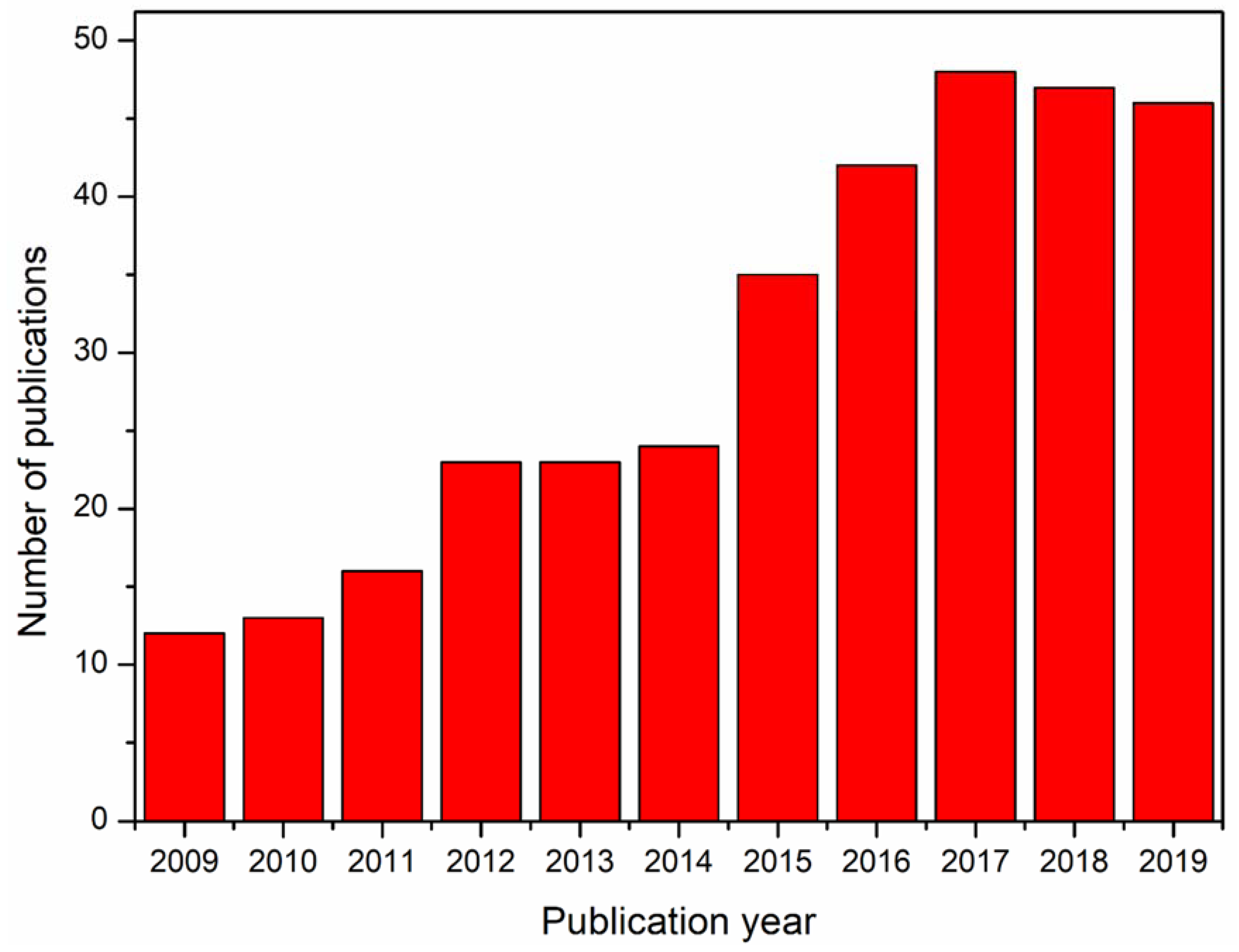
1. Introduction
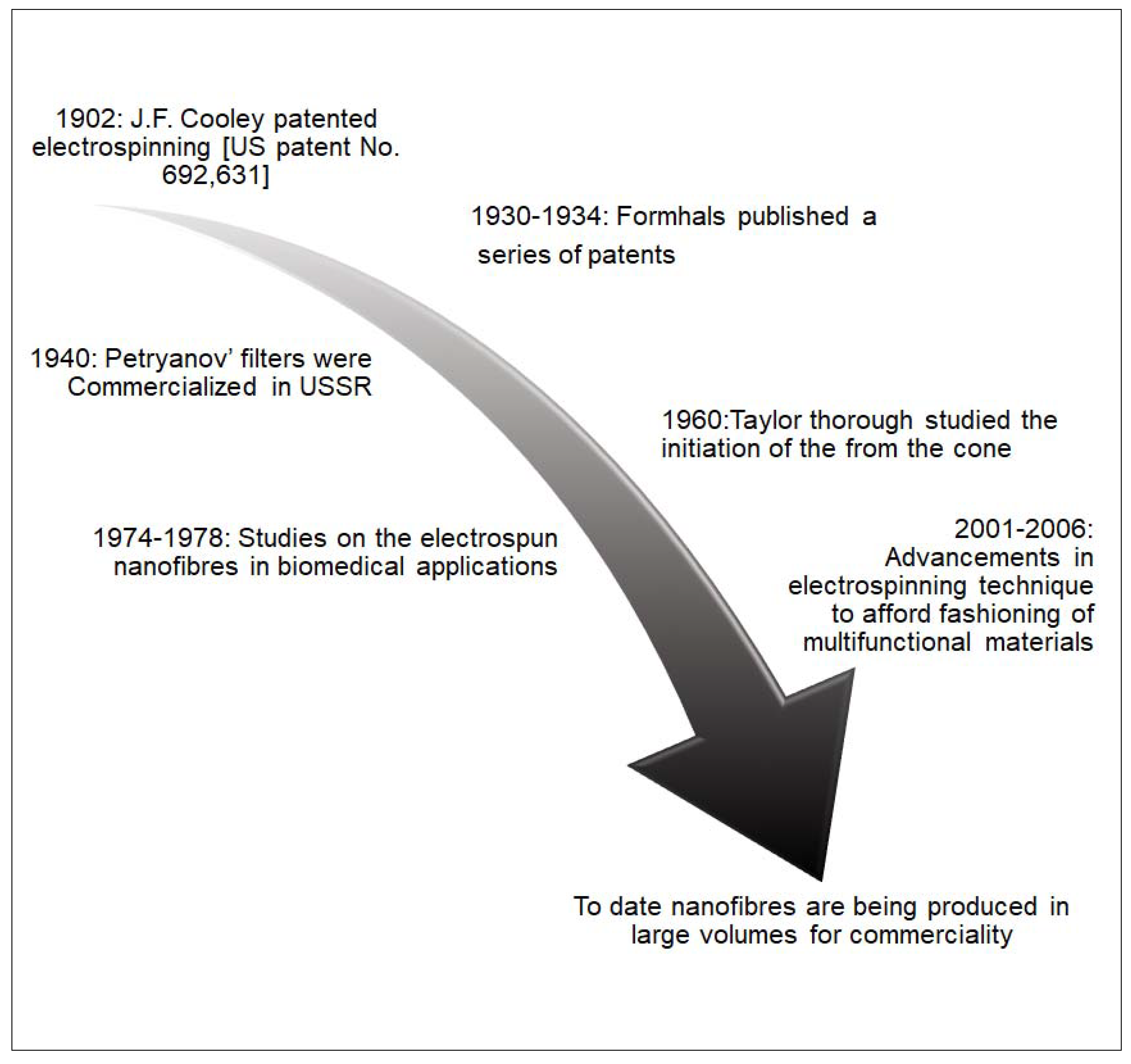
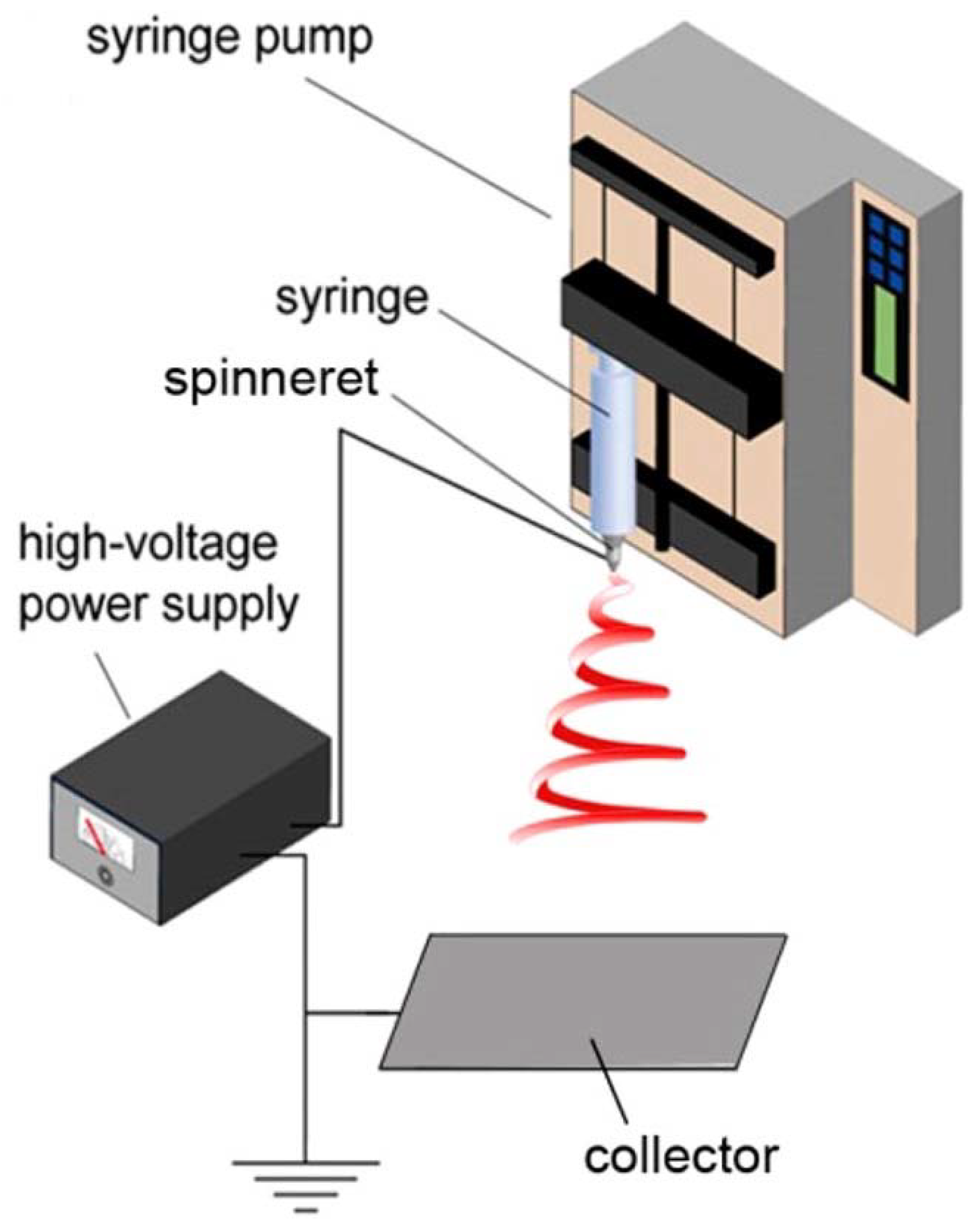
2. Electrospinning Technique
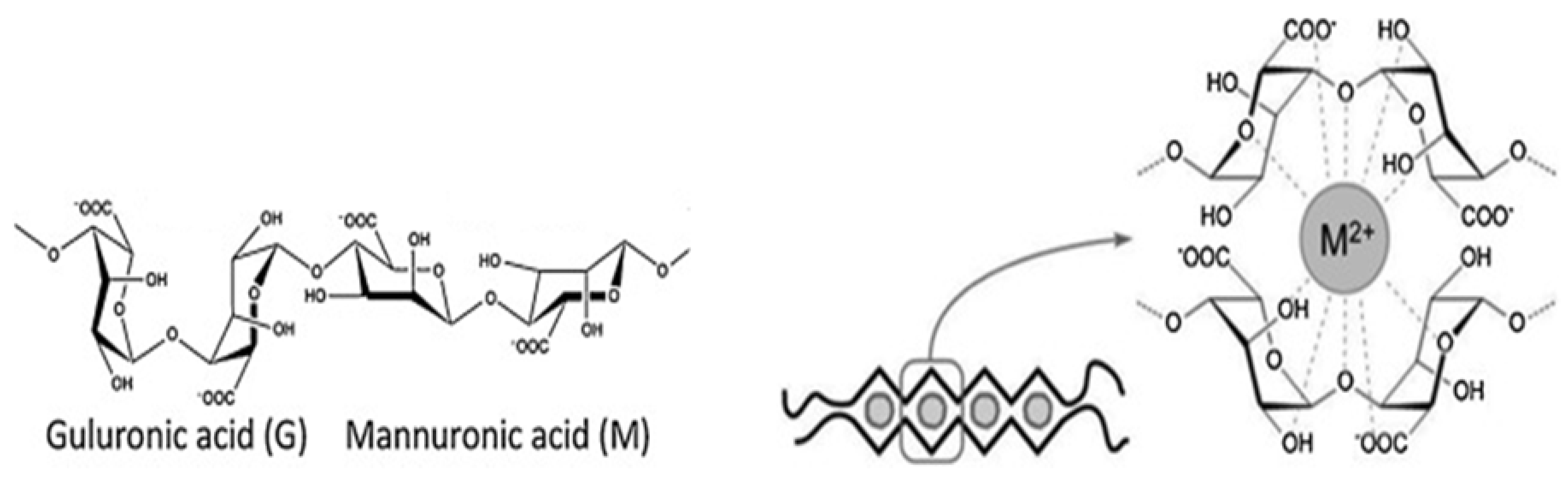
3. Electrospinnability of Alginate
3.1. Pure Alginate
3.2. Co-Solvent Systems
3.3. Hydrosoluble Polymers
3.4. Co-Solvent/Surfactant and Carrier Polymers
3.5. Structural Modification
3.6. Advanced Electrospinning Techniques
3.7. Other Polymers
4. Applications of Electrospun Alginate
4.1. Wastewater Treatment
4.1.1. Bioremediation
4.1.2. Filtration Membranes
4.2. Biomedical Applications
4.2.1. Wound Dressing
4.2.2. Tissue Engineering
4.2.3. Cancer Therapy
4.2.4. Delivery Systems
4.3. Sensors and Energy
5. Future Trends and Conclusions
Author Contributions
Funding
Conflicts of Interest
References
- Schiffman, J.D.; Schauer, C.L. A review: Electrospinning of Biopolymer Nanofibers and their Applications. Polym. Rev. 2008, 48, 317–352. [Google Scholar] [CrossRef]
- Hedayati, A.; Barnett, C.J.; Swan, G.; White, A.O. Chemical recycling of consumer-grade black plastic into electrically conductive carbon nanotubes. C J. Carbon Res. 2019, 5, 32. [Google Scholar] [CrossRef]
- Gönen, S.Ö.; Erol Taygun, M.; Küçükbayrak, S. Evaluation of the factors influencing the resultant diameter of the electrospun gelatin/sodium alginate nanofibers via Box–Behnken design. Mater. Sci. Eng. C 2016, 58, 709–723. [Google Scholar] [CrossRef] [PubMed]
- Venkatesan, J.; Bhatnagar, I.; Manivasagan, P.; Kang, K.-H.; Kim, S.-K. Alginate composites for bone tissue engineering: A review. Int. J. Biol. Macromol. 2015, 72, 269–281. [Google Scholar] [CrossRef] [PubMed]
- Jeong, S.I.; Krebs, M.D.; Bonino, C.A.; Khan, S.A.; Alsberg, E. Electrospun Alginate Nanofibers with Controlled Cell Adhesion for Tissue Engineeringa. Macromol. Biosci. 2010, 10, 934–943. [Google Scholar] [CrossRef] [PubMed]
- Sobhanian, P.; Khorram, M.; Hashemi, S.-S.; Mohammadi, A. Development of nanofibrous collagen-grafted poly (vinyl alcohol)/gelatin/alginate scaffolds as potential skin substitute. Int. J. Biol. Macromol. 2019, 130, 977–987. [Google Scholar] [CrossRef]
- Mohammadi, S.; Ramakrishna, S.; Laurent, S.; Shokrgozar, M.A.; Semnani, D.; Sadeghi, D.; Bonakdar, S.; Akbari, M. Fabrication of Nanofibrous PVA/Alginate-Sulfate Substrates for Growth Factor Delivery. J. Biomed. Mater. Res. Part A 2019, 107, 403–413. [Google Scholar] [CrossRef]
- Taemeh, M.A.; Shiravandi, A.; Korayem, M.A.; Daemi, H. Fabrication challenges and trends in biomedical applications of alginate electrospun nanofibers. Carbohydr. Polym. 2020, 228, 115419. [Google Scholar] [CrossRef]
- Hay, I.D.; Rehman, Z.U.; Moradali, M.F.; Wang, Y.; Rehm, B.H.A. Microbial alginate production, modification and its applications. Microb. Biotechnol. 2013, 6, 637–650. [Google Scholar] [CrossRef]
- Mokhena, T.C.; Jacobs, V.; Luyt, A.S. A review on electrospun bio-based polymers for water treatment. Express Polym. Lett. 2015, 9, 839–880. [Google Scholar] [CrossRef]
- Mirtič, J.; Balažic, H.; Zupančič, Š.; Kristl, J. Effect of Solution Composition Variables on Electrospun Alginate Nanofibers: Response Surface Analysis. Polymers 2019, 11, 692. [Google Scholar]
- Li, W.; Li, X.; Chen, Y.; Li, X.; Deng, H.; Wang, T.; Huang, R.; Fan, G. Poly(vinyl alcohol)/sodium alginate/layered silicate based nanofibrous mats for bacterial inhibition. Carbohydr. Polym. 2013, 92, 2232–2238. [Google Scholar] [CrossRef] [PubMed]
- Dodero, A.; Alloisio, M.; Vicini, S.; Castellano, M. Preparation of composite alginate-based electrospun membranes loaded with ZnO nanoparticles. Carbohydr. Polym. 2020, 227, 115371. [Google Scholar] [CrossRef] [PubMed]
- Sun, F.; Guo, J.; Liu, Y.; Yu, Y. Preparation, characterizations and properties of sodium alginate grafted acrylonitrile/polyethylene glycol electrospun nanofibers. Int. J. Biol. Macromol. 2019, 137, 420–425. [Google Scholar] [CrossRef]
- Phanikumar, V.V.N.; Rikka, R.V.; Das, B.; Gopalan, R.; Rao, B.V.A.; Prakash, R. Investigation of polyvinyl alcohol and sodium alginate as aqueous binders for lithium-titanium oxide anode in lithium-ion batteries. Ionics 2019, 25, 2549–2561. [Google Scholar] [CrossRef]
- Tan, R.; She, Z.; Wang, M.; Fang, Z.; Liu, Y.; Feng, Q. Thermo-sensitive alginate-based injectable hydrogel for tissue engineering. Carbohydr. Polym. 2012, 87, 1515–1521. [Google Scholar] [CrossRef]
- Zhou, W.; Zhang, H.; Liu, Y.; Zou, X.; Shi, J.; Zhao, Y.; Ye, Y.; Yu, Y.; Guo, J. Sodium alginate-polyethylene glycol diacrylate based double network fiber: Rheological properties of fiber forming solution with semi-interpenetrating network structure. Int. J. Biol. Macromol. 2020, 142, 535–544. [Google Scholar] [CrossRef]
- Pawar, S.N.; Edgar, K.J. Alginate derivation: A review of chemistry, properties and applications. Biomaterials 2012, 33, 3279–3305. [Google Scholar] [CrossRef]
- Nie, H.; He, A.; Zheng, J.; Xu, S.; Li, J.; Han, C.C. Effects of Chain Conformation and Entanglement on the Electrospinning of Pure Alginate. Biomacromolecules 2008, 9, 1362–1365. [Google Scholar] [CrossRef]
- Xue, J.; Wu, T.; Dai, Y.; Xia, Y. Electrospinning and Electrospun Nanofibers: Methods, Materials, and Applications. Chem. Rev. 2019, 119, 5298–5415. [Google Scholar] [CrossRef]
- Yang, J.M.; Yang, J.H.; Tsou, S.C.; Ding, C.H.; Hsu, C.C.; Yang, K.C.; Yang, C.C.; Chen, K.S.; Chen, S.W.; Wang, J.S. Cell proliferation on PVA/sodium alginate and PVA/poly(γ-glutamic acid) electrospun fiber. Mater. Sci. Eng. C 2016, 66, 170–177. [Google Scholar] [CrossRef] [PubMed]
- Xue, J.; Xie, J.; Liu, W.; Xia, Y. Electrospun Nanofibers: New Concepts, Materials, and Applications. Acc. Chem. Res. 2017, 50, 1976–1987. [Google Scholar] [CrossRef] [PubMed]
- Ray, S.S.; Chen, S.-S.; Li, C.-W.; Nguyen, N.C.; Nguyen, H.T. A comprehensive review: Electrospinning technique for fabrication and surface modification of membranes for water treatment application. RSC Adv. 2016, 6, 85495–85514. [Google Scholar] [CrossRef]
- Yalcinkaya, F. A review on advanced nanofiber technology for membrane distillation. J. Eng. Fibers Fabr. 2019, 14, 1558925018824901. [Google Scholar] [CrossRef]
- Wongkanya, R.; Chuysinuan, P.; Pengsuk, C.; Techasakul, S.; Lirdprapamongkol, K.; Svasti, J.; Nooeaid, P. Electrospinning of alginate/soy protein isolated nanofibers and their release characteristics for biomedical applications. J. Sci. Adv. Mater. Devices 2017, 2, 309–316. [Google Scholar] [CrossRef]
- Fang, D.; Liu, Y.; Jiang, S.; Nie, J.; Ma, G. Effect of intermolecular interaction on electrospinning of sodium alginate. Carbohydr. Polym. 2011, 85, 276–279. [Google Scholar] [CrossRef]
- Saquing, C.D.; Tang, C.; Monian, B.; Bonino, C.A.; Manasco, J.L.; Alsberg, E.; Khan, S.A. Alginate–Polyethylene Oxide Blend Nanofibers and the Role of the Carrier Polymer in Electrospinning. Ind. Eng. Chem. Res. 2013, 52, 8692–8704. [Google Scholar] [CrossRef]
- Mokhena, T.C.; Jacobs, N.V.; Luyt, A.S. Electrospun alginate nanofibres as potential bio-sorption agent of heavy metals in water treatment. Express Polym. Lett. 2017, 11, 652–663. [Google Scholar] [CrossRef]
- Shalumon, K.T.; Anulekha, K.H.; Nair, S.V.; Nair, S.V.; Chennazhi, K.P.; Jayakumar, R. Sodium alginate/poly(vinyl alcohol)/nano ZnO composite nanofibers for antibacterial wound dressings. Int. J. Biol. Macromol. 2011, 49, 247–254. [Google Scholar] [CrossRef]
- Vigani, B.; Rossi, S.; Milanesi, G.; Bonferoni, M.C.; Sandri, G.; Bruni, G.; Ferrari, F. Electrospun Alginate Fibers: Mixing of Two Different Poly(ethylene oxide) Grades to Improve Fiber Functional Properties. Nanomaterials 2018, 8, 971. [Google Scholar] [CrossRef]
- Bonino, C.A.; Krebs, M.D.; Saquing, C.D.; Jeong, S.I.; Shearer, K.L.; Alsberg, E.; Khan, S.A. Electrospinning alginate-based nanofibers: From blends to crosslinked low molecular weight alginate-only systems. Carbohydr. Polym. 2011, 85, 111–119. [Google Scholar] [CrossRef]
- Chang, J.-J.; Lee, Y.-H.; Wu, M.-H.; Yang, M.-C.; Chien, C.-T. Preparation of electrospun alginate fibers with chitosan sheath. Carbohydr. Polym. 2012, 87, 2357–2361. [Google Scholar] [CrossRef]
- Bhattarai, N.; Li, Z.; Edmondson, D.; Zhang, M. Alginate-Based Nanofibrous Scaffolds: Structural, Mechanical, and Biological Properties. Adv. Mater. 2006, 18, 1463–1467. [Google Scholar] [CrossRef]
- Velasco-Barraza, R.D.; Vera-Graziano, R.; López-Maldonado, E.A.; Oropeza-Guzmán, M.T.; Dastager, S.G.; Álvarez-Andrade, A.; Iglesias, A.L.; Villarreal-Gómez, L.J. Study of nanofiber scaffolds of PAA, PAA/CS, and PAA/ALG for its potential use in biotechnological applications. Int. J. Polym. Mater. Polym. Biomater. 2018, 67, 800–807. [Google Scholar] [CrossRef]
- Lee, Y.J.; Lyoo, W.S. Preparation and characterization of high-molecular-weight atactic poly(vinyl alcohol)/sodium alginate/silver nanocomposite by electrospinning. J. Polym. Sci. Part B Polym. Phys. 2009, 47, 1916–1926. [Google Scholar] [CrossRef]
- Bonino, C.A.; Efimenko, K.; Jeong, S.I.; Krebs, M.D.; Alsberg, E.; Khan, S.A. Three-Dimensional Electrospun Alginate Nanofiber Mats via Tailored Charge Repulsions. Small 2012, 8, 1928–1936. [Google Scholar] [CrossRef]
- Mokhena, T.C.; Jacobs, N.V.; Luyt, A.S. Nanofibrous alginate membrane coated with cellulose nanowhiskers for water purification. Cellulose 2018, 25, 417–427. [Google Scholar] [CrossRef]
- Mokhena, T.C.; Luyt, A.S. Electrospun alginate nanofibres impregnated with silver nanoparticles: Preparation, morphology and antibacterial properties. Carbohydr. Polym. 2017, 165, 304–312. [Google Scholar] [CrossRef]
- Mokhena, T.C.; Luyt, A.S. Development of multifunctional nano/ultrafiltration membrane based on a chitosan thin film on alginate electrospun nanofibres. J. Clean. Prod. 2017, 156, 470–479. [Google Scholar] [CrossRef]
- Castellano, M.; Alloisio Darawish, M.; Dodero, A.; Vicini, S. Electrospun composite mats of alginate with embedded silver nanoparticles. J. Therm. Anal. Calorim. 2019, 137, 767–778. [Google Scholar] [CrossRef]
- Hajaila, H.; Summa, M.; Russo, D.; Armirotti, A.; Brunetti, V.; Bertorelli, R.; Athanassiou, A.; Mele, E. Alginate–lavender nanofibers with antibacterial and anti-inflammatory activity to effectively promote burn healing. J. Mater. Chem. B 2016, 4, 1686–1695. [Google Scholar]
- Wang, S.; Ju, J.; Wu, S.; Lin, M.; Sui, K.; Xia, Y.; Tan, Y. Electrospinning of biocompatible alginate-based nanofiber membranes via tailoring chain flexibility. Carbohydr. Polym. 2020, 230, 115665. [Google Scholar] [CrossRef] [PubMed]
- Hajiali, H.; Heredia-Guerrero, J.A.; Liakos, I.; Athanassiou, A.; Mele, E. Alginate Nanofibrous Mats with Adjustable Degradation Rate for Regenerative Medicine. Biomacromolecules 2015, 16, 936–943. [Google Scholar] [CrossRef] [PubMed]
- Kyzioł, A.; Michna, J.; Moreno, I.; Gamez, E.; Irusta, S. Preparation and characterization of electrospun alginate nanofibers loaded with ciprofloxacin hydrochloride. Eur. Polym. J. 2017, 96, 350–360. [Google Scholar] [CrossRef]
- Daemi, H.; Mashayekhi, M.; Pezeshki Modaress, M. Facile fabrication of sulfated alginate electrospun nanofibers. Carbohydr. Polym. 2018, 198, 481–485. [Google Scholar] [CrossRef]
- Arlov, Ø.; Aachmann, F.L.; Sundan, A.; Espevik, T.; Skjåk-Bræk, G. Heparin-Like Properties of Sulfated Alginates with Defined Sequences and Sulfation Degrees. Biomacromolecules 2014, 15, 2744–2750. [Google Scholar] [CrossRef]
- Esfandiari, F.; Ashtiani, M.K.; Sharifi-Tabar, M.; Saber, M.; Daemi, H.; Ghanian, M.H.; Shahverdi, A.; Baharvand, H. Microparticle-Mediated Delivery of BMP4 for Generation of Meiosis-Competent Germ Cells from Embryonic Stem Cells. Macromol. Biosci. 2017, 17, 1600284. [Google Scholar] [CrossRef]
- Pei, M.; Jia, X.; Zhao, X.; Li, J.; Liu, P. Alginate-based cancer-associated, stimuli-driven and turn-on theranostic prodrug nanogel for cancer detection and treatment. Carbohydr. Polym. 2018, 183, 131–139. [Google Scholar] [CrossRef]
- Pegg, C.E.; Jones, G.H.; Athauda, T.J.; Ozer, R.R.; Chalker, J.M. Facile preparation of ammonium alginate-derived nanofibers carrying diverse therapeutic cargo. Chem. Commun. 2014, 50, 156–158. [Google Scholar] [CrossRef]
- Nista, S.V.G.; Bettini, J.; Mei, L.H.I. Coaxial nanofibers of chitosan–alginate–PEO polycomplex obtained by electrospinning. Carbohydr. Polym. 2015, 127, 222–228. [Google Scholar] [CrossRef]
- Yu, C.C.; Chang, J.J.; Lee, Y.H.; Lin, Y.C.; Wu, M.H.; Yang, M.C.; Chien, C.T. Electrospun scaffolds composing of alginate, chitosan, collagen and hydroxyapatite for applying in bone tissue engineering. Mater. Lett. 2013, 93, 133–136. [Google Scholar] [CrossRef]
- Fujita, S.; Wakuda, Y.; Matsumura, M.; Suye, S.-I. Geometrically customizable alginate hydrogel nanofibers for cell culture platforms. J. Mater. Chem. B 2019, 7, 6556–6563. [Google Scholar] [CrossRef] [PubMed]
- Hu, W.-W.; Yu, H.-N. Coelectrospinning of chitosan/alginate fibers by dual-jet system for modulating material surfaces. Carbohydr. Polym. 2013, 95, 716–727. [Google Scholar] [CrossRef] [PubMed]
- Hu, W.-W.; Lin, C.-H.; Hong, Z.-J. The enrichment of cancer stem cells using composite alginate/polycaprolactone nanofibers. Carbohydr. Polym. 2019, 206, 70–79. [Google Scholar] [CrossRef]
- Vicini, S.; Mauri, M.; Vita, S.; Castellano, M. Alginate and alginate/hyaluronic acid membranes generated by electrospinning in wet conditions: Relationship between solution viscosity and spinnability. J. Appl. Sci. 2018, 135, 46390. [Google Scholar] [CrossRef]
- Dodero, A.; Vicini, S.; Alloisio, M.; Castellano, M. Sodium alginate solutions: Correlation between rheological properties and spinnability. J. Mater. Sci. 2019, 54, 8034–8046. [Google Scholar] [CrossRef]
- Hu, W.-W.; Hu, Z.-C. The control of alginate degradation to dynamically manipulate scaffold composition for in situ transfection application. Int. J. Biol. Macromol. 2018, 117, 1169–1178. [Google Scholar] [CrossRef]
- Hu, W.-W.; Wu, Y.-C.; Hu, Z.-C. The development of an alginate/polycaprolactone composite scaffold for in situ transfection application. Carbohydr. Polym. 2018, 183, 29–36. [Google Scholar] [CrossRef]
- Xu, W.; Shen, R.; Yan, Y.; Gao, J. Preparation and characterization of electrospun alginate/PLA nanofibers as tissue engineering material by emulsion eletrospinning. J. Mech. Behav. Biomed. Mater. 2017, 65, 428–438. [Google Scholar] [CrossRef]
- Liu, X.; Nielsen, L.H.; Kłodzińska, S.N.; Nielsen, H.M.; Qu, H.; Christensen, L.P.; Rantanen, J.; Yang, M. Ciprofloxacin-loaded sodium alginate/poly (lactic-co-glycolic acid) electrospun fibrous mats for wound healing. Eur. J. Pharm. Biopharm. 2018, 123, 42–49. [Google Scholar] [CrossRef]
- Wang, Q.; Ju, J.; Tan, Y.; Hao, L.; Ma, Y.; Wu, Y.; Zhang, H.; Xia, Y.; Sui, K. Controlled synthesis of sodium alginate electrospun nanofiber membranes for multi-occasion adsorption and separation of methylene blue. Carbohydr. Polym. 2019, 205, 125–134. [Google Scholar] [CrossRef] [PubMed]
- De Silva, R.T.; Mantilaka, M.M.M.G.P.G.; Goh, K.L.; Ratnayake, S.P.; Amaratunga, G.A.J.; de Silva, K.M.N. Magnesium Oxide Nanoparticles Reinforced Electrospun Alginate-Based Nanofibrous Scaffolds with Improved Physical Properties. Int. J. Biomater. 2017, 2017, 9. [Google Scholar] [CrossRef] [PubMed]
- Wang, X.; Hsiao, B.S. Electrospun nanofiber membranes. Curr. Opin. Chem. Eng. 2016, 12, 62–81. [Google Scholar] [CrossRef]
- Wang, M.; Zhang, T.; Deng, L.; Li, P.; Wang, X.; Hsiao, B.S. Eco-friendly poly(acrylic acid)-sodium alginate nanofibrous hydrogel: A multifunctional platform for superior removal of Cu(II) and sustainable catalytic applications. Colloids Surf. A: Physicochem. Eng. Asp. 2018, 558, 228–241. [Google Scholar] [CrossRef]
- Tang, Y.; Lan, X.; Liang, C.; Zhong, Z.; Xie, R.; Zhou, Y.; Miao, X.; Wang, H.; Wang, W. Honey loaded alginate/PVA nanofibrous membrane as potential bioactive wound dressing. Carbohydr. Polym. 2019, 219, 113–120. [Google Scholar] [CrossRef]
- Cai, Z.; Zhu, C.; Xiong, P.; Guo, J.; Zhao, K. Calcium alginate-coated electrospun polyhydroxybutyrate/carbon nanotubes composite nanofibers as nanofiltration membrane for dye removal. J. Mater. Sci. 2018, 53, 14801–14820. [Google Scholar] [CrossRef]
- Anyfantis, G.C.; Hajiali, H.; Mele, E.; Marras, S.; Carzino Marini, L.; Papadopoulou, E.L.; Athanassiou, A. Investigation of the electro-spinnability of alginate solutions containing gold precursor HAuCl4. J. Colloid Interface Sci. 2016, 483, 60–66. [Google Scholar] [CrossRef]
- Wróblewska-Krepsztul, J.; Rydzkowski, T.; Michalska-Pożoga, I.; Thakur, V.K. Biopolymers for biomedical and pharmaceutical applications: recent advances and overview of alginate electrospinning. Nanometerials 2019, 9, 404. [Google Scholar]
- Jeong, S.I.; Jeon, O.; Krebs, M.D.; Hill, M.C.; Alsberg, E. Biodegradable photo-crosslinked alginate nanofibre scaffolds with tuneable physical properties, cell adhesivity and growth factor release. Eur. Cells Mater. 2012, 24, 331–343. [Google Scholar] [CrossRef]
- Dodero, A.; Scarfi, S.; Pozzolini, M.; Vicini, S.; Alloisio, M.; Castellano, M. Alginate-Based Electrospun Membranes Containing ZnO Nanoparticles as Potential Wound Healing Patches: Biological, Mechanical, and Physicochemical Characterization. ACS Appl. Mater. Interfaces 2019. [Google Scholar] [CrossRef]
- Ni, P.; Bi, H.; Zhao, G.; Han, Y.; Wickramaratne, M.N.; Dai, H.; Wang, X. Electrospun preparation and biological properties in vitro of polyvinyl alcohol/sodium alginate/nano-hydroxyapatite composite fiber membrane. Colloids Surf. Biointerfaces 2019, 173, 171–177. [Google Scholar] [CrossRef]
- Guo, J.; Zhang, Q.; Cai, Z.; Zhao, K. Preparation and dye filtration property of electrospun polyhydroxybutyrate–calcium alginate/carbon nanotubes composite nanofibrous filtration membrane. Sep. Purif. Technol. 2016, 161, 69–79. [Google Scholar] [CrossRef]
- Jeong, S.I.; Krebs, M.D.; Bonino, C.A.; Samorezov, J.E.; Khan, S.A.; Alsberg, E. Electrospun Chitosan–Alginate Nanofibers with In Situ Polyelectrolyte Complexation for Use as Tissue Engineering Scaffolds. Tissue Eng. Part A 2010, 17, 59–70. [Google Scholar] [CrossRef] [PubMed]
- De Silva, R.T.; Dissanayake, R.K.; Mantilaka, M.P.; Wijesinghe, W.S.; Kaleel, S.S.; Premachandra, T.N.; Weerasinghe, L.; Amaratunga, G.A.; de Silva, K.N. Drug-Loaded Halloysite Nanotube-Reinforced Electrospun Alginate-Based Nanofibrous Scaffolds with Sustained Antimicrobial Protection. ACS Appl. Mater. Interfaces 2018, 10, 33913–33922. [Google Scholar] [CrossRef] [PubMed]
- Golafshan, N.; Kharaziha, M.; Fathi, M. Tough and conductive hybrid graphene-PVA: Alginate fibrous scaffolds for engineering neural construct. Carbon 2016, 111, 752–763. [Google Scholar] [CrossRef]
- Chae, T.; Yang, H.; Leung, V.; Ko, F.; Troczynski, T. Novel biomimetic hydroxyapatite/alginate nanocomposite fibrous scaffolds for bone tissue regeneration. J. Mater. Sci. Mater. Med. 2013, 24, 1885–1894. [Google Scholar] [CrossRef]
- Chen, Y.-H.; Cheng, C.-H.; Chang, W.-J.; Lin, Y.-C.; Lin, F.-H.; Lin, J.-C. Studies of magnetic alginate-based electrospun matrices crosslinked with different methods for potential hyperthermia treatment. Mater. Sci. Eng. C 2016, 62, 338–349. [Google Scholar] [CrossRef]
- Chang, J.-J.; Lee, Y.-H.; Wu, M.-H.; Yang, M.-C.; Chien, C.-T. Electrospun anti-adhesion barrier made of chitosan alginate for reducing peritoneal adhesions. Carbohydr. Polym. 2012, 88, 1304–1312. [Google Scholar] [CrossRef]
- Gizaw, M.; Thompson, J.; Faglie, A.; Lee, S.-Y.; Neuenschwander Chou, S.-F. Electrospun fibers as a dressing material for drug and biological agent delivery in wound healing applications. Bioengineering 2018, 5, 9. [Google Scholar] [CrossRef]
- Hu, W.-W.; Ting, J.-C. Gene immobilization on alginate/polycaprolactone fibers through electrophoretic deposition to promote in situ transfection efficiency and biocompatibility. Int. J. Biol. Macromol. 2019, 121, 1337–1345. [Google Scholar] [CrossRef]
- Kamel, R.M. Prevention of postoperative peritoneal adhesions. Eur. J. Obstet. Gynecol. Reprod. Biol. 2010, 150, 111–118. [Google Scholar] [CrossRef] [PubMed]
- Kovalenko, I.; Zdyrko, B.; Magasinski, A.; Hertzberg, B.; Milicev, Z.; Burtovyy, R.; Luzinov, I.; Yushin, G. A Major Constituent of Brown Algae for Use in High-Capacity Li-Ion Batteries. Science 2011, 334, 75. [Google Scholar] [CrossRef]
- He, Y.; Du, E.; Zhou, J.; He, Y.; Ye, Y.; Wang, J.; Tang, B.; Wang, X. Wet-spinning of fluorescent fibers based on gold nanoclusters-loaded alginate for sensing of heavy metal ions and anti-counterfeiting. Spectrochim. Acta Part A Mol. Biomol. Spectrosc. 2020, 230, 18031. [Google Scholar] [CrossRef] [PubMed]
- Zhang, J.; Wang, X.-X.; Zhang, B.; Ramakrishna, S.; Yu, M.; Ma, J.W.; Long, Y.-Z. In Situ Assembly of Well-Dispersed Ag Nanoparticles throughout Electrospun Alginate Nanofibers for Monitoring Human Breath—Smart Fabrics. ACS Appl. Mater. Interfaces 2018, 10, 19863–19870. [Google Scholar] [CrossRef] [PubMed]
- Hu, W.-P.; Zhang, B.; Zhang, J.; Luo, W.-L.; Guo, Y.; Chen, S.-J.; Yun, M.-J.; Ramakrishna, S.; Long, Y.-Z. Ag/alginate nanofiber membrane for flexible electronic skin. Nanotechnology 2017, 28, 445502. [Google Scholar] [CrossRef] [PubMed]
- İspirli Doğaç, Y.; Deveci, İ.; Mercimek, B.; Teke, M. A comparative study for lipase immobilization onto alginate based composite electrospun nanofibers with effective and enhanced stability. Int. J. Biol. Macromol. 2017, 96, 302–311. [Google Scholar] [CrossRef] [PubMed]
- Ramakrishna, S.; Fujihara, K.; Teo, W.-E.; Yong, T.; Ma, Z.; Ramaseshan, R. Electrospun nanofibers: Solving global issues. Materialstoday 2006, 9, 40–50. [Google Scholar] [CrossRef]
- Dong, Z.; Kennedy, S.J.; Wu, Y. Electrospinning materials for energy-related applications and devices. J. Power Sources 2011, 196, 4886–4904. [Google Scholar] [CrossRef]


| Type | Additional/Carrier Polymer | Co-Solvent | Surfactant | Technique Type | Optimal Conditions | Highlights | Proposed Application | Refs. |
|---|---|---|---|---|---|---|---|---|
| Sodium alginate (SA) 22 KDa | Chitosan as coagulation bath for fiber collection | Glycerol | - | Single needle | Flow rate: 0.1–0.5 mL/hr Tip-to-collector distance (TOC): 7 cm Voltage:13–15 kV | Core–sheath morphology was achieved by electrospinning alginate directly into chitosan coagulation bath, and the fibers having diameter ranging between 600 and 900 nm can be obtained by changing processing parameters | Biomedical | [32] |
| SA (220 kDa) | Chitosan dissolved in 2.5 wt % acetic acid with 37.5 wt % ethanol co-solvent | Glycerol | - | Single needle | Flow rate: 0.4 mL/hr TOC: 3.5 cm Voltage: 10 kV | Core–sheath morphology coated with collagen/hydroxyapatite (HAp) | Bone tissue engineering | [49] |
| Sulfated SA (115 kDa) | Poly(vinyl alcohol) (PVA) a | - | - | Single needle | Flow rate: 0.2 mL/hr TOC: 12 cm Voltage: 20 kV | 5 mL/hr was demonstrated and obtained fibers with average diameters of 144 ± 21 nm | Biomedical | [43] |
| SA | Polyethylene oxide (PEO) a | Dimethylformamide | Pluronic F127 | Single needle | Flow rate: 0.5 mL/hr TOC: 20 cm Voltage: 25 kV | Cylindrical nanofibers with average diameter of 90 ± 20 nm were obtained and crosslinked with trifluroacetiic acid to afford their stability to more than 14 days in phosphate-buffered saline (PBS) solution at pH value of 7.4 | Biomedical | [38] |
| SA (80–120 kDa, M/G 1.56) | PEO a | Dimethylsulfoxide (DMSO) | Triton X-100 | Single needle | Flow rate: 0.8 mL/hr TOC: 17 cm Voltage: 17 kV | Polyelectrlyte complex composed of alginate nanofibers coated with chitosan–AgNPs | Wound dressing | [36] |
| SA (196 kDa, M/G:1.94) | PEO a | - | Triton X-100 | Single needle | Flow rate: 0.5–0.75 mL/hr TOC: 15 cm Voltage: 10–15 kV | Uniform fibers were obtained after addition of Triton X-100 (1 wt %) because of the reduction of surface tension from 55 mN/m to 29 mN/m (70/30 PEO/SA) | Tissue engineering | [31] |
| SA (196 kDa, M/G: 1.94) | PEO a | - | Pluronic F127 | Uniform fibers were obtained after the addition of Pluronic F127 (2 wt %) due to a reduction in surface tension 57 nN/m to 36 mN/m without changing the rheological properties and ionic conductivity of the PEO/SA blend (40/60 SA/PEO) | ||||
| SA (37 kDa) | PEO a | - | Pluronic F127 | Uniform fibers having diameters of about 150 nm with over 83% alginate in the blend (SA/PEO 8.0/16) | In vivo applications | |||
| SA | PVA a and ZnO nanoparticles | - | - | Singe needle | Flow rate: 0.1 mL/hr TOC: 5 cm Voltage:17 kV | SA/PVA fibers had diameter ranging between 190 and 240 nm, which increased to 220–360 nm with the inclusion of ZnO nanoparticles into the system | Wound dressing | [29] |
| SA | PEO a and Ciprofloxacin hydrochloride (antibiotic) | Triton X-100 or Pluronic F127 | Single needle | Flow rate: 0.1–1.0 mL/hr TOC: 15–20 cm Voltage: 6–10 kV | Addition of Triton X-100 led to heterogeneous diameters, while Pluronic resulted in more heterogeneous diametersAntibiotic-loaded fibers had an average of 109–161 nm with loading efficiency of 51% | Wound dressing | [39] | |
| SA (323 kDa, M/G ratio 1.25) | PEO a | Ethanol 10 wt % | Triton X-100 0.8 wt % | Single needle | Flow rate: 0.30 mL/hr TOC: 15–20 cm Voltage: 20–28 kV | Average diameter of 150 nm with mechanical and adsorption capacities were directly depended on the crosslinking agent employed | Dye removal | [59] |
| SA | Poly (acrylic acid) (PAA) a | - | - | Single needle | Flow rate: 16 µL/hr TOC: 16 cm Voltage: 20 kV Relative humidy (RH): 45 ± 6% Temperature: 35 ± 3 °C | The obtained membrane was thermally crosslinked at 150 °C for 3 h and exhibited an adsorption capacity of 591.7 mg/g | Bioremediation | [60] |
| SA (80–120 kDa M/G 1.56) | PEO a and PCL (as co-blended polymer) | Dimethyl sulfoxide (DMSO) | Triton X-100 | Dual jet system | Flow rate: 2 µL/min TOC: 20 cm Voltage: 15 kV | Composition with different properties can be achieved by fine tuning the perfusion rate | Gene-delivery | [53,54] |
| Sulfated alginate | PVA a | - | - | Single needle | Flow rate: 18 mL/hr TOC: 12 cm Voltage: 30 kV | Thermal crosslinking of the electrospun nanofibers improved their integrity and mechanical properties | Tissue engineering | [7] |
| SA | Poly (acrylic acid) (PAA) a | Single needle | Flow rate: 0.3 mL/hr TOC: 10 cm Voltage: 20 kV | Fibers with diameters of 278.52 ± 64.33 nm and porosity of 42.38 | Bone tissue engineering | [32] | ||
| SA | PVA a (MgO NPs as reinforcing agent) | - | - | Single needle | Flow rate: 8–10 µL/min TOC: 10 cm Voltage: 26 kV | Randomly oriented fibers with diameter of 83–230 nm with excellent mechanical properties due to the presence of the NPs | Tissue engineering | [61] |
| SA | PEO a (Soy protein as co-blend) | - | - | Single needle | Flow rate: 0.5 mL/min TOC: 15 cm Voltage: 15 kV | Fibers with diameters of 100–300 nm were obtained; however, an increase soy protein content led to beaded fibers | Biomedical | [25] |
| SA | PVA a (Honey (acacia as antibiotic) | - | - | Single needle | Flow rate: 0.4 mL/hr TOC: 10 cm Voltage: 15 kV | With honey content being >20% (v/v) the fibers were more uniform and the membrane exhibited excellent antimicrobial activity against E. coli and S. aureus without cytotoxicity to NIH/3T3 | Wound dressing | [62] |
| SA | PEO a (Lavender oil (LO) as antibiotic) | Dimethylformamide (DMF) | Pluronic F127 | Single needle | Flow rate: 0.5 mL/hr TOC: 20 cm Voltage: 25 kV | Bead-free fibers with diameters of 93 ± 22 nm were obtained and the membrane exhibited good antibacterial activity against S. aureus; meanwhile, inhibited the production of pro-inflammatory cytokines in vitro and in vivo | Wound dressing | [41] |
| SA | PEO a | DMF | Pluronic F127 | Single needle | Flow rate: 0.5 mL/hr TOC: 20 cm Voltage: 25 kV | Using trifluoroacetic acid (TFA), the degradation of electrospun alginate can be adjusted by immersing the fibers at different intervals, which is of significant importance for manufacturing biomedical devices | Regenerative medicine/drug delivery | [63] |
| Alginate dialdehyde (ADA) | - | Ethanol | - | Single needle | Flow rate: 0.3 mL/hr TOC: 15 cm Voltage: 20–25 kV Temperature: 25 °C Relative Humidity: 30–35% | Oxidation of sodium alginate for 4 h using sodium periodate (NaIO4) with the addition of ethanol as co-solvent resulted in bead-free fibers, with the electrospinnability window being improved by incorporating PEO and adipic acid dihydrazide (AAD, crosslinking agent) | Biomedical | [42] |
© 2020 by the authors. Licensee MDPI, Basel, Switzerland. This article is an open access article distributed under the terms and conditions of the Creative Commons Attribution (CC BY) license (http://creativecommons.org/licenses/by/4.0/).
Share and Cite
Mokhena, T.C.; Mochane, M.J.; Mtibe, A.; John, M.J.; Sadiku, E.R.; Sefadi, J.S. Electrospun Alginate Nanofibers Toward Various Applications: A Review. Materials 2020, 13, 934. https://doi.org/10.3390/ma13040934
Mokhena TC, Mochane MJ, Mtibe A, John MJ, Sadiku ER, Sefadi JS. Electrospun Alginate Nanofibers Toward Various Applications: A Review. Materials. 2020; 13(4):934. https://doi.org/10.3390/ma13040934
Chicago/Turabian StyleMokhena, Teboho Clement, Mokgaotsa Jonas Mochane, Asanda Mtibe, Maya Jacob John, Emmanuel Rotimi Sadiku, and Jeremia Shale Sefadi. 2020. "Electrospun Alginate Nanofibers Toward Various Applications: A Review" Materials 13, no. 4: 934. https://doi.org/10.3390/ma13040934
APA StyleMokhena, T. C., Mochane, M. J., Mtibe, A., John, M. J., Sadiku, E. R., & Sefadi, J. S. (2020). Electrospun Alginate Nanofibers Toward Various Applications: A Review. Materials, 13(4), 934. https://doi.org/10.3390/ma13040934