Article

A Nanosensor for Naked-Eye Identification and Adsorption of Cadmium Ion Based on Core–Shell Magnetic Nanospheres

1
Laboratory for Functional Materials, School of Electronics and Materials Engineering, Leshan Normal University, Leshan 614000, China
2
School of Textile Science and Engineering, National Engineering Laboratory for Advanced Yarn and Clean Production, Wuhan Textile University, Wuhan 430200, China
*
Authors to whom correspondence should be addressed.
Materials 2020, 13(17), 3678; https://doi.org/10.3390/ma13173678
Submission received: 7 July 2020 / Revised: 31 July 2020 / Accepted: 5 August 2020 / Published: 20 August 2020
(This article belongs to the Special Issue Electrocatalytic Nanomaterials for Energy Conversion and Storage)

Abstract

:
Fe3O4@SiO2 nanospheres with a core–shell structure were synthesized and functionalized with bis(2-pyridylmethyl)amine (BPMA). The photoresponses of the as-obtained Fe3O4@SiO2-BPMA for Cr3+, Cd2+, Hg2+ and Pb2+ ions were evaluated through irradiation with a 352 nm ultraviolet lamp, and Fe3O4@SiO2-BPMA exhibited remarkable fluorescence enhancement toward the Cd2+ ion. The adsorption experiments revealed that Fe3O4@SiO2-BPMA had rapid and effective adsorption toward the Cd2+ ion. The adsorption reaction was mostly complete within 30 min, the adsorption efficiency reached 99.3%, and the saturated adsorption amount was 342.5 mg/g based on Langmuir linear fitting. Moreover, Fe3O4@SiO2-BPMA displayed superparamagnetic properties with the saturated magnetization of 20.1 emu/g, and its strong magnetic sensitivity made separation simple and feasible. Our efforts in this work provide a potential magnetic functionalized nanosensor for naked-eye identification and adsorption toward the Cd2+ ion.

Graphical Abstract

1. Introduction

Cadmium (Cd) is an important rare element, which is widely used in the production of pigments [1], phosphors [2] and photocells [3]. Because of its strong toxicity, the Cd2+ ion can cause serious environmental and health issues, including itai-itai disease, lung cancer, renal cancer and prostatic cancer [4,5,6]. Therefore, some fluorescent chemosensors have been developed for the selective recognition and sensing of the Cd2+ ion. These chemosensors can be induced by the Cd2+ ion and exhibit naked-eye changes in fluorescence. High sensitivity, high selectivity, instantaneous response and simplicity are the greatest advantages of these probe molecules [7,8,9,10]. However, these chemosensors for Cd2+ are meant to be disposable and for single use only, and they are difficult if not impossible to recycle. On this basis, the incorporation of fluorescent chemosensors and nanomaterials into an inorganic/organic hybrid nanocomposite is presented.
Magnetic Fe3O4@SiO2 nanoparticles with a core–shell structure have attracted an increasing interest in biological and environmental fields [11,12,13,14]. The particles with nano size possess abundant surface active sites for surface modification, and the greatest strength of Fe3O4@SiO2 nanoparticles is their targeting ability. These magnetic particles can be attracted to a target zone under the action of an external magnetic field, which makes recovery feasible [15,16,17,18,19,20,21]. Moreover, the inert SiO2-shell can prevent the magnetic Fe3O4-core from oxidation, and the abundant surface hydroxyl of SiO2 is highly conducive to surface grafting with organic ligands [22,23,24,25].
In this work, we developed an inorganic/organic hybrid nanosensor for Cd2+ detection, in which the core–shell Fe3O4@SiO2 nanospheres were employed as a matrix, and bis(2-pyridylmethyl)amine (BPMA) was grafted onto the end of the Fe3O4@SiO2 surface through the “grafting from” method. BPMA is a tridentate ligand with two terminal nitrogen-donor coordination sites (pyridyl) and the central nitrogen donor site (amine), which can coordinate with some heavy metal ions to form complexes. The coordination behavior with metal ions caused an interesting photoresponse, especially ones with d10 metal centers. The as-obtained Fe3O4@SiO2-BPMA showed a significant change after binding with a Cd2+ ion in the excitation of ultraviolet light, which was visible to the naked eye. Moreover, Fe3O4@SiO2-BPMA also showed rapid and efficient adsorption toward the Cd2+ ion. It is worth noting that Fe3O4@SiO2-BPMA possesses superparamagnetism, and the strong magnetic sensitivity makes recycling easier.

2. Experimental

2.1. Materials

FeCl24H2O, FeCl36H2O, NH3H2O (25 wt.%), trisodium citrate and ethyl silicate were purchased from Shanghai Macklin Biochemical Co. Ltd. (Shanghai, China) Trichloromethylsilane, trichloro(3-chloropropyl)silane, branched polyethylenimine (Mw ~25,000), 4-bromonaphthalic anhydride and bis(2-pyridylmethyl)amine (BPMA) were purchased from Sigma-Aldrich Co. Ltd. (Shanghai, China)

2.2. Synthesis of Fe3O4@SiO2 Nanospheres

Firstly, Fe3O4 particles were synthesized based on a chemical co-precipitation strategy. Briefly, FeCl24H2O (10 mmol) and FeCl36H2O (20 mmol) were dissolved with distilled water (120 mL), and 60 mL of NH3H2O (25 wt.%) was added to the above solution under a N2 atmosphere with mechanical stirring. The mixture was kept for 30 min at 70 °C. After that, the black precipitate was collected and washed with distilled water. Subsequently, the as-synthesized Fe3O4 particles were added to a trisodium citrate solution (150 mL, 20 mmol/L), the mixture was kept with mechanical stirring for 12 h at room temperature, and N2 was bubbled throughout the reaction. After that, the citrate-capped Fe3O4 was washed with distilled water and ethanol.
Then, Fe3O4@SiO2 particles were synthesized using a sol–gel process by the hydrolysis and condensation of ethyl silicate on the Fe3O4 seed. Briefly, the citrate-capped Fe3O4 (0.56 g), NH3∙H2O (5.0 mL, 25 wt.%) and ethyl silicate (4.0 mL) were added to ethanol (120 mL) under mechanical stirring, and the mixture was kept for 8 h at room temperature under a N2 atmosphere. After that, the as-synthesized Fe3O4@SiO2 precipitate was collected and washed with ethanol.

2.3. Fe3O4@SiO2 Functionalized with BPMA

The surface modifications on Fe3O4@SiO2 are illustrated by a flow chart in Figure 1. Firstly, the Fe3O4@SiO2 surface was functionalized with chlorine groups (labeled as Fe3O4@SiO2-Cl). Fe3O4@SiO2 (0.60 g), trichloromethylsilane (17.6 mmol) and trichloro(3-chloropropyl)silane (1.4 mmol) were added to hexane (150 mL), and the mixture was kept for 24 h with mechanical stirring at room temperature in an atmosphere of hydrogen chloride. After that, the as-synthesized Fe3O4@SiO2-Cl particles were washed with ethanol.
Secondly, the Fe3O4@SiO2 surface was functionalized with amino groups (labeled as Fe3O4@SiO2-NH2). The above Fe3O4@SiO2-Cl, methanol (5.0 mL) and branched polyethylenimine (1.0 mL) were added to distilled water (120 mL) under a N2 atmosphere with mechanical stirring, and the mixture was kept for 48 h at 65 °C. After that, the as-synthesized Fe3O4@SiO2-NH2 particles were washed with distilled water and ethanol.
Thirdly, the Fe3O4@SiO2 surface was functionalized with bromine groups (labeled as Fe3O4@SiO2-Br). The above Fe3O4@SiO2-NH2 and 4-bromonaphthalic anhydride (5.4 mmol) were added to ethanol (80 mL) under a N2 atmosphere with mechanical stirring, and the mixture was refluxed for 36 h in darkness. After that, the as-synthesized Fe3O4@SiO2-Br particles were washed with ethanol.
Finally, the Fe3O4@SiO2 surface was functionalized with bis(2-pyridylmethyl)amine (labeled as Fe3O4@SiO2-BPMA). The above Fe3O4@SiO2-Br and BPMA (7.02 mmol) were added to toluene (120 mL) under a N2 atmosphere with mechanical stirring, and the mixture was refluxed for 36 h in darkness. After that, the as-synthesized Fe3O4@SiO2-BPMA particles were washed with ethanol and then dried in vacuum at 60 °C for 24 h.

2.4. Characterization

X-ray diffraction (XRD) analyses were carried out using a DX-2700 instrument (Dandong, China) with Cu Kα radiation (30 kV, 25 mA). Transmission electron microscopy (TEM) and scanning electron microscopy (SEM) images were obtained by means of JEM-2100 (Tokyo, Japan) and SU8020 (Tokyo, Japan) instruments, respectively. Fourier transform infrared (FTIR) spectra were obtained using a Spectrum One (Nicolet iS 10, Waltham, MA, USA). X-ray photoelectron spectroscopy (XPS) analyses were performed by an ESCALAB 250Xi electron spectrometer (Waltham, MA, USA). Magnetization curves were obtained using a vibrating sample magnetometer (VSM, Lakeshore 7307, Columbus, OH, USA).

2.5. Adsorption Study

The adsorption capacities of Fe3O4@SiO2-BPMA were evaluated by removing heavy metal ions with strong toxicity (Cr3+, Cd2+, Hg2+ and Pb2+ ions) from simulated wastewaters. Briefly, 50 mg Fe3O4@SiO2 or Fe3O4@SiO2-BPMA powders were dispersed in 50 mL of the simulated wastewaters without pH pre-adjustments. The mixture was stirred with a constant speed at room temperature. Then, a certain amount of suspension was withdrawn at regular intervals, and the qualitative analysis of metal ions was performed. The adsorption efficiencies (ηt, %) at a time t were calculated using Equation (1).
η t = C 0 C t C 0 × 100   %

3. Results and Discussion

Figure 2 shows the XRD patterns of Fe3O4 and Fe3O4@SiO2 powders. As observed, the XRD patterns of Fe3O4 showed several well-resolved peaks indexed to (220), (311), (400), (422), (511) and (440) planes, which are considered strongly consistent with the database of magnetite (JCPDS No. 65-3107). According to the Scherrer formula, the grain size of Fe3O4 was calculated, and the value was about 9.6 nm. For the XRD pattern of Fe3O4@SiO2, besides the diffraction peaks of the Fe3O4 phase, a broad and weak peak at 2θ = 11~27° (the orange dotted bordered rectangle) was observed, which was assigned to amorphous SiO2. This suggests that the Fe3O4 crystal was successfully composited with amorphous SiO2.
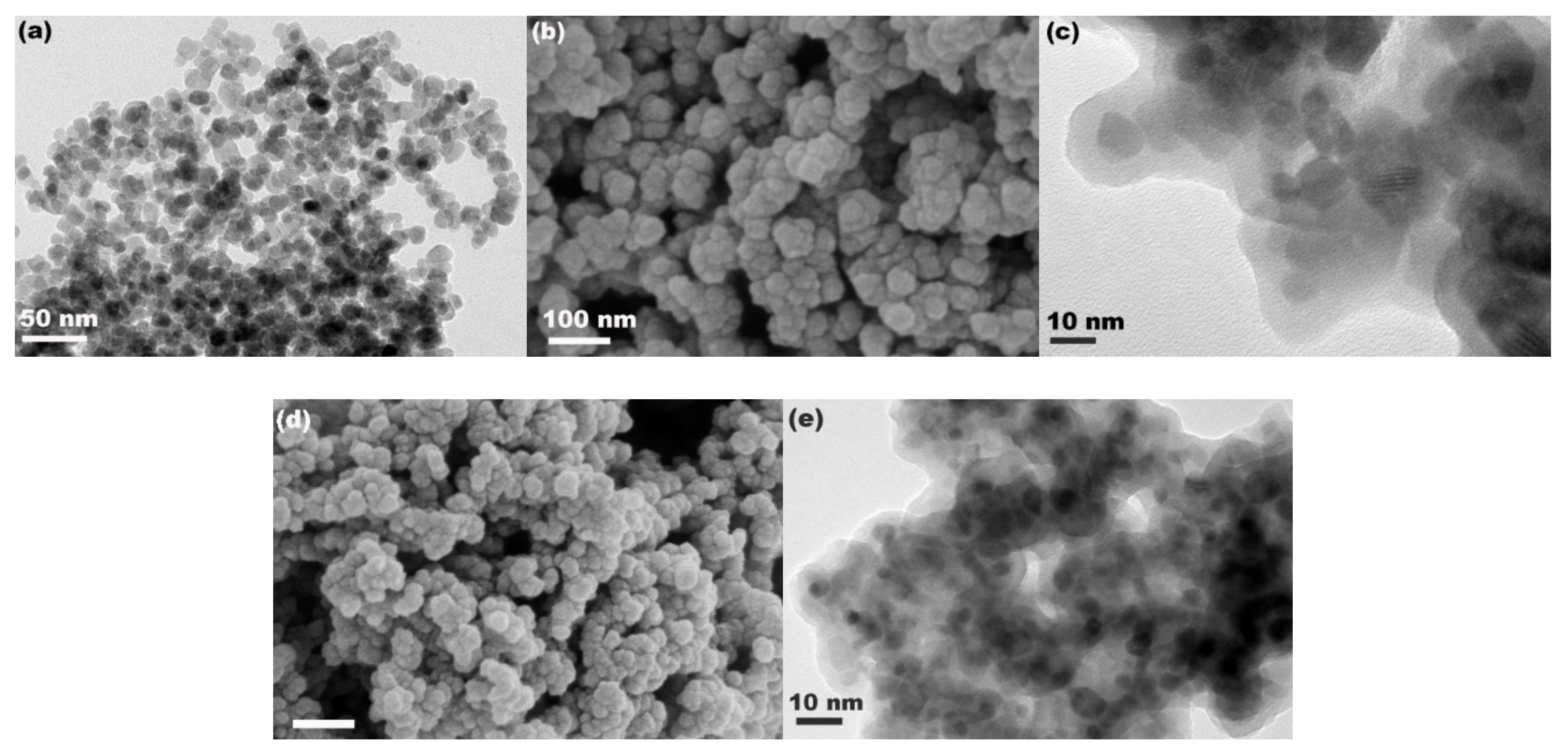
Figure 3a shows the TEM image of Fe3O4 particles. As observed, the Fe3O4 particles exhibited a mean size of about 10 nm, consistent with the calculation of XRD analysis. After compositing with SiO2, the size of particles increased to about 24.0 nm, as shown in the SEM image in Figure 3b, and the TEM image in Figure 3c reveals the obvious core–shell structure with a shell thickness of about 7.0 nm. This suggests that Fe3O4@SiO2 nanospheres with a core–shell structure were successfully synthesized using a sol–gel process by the hydrolysis and condensation of ethyl silicate on the Fe3O4 seed. However, the Fe3O4@SiO2 particles were aggregated slightly, which may be attributed to the enhancement of surface activity after coating SiO2 on the Fe3O4 seed because of more abundant surface hydroxyl from SiO2. Figure 3d,e show the SEM and TEM images of Fe3O4@SiO2-BPMA particles, respectively. There were no significant changes in either size or morphology compared with those of Fe3O4@SiO2 particles.
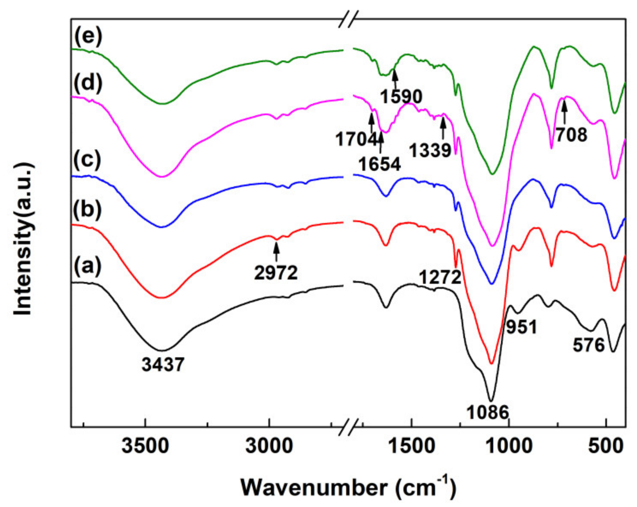
The grafted groups on the Fe3O4@SiO2 surface were characterized by FTIR. As observed in Figure 4a–e, the peaks for all samples at 3437, 1086 and 576 cm−1 were ascribed to the stretching vibration of O-H, Si-O and Fe-O bonds, further demonstrating the successful synthesis of Fe3O4@SiO2 with abundant surface hydroxyl. In the FTIR spectrum of Fe3O4@SiO2-Cl (Figure 4b), the absorption peaks at 2972 and 1272 cm−1 were ascribed to the stretching of C-H species and the symmetrical deformation vibration of Si-C species, respectively. Compared with the FTIR spectrum of Fe3O4@SiO2-Cl, there were no observable changes in that of Fe3O4@SiO2-NH2 in Figure 4c, but the signal of the Si-O bond vanished at 576 cm−1. For the FTIR spectrum of Fe3O4@SiO2-Br in Figure 4d, the absorption peaks at 1704, 1654 and 1339 cm−1 could be attributable to N-C=O species. In Figure 4d, the immobilization of BPMA on the Fe3O4@SiO2 surface is demonstrated by the new peak appearing at 1590 cm−1, which could be attributable to C=N species. Further analyses of Fe3O4@SiO2-BPMA were conducted by XPS, as discussed below.
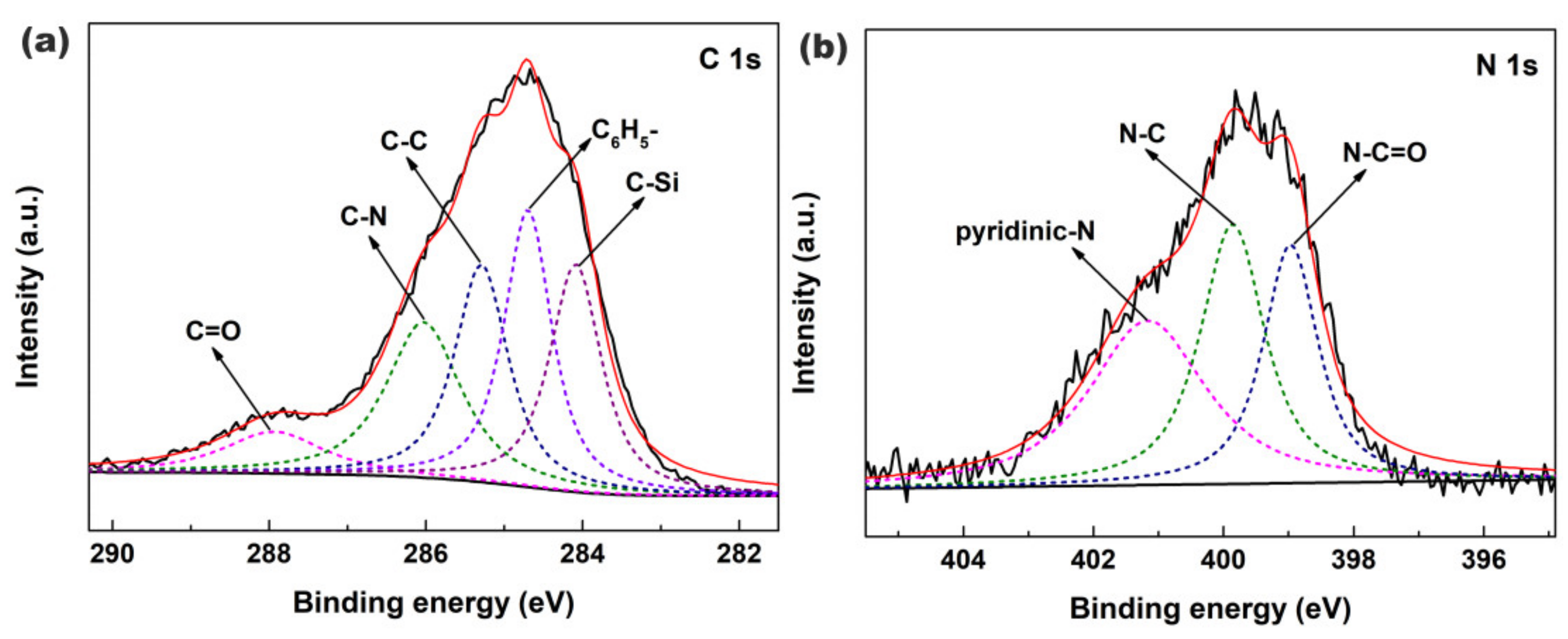
Figure 5a,b show the XPS spectra of C 1s and N 1s core-levels of Fe3O4@SiO2-BPMA powders, respectively. As observed in Figure 5a, the C 1s core-level spectrum of Fe3O4@SiO2-BPMA can be curve-fitted into five peak components with binding energies of 287.9, 286.0, 285.3, 284.7 and 284.1 eV, corresponding to C=O, C-N, C-C, (C6H5-) and C-Si bonds, respectively. Moreover, the N 1s core-level spectrum in Figure 5b can be curve-fitted into three peak components with the binding energies of 401.1 eV for pyridinic-N species, 399.8 eV for N-C species and 399.0 eV for N-C=O species, respectively. Combined with the results of FTIR analyses in Figure 4, it can be concluded that BPMA was successfully bonded onto the Fe3O4@SiO2 surface through a series of successive coupling reactions.
The magnetic properties of Fe3O4@SiO2 and Fe3O4@SiO2-BPMA powders were investigated using a VSM at 300 K. As observed in Figure 6, the saturated magnetization of Fe3O4@SiO2 and Fe3O4@SiO2-BPMA powders were 38.1 and 20.1 emu/g with neither remanence nor coercivity, indicating their superparamagnetism. Compared with Fe3O4@SiO2, the decrease in saturated magnetization for Fe3O4@SiO2-BPMA powders was attributed to the contribution of the non-magnetic grafted organic molecule on the Fe3O4@SiO2 surface. The insets in Figure 6 show the photographs of the magnetic response using a magnet and re-dispersion by shaking the Fe3O4@SiO2-BPMA suspension. From the Figure 6 insets, it can be seen that Fe3O4@SiO2-BPMA particles were well dispersed in an aqueous solution, and these particles were quickly and completely separated from solution under the attraction of an external magnet within about 30 seconds in this work. Moreover, the re-dispersion of Fe3O4@SiO2-BPMA in aqueous solution occurred simply and quickly through slight shaking once the external magnet was removed. This demonstrates that the as-synthesized Fe3O4@SiO2-BPMA possesses strong magnetic sensitivity, which could make recycling easy and feasible.
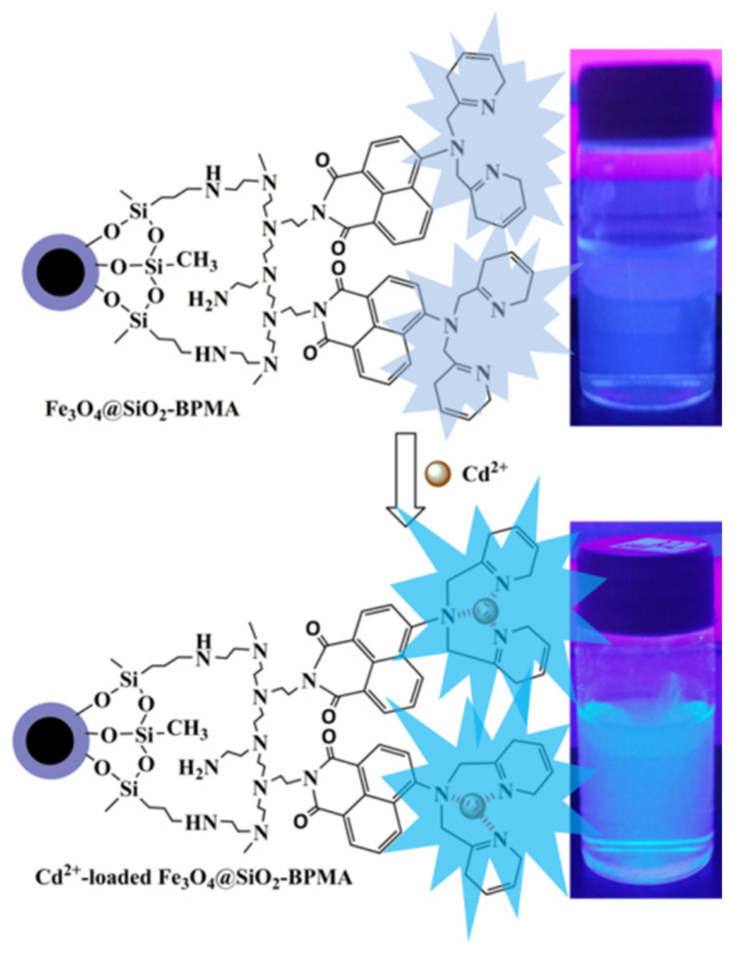
To evaluate the photoresponse of the Fe3O4@SiO2-BPMA nanosensor for heavy metal ions such as Cr3+, Cd2+, Hg2+ and Pb2+ ions, the fluorescence changes to various heavy metal ions with equal concentrations were measured using a 352 nm fluorescent lamp. The top of Figure 7 shows the changes in color of the Fe3O4@SiO2-BPMA suspension upon the addition of Men+ (Men+ = Cr3+, Cd2+, Hg2+ and Pb2+) illuminated by natural light; the Fe3O4@SiO2-BPMA suspension had no significant changes in the appearance of color after the addition of Cr3+, Cd2+, Hg2+ and Pb2+ ions in comparison with that of only Fe3O4@SiO2-BPMA (blank in Figure 7, top). The bottom of Figure 7 shows the eye-perceived fluorescence changes in Fe3O4@SiO2-BPMA toward Cr3+, Cd2+, Hg2+ and Pb2+ ions through irradiation with a 352 nm ultraviolet lamp. The considerable fluorescence enhancements can be observed by the naked eye in the vial with the Cd2+ ion, and the detection limit for Cd2+ ions reached up to 8.0 × 10⁻⁷ mol/L. In contrast, no significant fluorescent changes in emission were observed for the other vials containing Cr3+, Hg2+ and Pb2+ ions. However, the coordination diagrammatic sketch of Fe3O4@SiO2-BPMA with Cd2+ is shown in Figure 8. Upon the addition of the Cd2+ ion, the grafted tridentate receptor BPMA molecule coordinated with the Cd2+ ion, which decreased the electron-donating ability of the nitrogen atom from BPMA during the photo-induced charge transfer process, and thus, significant fluorescence enhancement occurred [26,27,28].
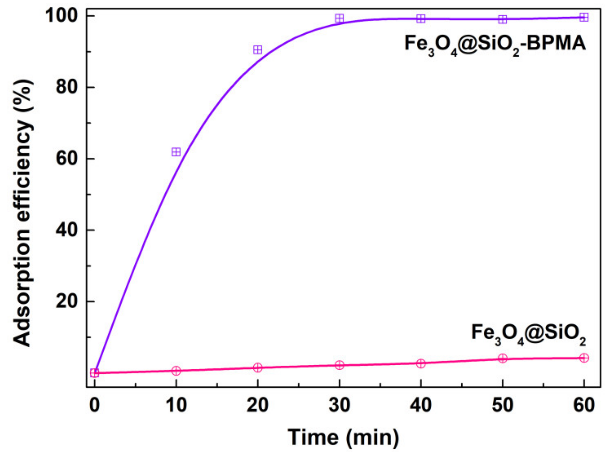
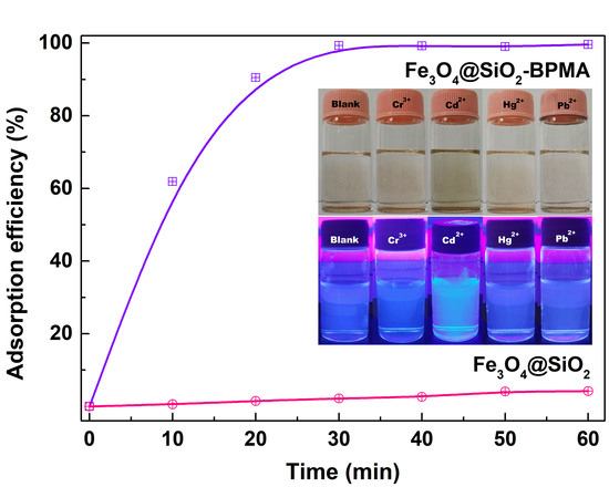
The adsorption capacity of Fe3O4@SiO2-BPMA nanocomposites toward the Cd2+ ion was evaluated at room temperature without pH pre-adjustments. Figure 9 shows the time-dependence of adsorption profiles of the Cd2+ ion onto Fe3O4@SiO2-BPMA. As observed, Fe3O4@SiO2-BPMA exhibited rapid adsorption to Cd2+ ions within 20 min, and the adsorption efficiency within 20 min reached 90.5%. Moreover, the adsorption reaction was mostly complete within 30 min, and the adsorption efficiency within 30 min reached 99.3%. As a comparison, the adsorption capacity of Fe3O4@SiO2 without BPMA modification was also tested, and the adsorption efficiency within 60 min was below 5.0% under the same conditions. This indicates that the absorption of the Cd2+ ion was mainly attributed to the grafted BPMA molecule, not Fe3O4@SiO2 particles.
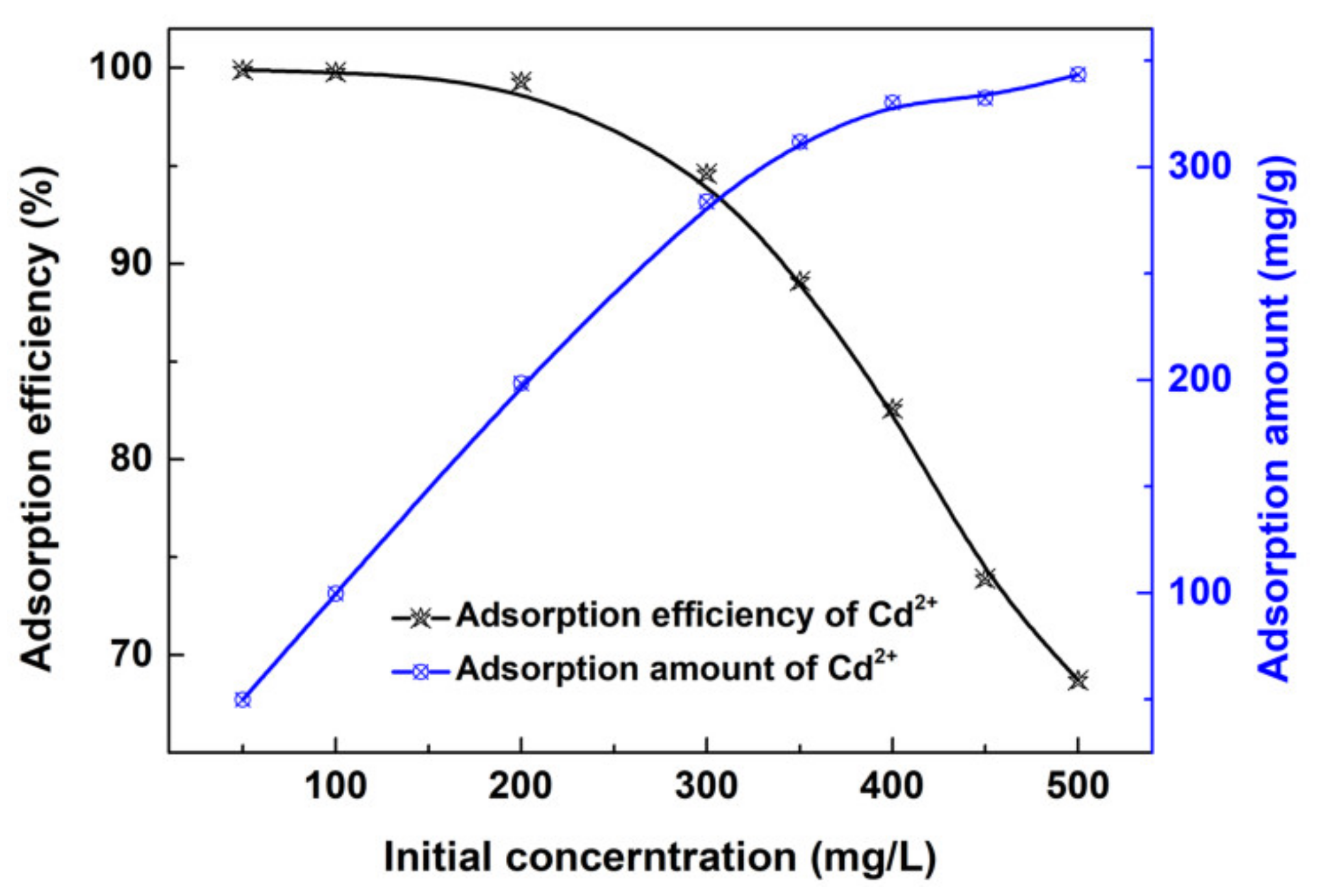
Figure 10 shows the effects of Cd2+ initial concentration on the adsorption efficiency and adsorption amount of the Fe3O4@SiO2-BPMA adsorbent. As observed, the adsorption efficiency decreased with the increasing Cd2+ initial concentration, the decrease being especially sharp when the Cd2+ initial concentration was higher than 300 mg/L, which could be due to the saturation of the Fe3O4@SiO2-BPMA adsorbent. The adsorption efficiency remained at more than 94.0% when the Cd2+ initial concentration was lower than 300 mg/L. However, the adsorption amount increased with the increasing Cd2+ initial concentration, and the increasing trend receded gradually. The mass of the Fe3O4@SiO2-BPMA adsorbent was certain, and the relative adsorption sites gradually decreased with the increasing Cd2+ initial concentration and finally reached an adsorption–desorption equilibrium between the Cd2+ ion and the adsorbent. The saturated adsorption amount (qm, mg/g) of Cd2+ ions can be evaluated by the Langmuir isotherm model using Equations (2) and (3).
q e = ( C 0 C e ) V m
C e q e = 1 q m C e + 1 K L q m
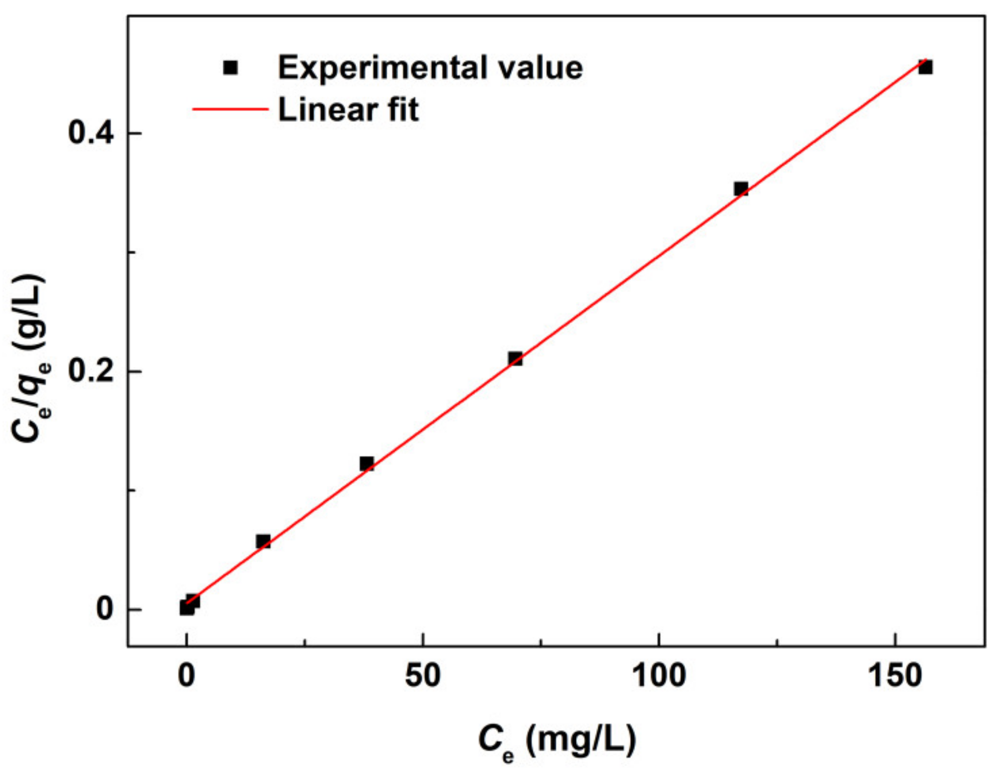
where C0 (mg/L) is the initial concentration of Cd2+ ions, Ce (mg/L) is the concentration of Cd2+ ions at equilibrium, m (g) is the mass of Fe3O4@SiO2-BPMA powders, V (L) is the volume of Cd2+ aqueous solution, qe (mg/g) is the equilibrium adsorption amount of Fe3O4@SiO2-BPMA, qm (mg/g) is the saturated adsorption amount of Fe3O4@SiO2-BPMA, and KL is the Langmuir adsorption constant. The Langmuir linear fit of the adsorption of Cd2+ ions onto Fe3O4@SiO2-BPMA is shown in Figure 11. The corresponding Langmuir parameters calculated at room temperature were as follows: qm = 342.5 mg/g and KL = 0.5478. Moreover, a high associated correlation coefficient R2 of 0.9991 was obtained, indicating that the Langmuir isotherm model was a good fit for modeling the adsorption of Cd2+ ions onto Fe3O4@SiO2-BPMA.

4. Conclusions

A sensitive fluorescence sensor based on functionalized Fe3O4@SiO2 nanospheres was used for simultaneously detecting and removing Cd2+ ions. The as-synthesized Fe3O4@SiO2-BPMA underwent an eye-perceived fluorescence enhancement induced by Cd2+ ions. Meanwhile, Fe3O4@SiO2-BPMA exhibited rapid and effective adsorption toward the Cd2+ ion, and the adsorption reaction was mostly complete within 30 min. The Cd2+ adsorption capacity of Fe3O4@SiO2-BPMA was determined by fitting the experimental data with the Langmuir model, and the saturated adsorption amount was 342.5 mg/g at room temperature. Moreover, Fe3O4@SiO2-BPMA showed superparamagnetic properties with a saturated magnetization of 20.1 emu/g, which could help to separate these particles after capturing Cd2+ ions. The nanocomposites presented here have great potential applications for the naked-eye identification, adsorption and separation of the Cd2+ ion.

Author Contributions

Y.X. project administration; writing—original draft preparation; Z.X. and C.C. data curation; X.L., formal analysis; Q.J. funding acquisition; Y.Z. and C.D. supervision. All authors have read and agreed to the published version of the manuscript.

Funding

This research was funded by Leshan Normal University Research Program, China (No. LZD021).

Conflicts of Interest

The authors declare no conflict of interest.

References

  1. Makarewicz, E.; Cysewski, P.; Michalik, A.; Ziółkowska, D. Properties of Acid or Alkali Treated Cadmium Pigments. Dyes Pigment. 2013, 96, 338–348. [Google Scholar] [CrossRef]
  2. İlhan, M.; Keskin, İ.Ç. Photoluminescence, Radioluminescence and Thermoluminescence Properties of Eu3+ Doped Cadmium Tantalate Phosphor. Dalton Trans. 2018, 47, 13939–13948. [Google Scholar] [CrossRef]
  3. Ma, J.; Ni, S.; Zhang, J.; Yang, X.; Zhang, L. The Electrochemical Performance of Nickel Chromium Oxide as a New Anode Material for Lithium Ion Batteries. Electrochim. Acta 2015, 176, 1420–1426. [Google Scholar] [CrossRef]
  4. Vizuete, J.; Pérez-López, M.; Míguez-Santiyán, M.P.; Hernández-Moreno, D. Mercury (Hg), Lead (Pb), Cadmium (Cd), Selenium (Se), and Arsenic (As) in Liver, Kidney, and Feathers of Gulls: A Review. In Reviews of Environmental Contamination and Toxicology Volume 247; Springer International Publishing: Cham, Switzerland, 2008; Volume 247, pp. 85–146. [Google Scholar] [CrossRef]
  5. Ur Rehman, M.Z.; Rizwan, M.; Hussain, A.; Saqib, M.; Ali, S.; Sohail, M.I.; Shafiq, M.; Hafeez, F. Alleviation of Cadmium (Cd) Toxicity and Minimizing its Uptake in Wheat (Triticum Aestivum) by Using Organic Carbon Sources in Cd-Spiked Soil. Environ. Pollut. 2018, 241, 557–565. [Google Scholar] [CrossRef] [PubMed]
  6. D’Haese, P.C.; Couttenye, M.M.; Lamberts, L.V.; Elseviers, M.M.; Goodman, W.G.; Schrooten, I.; Cabrera, W.E.; De Broe, M.E. Aluminum, Iron, Lead, Cadmium, Copper, Zinc, Chromium, Magnesium, Strontium, and Calcium Content in Bone of End-Stage Renal Failure Patients. Clin. Chem. 1999, 45, 1548–1556. [Google Scholar] [CrossRef] [PubMed]
  7. Shi, Y.S.; Li, Y.H.; Cui, G.H.; Dong, G.Y. New Two-Dimensional Cd(II) Coordination Networks Bearing Benzimidazolyl-Based Linkers as Bifunctional Chemosensors for Detection of Acetylacetone and Fe3+. CrystEngComm 2020, 22, 905–914. [Google Scholar] [CrossRef]
  8. Quan, C.; Liu, J.; Sun, W.; Cheng, X. Highly Sensitive and Selective Fluorescence Chemosensors Containing Phenanthroline Moieties for Detection of Zn2+ and Cd2+ Ions. Chem. Pap. 2019, 74, 485–497. [Google Scholar] [CrossRef]
  9. Eseola, A.O.; Görls, H.; Bangesh, M.; Plass, W. ESIPT-capable 2,6-di(1H-imidazol-2-yl)phenols with Very Strong Fluorescent Sensing Signals towards Cr(III), Zn(II) and Cd(II): Molecular Variation Effects on Turn-On Efficiency. New J. Chem. 2018, 42, 7884–7900. [Google Scholar] [CrossRef]
  10. Vera, M.; Santacruz Ortega, H.; Inoue, M.; Machi, L. Bichromophoric Naphthalene Derivatives of Diethylenetriaminepentaacetate (DTPA): Fluorescent Response to H+, Ca2+, Zn2+, and Cd2+. Supramol. Chem. 2019, 31, 336–348. [Google Scholar] [CrossRef]
  11. Sun, M.; Zhao, A.; Wang, D.; Wang, J.; Chen, P.; Sun, H. Cube-like Fe3O4@SiO2@Au@Ag Magnetic Nanoparticles: A Highly Efficient SERS Substrate for Pesticide Detection. Nanotechnology 2018, 29, 165302. [Google Scholar] [CrossRef]
  12. Yuan, Z.; Xu, R.; Li, J.; Chen, Y.; Wu, B.; Feng, J.; Chen, Z. Biological Responses to Core-Shell-Structured Fe3O4@SiO2-NH2 Nanoparticles in Rats by a Nuclear Magnetic Resonance-Based Metabonomic Strategy. Int. J. Nanomed. 2018, 13, 2447–2462. [Google Scholar] [CrossRef] [PubMed] [Green Version]
  13. Chen, S.; Chen, J.; Zhu, X. Solid Phase Extraction of Bisphenol A Using Magnetic Core-Shell (Fe3O4@SiO2) Nanoparticles Coated with an Ionic liquid, and its Quantitation by HPLC. Microchim. Acta 2016, 183, 1315–1321. [Google Scholar] [CrossRef]
  14. Jiang, H.; Liu, Y.; Luo, W.; Wang, Y.; Tang, X.; Dou, W.; Cui, Y.; Liu, W. A Resumable Two-Photon Fluorescent Probe for Cu2+ And S2− Based on Magnetic Silica Core-Shell Fe3O4@SiO2 Nanoparticles and its Application in Bioimaging. Anal. Chim. Acta 2018, 1014, 91–99. [Google Scholar] [CrossRef] [PubMed]
  15. Yang, Y.; Liu, Y.; Cheng, C.; Shi, H.; Yang, H.; Yuan, H.; Ni, C. Rational Design of GO-Modified Fe3O4/SiO2 Nanoparticles with Combined Rhenium-188 and Gambogic Acid for Magnetic Target Therapy. ACS Appl. Mater. Interfaces 2017, 9, 28195–28208. [Google Scholar] [CrossRef] [PubMed]
  16. Ding, Z.; Shaw, L.L. Enhancement of Hydrogen Desorption from Nano-Composite Prepared by Ball-Milling MgH2 with in-situ Aerosol-Spraying LiBH4. ACS Sustain. Chem. Eng. 2019, 7, 15064–15072. [Google Scholar] [CrossRef]
  17. Morita, Y.; Sakurai, R.; Wakimoto, T.; Kobayashi, K.; Xu, B.; Toku, Y.; Song, G.; Luo, Q.; Ju, Y. tLyP-1-Conjugated Core-Shell Nanoparticles, Fe3O4NPs@mSiO2, for Tumor-Targeted Drug Delivery. Appl. Surf. Sci. 2019, 474, 17–24. [Google Scholar] [CrossRef]
  18. Ding, Z.; Li, H.; Shaw, L. New Insights into the Solid-State Hydrogen Storage of Nanostructured LiBH4-MgH2 System. Chem. Eng. J. 2019, 385, 123856. [Google Scholar] [CrossRef]
  19. Du, Q.; Zhang, W.; Ma, H.; Zheng, J.; Zhou, B.; Li, Y. Immobilized Palladium on Surface-Modified Fe3O4/SiO2 Nanoparticles: As a Magnetically Separable and Stable Recyclable High-Performance Catalyst for Suzuki and Heck Cross-Coupling Reactions. Tetrahedron 2012, 68, 3577–3584. [Google Scholar] [CrossRef]
  20. Ding, Z.; Lu, Y.; Li, L.; Shaw, L. High Reversible Capacity Hydrogen Storage Through Nano-LiBH4 + Nano-MgH2 System. Energy Storage Mater. 2019, 20, 24–35. [Google Scholar] [CrossRef]
  21. Zhang, S.; Tang, Y.; Nguyen, L.; Zhao, Y.; Wu, Z.; Goh, T.W.; Liu, J.J.; Li, Y.; Zhu, T.; Huang, W.; et al. Catalysis on Singly Dispersed Rh Atoms Anchored on an Inert Support. ACS Catal. 2017, 8, 110–121. [Google Scholar] [CrossRef]
  22. Ding, Z.; Chen, Z.; Ma, T.; Lu, C.-T.; Ma, W.; Shaw, L. Predicting the Hydrogen Release Ability of LiBH4-based Mixtures by Ensemble Machine Learning. Energy Storage Mater. 2019. [Google Scholar] [CrossRef]
  23. Peralta-Perez, M.R.; Saucedo-Castañeda, G.; Gutierrez-Rojas, M.; Campero, A. SiO2 Xerogel: A Suitable Inert Support for Microbial Growth. J. Sol-Gel Sci. Technol. 2001, 20, 105–110. [Google Scholar] [CrossRef]
  24. Ding, Z.; Ma, W.; Wei, K.; Wu, J.; Zhou, Y.; Xie, K. Boron Removal from Metallurgical-Grade Silicon Using Lithium Containing Slag. J. Non-Cryst. Solids 2012, 358, 2708–2712. [Google Scholar] [CrossRef]
  25. Wu, Y.; Ma, J.; Liu, C.; Yan, H. Surface Modification Design for Improving the Strength and Water Vapor Permeability of Waterborne Polymer/SiO2 Composites: Molecular Simulation and Experimental Analyses. Polymers 2020, 12, 170. [Google Scholar] [CrossRef] [PubMed] [Green Version]
  26. Eom, G.H.; Kim, J.H.; Jo, Y.D.; Kim, E.Y.; Bae, J.M.; Kim, C.; Kim, S.J.; Kim, Y. Anion Effects on Construction of Cadmium(II) Compounds with A Chelating Ligand Bis(2-Pyridylmethyl)Amine: Their Photoluminescence and Catalytic Activities. Inorg. Chim. Acta 2012, 387, 106–116. [Google Scholar] [CrossRef]
  27. Davies, C.J.; Solan, G.A.; Fawcett, J. Synthesis and Structural Characterisation of Cobalt(II) and Iron(II) Chloride Complexes Containing Bis(2-Pyridylmethyl)Amine and Tris(2-Pyridylmethyl)Amine Ligands. Polyhedron 2004, 23, 3105–3114. [Google Scholar] [CrossRef]
  28. Park, B.K.; Lee, S.H.; Lee, E.Y.; Kwak, H.; Lee, Y.M.; Lee, Y.J.; Jun, J.Y.; Kim, C.; Kim, S.J.; Kim, Y. Zinc(II) polymeric compounds with a chelating ligand bis(2-pyridylmethyl)amine (bispicam) directed by intermolecular C/N/O–HX (X=Cl, Br, I) interactions: Catalytic Activities. J. Mol. Struct. 2008, 890, 123–129. [Google Scholar] [CrossRef]
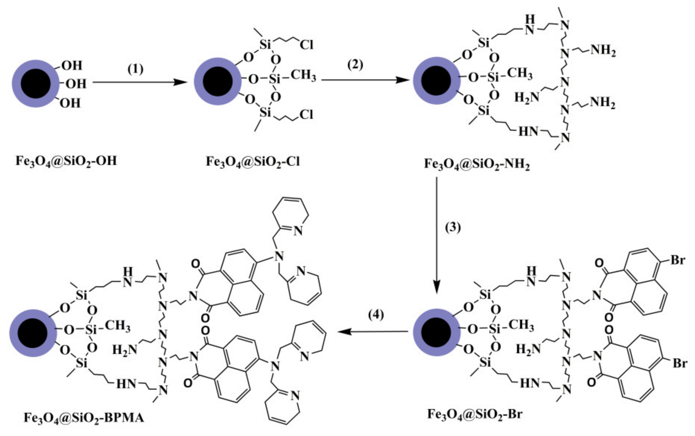
Figure 1. Synthesis of bis(2-pyridylmethyl)amine (BPMA)-functionalized Fe3O4@SiO2 by a “grafting-from” approach.
Materials 13 03678 g001
Figure 2. XRD patterns of Fe3O4 and Fe3O4@SiO2 powders.
Materials 13 03678 g002
Figure 3. (a) TEM image of Fe3O4 particles, (b) SEM and (c) TEM images of Fe3O4@SiO2 particles, (d) SEM and (e) TEM images of Fe3O4@SiO2-BPMA particles.
Materials 13 03678 g003
Figure 4. FTIR spectra of (a) Fe3O4@SiO2, (b) Fe3O4@SiO2-Cl, (c) Fe3O4@SiO2-NH2, (d) Fe3O4@SiO2-Br and (e) Fe3O4@SiO2-BPMA powders.
Materials 13 03678 g004
Figure 5. XPS spectra of (a) C 1s and (b) N 1s core-levels of Fe3O4@SiO2-BPMA powders.
Materials 13 03678 g005
Figure 6. Magnetization hysteresis loops of Fe3O4@SiO2 (a) and Fe3O4@SiO2-BPMA (b) powders (insets are the Fe3O4@SiO2-BPMA suspension, [Fe3O4@SiO2-BPMA] = 1.25 g/L, V = 8.0 mL, aqueous medium).
Materials 13 03678 g006
Figure 7. Changes in color (top) illuminated by natural light and fluorescence (bottom) illuminated with a fluorescent lamp (λex = 352 nm, 8 W) of Fe3O4@SiO2-BPMA upon the addition of Men+ (Men+ = Cr3+, Cd2+, Hg2+ and Pb2+) ([Fe3O4@SiO2-BPMA] = 0.1 g/L; [Men+] = 10−5 mol/L; V = 6 mL, Vacetonitrile/Vwater = 4/1).
Materials 13 03678 g007
Figure 8. Binding mechanism of Fe3O4@SiO2-BPMA toward Cd2+ ions.
Materials 13 03678 g008
Figure 9. Time-dependence of adsorption profiles of Cd2+ ion onto Fe3O4@SiO2 and Fe3O4@SiO2-BPMA powders (adsorption conditions: room temperature; no pH pre-adjustments; [Fe3O4@SiO2-BPMA] = 1.0 g/L; [Cd2+] = 200 mg/L; V = 50 mL; t = 0–60 min).
Materials 13 03678 g009
Figure 10. Effects of Cd2− initial concentration on the adsorption efficiency and adsorption amount (adsorption conditions: room temperature; no pH pre-adjustments; [Fe3O4@SiO2-BPMA] = 1.0 g/L; [Cd2+] = 50–500 mg/L; V = 50 mL; t = 30 min).
Materials 13 03678 g010
Figure 11. Langmuir linear fit of Cd2+ adsorbed onto Fe3O4@SiO2-BPMA.
Materials 13 03678 g011