Article

Sintering Temperature Effect on Structural and Optical Properties of Heat Treated Coconut Husk Ash Derived SiO2 Mixed with ZnO Nanoparticles

by
Muhammad Fahmi Anuar
1,*,
Yap Wing Fen
1,2,*,
Mohd Hafiz Mohd Zaid
1,
Nur Alia Sheh Omar
2 and
Rahayu Emilia Mohamed Khaidir
2
1
Department of Physics, Faculty of Science, University Putra Malaysia, UPM Serdang, Selangor 43400, Malaysia
2
Functional Devices Laboratory, Institute of Advanced Technology, University Putra Malaysia, UPM Serdang, Selangor 43400, Malaysia
*
Authors to whom correspondence should be addressed.
Materials 2020, 13(11), 2555; https://doi.org/10.3390/ma13112555
Submission received: 17 March 2020 / Revised: 22 April 2020 / Accepted: 24 April 2020 / Published: 4 June 2020

Abstract

:
In this work, waste coconut husk ash was used to prepare a ZnO-SiO2 composite. Solid-state technique was used to fabricate the composite due to its producibility, simple procedure as well as lower production cost. At high sintering temperatures ranging from 600 °C to 1000 °C, the X-ray diffraction (XRD) peaks of the Zn2SiO4 showed high intensity, which indicated high crystallinity. Furthermore, the formation of broad bands of ZnO4, Si-O-Si, and SiO4 were detected by Fourier transform infrared (FTIR) spectroscopy and the bands became narrower with the increment of sintering temperature. Besides, the morphological image from field emission scanning electron microscopy (FESEM) showed the formation of densely packed grains and smooth surface composite with the increase of sintering temperature. Upon obtaining the absorbance spectrum from Ultraviolet–Visible (UV–Vis) spectroscopy, the optical band gap was calculated to be 4.05 eV at 1000 °C. The correlation between the structural and optical properties of ZnO-SiO2 composite was discussed in detail.

1. Introduction

In recent years, the demands of ‘green’ environmentally friendly processes are growing higher and are much needed as a result of environmental concerns [1,2]. The increasing demand for using the biosynthesis method is due to its advantages as it is simple, environmentally friendly, cost-effective, easy to reproduce, and often more stable [3]. A number of agricultural wastes have been studied that are able to produce biosynthesis materials such as SiO2 [4,5,6,7,8]. Among the prominence agricultural sources of silica, coconut husk is believed to have great potential in the optical industry due to the fact that 91.76% of silica can be extracted from coconut husk ash (CHA) [9]. In addition, different types of silica can be extracted by using chemical treatments that are flexible and can be used in different kinds of applications [10]. Despite the advantages, there have been fewer studies done on CHA as more focus has been given to its ability as a good absorbent material toward wastewater treatment problems. Undoubtedly, these biosynthesis materials have great potential to be utilized, for example, as a ZnO dopant to modify its properties.
Zinc oxide (ZnO), which in white powder is an inorganic compound that is insoluble in water, is commonly used as an additive in a number of materials and products such as plastics, rubbers, and ceramics. It is also known for its wide-band gap and is used widely in various applications including devices in ultraviolet light-emitting diodes (UV LEDs), blue laminating devices, and UV lasers [11,12]. ZnO based material has also caught the attention of researchers because of its non-toxicity, environment-friendly, and low-cost, causing it to be highly valued by industry and researchers alike [13]. Furthermore, it is also known that doping with different types of materials can alter and modify its promising properties [14,15,16]. However, ZnO is unstable in most specialized applications and causes red-shift in absorption spectra [17]. Obviously, new synthesis methods that utilize ‘green’ methodology are preferable as some researchers have proven them to be safer and they can be extracted from plant-based materials.
In the past decades, numerous studies have been conducted on a mixture of ZnO and SiO2. SiO2 has generally been used as a host material for ZnO nanoparticles due to its chemical and physical stability properties [18]. Traditionally, zinc silicate forms when heat treatment is applied at a certain temperature [19,20]. Zinc silicate is one of the most promising phosphors materials known today because of its excellent luminescence properties in blue, green, and red spectral regions [21]. Wide energy band gap (5.5 eV), exceptional and high luminescent efficiency are also the benefitting factors that are vital in optoelectronic devices. In addition, zinc silicate has been established as a good host material and is suitable for applications in cathode ray tubes, plasma display panels, laser crystal, and electroluminescent devices [22,23,24]. Furthermore, the optical properties of zinc silicate can also be enhanced and modified by doping with different types of metals and applying heat treatment for preferred properties in different applications.
Moreover, the physical, chemical, and optical properties of ZnO-SiO2 are dependent on the preparation process and starting materials [20]. Consequently, various methods to synthesize ZnO-SiO2 have been developed such as hydrothermal [25], sol-gel [23,26], spray pyrolysis [17], and the conventional solid-state method [27,28]. The solid-state method is much simpler, and the materials can be produced in large quantities compared to other methods that require complicated steps, long preparation periods, and high equipment costs.
To the best of our knowledge, there have been limited studies on producing ZnO-based composites based on agricultural waste, especially coconut husk using the solid-state method. Hence, the aim of this work was to prepare a ZnO-SiO2 composite by incorporating silica derived from coconut husk into ZnO, and to investigate their structural and optical properties subjected to sintering temperature.

2. Materials and Methods
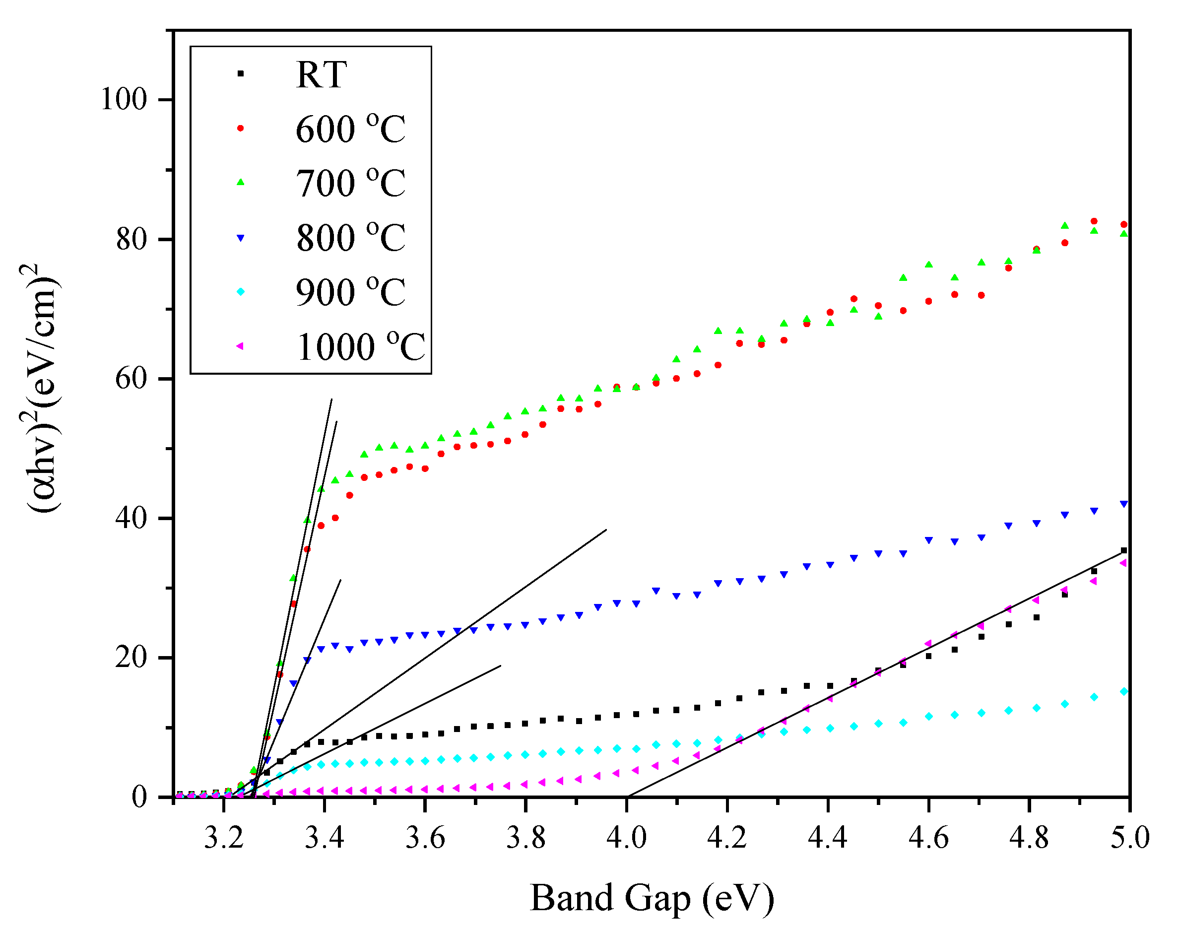
The sample of coconut husk ash (CHA) used in the study was obtained from the process of burning the coconut husk at 700 °C and treating as described in the previous report [10]. CHA was used as the source of SiO2 with 91.76% purity after treatment, which was confirmed by XRF as reported. In this study, ZnO nanoparticles (US Research Nanomaterials Inc., Houston, TX USA, 99%, 10–30 nm) were added with CHA to form a mixture of 20.0 g with the ratio of 1:1 using a ball milling jar for 24 h. After that, the mixed composition was pressed at 5 tons pressure to form a pellet. Next, the pellet was sintered in an alumina crucible at 600 °C to 1000 °C with a constant heat rate for 2 h.
X-ray diffraction (XRD) analysis was carried out by using a Philips X’Pert HighPro PANanalytical Diffractometer (Malvern Pananalytical, Almelo (The Netherland) and Malvern (UK)) with a copper (Cu) anode and Cu k-α radiation with wavelength 1.5406 Å operated at 40 mA and 40 kV. A Thermo Nicolet Nexus FTIR (Thermofisher Scientific, Waltham, MA, US) was used in the range of 280 to 4000 cm−1 to determine the bonds in the composite. Nova NanoSEM 30 (FEI, Hillsboro, OR, USA) was used to study the morphological structure of the composite sample with a 50,000 magnification level. Furthermore, the optical properties were studied using a UV-3600 Shimadzu (Shimadzu, Kyoto, Japan).

3. Results

The phase identification of the sample was carried out by using XRD analysis and X’Pert Highscore Plus software was used to identify the diffraction peaks obtained in Figure 1. ZnO (JCPDS 14-0653) and SiO2 (JCPDS 82-0511) were identified on the peaks of the unsintered (RT) composite. At 600 °C, the zinc silicate (Zn2SiO4) diffraction peak appeared (JCPDS 37-1485) at 2θ = 25.44°. The zinc silicate had a rhombohedral structure with lattice parameter a = b = 13.938 (Å) and c = 9.3100 (Å), space group of R 3 ¯ , and space group number of 148, as also reported in previous research [6,29,30]. Some zinc oxide (ZnO) (JCPDS 14-0653) peaks also appeared at 2θ = 31.78°, 34.44°, 36.26°, 47.58°, 56.59°, 62.95°, 68.08°, and 69.06°. As the temperature increased to 700 °C and 800 °C, more Zn2SiO4 peaks appeared and less ZnO peaks existed in the spectrum. Based on the XRD spectrum, the zinc silicate diffraction peaks appeared at 2θ = 22.02°, 25.56°, 31.88°, and 38.96° after a sintering temperature of 700 °C, and at 800 °C, it appeared at 2θ = 45.09°, 48.99°, 65.07°. Next, the diffraction peaks of ZnO located at 2θ = 36.26°, 47.58°, 56.69°, 62.95°, and 68.08° started to disappear at the higher temperature of 900 °C and was completely gone at 1000 °C. This phenomenon indicated greater fusion of ZnO and SiO2 to form zinc silicate at 900 °C and was completed at 1000 °C. Upon reaching the sintering temperature of 1000 °C, the zinc silicate diffraction peaks located at 2θ = 22.19°, 25.66°, 31.65°, 34.12°, 38.94°, 45.14°, 47.07°, 49.03°, 54.39°, 56.14°, 57.70°, 59.63°, 60.97°, 65.73°, 68.77°, and 70.45° became more intensified and sharper due to the higher sintering temperature increasing the crystallinity of the zinc silicate [31,32].
The FTIR spectrum of the ZnO-SiO2 composite derived from coconut husk ash sintered at several temperatures was observed in the range of 400–4000 cm−1, as illustrated in Figure 2. The pre-sintering sample (RT) showed a broad absorption band of Si-O-Si asymmetric stretching vibration, which resides at 1060 cm−1, probably due to the change in bonding around the tetrahedral SiO4. These results are in a good agreement with previous studies [29,33]. Upon the sintering temperatures of 600 °C and 700 °C, a few weak absorption bands were observed at wavenumbers 570 cm−1 and 900 cm−1. These two bands can be attributed to the ZnO4 symmetric stretching and SiO4 asymmetric stretching, respectively [29,34,35,36]. At a temperature of 800 °C, a band attributed to ZnO4, was observed at 450 cm−1 [36]. Furthermore, from 900 °C up to 1000 °C, the broad peaks of ZnO4 (570 cm−1) and SiO4 (900 cm−1) became narrower and stronger as the sintering temperature increased, since high temperature increased the crystallinity of the composite, as indicated by the XRD analysis. In a nutshell, the observed appearances of the band attributed to SiO4 and ZnO4 indicate the successful formation of the crystalline ZnO-SiO2 composite, especially starting at a temperature of 800 °C and above, which was also supported by the XRD spectrum.
The microstructure images of the ZnO-SiO2 composite that was heat treated at various sintering temperatures of 600–1000 °C were studied using field emission scanning electron microscopy (FESEM) at a 50,000 magnification level. As shown in Figure 3, it was observed that the composite sample did not have a uniform shape or structure. Additionally, it can be observed from the images that the particle size distribution of the composite samples was irregular and not consistent when it was not heat-treated. There was also a rod-like formation observed on the untreated (RT) composite at 600 °C that originated from the coconut husk ash structure [10]. Based on Figure 3, the average grain sizes were measured and tabulated in Table 1. The average grain sizes of the sample at RT was measured to be 72.23 nm and increased to 94.23 nm after heat-treated at 600 °C. Next, at a temperature of 700 °C, the average grain sizes increased to 121.37 nm and aggregated due to an increase in the sintering temperature. Furthermore, the samples sintered at 800 °C and 900 °C showed average grain sizes of 299.37 nm and 326.83 nm, respectively, due to the increase in grain growth and crystallinity of the sample that was caused by a higher sintering temperature. However, the sample sintered at 900 °C showed densely packed grains of different sizes with less porosity present in the sample. Particles in the samples started to form necks between each other and agglomerated to form bigger particles. The sample sintered at 1000 °C showed a smooth surface with well-distinct boundaries in the images and the average grain sizes were measured at 515.70 nm. The image indicated that the composite sample was well-crystallized after being sintered at 1000 °C. The formation of rhombohedral-like particles also confirmed the formation of the high crystallinity of Zn2SiO4 when sintered at high temperature.
It can also be observed from the images, where the particle grain sizes increased with the sintering temperatures as a result of grain growth. Grain growth is known as a temperature-dependent phenomenon and thus the composite samples increased in sizes along with the sintering temperature [37]. The grain growth with well-distinct characteristics with the sintering temperatures suggested that sintering also increased the crystallization of the composite sample of ZnO-SiO2 and produced high crystallinity at 1000 °C.
For the optical properties, the absorption spectra of ZnO-SiO2 sintered at various temperatures are shown in Figure 4. From the absorbance spectrum, a sharp absorption edge was observed at a wavelength of about 400 nm, indicating that the absorption properties of the ZnO-SiO2 composite at room temperature occurred in the UV region. When the prepared composite was sintered at 600 °C to 1000 °C, the absorbance intensity starting to decrease with the increase in temperature. This occurrence was due to the deformation of the ZnO hexagonal structure in the sample and started to form a zinc silicate composite structure. Furthermore, the absorption edge of the sintered samples shifted to a longer wavelength (red-shifted) when at 600 °C to 800 °C. This occurrence suggests that the crystallization process between temperatures of 600 °C and 800 °C enhanced the absorption edge toward the red-shift. However, when the sintering temperatures were 900 °C and 1000 °C, the absorption edge shifted back to the lower wavelength, which can be attributed to the high crystallinity of the zinc silicate composite sample [36].
The optical band gap of all the ZnO-SiO2 composite samples was determined by using the following relation between absorption coefficient, α, and photon energy, [38]:
α = k [ h v E gap ] n h v
By rearranging Equation (1) above, it gives:
( α h v ) 1 / n = h v E gap
where ν is the frequency; h is the Plank constant; n is the type of transition; and Egap is the optical band gap. In this work, n = 1/2 was used as the direct allowed transition, and by inserting the value of n into Equation (2), it becomes:
( α h v ) 2 = h v E gap
By using the Equation (3) relation, the optical band gap can be obtained by extrapolating the linear fitted region of the graph of (αhν)2 against hv to the position at which (αhν)2 = 0. The variations of (αhν)2 of ZnO-SiO2 as a function of photon energy sintered at different temperatures are shown in Figure 5.
When the sintering temperature increased, the amount of vibration energy provided to the atom increased and caused the interatomic spacing between the atoms to weaken. Consequently, a weak interatomic spacing allowed weak energy to break the bond and caused the gap between the conduction and valence band to shrink [39]. Inversely, the band gap value promptly increased when the temperature reached 1000 °C. The high crystallinity of the composite at 1000 °C caused the band gap value to increase rapidly to 4.05 eV [40]. The improvement of the composite crystallinity allowed the reduction of delocalized states, resulting in a composite with less defects, and thus increased the optical band gap value [39,41]. The formation of the rhombohedral phase (Zn2SiO4) was also the justification of the phenomenon that occurred at a sintering temperature of 1000 °C. Consequently, the absorption edge became broad and increased the optical band gap values of the ZnO-SiO2 composite due to the comparatively large band gap of zinc silicate.

4. Conclusions

The composite ZnO-SiO2 was prepared and the zinc silicate started to form at a temperature of 600 °C and increased in the diffraction peak intensity at a higher temperature until 1000 °C. The formation of bands assigned to ZnO4 and SiO4 was detected in the FTIR spectrum and became stronger at 1000 °C. Next, the FESEM images of the ZnO-SiO2 composite revealed that at 1000 °C, a well-distinct crystal formation with densely packed grains was observed, indicating the high crystallinity of ZnO-SiO2. Moreover, the optical band gap of the ZnO-SiO2 composite at room temperature was 3.22 eV, and this value increased to 4.05 eV at 1000 °C due to the formation of highly crystalline zinc silicate composite. Finally, with all the evidence obtained, we can conclude that zinc silicate was formed after the heat treatment. Zinc silicate is known to be a good host material for doping with rare-earth ions and transition metals. Therefore, it is of interest to further expand this study by exploring the possibility of turning this zinc silicate from coconut husk into a phosphor material.

Author Contributions

Data curation, N.A.S.O.; Formal analysis, M.F.A. and N.A.S.O.; Investigation, M.F.A., N.A.S.O. and R.E.M.K.; Methodology, Y.W.F., M.H.M.Z., and R.E.M.K.; Supervision, Y.W.F. and M.H.M.Z.; Writing—original draft, M.F.A.; Writing—review & editing, Y.W.F. All authors have read and agreed to the published version of the manuscript.

Funding

The work was funded by the Malaysian Ministry of Education (MOE) and the Universiti Putra Malaysia (UPM) through the Putra Grant 9642800. The fund was used for purchasing materials, apparatus, and the characterization processing fee.

Acknowledgments

The authors gratefully acknowledge the financial support for this study from the Malaysian Ministry of Education (MOE) and the Universiti Putra Malaysia (UPM) through the Putra Grant. The laboratory facilities provided by the Institute of Advanced Technology and Faculty of Science, Universiti Putra Malaysia, are also acknowledged.

Conflicts of Interest

The authors declare no conflicts of interest.

References

  1. Ahmed, S.; Ullah, S.; Ahmad, M.; Swami, B.L.; Ikram, S. Green synthesis of silver nanoparticles using Azadirachta indica aqueous leaf extract. J. Radiat. Res. Appl. Sci. 2016, 9, 1–7. [Google Scholar] [CrossRef] [Green Version]
  2. Thuesombat, P.; Hannongbua, S.; Akasit, S.; Chadchawan, S. Effect of silver nanoparticles on rice (Oryza sativa L. cv. KDML 105) seed germination and seedling growth. Ecotoxicol. Environ. Saf. 2014, 104, 302–309. [Google Scholar] [CrossRef] [PubMed]
  3. Mittal, J.; Batra, A.; Singh, A.; Sharma, M.M. Phytofabrication of nanoparticles through plant as nanofactories. Adv. Nat. Sci. Nanosci. Nanotechnol. 2014, 5, 043002. [Google Scholar] [CrossRef]
  4. Aisyah, S.; Wahab, A.; Amin, K.; Hj, S.; Aziz, A.; Zakiah, A.; Azman, K.; Emilia, R.; Khaidir, M.; Zulhasif, M.; et al. Synthesis of cobalt oxide Co3O4 doped zinc silicate based glass- ceramic derived for LED applications. Optik 2019, 179, 919–926. [Google Scholar]
  5. Khaidir, R.E.M.; Fen, Y.W.; Mohd Zaid, M.H.; Matori, K.A.; Omar, N.A.S.; Anuar, M.F.; Abdul Wahab, S.A.; Khirel Azman, A.Z. Addition of ZnO nanoparticles on waste rice husk as potential host material for red-emitting phosphor. Mater. Sci. Semicond. Process. 2020, 106, 104774. [Google Scholar] [CrossRef]
  6. Omar, N.A.S.; Fen, Y.W.; Matori, K.A.; Aziz, S.H.A.; Alassan, Z.N.; Samsudin, N.F. Development and characterization studies of Eu3+-doped Zn2SiO4 phosphors with waste silicate sources. Procedia Chem. 2016, 19, 21–29. [Google Scholar] [CrossRef] [Green Version]
  7. Faizul, C.P.; Chik, A.; Bari, M.F. Palm Ash as an alternative source for silica production. MATEC Web Conf. 2016, 78, 01062. [Google Scholar]
  8. Norsuraya, S.; Fazlena, H.; Norhasyimi, R. Sugarcane bagasse as a renewable source of silica to synthesize Santa Barbara Amorphous-15 (SBA-15). Procedia Eng. 2016, 148, 839–846. [Google Scholar] [CrossRef] [Green Version]
  9. Anuar, M.F.; Fen, Y.W.; Matori, K.A.; Mohd, M.H. The physical and optical studies of crystalline silica derived from green synthesis of coconut husk ash. Appl. Sci. 2020, 10, 2128. [Google Scholar] [CrossRef] [Green Version]
  10. Anuar, M.F.; Fen, Y.W.; Zaid, M.H.M.; Matori, K.A.; Khaidir, R.E.M. Synthesis and structural properties of coconut husk as potential silica source. Results Phys. 2018, 11, 1–4. [Google Scholar] [CrossRef]
  11. Kundu, T.K.; Karak, N.; Barik, P.; Saha, S. Optical properties of ZnO nanoparticles prepared by chemical method using poly vinyl alcohol (PVA) as capping agent. Int. J. Soft Comput. Eng. 2011, 1, 19–24. [Google Scholar]
  12. Yang, Y.; Yang, B.; Fu, Z.; Yan, H.; Zhen, W.; Dong, W.; Xia, L.; Liu, W.; Jian, Z.; Li, F. Enhanced yellow-green photoluminescence from ZnO-SiO2 composite opal. J. Phys. Condens. Matter 2004, 16, 7277–7286. [Google Scholar] [CrossRef]
  13. Yuan, Q.; Li, N.; Tu, J.; Li, X.; Wang, R.; Zhang, T.; Shao, C. Preparation and humidity sensitive property of mesoporous ZnO-SiO 2 composite. Sens. Actuators B Chem. 2010, 149, 413–419. [Google Scholar] [CrossRef]
  14. Meyer, M.; Damonte, L.C. Study of Co and Fe-doped ZnO milled nanopowders. Powder Technol. 2015, 286, 371–377. [Google Scholar] [CrossRef]
  15. McCluskey, M.D.; Jokela, S.J. Defects in ZnO. J. Appl. Phys. 2009, 106, 10. [Google Scholar] [CrossRef] [Green Version]
  16. Pearton, S.J.; Norton, D.P.; Ip, K.; Heo, Y.W.; Steiner, T. Recent progress in processing and properties of ZnO. Prog. Mater. Sci. 2005, 50, 293–340. [Google Scholar] [CrossRef]
  17. Tani, T.; Mädler, L.; Pratsinis, S.E. Synthesis of zinc oxide/silica composite nanoparticles by flame spray pyrolysis. J. Mater. Sci. 2002, 37, 4627–4632. [Google Scholar] [CrossRef]
  18. Wang, Y.; Zhu, G.; Xin, S.; Wang, Q.; Li, Y.; Wu, Q.; Wang, C.; Wang, X.; Ding, X.; Geng, W. Recent development in rare earth doped phosphors for white light emitting diodes. J. Rare Earths 2015, 33, 1–12. [Google Scholar] [CrossRef]
  19. Takesue, M.; Hayashi, H.; Smith, R.L. Thermal and chemical methods for producing zinc silicate (willemite): A review. Prog. Cryst. Growth Charact. Mater. 2009, 55, 98–124. [Google Scholar] [CrossRef]
  20. Engku Ali, E.A.G.; Matori, K.A.; Saion, E.; Aziz, S.H.A.; Zaid, M.H.M.; Alibe, I.M. Structural and Optical Properties of Heat Treated Zn2SiO4 Composite Prepared by Impregnation of ZnO on SiO2 Amorphous Nanoparticles. ASM Sci. J. Spec. Issue 2018, 1, 75–85. [Google Scholar]
  21. Zhang, Q.Y.; Pita, K.; Kam, C.H. Sol-gel derived zinc silicate phosphor films for full-color display applications. J. Phys. Chem. Solids 2003, 64, 333–338. [Google Scholar] [CrossRef]
  22. Yang, J.; Sun, Y.; Chen, Z.; Zhao, X. Hydrothermal synthesis and optical properties of zinc silicate hierarchical superstructures. Mater. Lett. 2011, 65, 3030–3033. [Google Scholar] [CrossRef]
  23. Rasdi, N.M.; Fen, Y.W.; Azis, R.S.; Omar, N.A.S. Photoluminescence studies of cobalt (II) doped zinc silicate nanophosphors prepared via sol-gel method. Optik 2017, 149, 409–415. [Google Scholar] [CrossRef]
  24. Ouyang, X.; Kitai, A.H.; Xiao, T. Electroluminescence of the oxide thin film phosphors Zn2SiO4 and Y2SiO5. J. Appl. Phys. 1996, 79, 3229–3234. [Google Scholar] [CrossRef]
  25. Li, Q.H.; Komarneni, S.; Roy, R. Control of morphology of Zn2SiO4 by hydrothermal preparation. J. Mater. Sci. 1995, 30, 2358–2363. [Google Scholar] [CrossRef]
  26. He, H.; Wang, Y.; Zou, Y. Photoluminescence property of ZnO-SiO2 composites synthesized by sol-gel method. J. Phys. D. Appl. Phys. 2003, 36, 2972–2975. [Google Scholar] [CrossRef]
  27. Syamimi, N.F.; Matori, K.A.; Lim, W.F.; Aziz, S.A.; Hafiz, M.; Zaid, M. Effect of sintering temperature on structural and morphological properties of europium (III oxide doped willemite. J. Spectrosc. 2014, 2014, 1–9. [Google Scholar] [CrossRef]
  28. Effendy, N.; Abdul, Z.; Mohamed, H.; Amin, K.; Hj, S.; Aziz, A.; Hafiz, M.; Zaid, M. Structural and optical properties of Er 3 + -doped willemite glass-ceramics from waste materials. Opt. Int. J. Light Electron Opt. 2016, 127, 11698–11705. [Google Scholar] [CrossRef]
  29. Rasdi, N.M.; Fen, Y.W.; Omar, N.A.S.; Azis, R.S.; Zaid, M.H.M. Effects of cobalt doping on structural, morphological, and optical properties of Zn2SiO4 nanophosphors prepared by sol-gel method. Results Phys. 2017, 7, 3820–3825. [Google Scholar] [CrossRef]
  30. Omar, N.A.S.; Fen, Y.W.; Matori, K.A.; Zaid, M.H.M.; Norhafizah, M.R.; Nurzilla, M.; Zamratul, M.I.M. Synthesis and optical properties of europium doped zinc silicate prepared using low cost solid state reaction method. J. Mater. Sci. Mater. Electron. 2016, 27, 1092–1099. [Google Scholar] [CrossRef]
  31. Tarafder, A.; Molla, A.R.; Dey, C.; Karmakar, B. Thermal, structural, and enhanced photoluminescence properties of Eu3+-doped transparent willemite glass-ceramic nanocomposites. J. Am. Ceram. Soc. 2013, 96, 2424–2431. [Google Scholar] [CrossRef]
  32. Azman, A.Z.K.; Matori, K.A.; Ab Aziz, S.H.; Zaid, M.H.M.; Wahab, S.A.A.; Khaidir, R.E.M. Comprehensive study on structural and optical properties of Tm2O3 doped zinc silicate based glass–ceramics. J. Mater. Sci. Mater. Electron. 2018, 29, 19861–19866. [Google Scholar] [CrossRef]
  33. Sanaeishoar, H.; Sabbaghan, M.; Mohave, F. Synthesis and characterization of micro-mesoporous MCM-41 using various ionic liquids as co-templates. Microporous Mesoporous Mater. 2015, 217, 219–224. [Google Scholar] [CrossRef]
  34. Babu, B.C.; Rao, B.V.; Ravi, M.; Babu, S. Structural, microstructural, optical, and dielectric properties of Mn2+: Willemite Zn2SiO4 nanocomposites obtained by a sol-gel method. J. Mol. Struct. 2017, 1127, 6–14. [Google Scholar] [CrossRef]
  35. Sarrigani, G.V.; Quah, H.J.; Lim, W.F.; Matori, K.A.; Mohd Razali, N.S.; Kharazmi, A.; Hashim, M.; Bahari, H.R. Characterization of waste material derived willemite-based glass-ceramics doped with erbium. Adv. Mater. Sci. Eng. 2015, 201, 1–7. [Google Scholar] [CrossRef] [Green Version]
  36. Rashid, S.S.A.; Aziz, S.H.A.; Matori, K.A.; Zaid, M.H.M.; Mohamed, N. Comprehensive study on effect of sintering temperature on the physical, structural and optical properties of Er 3+ doped ZnO-GSLS glasses. Results Phys. 2017, 7, 2224–2231. [Google Scholar] [CrossRef]
  37. Naceur, H.; Megriche, A.; El Maaoui, M. Effect of sintering temperature on microstructure and electrical properties of Sr 1−x (Na 0.5 Bi 0.5) x Bi 2 Nb 2 O 9 solid solutions. J. Adv. Ceram. 2014, 3, 17–30. [Google Scholar] [CrossRef] [Green Version]
  38. Tauc, J.; Grigorovici, R.; Vancu, A. Optical properties and electronic structure of amorphous germanium. Phys. Status Solidi 1966, 15, 627–636. [Google Scholar] [CrossRef]
  39. Khaidir, R.E.M.; Fen, Y.W.; Zaid, M.H.M.; Matori, K.A.; Omar, N.A.S.; Anuar, M.F.; Wahab, S.A.A.; Azman, A.Z.K. Optical band gap and photoluminescence studies of Eu 3+ -doped zinc silicate derived from waste rice husks. Optik 2019, 182, 486–495. [Google Scholar] [CrossRef]
  40. Omar, N.A.S.; Fen, Y.W.; Matori, K.A.; Zaid, M.H.M.; Samsudin, N.F. Structural and optical properties of Eu3+activated low cost zinc soda lime silica glasses. Results Phys. 2016, 6, 640–644. [Google Scholar] [CrossRef] [Green Version]
  41. Nazrin, S.N.; Halimah, M.K.; Muhammad, F.D.; Yip, J.S.; Hasnimulyati, L.; Faznny, M.F.; Hazlin, M.A.; Zaitizila, I. The effect of erbium oxide in physical and structural properties of zinc tellurite glass system. J. Non. Cryst. Solids 2018, 490, 35–43. [Google Scholar] [CrossRef]
Figure 1. X-ray diffraction (XRD) spectrum of the ZnO-SiO2 composite sintered at different temperatures.
Materials 13 02555 g001
Figure 2. Fourier transform infrared (FTIR) spectra of ZnO-SiO2 at different sintering temperatures.
Materials 13 02555 g002
Figure 3. Field emission scanning electron microscopy (FESEM) images of composite sample ZnO-SiO2 at (a) RT, (b) 600 °C, (c) 700 °C, (d) 800 °C, (e) 900 °C, and (f) 1000 °C.
Materials 13 02555 g003
Figure 4. Optical absorbance spectra for the ZnO-SiO2 composite sintered at different sintering temperatures.
Materials 13 02555 g004
Figure 5. The optical band gap of the ZnO-SiO2 composite at different sintering temperatures.
Materials 13 02555 g005
Table 1. Average grain sizes of ZnO-SiO2 at different sintering temperatures.
Sintering Temperature (°C)Average Grain Sizes (nm)
RT72.23
60094.23
700121.37
800299.37
900326.83
1000515.70

