Effect of Exposed Surface Area, Volume and Environmental pH on the Calcium Ion Release of Three Commercially Available Tricalcium Silicate Based Dental Cements

Abstract
:1. Introduction
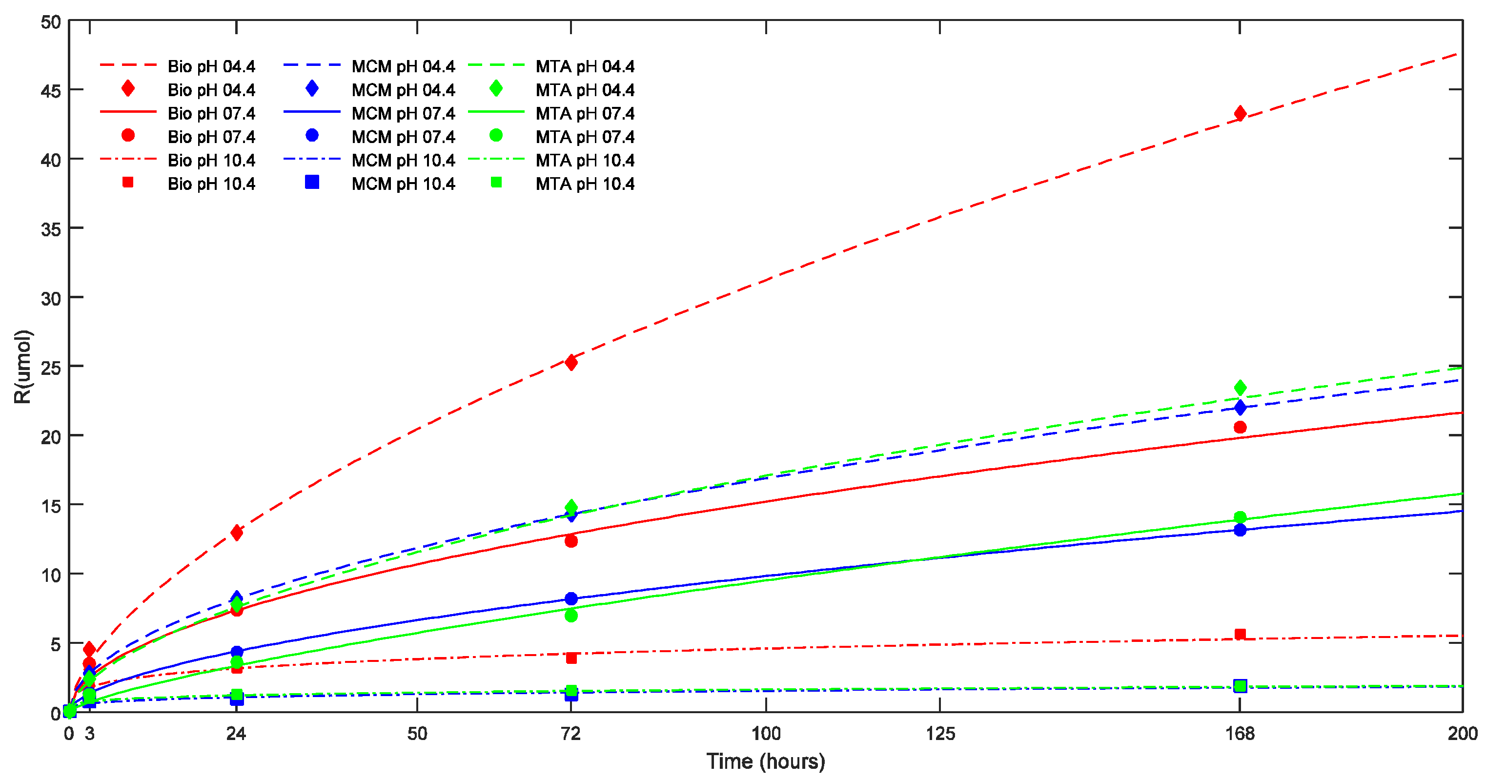
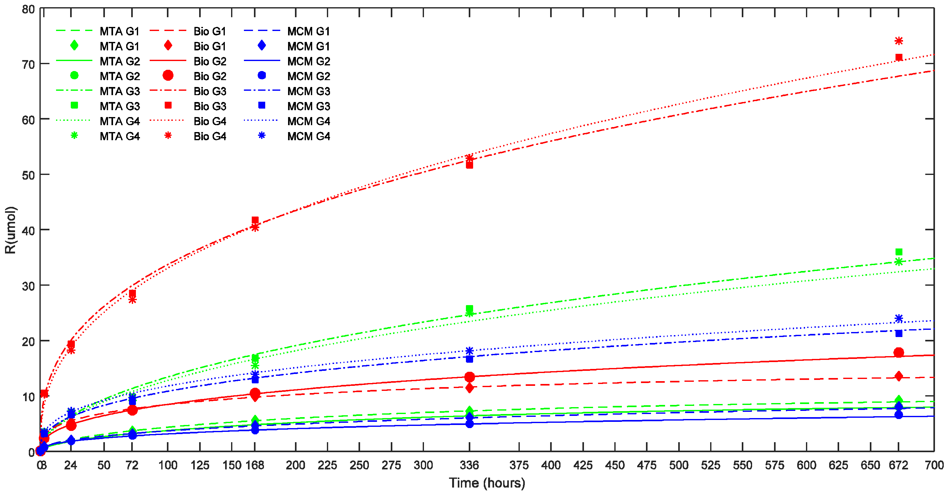
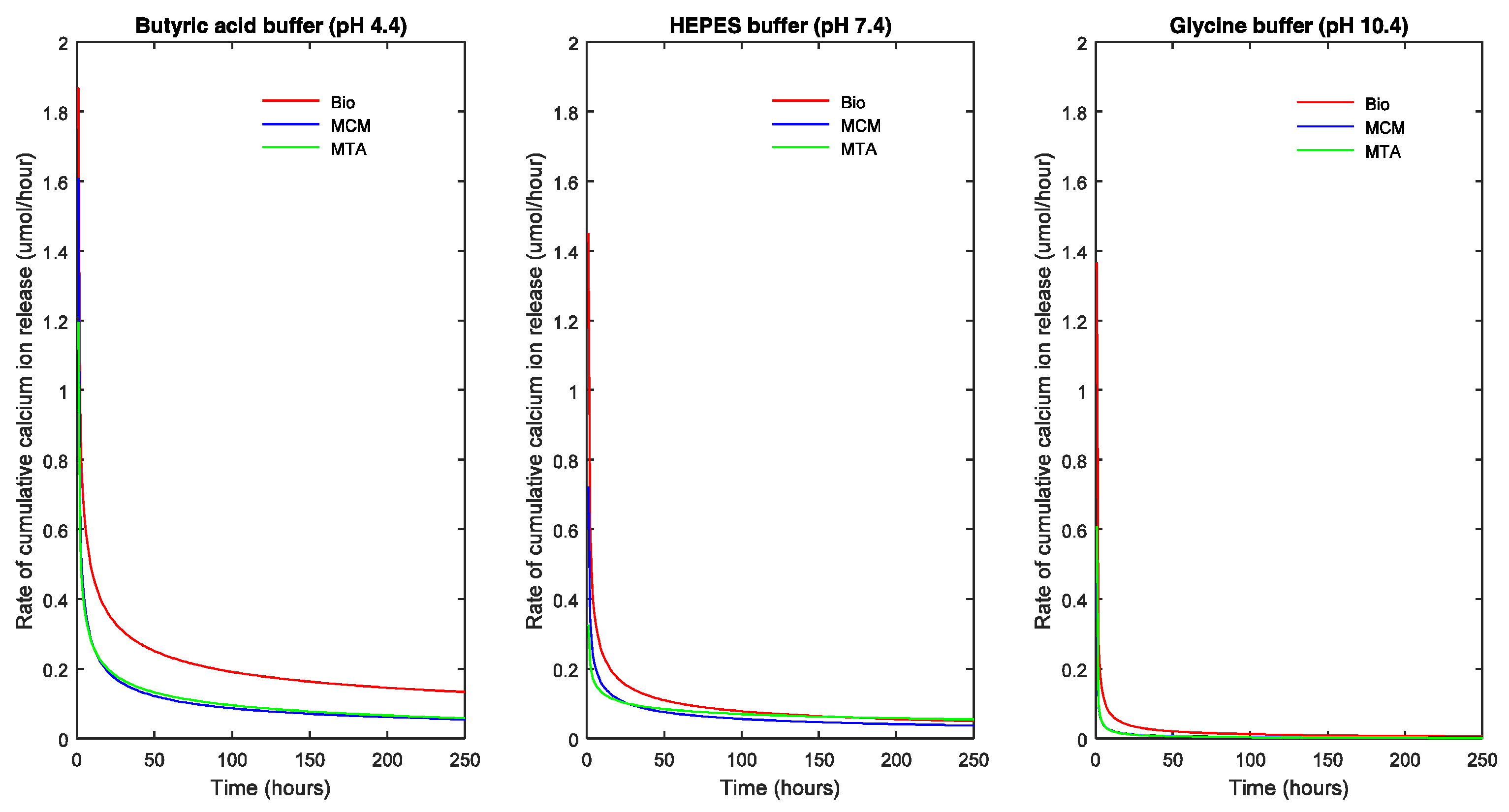
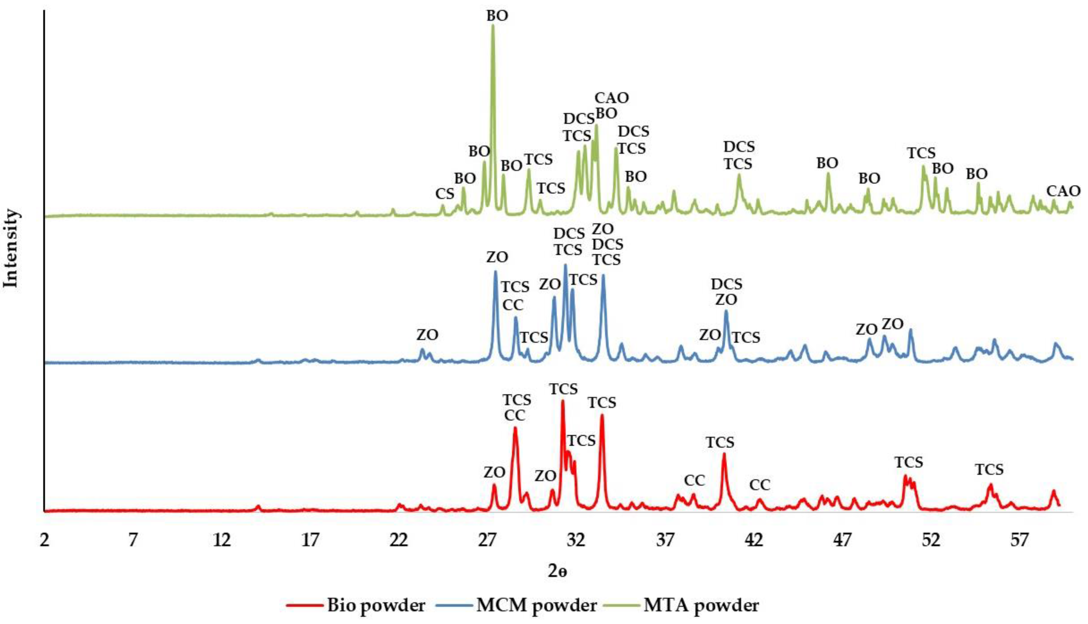
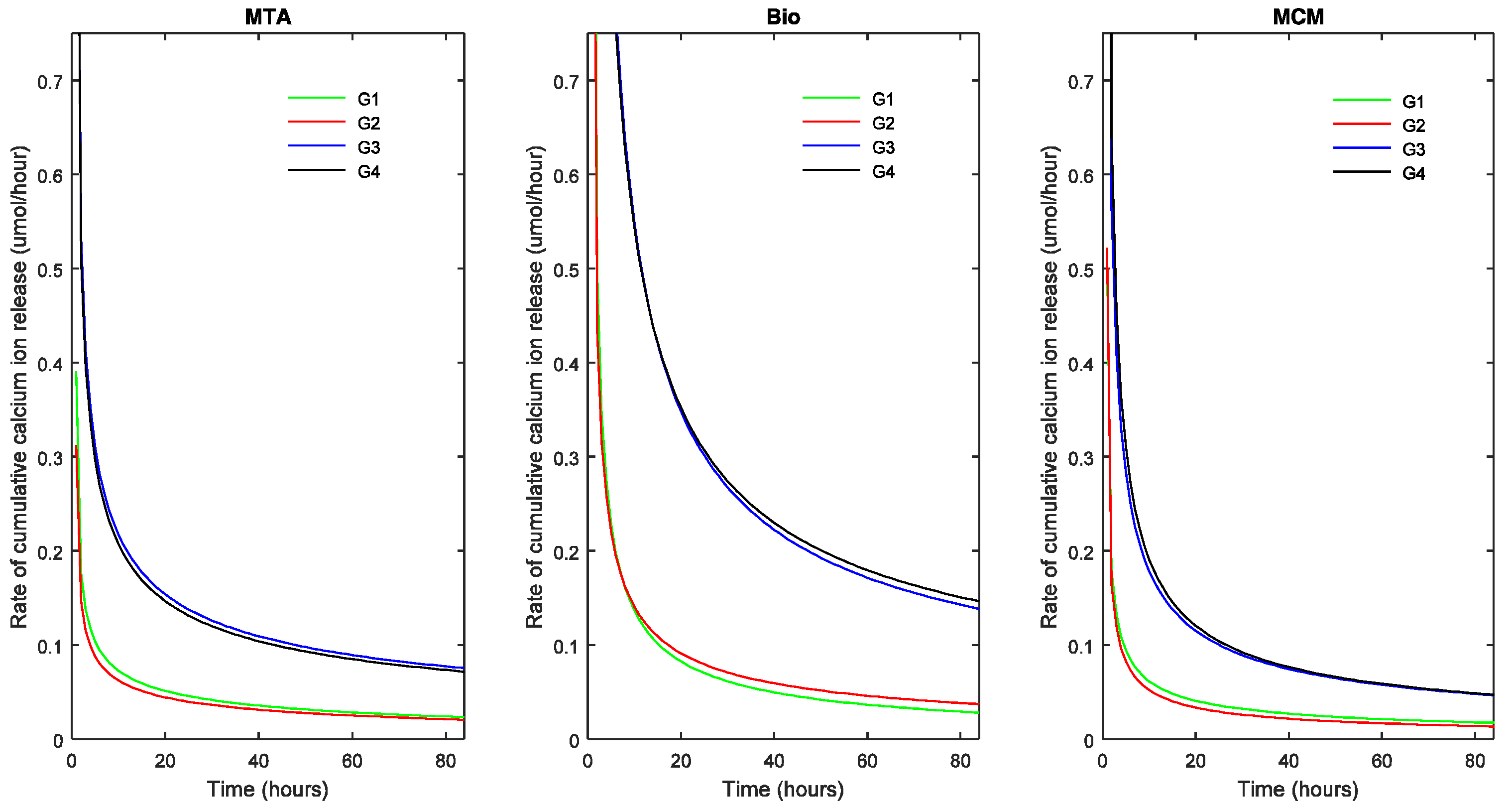
2. Results
3. Discussion
4. Materials and Methods
4.1. XRD Procedure and Analysis
4.2. Sample Preparation for Effect of ESA and Vol on Calcium Ion Release and pH
4.3. Sample Preparation for Effect of pH on Calcium Ion Release
4.4. pH and Calcium Ion Release Measurement
4.5. Calcium Ion Release Analysis
5. Conclusions
Acknowledgments
Author Contributions
Conflicts of Interest
References
- Okiji, T.; Yoshiba, K. Reparative dentinogenesis induced by mineral trioxide aggregate: A review from the biological and physicochemical points of view. Int. J. Dent. 2009, 2009, 464280. [Google Scholar] [CrossRef] [PubMed]
- Guerreiro-Tanomaru, J.M.; Chula, D.G.; de Pontes Lima, R.K.; Berbert, F.L.; Tanomaru-Filho, M. Release and diffusion of hydroxyl ion from calcium hydroxide-based medicaments. Dent. Traumatol. 2012, 28, 320–323. [Google Scholar] [CrossRef] [PubMed]
- Maeno, S.; Niki, Y.; Matsumoto, H.; Morioka, H.; Yatabe, T.; Funayama, A.; Toyama, Y.; Taguchi, T.; Tanaka, J. The effect of calcium ion concentration on osteoblast viability, proliferation and differentiation in monolayer and 3d culture. Biomaterials 2005, 26, 4847–4855. [Google Scholar] [CrossRef] [PubMed]
- Gandolfi, M.G.; Siboni, F.; Primus, C.M.; Prati, C. Ion release, porosity, solubility, and bioactivity of MTA plus tricalcium silicate. J. Endod. 2014, 40, 1632–1637. [Google Scholar] [CrossRef] [PubMed]
- Kahler, B.; Chugal, N.; Lin, L.M. Alkaline materials and regenerative endodontics: A review. Materials 2017, 10, E1389. [Google Scholar] [CrossRef] [PubMed]
- Ausiello, P.; Ciaramella, S.; Martorelli, M.; Lanzotti, A.; Zarone, F.; Watts, D.C.; Gloria, A. Mechanical behavior of endodontically restored canine teeth: Effects of ferrule, post material and shape. Dent. Mater. 2017, 33, 1466–1472. [Google Scholar] [CrossRef] [PubMed]
- Vayron, R.; Karasinski, P.; Mathieu, V.; Michel, A.; Loriot, D.; Richard, G.; Lambert, G.; Haiat, G. Variation of the ultrasonic response of a dental implant embedded in tricalcium silicate-based cement under cyclic loading. J. Biomech. 2013, 46, 1162–1168. [Google Scholar] [CrossRef] [PubMed]
- Malkondu, O.; Karapinar Kazandag, M.; Kazazoglu, E. A review on biodentine, a contemporary dentine replacement and repair material. Biomed. Res. Int. 2014, 2014, 160951. [Google Scholar] [CrossRef] [PubMed]
- Koubi, G.; Colon, P.; Franquin, J.C.; Hartmann, A.; Richard, G.; Faure, M.O.; Lambert, G. Clinical evaluation of the performance and safety of a new dentine substitute, biodentine, in the restoration of posterior teeth—A prospective study. Clin. Oral Investig. 2013, 17, 243–249. [Google Scholar] [CrossRef] [PubMed]
- Massi, S.; Tanomaru-Filho, M.; Silva, G.F.; Duarte, M.A.; Grizzo, L.T.; Buzalaf, M.A.; Guerreiro-Tanomaru, J.M. Ph, calcium ion release, and setting time of an experimental mineral trioxide aggregate-based root canal sealer. J. Endod. 2011, 37, 844–846. [Google Scholar] [CrossRef] [PubMed]
- Ballal, N.V.; Shavi, G.V.; Kumar, R.; Kundabala, M.; Bhat, K.S. In vitro sustained release of calcium ions and pH maintenance from different vehicles containing calcium hydroxide. J. Endod. 2010, 36, 862–866. [Google Scholar] [CrossRef] [PubMed]
- Duarte, M.A.; Midena, R.Z.; Zeferino, M.A.; Vivan, R.R.; Weckwerth, P.H.; Dos Santos, F.; Guerreiro-Tanomaru, J.M.; Tanomaru-Filho, M. Evaluation of pH and calcium ion release of calcium hydroxide pastes containing different substances. J. Endod. 2009, 35, 1274–1277. [Google Scholar] [CrossRef] [PubMed]
- Peng, W.; Liu, W.; Zhai, W.; Jiang, L.; Li, L.; Chang, J.; Zhu, Y. Effect of tricalcium silicate on the proliferation and odontogenic differentiation of human dental pulp cells. J. Endod. 2011, 37, 1240–1246. [Google Scholar] [CrossRef] [PubMed]
- Holland, R.; de Souza, V.; Nery, M.J.; Bernabe o, F.; Filho, J.A.; Junior, E.D.; Murata, S.S. Calcium salts deposition in rat connective tissue after the implantation of calcium hydroxide-containing sealers. J. Endod. 2002, 28, 173–176. [Google Scholar] [CrossRef] [PubMed]
- Shen, Q.; Sun, J.; Wu, J.; Liu, C.; Chen, F. An in vitro investigation of the mechanical-chemical and biological properties of calcium phosphate/calcium silicate/bismutite cement for dental pulp capping. J. Biomed. Mater. Res. B. Appl. Biomater. 2010, 94, 141–148. [Google Scholar] [PubMed]
- Parirokh, M.; Torabinejad, M. Mineral trioxide aggregate: A comprehensive literature review—Part iii: Clinical applications, drawbacks, and mechanism of action. J. Endod. 2010, 36, 400–413. [Google Scholar] [CrossRef] [PubMed]
- Rajasekharan, S.; Martens, L.C.; Cauwels, R.G.; Verbeeck, R.M. Biodentine material characteristics and clinical applications: A review of the literature. Eur. Arch. Paediatr. Dent. 2014, 15, 147–158. [Google Scholar] [CrossRef] [PubMed]
- Duarte, M.A.H.; Minotti, P.G.; Rodrigues, C.T.; Zapata, R.O.; Bramante, C.M.; Tanomaru Filho, M.; Vivan, R.R.; de Moraes, I.G.; de Andrade, F.B. Effect of different radiopacifying agents on the physicochemical properties of white portland cement and white mineral trioxide aggregate. J. Endod. 2012, 38, 394–397. [Google Scholar] [CrossRef] [PubMed]
- Kouzmanova, Y.I.; Dimitrova, I.V.; Gentscheva, G.D.; Aleksandrov, L.I.; Markova-Velichkova, M.G.; Kovacheva, D.G. Comparative study of the phase formation and interaction with water of calcium-silicate cements with dental applications. Bulg. Chem. Commun. 2015, 47, 239–244. [Google Scholar]
- Han, L.; Kodama, S.; Okiji, T. Evaluation of calcium-releasing and apatite-forming abilities of fast-setting calcium silicate-based endodontic materials. Int. Endod. J. 2015, 48, 124–130. [Google Scholar] [CrossRef] [PubMed]
- Duarte, M.A.; Demarchi, A.C.; Yamashita, J.C.; Kuga, M.C.; Fraga Sde, C. Ph and calcium ion release of 2 root-end filling materials. Oral Surg. Oral Med. Oral Pathol. Oral Radiol. Endod. 2003, 95, 345–347. [Google Scholar] [CrossRef] [PubMed]
- Amini Ghazvini, S.; Abdo Tabrizi, M.; Kobarfard, F.; Akbarzadeh Baghban, A.; Asgary, S. Ion release and pH of a new endodontic cement, MTA and portland cement. Iran. Endod. J. 2009, 4, 74–78. [Google Scholar] [PubMed]
- Gandolfi, M.G.; Siboni, F.; Prati, C. Chemical-physical properties of theracal, a novel light-curable mta-like material for pulp capping. Int. Endod. J. 2012, 45, 571–579. [Google Scholar] [CrossRef] [PubMed]
- Gandolfi, M.G.; Taddei, P.; Siboni, F.; Modena, E.; Ciapetti, G.; Prati, C. Development of the foremost light-curable calcium-silicate MTA cement as root-end in oral surgery. Chemical-physical properties, bioactivity and biological behavior. Dent. Mater. 2011, 27, e134–e157. [Google Scholar] [CrossRef] [PubMed]
- Han, L.; Okiji, T. Bioactivity evaluation of three calcium silicate-based endodontic materials. Int. Endod. J. 2013, 46, 808–814. [Google Scholar] [CrossRef] [PubMed]
- Gandolfi, M.G.; Taddei, P.; Modena, E.; Siboni, F.; Prati, C. Biointeractivity-related versus chemi/physisorption-related apatite precursor-forming ability of current root end filling materials. J. Biomed. Mater. Res. B. Appl. Biomater. 2013, 101, 1107–1123. [Google Scholar] [CrossRef] [PubMed]
- Gandolfi, M.G.; Siboni, F.; Botero, T.; Bossu, M.; Riccitiello, F.; Prati, C. Calcium silicate and calcium hydroxide materials for pulp capping: Biointeractivity, porosity, solubility and bioactivity of current formulations. J. Appl. Biomater. Funct. Mater. 2015, 13, 43–60. [Google Scholar] [CrossRef] [PubMed]
- Staehle, H.J.; Spiess, V.; Heinecke, A.; Muller, H.P. Effect of root canal filling materials containing calcium hydroxide on the alkalinity of root dentin. Endod. Dent. Traumatol. 1995, 11, 163–168. [Google Scholar] [CrossRef] [PubMed]
- Tronstad, L.; Andreasen, J.O.; Hasselgren, G.; Kristerson, L.; Riis, I. pH changes in dental tissues after root canal filling with calcium hydroxide. J. Endod. 1981, 7, 17–21. [Google Scholar] [CrossRef]
- Lee, Y.L.; Lee, B.S.; Lin, F.H.; Yun Lin, A.; Lan, W.H.; Lin, C.P. Effects of physiological environments on the hydration behavior of mineral trioxide aggregate. Biomaterials 2004, 25, 787–793. [Google Scholar] [CrossRef]
- Namazikhah, M.S.; Nekoofar, M.H.; Sheykhrezae, M.S.; Salariyeh, S.; Hayes, S.J.; Bryant, S.T.; Mohammadi, M.M.; Dummer, P.M. The effect of pH on surface hardness and microstructure of mineral trioxide aggregate. Int. Endod. J. 2008, 41, 108–116. [Google Scholar] [CrossRef] [PubMed]
- Saghiri, M.A.; Lotfi, M.; Saghiri, A.M.; Vosoughhosseini, S.; Fatemi, A.; Shiezadeh, V.; Ranjkesh, B. Effect of pH on sealing ability of white mineral trioxide aggregate as a root-end filling material. J. Endod. 2008, 34, 1226–1229. [Google Scholar] [CrossRef] [PubMed]
- Shokouhinejad, N.; Nekoofar, M.H.; Iravani, A.; Kharrazifard, M.J.; Dummer, P.M. Effect of acidic environment on the push-out bond strength of mineral trioxide aggregate. J. Endod. 2010, 36, 871–874. [Google Scholar] [CrossRef] [PubMed]
- Shie, M.Y.; Huang, T.H.; Kao, C.T.; Huang, C.H.; Ding, S.J. The effect of a physiologic solution pH on properties of white mineral trioxide aggregate. J. Endod. 2009, 35, 98–101. [Google Scholar] [CrossRef] [PubMed]
- Saghiri, M.A.; Shokouhinejad, N.; Lotfi, M.; Aminsobhani, M.; Saghiri, A.M. Push-out bond strength of mineral trioxide aggregate in the presence of alkaline pH. J. Endod. 2010, 36, 1856–1859. [Google Scholar] [CrossRef] [PubMed]
- Saghiri, M.A.; Lotfi, M.; Saghiri, A.M.; Vosoughhosseini, S.; Aeinehchi, M.; Ranjkesh, B. Scanning electron micrograph and surface hardness of mineral trioxide aggregate in the presence of alkaline ph. J. Endod. 2009, 35, 706–710. [Google Scholar] [CrossRef] [PubMed]
- Duarte, M.A.; Martins, C.S.; de Oliveira Cardoso Demarchi, A.C.; de Godoy, L.F.; Kuga, M.C.; Yamashita, J.C. Calcium and hydroxide release from different pulp-capping materials. Oral Surg. Oral Med. Oral Pathol. Oral Radiol. Endod. 2007, 104, e66–e69. [Google Scholar] [CrossRef] [PubMed]
- Maeda, H.; Nakano, T.; Tomokiyo, A.; Fujii, S.; Wada, N.; Monnouchi, S.; Hori, K.; Akamine, A. Mineral trioxide aggregate induces bone morphogenetic protein-2 expression and calcification in human periodontal ligament cells. J. Endod. 2010, 36, 647–652. [Google Scholar] [CrossRef] [PubMed]
- Rashid, F.; Shiba, H.; Mizuno, N.; Mouri, Y.; Fujita, T.; Shinohara, H.; Ogawa, T.; Kawaguchi, H.; Kurihara, H. The effect of extracellular calcium ion on gene expression of bone-related proteins in human pulp cells. J. Endod. 2003, 29, 104–107. [Google Scholar] [CrossRef] [PubMed]
- Yavari, H.R.; Borna, Z.; Rahimi, S.; Shahi, S.; Valizadeh, H.; Ghojazadeh, M. Placement in an acidic environment increase the solubility of white mineral trioxide aggregate. J. Conserv. Dent. 2013, 16, 257–260. [Google Scholar] [PubMed]
- Dawood, A.E.; Manton, D.J.; Parashos, P.; Wong, R.H.; Palamara, J.E.; Stanton, D.P.; Reynolds, E.C. The physical properties and ion release of CPP-ACP-modified calcium silicate-based cements. Aust. Dent. J. 2015, 60, 434–444. [Google Scholar] [CrossRef] [PubMed]
- De Vasconcelos, B.C.; Bernardes, R.A.; Cruz, S.M.; Duarte, M.A.; Padilha Pde, M.; Bernardineli, N.; Garcia, R.B.; Bramante, C.M.; de Moraes, I.G. Evaluation of pH and calcium ion release of new root-end filling materials. Oral Surg. Oral Med. Oral Pathol. Oral Radiol. Endod. 2009, 108, 135–139. [Google Scholar] [CrossRef] [PubMed]
- Vivan, R.R.; Zapata, R.O.; Zeferino, M.A.; Bramante, C.M.; Bernardineli, N.; Garcia, R.B.; Hungaro Duarte, M.A.; Tanomaru Filho, M.; Gomes de Moraes, I. Evaluation of the physical and chemical properties of two commercial and three experimental root-end filling materials. Oral Surg. Oral Med. Oral Pathol. Oral Radiol. Endod. 2010, 110, 250–256. [Google Scholar] [CrossRef] [PubMed]
- Shayegan, A.; Jurysta, C.; Atash, R.; Petein, M.; Abbeele, A.V. Biodentine used as a pulp-capping agent in primary pig teeth. Pediatr. Dent. 2012, 34, e202–e208. [Google Scholar] [PubMed]
- Grech, L.; Mallia, B.; Camilleri, J. Characterization of set intermediate restorative material, biodentine, bioaggregate and a prototype calcium silicate cement for use as root-end filling materials. Int. Endod. J. 2013, 46, 632–641. [Google Scholar] [CrossRef] [PubMed]
- Saghiri, M.A.; Asatourian, A.; Orangi, J.; Lotfi, M.; Soukup, J.W.; Garcia-Godoy, F.; Sheibani, N. Effect of particle size on calcium release and elevation of pH of endodontic cements. Dent. Traumatol. 2015, 31, 196–201. [Google Scholar] [CrossRef] [PubMed]
- Kaup, M.; Schafer, E.; Dammaschke, T. An in vitro study of different material properties of biodentine compared to proroot mta. Head Face Med. 2015, 11, 16. [Google Scholar] [CrossRef] [PubMed]
- Candeiro, G.T.; Correia, F.C.; Duarte, M.A.; Ribeiro-Siqueira, D.C.; Gavini, G. Evaluation of radiopacity, ph, release of calcium ions, and flow of a bioceramic root canal sealer. J. Endod. 2012, 38, 842–845. [Google Scholar] [CrossRef] [PubMed]
- Bozeman, T.B.; Lemon, R.R.; Eleazer, P.D. Elemental analysis of crystal precipitate from gray and white mta. J. Endod. 2006, 32, 425–428. [Google Scholar] [CrossRef] [PubMed]
- Bortoluzzi, E.A.; Broon, N.J.; Duarte, M.A.H.; Demarchi, C.C.D.; Bramante, C.M. The use of a setting accelerator and its effect on ph and calcium ion release of mineral trioxide aggregate and white portland cement. J. Endod. 2006, 32, 1194–1197. [Google Scholar]
- Persson, C.; Engqvist, H. Premixed calcium silicate cement for endodontic applications: Injectability, setting time and radiopacity. Biomatter 2011, 1, 76–80. [Google Scholar] [CrossRef] [PubMed]


| Group | TSC Variant (1.5 × 10 mm) | 3 h | 24 h | 72 h | 168 h |
|---|---|---|---|---|---|
| pH 4.4 | ProRoot® white MTA | 2.41 (0.68) | 5.39 (0.71) | 6.96 (0.82) | 8.69 (0.70) |
| Medcem MTA® | 2.83 (1.27) | 5.39 (0.19) | 6.04 (0.19) | 7.79 (0.40) | |
| Biodentine™ | 4.49 (1.21) | 8.49 (0.66) | 12.31 (1.01) | 17.98 (1.44) | |
| pH 7.4 | ProRoot® white MTA | 1.23 (0.09) | 2.40 (0.22) | 3.31 (0.09) | 7.15 (0.50) |
| Medcem MTA® | 1.38 (0.23) | 2.97 (0.30) | 3.81 (0.24) | 4.97 (0.17) | |
| Biodentine™ | 3.54 (0.60) | 3.80 (0.34) | 5.02 (0.72) | 8.19 (0.71) | |
| pH 10.4 | ProRoot® white MTA | 0.92 (0.19) | 0.39 (0.10) | 0.28 (0.05) | 0.30 (0.05) |
| Medcem MTA® | 0.75 (0.12) | 0.21 (0.03) | 0.34 (0.03) | 0.55 (0.09) | |
| Biodentine™ | 2.13 (0.91) | 1.07 (0.31) | 0.72 (0.65) | 1.74 (0.82) |
| Group | Vol (mm3) | ESA (mm2) | TSC Variant | 3 h | 24 h | 72 h | 168 h | 336 h | 672 h |
|---|---|---|---|---|---|---|---|---|---|
| 1.5 × 5 mm (G1) | 8.835 | 1.77 | ProRoot® white MTA | 0.75 (0.03) | 1.34 (0.45) | 1.55 (0.06) | 1.89 (0.12) | 1.69 (0.10) | 1.91 (0.13) |
| Medcem MTA® | 0.81 (0.07) | 1.20 (0.04) | 1.17 (0.03) | 1.36 (0.04) | 1.50 (0.13) | 2.06 (0.10) | |||
| Biodentine™ | 2.91 (0.86) | 2.38 (0.27) | 2.62 (0.32) | 1.97 (0.33) | 1.55 (0.12) | 2.12 (0.12) | |||
| r1.5 × 10 mm (G2) | 17.67 | 1.77 | ProRoot® white MTA | 0.67 (0.03) | 1.13 (0.09) | 1.31 (0.11) | 1.69 (0.08) | 1.69 (0.15) | 1.63 (0.25) |
| Medcem MTA® | 0.76 (0.07) | 1.04 (0.12) | 0.97 (0.11) | 1.07 (0.10) | 1.07 (0.14) | 1.63 (0.12) | |||
| Biodentine™ | 2.33 (0.09) | 2.32 (0.26) | 2.69 (0.12) | 3.10 (0.09) | 3.04 (0.17) | 4.40 (1.27) | |||
| 3.5 × 5 mm (G3) | 48.11 | 17.62 | ProRoot® white MTA | 3.52 (0.75) | 3.52 (0.64) | 2.85 (0.76) | 6.93 (1.07) | 8.92 (0.33) | 10.23 (0.52) |
| Medcem MTA® | 3.25 (0.37) | 3.29 (0.50) | 2.45 (0.45) | 3.89 (1.26) | 3.76 (0.99) | 4.63 (2.02) | |||
| Biodentine™ | 10.50 (1.40) | 8.93 (1.19) | 9.17 (1.16) | 13.06 (1.42) | 9.92 (1.94) | 19.52 (1.03) | |||
| 3.5 × 10 mm (G4) | 96.22 | 17.62 | ProRoot® white MTA | 3.19 (0.44) | 4.04 (0.45) | 1.89 (0.13) | 6.42 (0.45) | 9.42 (0.38) | 9.17 (0.86) |
| Medcem MTA® | 3.42 (0.36) | 3.76 (0.71) | 2.54 (0.37) | 4.03 (0.72) | 4.20 (1.19) | 5.94 (2.55) | |||
| Biodentine™ | 10.32 (1.02) | 7.96 (0.68) | 9.18 (0.86) | 12.98 (0.89) | 12.44 (2.12) | 21.21 (0.94) |
| Group | Vol (mm3) | ESA (mm2) | TSC Variant | 3 h | 24 h | 72 h | 168 h | 336 h | 672 h |
|---|---|---|---|---|---|---|---|---|---|
| 1.5 × 5 mm (G1) | 8.835 | 1.77 | ProRoot® white MTA | 0.64 (0.01) | 0.76 (0.04) | 1.43 (0.02) | 1.51 (0.17) | 0.56 (0.08) | 0.86 (0.10) |
| Medcem MTA® | 0.27 (0.01) | 0.23 (0.12) | 1.35 (0.05) | 1.31 (0.04) | 1.46 (0.05) | 1.05 (0.05) | |||
| Biodentine™ | 1.00 (0.36) | 1.05 (0.06) | 1.07 (0.18) | 1.38 (0.20) | 0.37 (0.01) | 0.63 (0.05) | |||
| 1.5 × 10 mm (G2) | 17.67 | 1.77 | ProRoot® white MTA | 0.96 (0.25) | 1.01 (0.17) | 1.42 (0.35) | 1.32 (0.43) | 1.35 (0.12) | 1.50 (0.06) |
| Medcem MTA® | 0.12 (1.24) | 0.93 (0.94) | 1.35 (0.30) | 1.53 (0.57) | 1.57 (0.64) | 1.32 (1.06) | |||
| Biodentine™ | 1.15 (0.35) | 0.76 (0.70) | 0.65 (0.24) | 1.83 (0.12) | 1.45 (0.32) | 1.60 (0.05) | |||
| 3.5 × 5 mm (G3) | 48.11 | 17.62 | ProRoot® white MTA | 3.56 (0.65) | 4.11 (0.32) | 2.53 (0.34) | 3.91 (0.64) | 4.93 (0.10) | 5.18 (0.08) |
| Medcem MTA® | 2.65 (1.12) | 2.99 (0.80) | 1.86 (0.81) | 3.14 (0.53) | 3.95 (0.58) | 3.67 (0.94) | |||
| Biodentine™ | 2.07 (0.15) | 3.07 (0.50) | 3.95 (0.66) | 4.71 (0.12) | 4.62 (0.61) | 5.38 (0.05) | |||
| 3.5 × 10 mm (G4) | 96.22 | 17.62 | ProRoot® white MTA | 4.09 (0.25) | 4.27 (0.17) | 3.32 (0.35) | 4.24 (0.43) | 4.89 (0.12) | 5.13 (0.05) |
| Medcem MTA® | 2.99 (1.24) | 3.22 (0.94) | 2.59 (0.30) | 3.35 (0.57) | 3.82 (0.64) | 4.01 (1.06) | |||
| Biodentine™ | 1.10 (0.35) | 2.91 (0.70) | 4.18 (0.24) | 4.67 (0.12) | 4.87 (0.32) | 5.32 (0.05) |
| Group | TSC Variant | R∞ | β × 104 | δ | t1/2 (days) | r2 |
|---|---|---|---|---|---|---|
| pH 4.4 | ProRoot® white MTA | 68.67 (0.58) | 9.65 (0.84) | 0.58 (0.00) | 15.78 (1.20) | 0.98 (0.04) |
| Medcem MTA® | 197.33 (0.51) | 0.82 (0.01) | 0.51 (0.00) | 151.92 (1.77) | 0.98 (0.03) | |
| Biodentine™ | 993.66 (0.61) | 0.40 (0.01) | 0.61 (0.00) | 463.19 (11.05) | 0.99 (0.01) | |
| pH 7.4 | ProRoot® white MTA | 792.74 (0.73) | 0.24 (0.01) | 0.73 (0.00) | 856.16 (23.31) | 0.98 (0.00) |
| Medcem MTA® | 139.40 (0.57) | 0.93 (0.11) | 0.57 (0.00) | 156.79 (13.77) | 0.99 (0.00) | |
| Biodentine™ | 520.67 (0.51) | 0.10 (0.01) | 0.51 (0.00) | 1259.90 (31.09) | 0.98 (0.02) | |
| pH 10.4 | ProRoot® white MTA | 8.45 (0.21) | 0.05 (0.00) | 0.21 (0.00) | 363.05 (7.10) | 0.90 (0.11) |
| Medcem MTA® | 14.39 (0.26) | 0.01 (0.00) | 0.26 (0.00) | 1668.10 (75.16) | 0.93 (0.04) | |
| Biodentine™ | 37.67 (0.26) | 0.03 (0.00) | 0.26 (0.00) | 917.92 (42.99) | 0.82 (0.09) |
| Group | Vol (mm3) | ESA (mm2) | TSC Variant | R∞ | β × 104 | δ | t1/2 (days) | r2 |
|---|---|---|---|---|---|---|---|---|
| 1.5 × 5 mm (G1) | 8.835 | 1.77 | ProRoot® white MTA | 10.11 (0.25) | 23.42 (0.63) | 0.54 (0.01) | 5.73 (0.18) | 0.99 (0.00) |
| Medcem MTA® | 11.49 (0.42) | 7.72 (0.66) | 0.44 (0.01) | 12.37 (0.99) | 0.99 (0.00) | |||
| Biodentine™ | 14.67 (0.41) | 19.53 (6.62) | 0.32 (0.00) | 2.89 (1.09) | 0.99 (0.01) | |||
| 1.5 × 10 mm (G2) | 17.67 | 1.77 | ProRoot® white MTA | 9.04 (0.35) | 22.97 (0.93) | 0.55 (0.01) | 6.12 (0.28) | 0.99 (0.00) |
| Medcem MTA® | 9.61 (0.37) | 6.09 (1.01) | 0.39 (0.01) | 13.24 (2.23) | 0.99 (0.00) | |||
| Biodentine™ | 26.83 (0.52) | 5.93 (0.61) | 0.40 (0.01) | 14.00 (1.28) | 0.99 (0.01) | |||
| 3.5 × 5 mm (G3) | 48.11 | 17.62 | ProRoot® white MTA | 70.43 (0.47) | 4.32 (0.81) | 0.52 (0.01) | 30.78 (6.02) | 0.99 (0.01) |
| Medcem MTA® | 37.33 (0.52) | 4.34 (0.81) | 0.39 (0.02) | 18.47 (3.47) | 0.93 (0.07) | |||
| Biodentine™ | 526.17 (0.52) | 0.06 (0.01) | 0.37 (0.00) | 1242.90 (118.64) | 0.99 (0.01) | |||
| 3.5 × 10 mm (G4) | 96.22 | 17.62 | ProRoot® white MTA | 64.333 (0.52) | 4.67 (0.20) | 0.52 (0.01) | 27.63 (1.32) | 0.98 (0.00) |
| Medcem MTA® | 126.50 (0.55) | 0.13 (0.01) | 0.36 (0.01) | 501.00 (61.99) | 0.92 (0.10) | |||
| Biodentine™ | 711.49 (0.54) | 0.04 (0.00) | 0.40 (0.00) | 1820.70 (152.02) | 0.99 (0.00) |
| Groups | Length (L) | Diameter (ϕ) | ESA | Vol |
|---|---|---|---|---|
| Group 1 (G1) | 5 mm | 1.5 mm | 1.77 mm2 | 8.835 mm3 |
| Group 2 (G2) | 10 mm | 1.5 mm | 1.77 mm2 | 17.67 mm3 |
| Group 3 (G3) | 5 mm | 3.5 mm | 17.62 mm2 | 48.11 mm3 |
| Group 4 (G4) | 10 mm | 3.5 mm | 17.62 mm2 | 96.22 mm3 |
© 2018 by the authors. Licensee MDPI, Basel, Switzerland. This article is an open access article distributed under the terms and conditions of the Creative Commons Attribution (CC BY) license (http://creativecommons.org/licenses/by/4.0/).
Share and Cite
Rajasekharan, S.; Vercruysse, C.; Martens, L.; Verbeeck, R. Effect of Exposed Surface Area, Volume and Environmental pH on the Calcium Ion Release of Three Commercially Available Tricalcium Silicate Based Dental Cements. Materials 2018, 11, 123. https://doi.org/10.3390/ma11010123
Rajasekharan S, Vercruysse C, Martens L, Verbeeck R. Effect of Exposed Surface Area, Volume and Environmental pH on the Calcium Ion Release of Three Commercially Available Tricalcium Silicate Based Dental Cements. Materials. 2018; 11(1):123. https://doi.org/10.3390/ma11010123
Chicago/Turabian StyleRajasekharan, Sivaprakash, Chris Vercruysse, Luc Martens, and Ronald Verbeeck. 2018. "Effect of Exposed Surface Area, Volume and Environmental pH on the Calcium Ion Release of Three Commercially Available Tricalcium Silicate Based Dental Cements" Materials 11, no. 1: 123. https://doi.org/10.3390/ma11010123
APA StyleRajasekharan, S., Vercruysse, C., Martens, L., & Verbeeck, R. (2018). Effect of Exposed Surface Area, Volume and Environmental pH on the Calcium Ion Release of Three Commercially Available Tricalcium Silicate Based Dental Cements. Materials, 11(1), 123. https://doi.org/10.3390/ma11010123