
An Overview of Scaffold Design and Fabrication Technology for Engineered Knee Meniscus


Abstract
:1. Introduction
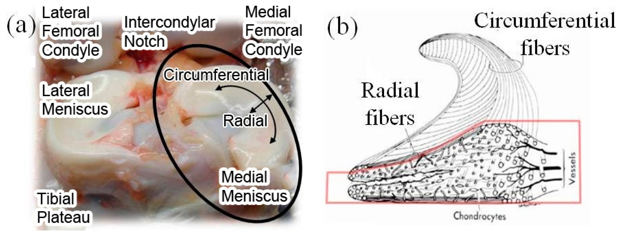
1.1. Meniscus
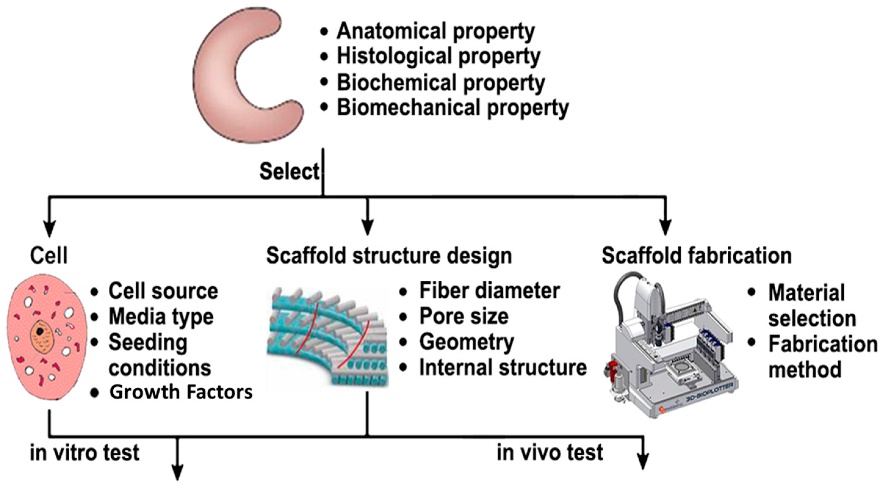
1.2. Meniscal Scaffold
1.3. Cell Sources for Meniscal Scaffold
2. Materials for Meniscal Scaffold and Cell Source
2.1. Tissue-Derived Materials
2.2. Extra Cellular Matrix (ECM) Components
2.3. Synthetic Polymers
2.4. Hydrogels
3. Scaffold Structure Characteristics and Meniscal Scaffold Structure Design
3.1. General Requirements for Meniscal Scaffolds
3.2. Sponge Scaffold Structure Characteristics
3.3. Fibrous Scaffold Structure Characteristics
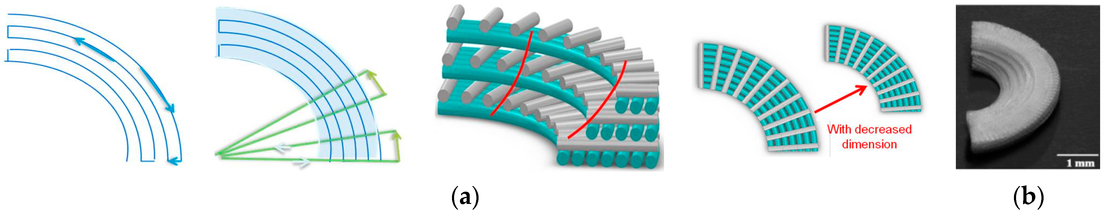
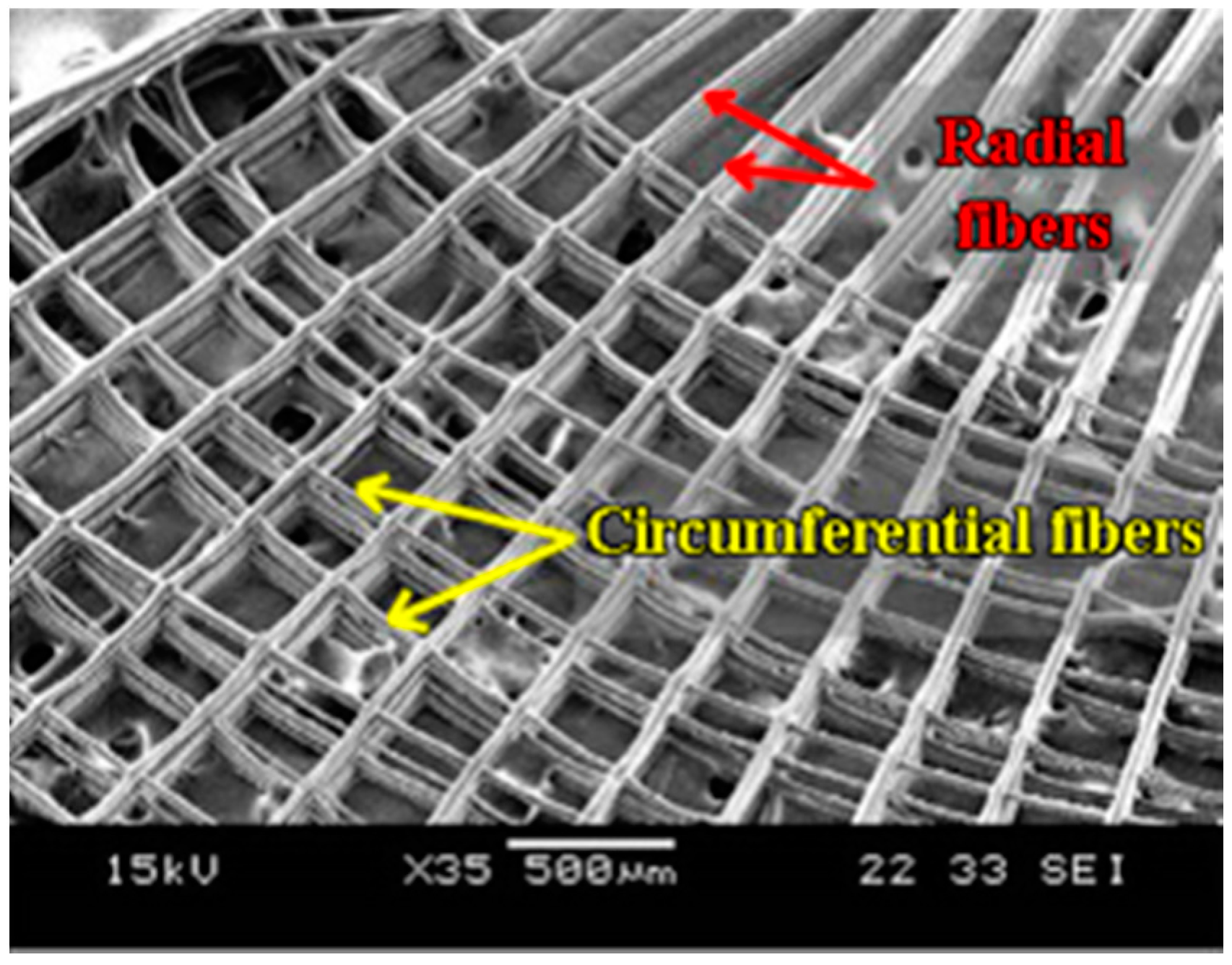
3.4. Fibrous Scaffold Structure Design with Circumferential Pattern
4. Meniscal Scaffold Fabrication Technologies
4.1. Sponge Scaffold Fabrication Technologies
4.2. Non-Woven Fibrous Scaffold Fabrication with Orientation Control
4.2.1. Electrospinning Technology in Fibrous Scaffold Fabrication
4.2.2. Rotating Devices to Align Electrospun Fibers
4.3. Additive Manufacturing (AM) in Woven Meniscal Scaffold Fabrication
4.3.1. Electrohydrodynamic-Jetting (EHD)-Based AM Platform for Fiber Orientation Control
4.3.2. Other AM Techniques for Biomimetic Scaffold Fabrication
5. Scaffold Properties
5.1. Static Mechanical Properties
5.2. Scaffold Properties under Mechanical Stimulation
5.3. Scaffold Properties under Degradation
5.4. Challenges
6. Conclusions
Acknowledgments
Conflicts of Interest
References
- Joint Surgery. Available online: http://www.jointsurgery.in/knee-arthoscopy/meniscal-tears/ (accessed on 10 October 2016).
- Fithian, D.C.; Kelly, M.A.; Mow, V.C. Material properties and structure-function relationships in the menisci. Clin. Orthop. Relat. Res. 1990, 252, 19–31. [Google Scholar] [CrossRef]
- Abrams, G.D.; Frank, R.M.; Gupta, A.K.; Harris, J.D.; McCormick, F.M.; Cole, B.J. Trends in meniscus repair and meniscectomy in the United States, 2005–2011. Am. J. Sports Med. 2013, 41, 2333–2339. [Google Scholar] [CrossRef] [PubMed]
- Nelson, C.G.; Bonner, K.F. Inside-out meniscus repair. Arthrosc. Tech. 2013, 2, e453–e460. [Google Scholar] [CrossRef] [PubMed]
- Jeong, H.J.; Lee, S.H.; Ko, C.S. Meniscectomy. Knee Surg. Relat. Res. 2012, 24, 129–136. [Google Scholar] [CrossRef] [PubMed]
- Roos, E.M.; Östenberg, A.; Roos, H.; Ekdahl, C.; Lohmander, L.S. Long-term outcome of meniscectomy: Symptoms, function, and performance tests in patients with or without radiographic osteoarthritis compared to matched controls. Osteoarthr. Cartil. 2001, 9, 316–324. [Google Scholar] [CrossRef] [PubMed]
- Johnson, R.J.; Kettelkamp, D.B.; Clark, W.; Leaverton, P. Factors affecting late results after meniscectomy. J. Bone Jt. Surg. Am. 1974, 56, 719–729. [Google Scholar] [CrossRef]
- Crook, T.B.; Ardolino, A.; Williams, L.A.P.; Barlow, I.W. Meniscal allograft transplantation: A review of the current literature. Ann. R. Coll. Surg. Engl. 2009, 91, 361–365. [Google Scholar] [CrossRef] [PubMed]
- Lee, S.R.; Kim, J.G.; Nam, S.W. The tips and pitfalls of meniscus allograft transplantation. Knee Surg. Relat. Res. 2012, 24, 137–145. [Google Scholar] [CrossRef] [PubMed]
- Chiari, C.; Koller, U.; Kapeller, B.; Dorotka, R.; Bindreiter, U.; Nehrer, S. Different behavior of meniscal cells in collagen II/I, III and Hyaff-11 scaffolds in vitro. Tissue Eng. Part A 2008, 14, 1295–1304. [Google Scholar] [CrossRef] [PubMed]
- Papalia, R.; Franceschi, F.; Balzani, L.D.; D’Adamio, S.; Maffulli, N.; Denaro, V. Scaffolds for partial meniscal replacement: An updated systematic review. Br. Med. Bull. 2013, 107, 19–40. [Google Scholar] [CrossRef] [PubMed]
- Vrancken, A.C.T.; Buma, P.; van Tienen, T.G. Synthetic meniscus replacement: A review. Int. Orthop. 2013, 37, 291–299. [Google Scholar] [CrossRef] [PubMed]
- Elsner, J.J.; Shemesh, M.; Shefy-Peleg, A.; Gabet, Y.; Zylberberg, E.; Linder-Ganz, E. Quantification of in vitro wear of a synthetic meniscus implant using gravimetric and micro-CT measurements. J. Mech. Behav. Biomed. Mater. 2015, 49, 310–320. [Google Scholar] [CrossRef] [PubMed]
- Caplan, A.I. Mesenchymal stem cells. J. Orthop. Res. 1991, 9, 641–650. [Google Scholar] [CrossRef] [PubMed]
- Walsh, C.J.; Goodman, D.; Caplan, A.I.; Goldberg, V.M. Meniscus regeneration in a rabbit partial meniscectomy model. Tissue Eng. 1999, 5, 327–337. [Google Scholar] [CrossRef] [PubMed]
- Yamasaki, T.; Deie, M.; Shinomiya, R.; Yasunaga, Y.; Yanada, S.; Ochi, M. Transplantation of meniscus regenerated by tissue engineering with a scaffold derived from a rat meniscus and mesenchymal stromal cells derived from rat bone marrow. Artif. Organs 2008, 32, 519–524. [Google Scholar] [CrossRef] [PubMed]
- Angele, P.; Müller, R.; Schumann, D.; Englert, C.; Zellner, J.; Johnstone, B.; Yoo, J.; Hammer, J.; Fierlbeck, J.; Angele, M.K.; et al. Characterization of esterified hyaluronan-gelatin polymer composites suitable for chondrogenic differentiation of mesenchymal stem cells. J. Biomed. Mater. Res. Part A 2009, 91, 416–427. [Google Scholar] [CrossRef] [PubMed]
- Johns, D.; Athanasiou, K.A. Growth factor effects on costal chondrocytes for tissue engineering fibrocartilage. Cell Tissue Res. 2008, 333, 439–447. [Google Scholar] [CrossRef]
- Shalumon, K.; Anulekha, K.; Chennazhi, K.; Tamura, H.; Nair, S.; Jayakumar, R. Fabrication of chitosan/poly (caprolactone) nanofibrous scaffold for bone and skin tissue engineering. Int. J. Biol. Macromol. 2011, 48, 571–576. [Google Scholar] [CrossRef] [PubMed]
- Cook, J.L.; Tomlinson, J.L.; Kreeger, J.M.; Cook, C.R. Induction of meniscal regeneration in dogs using a novel biomaterial. Am. J. Sports Med. 1999, 27, 658–665. [Google Scholar] [PubMed]
- Stapleton, T.W.; Ingram, J.; Katta, J.; Knight, R.; Korossis, S.; Fisher, J.; Ingham, E. Development and characterization of an acellular porcine medial meniscus for use in tissue engineering. Tissue Eng. Part A 2008, 14, 505–518. [Google Scholar] [CrossRef] [PubMed]
- Sweigart, M.A.; Athanasiou, K.A. Toward tissue engineering of the knee meniscus. Tissue Eng. 2001, 7, 111–129. [Google Scholar] [CrossRef] [PubMed]
- Makris, E.A.; Hadidi, P.; Athanasiou, K.A. The knee meniscus: Structure–function, pathophysiology, current repair techniques, and prospects for regeneration. Biomaterials 2011, 32, 7411–7431. [Google Scholar] [CrossRef] [PubMed]
- Beatty, M.W.; Ojha, A.K.; Cook, J.L.; Alberts, L.R.; Mahanna, G.K.; Iwasaki, L.R.; Nickel, J.C. Small intestinal submucosa versus salt-extracted polyglycolic acid-poly-L-lactic acid: A comparison of neocartilage formed in two scaffold materials. Tissue Eng. 2002, 8, 955–968. [Google Scholar] [CrossRef] [PubMed]
- Cook, J.L.; Fox, D.B.; Kuroki, K.; Jayo, M.; De Deyne, P.G. In vitro and in vivo comparison of five biomaterials used for orthopedic soft tissue augmentation. Am. J. Vet. Res. 2008, 69, 148–156. [Google Scholar] [CrossRef]
- Klompmaker, J.; Jansen, H.W.; Veth, R.H.; Nielsen, H.K.; de Groot, J.H.; Pennings, A.J. Porous implants for knee joint meniscus reconstruction: A preliminary study on the role of pore sizes in ingrowth and differentiation of fibrocartilage. Clin. Mater. 1993, 14, 1–11. [Google Scholar] [CrossRef]
- Yamasaki, T.; Deie, M.; Shinomiya, R.; Izuta, Y.; Yasunaga, Y.; Yanada, S.; Sharman, P.; Ochi, M. Meniscal regeneration using tissue engineering with a scaffold derived from a rat meniscus and mesenchymal stromal cells derived from rat bone marrow. J. Biomed. Mater. Res. Part A 2005, 75, 23–30. [Google Scholar] [CrossRef] [PubMed]
- Pabbruwe, M.B.; Kafienah, W.; Tarlton, J.F.; Mistry, S.; Fox, D.J.; Hollander, A.P. Repair of meniscal cartilage white zone tears using a stem cell/collagen-scaffold implant. Biomaterials 2010, 31, 2583–2591. [Google Scholar] [CrossRef] [PubMed]
- Monllau, J.C.; Gelber, P.E.; Abat, F.; Pelfort, X.; Abad, R.; Hinarejos, P.; Tey, M. Outcome after partial medial meniscus substitution with the collagen meniscal implant at a minimum of 10 years’ follow-up. Arthroscopy 2011, 27, 933–943. [Google Scholar] [CrossRef] [PubMed]
- Zaffagnini, S.; Muccioli, G.M.M.; Lopomo, N.; Bruni, D.; Giordano, G.; Ravazzolo, G.; Molinari, M.; Marcacci, M. Prospective Long-Term Outcomes of the Medial Collagen Meniscus Implant Versus Partial Medial Meniscectomy A Minimum 10-Year Follow-Up Study. Am. J. Sports Med. 2011, 39, 977–985. [Google Scholar] [CrossRef] [PubMed]
- Lee, B.S.; Chung, J.W.; Kim, J.M.; Cho, W.J.; Kim, K.A.; Bin, S.I. Morphologic changes in fresh-frozen meniscus allografts over 1 year a prospective magnetic resonance imaging study on the width and thickness of transplants. Am. J. Sports Med. 2012, 40, 1384–1391. [Google Scholar] [CrossRef] [PubMed]
- Harston, A.; Nyland, J.; Brand, E.; McGinnis, M.; Caborn, D.N. Collagen meniscus implantation: A systematic review including rehabilitation and return to sports activity. Knee Surg. Sports Traumatol. Arthrosc. 2012, 20, 135–146. [Google Scholar] [CrossRef] [PubMed]
- Park, S.H.; Kim, T.G.; Kim, H.C.; Yang, D.Y.; Park, T.G. Development of dual scale scaffolds via direct polymer melt deposition and electrospinning for applications in tissue regeneration. Acta Biomater. 2008, 4, 1198–1207. [Google Scholar] [CrossRef] [PubMed]
- Jawad, H.; Lyon, A.R.; Harding, S.E.; Ali, N.N.; Boccaccini, A.R. Myocardial tissue engineering. Br. Med. Bull. 2008, 87, 31–47. [Google Scholar] [CrossRef]
- Annandale, T. The Classic: The Classic an Operation for Displaced Semilunar Cartilage. Clin. Orthop. Relat. Res. 1990, 260, 3–5. [Google Scholar] [CrossRef]
- De Groot, J. Actifit, Polyurethane meniscus implant: Basic science. In The Meniscus; Springer: Berlin, Germany, 2010; pp. 383–387. [Google Scholar]
- Tienen, T.G.; Heijkants, R.G.; de Groot, J.H.; Pennings, A.J.; Schouten, A.J.; Veth, R.P.; Buma, P. Replacement of the knee meniscus by a porous polymer implant a study in dogs. Am. J. Sports Med. 2006, 34, 64–71. [Google Scholar] [CrossRef] [PubMed]
- Verdonk, R.; Verdonk, P.; Huysse, W.; Forsyth, R.; Heinrichs, E.L. Tissue ingrowth after implantation of a novel, biodegradable polyurethane scaffold for treatment of partial meniscal lesions. Am. J. Sports Med. 2011, 39, 774–782. [Google Scholar] [CrossRef] [PubMed]
- Zaffagnini, S.; Fink, C.; Grassi, A.; Muccioli, G.M.; Marcacci, M. Meniskusimplantate. Arthroskopie 2015, 28, 38–42. [Google Scholar] [CrossRef]
- Mandal, B.B.; Park, S.H.; Gil, E.S.; Kaplan, D.L. Multilayered silk scaffolds for meniscus tissue engineering. Biomaterials 2011, 32, 639–651. [Google Scholar] [CrossRef] [PubMed]
- Yan, L.P.; Oliveira, J.M.; Oliveira, A.L.; Caridade, S.G.; Mano, J.F.; Reis, R.L. Macro/microporous silk fibroin scaffolds with potential for articular cartilage and meniscus tissue engineering applications. Acta Biomater. 2012, 8, 289–301. [Google Scholar] [CrossRef] [PubMed]
- Gruchenberg, K.; Ignatius, A.; Friemert, B.; von Lübken, F.; Skaer, N.; Gellynck, K.; Kessler, O.; Dürselen, L. In vivo investigation of an innovative silk scaffold for partial meniscus replacement. J. Biomech. 2012, 45. [Google Scholar] [CrossRef]
- Ronken, S.; Wirz, D.; Daniels, A.; Kurokawa, T.; Gong, J.; Arnold, M. Double-network acrylamide hydrogel compositions adapted to achieve cartilage-like dynamic stiffness. Biomech. Model. Mechanobiol. 2013, 12, 243–248. [Google Scholar] [CrossRef] [PubMed]
- Nguyen, M.K.; Alsberg, E. Bioactive factor delivery strategies from engineered polymer hydrogels for therapeutic medicine. Prog. Polym. Sci. 2014, 39, 1235–1265. [Google Scholar] [CrossRef] [PubMed]
- Popowics, T.E.; Zhu, Z.; Herring, S.W. Mechanical properties of the periosteum in the pig, Sus scrofa. Arch. Oral Biol. 2002, 47, 733–741. [Google Scholar] [CrossRef]
- Hiles, M.C.; Badylak, S.F.; Lantz, G.C.; Kokini, K.; Geddes, L.A.; Morff, R.J. Mechanical properties of xenogeneic small-intestinal submucosa when used as an aortic graft in the dog. J. Biomed. Mater. Res. 1995, 29, 883–891. [Google Scholar] [CrossRef] [PubMed]
- Gao, S.; Yuan, Z.; Xi, T.; Wei, X.; Guo, Q. Characterization of decellularized scaffold derived from porcine meniscus for tissue engineering applications. Front. Mater. Sci. 2016, 10, 101–112. [Google Scholar] [CrossRef]
- Abdelgaied, A.; Stanley, M.; Galfe, M.; Berry, H.; Ingham, E.; Fisher, J. Comparison of the biomechanical tensile and compressive properties of decellularised and natural porcine meniscus. J. Biomech. 2015, 48, 1389–1396. [Google Scholar] [CrossRef] [PubMed]
- Hadidi, P.; Athanasiou, K.A. Enhancing the mechanical properties of engineered tissue through matrix remodeling via the signaling phospholipid lysophosphatidic acid. Biochem. Biophys. Res. Commun. 2013, 433, 133–138. [Google Scholar] [CrossRef] [PubMed]
- Broomell, C.C.; Mattoni, M.A.; Zok, F.W.; Waite, J.H. Critical role of zinc in hardening of Nereis jaws. J. Exp. Biol. 2006, 209, 3219–3225. [Google Scholar] [CrossRef] [PubMed]
- Li, J.; Suo, Z.; Vlassak, J.J. Stiff, strong, and tough hydrogels with good chemical stability. J. Mater. Chem. B 2014, 2, 6708–6713. [Google Scholar] [CrossRef]
- Moroni, L.; Lambers, F.M.; Wilson, W.; van Donkelaar, C.; De Wijn, J.; Huiskes, R.; van Blitterswijk, C. Finite element analysis of meniscal anatomical 3D scaffolds: Implications for tissue engineering. Open Biomed. Eng. J. 2007, 1, 23–34. [Google Scholar] [CrossRef] [PubMed]
- Tanaka, Y.; Yamaoka, H.; Nishizawa, S.; Nagata, S.; Ogasawara, T.; Asawa, Y.; Fujihara, Y.; Takato, T.; Hoshi, K. The optimization of porous polymeric scaffolds for chondrocyte/atelocollagen based tissue-engineered cartilage. Biomaterials 2010, 31, 4506–4516. [Google Scholar] [CrossRef] [PubMed]
- Woodfield, T.; Blitterswijk, C.V.; Wijn, J.D.; Sims, T.; Hollander, A.; Riesle, J. Polymer scaffolds fabricated with pore-size gradients as a model for studying the zonal organization within tissue-engineered cartilage constructs. Tissue Eng. 2005, 11, 1297–1311. [Google Scholar] [CrossRef] [PubMed]
- Xie, J.; Ihara, M.; Jung, Y.; Kwon, I.K.; Kim, S.H.; Kim, Y.H.; Matsuda, T. Mechano-active scaffold design based on microporous poly (l-lactide-co-ε-caprolactone) for articular cartilage tissue engineering: Dependence of porosity on compression force-applied mechanical behaviors. Tissue Eng. 2006, 12, 449–458. [Google Scholar] [CrossRef] [PubMed]
- Welsing, R.T.; van Tienen, T.G.; Ramrattan, N.; Heijkants, R.; Schouten, A.J.; Veth, R.P.; Buma, P. Effect on Tissue Differentiation and Articular Cartilage Degradation of a Polymer Meniscus Implant A 2-Year Follow-up Study in Dogs. Am. J. Sports Med. 2008, 36, 1978–1989. [Google Scholar] [CrossRef] [PubMed]
- Yannas, I.; Lee, E.; Orgill, D.; Skrabut, E.; Murphy, G. Synthesis and characterization of a model extracellular matrix that induces partial regeneration of adult mammalian skin. Proc. Natl. Acad. Sci. USA 1989, 86, 933–937. [Google Scholar] [CrossRef] [PubMed]
- Whang, K.; Healy, K.; Elenz, D.; Nam, E.; Tsai, D.; Thomas, C.; Nuber, G.; Glorieux, F.; Travers, R.; Sprague, S. Engineering bone regeneration with bioabsorbable scaffolds with novel microarchitecture. Tissue Eng. 1999, 5, 35–51. [Google Scholar] [CrossRef] [PubMed]
- Zardiackas, L.D.; Parsell, D.E.; Dillon, L.D.; Mitchell, D.W.; Nunnery, L.A.; Poggie, R. Structure, metallurgy, and mechanical properties of a porous tantalum foam. J. Biomed. Mater. Res. 2001, 58, 180–187. [Google Scholar] [CrossRef]
- Zeltinger, J.; Sherwood, J.K.; Graham, D.A.; Müeller, R.; Griffith, L.G. Effect of pore size and void fraction on cellular adhesion, proliferation, and matrix deposition. Tissue Eng. 2001, 7, 557–572. [Google Scholar] [CrossRef] [PubMed]
- Lanza, R.; Langer, R.; Vacanti, J.P. Principles of Tissue Engineering; Academic Press: New York, NY, USA, 2011. [Google Scholar]
- Yamane, S.; Iwasaki, N.; Kasahara, Y.; Harada, K.; Majima, T.; Monde, K.; Nishimura, S.I.; Minami, A. Effect of pore size on in vitro cartilage formation using chitosan-based hyaluronic acid hybrid polymer fibers. J. Biomed. Mater. Res. Part A 2007, 81, 586–593. [Google Scholar] [CrossRef] [PubMed]
- Hutmacher, D.W.; Schantz, T.; Zein, I.; Ng, K.W.; Teoh, S.H.; Tan, K.C. Mechanical properties and cell cultural response of polycaprolactone scaffolds designed and fabricated via fused deposition modeling. J. Biomed. Mater. Res. 2001, 55, 203–216. [Google Scholar] [CrossRef]
- Williams, J.M.; Adewunmi, A.; Schek, R.M.; Flanagan, C.L.; Krebsbach, P.H.; Feinberg, S.E.; Hollister, S.J.; Das, S. Bone tissue engineering using polycaprolactone scaffolds fabricated via selective laser sintering. Biomaterials 2005, 26, 4817–4827. [Google Scholar] [CrossRef]
- Geiger, B. A role for p130Cas in mechanotransduction. Cell 2006, 127, 879–881. [Google Scholar] [CrossRef] [PubMed]
- Jeong, C.G.; Hollister, S.J. Mechanical, permeability, and degradation properties of 3D designed poly (1, 8 octanediol-co-citrate) scaffolds for soft tissue engineering. J. Biomed. Mater. Res. Part B Appl. Biomater. 2010, 93, 141–149. [Google Scholar] [CrossRef] [PubMed]
- Balint, E.; Gatt, C.J.; Dunn, M.G. Design and mechanical evaluation of a novel fiber-reinforced scaffold for meniscus replacement. J. Biomed. Mater. Res. Part A 2012, 100, 195–202. [Google Scholar] [CrossRef] [PubMed]
- Li, J. Biofabrication and Characterization of Meniscal Scaffolds via an Electrohydrodynamic Jet Printing Technique. Ph.D. Thesis, National University of Singapore, Singapore, 2014. [Google Scholar]
- Wang, H.; Vijayavenkataraman, S.; Wu, Y.; Shu, Z.; Sun, J.; Hsi, J.F.Y. Investigation of process parameters of electrohydro-dynamic jetting for 3D printed PCL fibrous scaffolds with complex geometries. Int. J. Biopr. 2016, 2, 63–71. [Google Scholar] [CrossRef]
- Hosseinkhani, H.; Hosseinkhani, M.; Kobayashi, H. Design of tissue-engineered nanoscaffold through self-assembly of peptide amphiphile. J. Bioact. Compat. Polym. 2006, 21, 277–296. [Google Scholar] [CrossRef]
- Nam, Y.S.; Yoon, J.J.; Park, T.G. A novel fabrication method of macroporous biodegradable polymer scaffolds using gas foaming salt as a porogen additive. J. Biomed. Mater. Res. 2000, 53, 1–7. [Google Scholar] [CrossRef]
- Barbetta, A.; Rizzitelli, G.; Bedini, R.; Pecci, R.; Dentini, M. Porous gelatin hydrogels by gas-in-liquid foam templating. Soft Matter 2010, 6, 1785–1792. [Google Scholar] [CrossRef]
- Elsner, J.J.; Portnoy, S.; Zur, G.; Guilak, F.; Shterling, A.; Linder-Ganz, E. Design of a free-floating polycarbonate-urethane meniscal implant using finite element modeling and experimental validation. J. Biomech. Eng. 2010, 132. [Google Scholar] [CrossRef] [PubMed]
- Bang, S.H.; Kim, T.H.; Lee, H.Y.; Shin, U.S.; Kim, H.W. Nanofibrous-structured biopolymer scaffolds obtained by a phase separation with camphene and initial cellular events. J. Mater. Chem. 2011, 21, 4523–4530. [Google Scholar] [CrossRef]
- De Mulder, E.; Hannink, G.; Verdonschot, N.; Buma, P. Effect of polyurethane scaffold architecture on ingrowth speed and collagen orientation in a subcutaneous rat pocket model. Biomed. Mater. 2013, 8. [Google Scholar] [CrossRef] [PubMed]
- Esposito, A.R.; Moda, M.; Cattani, S.M.; de Santana, G.M.; Barbieri, J.A.; Munhoz, M.M.; Cardoso, T.P.; Barbo, M.L.P.; Russo, T.; D’Amora, U.; et al. PLDLA/PCL-T scaffold for meniscus tissue engineering. BioRes. Open Access 2013, 2, 138–147. [Google Scholar] [CrossRef] [PubMed]
- Chiari, C.; Koller, U.; Dorotka, R.; Eder, C.; Plasenzotti, R.; Lang, S.; Ambrosio, L.; Tognana, E.; Kon, E.; Salter, D.; et al. A tissue engineering approach to meniscus regeneration in a sheep model. Osteoarthr. Cartil. 2006, 14, 1056–1065. [Google Scholar] [CrossRef] [PubMed]
- Megelski, S.; Stephens, J.S.; Chase, D.B.; Rabolt, J.F. Micro-and nanostructured surface morphology on electrospun polymer fibers. Macromolecules 2002, 35, 8456–8466. [Google Scholar] [CrossRef]
- Shih, Y.R.V.; Chen, C.N.; Tsai, S.W.; Wang, Y.J.; Lee, O.K. Growth of mesenchymal stem cells on electrospun type I collagen nanofibers. Stem Cells 2006, 24, 2391–2397. [Google Scholar] [CrossRef] [PubMed]
- Chen, C.H.; Saville, D.; Aksay, I. Scaling laws for pulsed electrohydrodynamic drop formation. Appl. Phys. Lett. 2006, 89. [Google Scholar] [CrossRef]
- Kim, Y.J.; Lee, J.S.; Kim, S.Y.; Park, S.E.; Hwang, J.; Kim, Y.J. Hybrid on demand jetting system for ultra fine droplet based on electrohydrodynamic and piezoelectric actuation. In Proceedings of the IEEE 22nd International Conference on Micro Electro Mechanical Systems (MEMS 2009), Sorrento, Italy, 25–29 January 2009; pp. 491–494.
- Ionescu, L.C.; Mauck, R.L. Porosity and cell preseeding influence electrospun scaffold maturation and meniscus integration in vitro. Tissue Eng. Part A 2012, 19, 538–547. [Google Scholar] [CrossRef]
- Fisher, M.B.; Henning, E.A.; Söegaard, N.; Esterhai, J.L.; Mauck, R.L. Organized nanofibrous scaffolds that mimic the macroscopic and microscopic architecture of the knee meniscus. Acta Biomater. 2013, 9, 4496–4504. [Google Scholar] [CrossRef] [PubMed]
- Schantz, J.T.; Brandwood, A.; Hutmacher, D.W.; Khor, H.L.; Bittner, K. Osteogenic differentiation of mesenchymal progenitor cells in computer designed fibrin-polymer-ceramic scaffolds manufactured by fused deposition modeling. J. Mater. Sci. Mater. Med. 2005, 16, 807–819. [Google Scholar] [CrossRef] [PubMed]
- Wei, C.; Cai, L.; Sonawane, B.; Wang, S.; Dong, J. High-precision flexible fabrication of tissue engineering scaffolds using distinct polymers. Biofabrication 2012, 4. [Google Scholar] [CrossRef] [PubMed]
- Shor, L.; Güçeri, S.; Chang, R.; Gordon, J.; Kang, Q.; Hartsock, L.; An, Y.; Sun, W. Precision extruding deposition (PED) fabrication of polycaprolactone (PCL) scaffolds for bone tissue engineering. Biofabrication 2009, 1. [Google Scholar] [CrossRef] [PubMed]
- Hashimdeen, S.H.; Miodownik, M.; Edirisinghe, M.J. The design and construction of an electrohydrodynamic Cartesian robot for the preparation of tissue engineering constructs. PLoS ONE 2014, 9, e112166. [Google Scholar] [CrossRef] [PubMed]
- Caterson, E.J.; Nesti, L.J.; Li, W.J.; Danielson, K.G.; Albert, T.J.; Vaccaro, A.R.; Tuan, R.S. Three-dimensional cartilage formation by bone marrow-derived cells seeded in polylactide/alginate amalgam. J. Biomed. Mater. Res. 2001, 57, 394–403. [Google Scholar] [CrossRef]
- Chang, R.; Nam, J.; Sun, W. Direct cell writing of 3D microorgan for in vitro pharmacokinetic model. Tissue Eng. Part C Methods 2008, 14, 157–166. [Google Scholar] [CrossRef] [PubMed]
- Shim, J.H.; Kim, J.Y.; Park, M.; Park, J.; Cho, D.W. Development of a hybrid scaffold with synthetic biomaterials and hydrogel using solid freeform fabrication technology. Biofabrication 2011, 3. [Google Scholar] [CrossRef] [PubMed]
- Schuurman, W.; Khristov, V.; Pot, M.; Van Weeren, P.; Dhert, W.; Malda, J. Bioprinting of hybrid tissue constructs with tailorable mechanical properties. Biofabrication 2011, 3. [Google Scholar] [CrossRef]
- Zhao, X.; He, J.; Xu, F.; Liu, Y.; Li, D. Electrohydrodynamic printing: A potential tool for high-resolution hydrogel/cell patterning. Virtual Phys. Prototyp. 2016, 11, 57–63. [Google Scholar] [CrossRef]
- Lee, H.; Yeo, M.; Ahn, S.; Kang, D.O.; Jang, C.H.; Lee, H.; Park, G.M.; Kim, G.H. Designed hybrid scaffolds consisting of polycaprolactone microstrands and electrospun collagen-nanofibers for bone tissue regeneration. J. Biomed. Mater. Res. Part B Appl. Biomater. 2011, 97, 263–270. [Google Scholar] [CrossRef] [PubMed]
- Zhang, Z.; Ni, J.; Chen, L.; Yu, L.; Xu, J.; Ding, J. Biodegradable and thermoreversible PCLA-PEG-PCLA hydrogel as a barrier for prevention of post-operative adhesion. Biomaterials 2011, 32, 4725–4736. [Google Scholar] [CrossRef] [PubMed]
- Martínez, H.; Brackmann, C.; Enejder, A.; Gatenholm, P. Mechanical stimulation of fibroblasts in micro-channeled bacterial cellulose scaffolds enhances production of oriented collagen fibers. J. Biomed. Mater. Res. Part A 2012, 100, 948–957. [Google Scholar] [CrossRef] [PubMed]
- Gunja, N.J.; Athanasiou, K.A. Additive and synergistic effects of bFGF and hypoxia on leporine meniscus cell-seeded PLLA scaffolds. J. Tissue Eng. Regen. Med. 2010, 4, 115–122. [Google Scholar] [CrossRef]
- Marsano, A.; Wendt, D.; Raiteri, R.; Gottardi, R.; Stolz, M.; Wirz, D.; Daniels, A.U.; Salter, D.; Jakob, M.; Quinn, T.M.; et al. Use of hydrodynamic forces to engineer cartilaginous tissues resembling the non-uniform structure and function of meniscus. Biomaterials 2006, 27, 5927–5934. [Google Scholar] [CrossRef] [PubMed]
- Huey, D.J.; Athanasiou, K.A. Maturational growth of self-assembled, functional menisci as a result of TGF-β1 and enzymatic chondroitinase-ABC stimulation. Biomaterials 2011, 32, 2052–2058. [Google Scholar] [CrossRef] [PubMed]
- Petri, M.; Ufer, K.; Toma, I.; Becher, C.; Liodakis, E.; Brand, S.; Haas, P.; Liu, C.; Richter, B.; Haasper, C.; et al. Effects of perfusion and cyclic compression on in vitro tissue engineered meniscus implants. Knee Surg. Sports Traumatol. Arthrosc. 2012, 20, 223–231. [Google Scholar] [CrossRef] [PubMed]
- Odelius, K.; Höglund, A.; Kumar, S.; Hakkarainen, M.; Ghosh, A.K.; Bhatnagar, N.; Albertsson, A.C. Porosity and pore size regulate the degradation product profile of polylactide. Biomacromolecules 2011, 12, 1250–1258. [Google Scholar] [CrossRef]

| Materials | Properties | Reference | ||||
|---|---|---|---|---|---|---|
| Mechanics | Bioactivity | Logistics | ||||
| Mechanical Properties (Elastic Modulus) | Anisotropy | Geometry (Biomimetic) | ||||
| Tissue-derived Materials | Periosteal tissue: 8–12 MPa | Highly anisotropic | Highly biomimetic | High | Low | [45] |
| SIS: 12–25 MPa | [46] | |||||
| Porcine meniscus: 110–200 MPa | [47,48] | |||||
| ECM Components | 250–500 kPa | Anisotropic | Biomimetic | High | Medium | [49] |
| Synthetic Polymers | 200–5000 MPa | Highly anisotropic | Depends on the fabrication method | Low | High | [50] |
| Hydrogels | 0.01–10 MPa | Isotropic | Depends on the fabrication method | Medium | High | [51] |
| Scaffold Structure | Fabrication Method | Pros & Cons | Reference |
|---|---|---|---|
| Sponge scaffold | Particulate leaching | (+) highly porous scaffolds with porosity values up to 93% | [70] |
| (−) only used to produce thin membranes up to 3 mm thick | |||
| Gas foaming | (+) organic solvent-free process | [71,72] | |
| (−) a structure with largely unconnected pores | |||
| (−) non-porous external surface | |||
| Freeze drying | (+) highly porous scaffolds with porosity values >90% | [73] | |
| (+) reduction of toxic solvents use | |||
| (+) elimination of time-consuming drying and leaching processes of porogen components | |||
| (−) instability of the emulsion | |||
| (−) difficulty in controlling the pore size and porosity | |||
| Phase separation | (+) highly porous scaffolds with porosity values >90% | [74,75] | |
| (−) limited range of pore size (<200 um) | |||
| (−) difficult to control the micro- and macro-structure of the scaffold | |||
| Non-woven fibrous scaffold | Electrospinning | (+) nanofibrous architectures | [79,80,81,82,83] |
| (+) wide range of fiber diameters | |||
| (+) wide range of polymers can be used | |||
| (−) used solvents can be toxic | |||
| (−) limited capability to fabricate biomimetic structure | |||
| Oriented/woven fibrous scaffold | FDM/PED | (+) layer by layer architecture | [84,85,86] |
| (+) ability to fabricate complex structures | |||
| (−) low resolution | |||
| (−) limited range of materials | |||
| EHD-jetting | (+) layer-by-layer architecture | [68,87] | |
| (+) ability to fabricate complex structures | |||
| (−) used solvents can be toxic |
© 2017 by the authors. Licensee MDPI, Basel, Switzerland. This article is an open access article distributed under the terms and conditions of the Creative Commons Attribution (CC-BY) license ( http://creativecommons.org/licenses/by/4.0/).
Share and Cite
Sun, J.; Vijayavenkataraman, S.; Liu, H. An Overview of Scaffold Design and Fabrication Technology for Engineered Knee Meniscus. Materials 2017, 10, 29. https://doi.org/10.3390/ma10010029
Sun J, Vijayavenkataraman S, Liu H. An Overview of Scaffold Design and Fabrication Technology for Engineered Knee Meniscus. Materials. 2017; 10(1):29. https://doi.org/10.3390/ma10010029
Chicago/Turabian StyleSun, Jie, Sanjairaj Vijayavenkataraman, and Hang Liu. 2017. "An Overview of Scaffold Design and Fabrication Technology for Engineered Knee Meniscus" Materials 10, no. 1: 29. https://doi.org/10.3390/ma10010029
APA StyleSun, J., Vijayavenkataraman, S., & Liu, H. (2017). An Overview of Scaffold Design and Fabrication Technology for Engineered Knee Meniscus. Materials, 10(1), 29. https://doi.org/10.3390/ma10010029