Article

Tuning Solid-State Reaction Pathways Using Molecular Sulfur Precursors to Synthesize FeS Anodes of Li-Ion Batteries for Boosted Electrochemical Performance

1
School of Materials Science and Engineering, Dalian Jiaotong University, Dalian 116021, China
2
School of Electrical Engineering, Dalian Jiaotong University, Dalian 116021, China
3
State Grid Gansu Electric Power Company Qingyang Power Supply Branch Company, Qingyang 745000, China
*
Authors to whom correspondence should be addressed.
Energies 2025, 18(17), 4623; https://doi.org/10.3390/en18174623 (registering DOI)
Submission received: 7 August 2025 / Revised: 26 August 2025 / Accepted: 29 August 2025 / Published: 30 August 2025

Abstract

The solid-state reactions between Fe2O3 and molecular sulfur sources could produce FeS nanoparticles efficiently, while the functions of these molecules have been ignored except for the role as sulfur sources. In this work, thioacetamide and thiourea were employed as sulfur sources for the solid-state reactions with Fe2O3 to explore their effects on the microstructure and electrochemical performance of the produced FeS. Despite the slight difference in one functional group for two molecules (−CH3 against −NH2), thiourea leads to a more complex reaction pathway with FeS2 as the intermediate phase, while no such an intermediate phase is observed in the reaction with thioacetamide. The former yields FeS of 2D nanoflakes as the final products, compared with the aggregated nanoparticles in reactions with thiourea. As a result, the nanoflakes exhibit a higher discharge capacity with enhanced stability (388.9 mAh∙g−1 vs. 374.7 mAh∙g−1 above 1 V). According to the reaction pathways, the formation of FeS nanoflakes and superior electrochemical performance were addressed, paving a route for the solid-state reactions with molecules to develop high-performance sulfide electrode materials.

1. Introduction

The safety issue associated with the self-ignition of Li-ion batteries (LIBs) is receiving more attention, while the self-ignition is usually caused by Li dendrites leading to the short-circuit between cathodes and anodes. The physical origin of the Li dendrites lies in the low intercalation potential of commercial graphite (0.1 V against Li+/Li), very close to that for Li precipitation [1,2]. Li4Ti5O12, another commercial anode material, has an interaction potential of 1.4 V, and thus the Li dendrites could be inhibited in the Li-ion batteries based on it [3]. However, the high cost and low theoretical capacity (175 mAh∙g−1) of Li4Ti5O12 limit its market share.
FeS (usually Fe-deficient as Fe1-xS), as the anode for LIBs, has a similar plateau voltage (1.4 V) to that of Li4Ti5O12, and its discharge capacity above 1 V is more than 400 mAh∙g−1, much higher than that of Li4Ti5O12 [4,5]. Therefore, FeS could be a promising candidate to replace Li4Ti5O12. In fact, FeS had been used as electrodes for thermal batteries in the early 1970s [6,7]. The thermal battery uses molten LiCl salts as electrolytes and Li/LiAl as counter electrodes, yielding a working voltage of ca. 1.4 V. The batteries were already used in electrical grids and electrical vans then, due to low cost and high capacity [6,7]. However, the lower working voltage and high working temperature (above 300 °C to melt LiCl) limit its applications, compared with the LIBs based on organic electrolytes nowadays. Thus, more attentions are paid to the development of FeS as anodes for the LIBs over 2 decades. For instance, all-solid-state batteries of LiCoO2/FeS (LiCoO2 cathode and FeS anode) have been demonstrated [8].
The high capacity usually results in a large volume change of FeS during (de)lithiation, and the nano-sized powders are necessary to alleviate such volume change. Electrodes made from nanoscale materials form porous, flexible networks with more binder contact and smaller absolute dimensional change per particle, so the whole composite is less prone to delamination and loss of contact. In the all-solid-state batteries of LiCoO2/FeS, the nanoscale FeS particles of 300 nm in size show much better cyclic stability than the large particles of 10 μm in size (95% against 84% capacity retention after 15 cycles) [8]. In contrast, more superior cyclic stability has been reported for FeS nanoparticles of less than 100 nm (i.e., 90% capacity retention after 50 cycles) [9,10].
Typical methodologies to synthesize FeS nano powders include hydrothermal [11,12], solvothermal [13,14], solution [15], electrospinning [16], etc. In these methods, FeCl2 or FeCl3 is usually used as the main precursor, and the washing of final products is necessary to remove Cl ions. Therefore, it is difficult to scale up.
Compared with these conventional methods, the solid-state reactions between inorganic Fe sources and molecular S sources could produce nano-sized FeS powders at a temperature of no more than 700 °C in a facile way. Typical Fe sources include Fe [17,18,19], Fe2O3 [16,20], Ferrocene [21,22], FeS2 [18,19], etc., and sulfur sources include S [17,18,19,21], thioacetamide (TAA) [20], thiourea (TA) [16], ammonium persulfate [22], etc. For instance, reactions between Fe2O3 nanofibers and the gases from the TA pyrolysis produce porous FeS nanofibers at 400 °C, with a discharge capacity of 561 mAh∙g−1 [16]. The direction calcination of Fe2O3 nano cubes and TAA at 500 °C yields FeS nano cubes.
Reactions between ferrocene (5 mmol) and ammonium persulfate (10 mmol) at 500 °C give carbon-coated nanospheres of Fe7S8 [22], while a higher temperature is necessary for the reactions between ferrocene and sulfur to get carbon-coated Fe7S8 nanoparticles [18].
Obviously, different sulfur sources could modify the solid-state reactions (i.e., the reaction temperatures at least), rather than only the role of supplying sulfur. However, their functions other than sulfur source have been ignored in the literature. In this work, two similar molecular S precursors, thiourea and thioacetamide, have been employed as sulfur sources to explore their functions to modify the solid-state reactions, as well as microstructures and properties of the FeS products. As a result, two molecules reacted with Fe2O3 in different pathways to achieve the final product of FeS. However, the morphologies and electrochemical performance of the resulting FeS are also distinct.

2. Experimental

Synthesis. FeS powders were synthesized by the solid-state reactions of micrometer-size Fe2O3 (Macklin, Shanghai, China, AR, 99%) with two kinds of S sources: TAA (Macklin, AR, 99%) and TA (Macklin, AR, 99%), and named as FeS-TAA and FeS-TA, respectively. In the typical synthesis, Fe2O3 (9 g) and TAA/TA (18 g) were mixed in a mass ratio of 1:2 using an agate mortar, and then loaded into alumina semi-circular crucibles for calcination in a tube furnace. The furnace was first purged with an N2 flow of 100 mL/min with 99.998% grade high-purity gas for half an hour, and the calcination temperature of 650 °C was achieved at a rate of 2 °C/min and kept for 30 min under the N2 atmosphere. After cooling with furnace to 25 °C, black FeS powders were obtained. The temperature of the calcination is determined according to thermal analyses. For comparison, a lower calcination temperature of 400 °C is also used.
Characterization. All samples were analyzed using powder X-ray diffraction (XRD, PANalytical B.V., Almelo, Netherlands) using a laboratory Empyrean diffractometer with Cu Kα radiation with a Ni filter (wavelength 1.5406 Å, with 40 kV of accelerating voltage, and 40 mA of accelerating current). The morphologies and microstructures were inspected with field emission scanning electron microscopy (FESEM, JEOL, Tokyo Metropolis, Japan, SUPRA 55) and transmission electron microscopy (TEM, Hitathi, Tokyo Metropolis, Japan, JEM 2100 F), coupled with an Energy-dispersive X-ray spectroscopy (EDS, Bruker, Quantax, JEOL, Tokyo Metropolis, Japan). The reaction of Fe2O3 and TAA/TA was inspected using a Synchronous Thermal Analyzer (TG/DSC, NETZSCH, Free State of Bavaria, Germany, Netzsch STA449) at a heating rate of 5 °C/min under N2 atmosphere. Raman spectra were performed with a Confocal Raman microspectrometer (Raman, Renishaw, Gloucestershire, UK, Invia Qontor, with an excitation laser of 532 nm in wavelength) in the range of 50–4000 cm−1. X-ray Photoelectron Spectroscopy (XPS, Ulvac-Phi, Chigasaki-shi, Japan) analysis was performed using a ULVAC-PHI 5802 spectrometer (monochromatic Al Kα radiation, hv = 1486.6 eV, vacuum pressure ~10−9 mbar), with calibration referenced to the contaminant carbon C 1s peak (284.8 eV). The high-resolution spectra achieved a resolution of 0.1 eV.
Electrochemical measurements. In a glovebox under an Ar atmosphere, all coin cells were assembled using CR2032 coin cell components and using metallic lithium foils as counter electrodes. For anode preparation, the FeS materials (80 wt. %), polyvinylidene fluoride (PVDF, 10 wt. %), and carbon super P (10 wt. %) were mixed with n-methylpyrrolidone (NMP). The resulting slurry (ca. 500 mg) was stirred at 500 rpm for 12 h for homogeneity, cast on a copper foil, and dried at 120 °C for one night to produce FeS anodes, which were cut into discs of 1.2 cm in diameter as the working electrodes in coin cells. The surface density of the working electrodes is 2.9~3.1 mg∙cm−2. A solution of 1 M LiPF6 in ethylene carbonate (EC) and diethyl carbonate (DEC) (1:1 by volume) was employed as the electrolyte, and a Celgard 2400 microporous polypropylene membrane was used as the separator. All the electrochemical measurements were performed by a Neware battery testing system within 1–2.7 V. Electrochemical impedance spectroscopy (EIS) was performed using a DH7000 electrochemical working station in the frequency range from 10 kHz to 0.005 Hz. The electrochemical measurements of these samples were performed at room temperature.

3. Results and Discussions

To determine the reaction temperatures for Fe2O3 and TA/TAA, thermal analyses were performed on their mixtures with a mass ratio of 1:2. The mixture with TAA is largely decomposed with two endothermic reactions: the first at 111 °C stems from TAA melting, and the second at 155 °C corresponds to the TAA decomposition (Figure 1a). The reaction temperatures are slightly lower than those reported for pure TAA (115 °C and 170 °C, respectively) [23], but the data of TG and DSC are very similar to those of pure TAA. Metal oxides can catalyze TAA breakdown and thereby lower the temperature at which it decomposes, as demonstrated by Au-modified ZnO [24]. Thus, it is likely that Fe2O3 functions as a catalyst to reduce the decomposition temperature of TAA here.
The decomposition products are H2S gas and acetonitrile liquid, based on the reaction [25]:
CH3CSNH2 (s) → H2S(g) + CH3CN (l)
However, there are very few residues (<5%) after the decomposition, because most acetonitrile liquid will be evaporated out at the decomposition temperature (much higher than its boiling temperature). According to Figure 1a, the weight loss of the mixture is 64% after the decomposition, close to the weight of TAA. It is also possible that some of the H2S and acetonitrile were absorbed by Fe2O3 for sulfurization at higher temperatures.
The weight loss of the mixture with TA occurs mainly in two temperature regions: (1) 41.01% loss in the regions from 180 °C to 300 °C, with three endothermic reactions (174 °C, 189 °C, and 250 °C); (2) 23% loss in the regions from 450 °C to 700 °C, with two endothermic reactions (575 °C and 640 °C). The low-temperature region actually dominates the weight loss (>80%) of pure TA, while the weight loss in the high-temperature region is usually no more than 5% [26,27]. Evidently, the reaction between Fe2O3 and TA proceeds in a pathway different from that for the Fe2O3-TAA reaction.
Unlike the direction decomposition of TAA, TA will be converted into thiocyanate via the isomeric reaction above the melting temperature (Reaction (2)) [27,28,29]:
SC(NH2)2 (s) → NH4SCN (s)
In the second (Reaction (3)) and third (Reaction (4)) endothermic reactions, thiocyanate and residual TA will be decomposed into gases [27,30]:
2SC(NH2)2 (s) → CS2(g) + H2NCN(g) + 2NH3(g)
NH4SCN(s) → NH3(g) + HNCS(g)
TA will be completely lost as gases after these reactions, according to the above reactions. Polymerizations are also possible for TA and/or thiocyanate [31], to account for the residual weight and the further weight loss at 500 °C in TA [26]. For the mixture of TA and Fe2O3, the weight loss in the low-temperature region is heavily depressed. There are two possible reasons: (1) the decomposed gases react quickly with Fe2O3, and (2) Fe2O3 catalyzes the heteroaromatic formation from the N/C fragments produced by the pyrolysis of thioacetamide, because the temperature favors radical chain reactions [32], especially with reports on oxide catalysts for oligomerization and ring-forming chemistry [33,34]. Because the mixture shows the two-step weight loss even in the high-temperature region, compared with the single-step character in TAA [25], both the sulfurization and heteroaromatics could coexist.
According to the thermal analysis, a temperature higher than 640 °C is necessary for the reaction between Fe2O3 and TA to achieve the stable phase. However, it has been reported that Fe2O3 nanofibers in the gases from the TAA pyrolysis could be converted into FeS at 400 °C. Therefore, two reaction temperatures, 650 °C and 400 °C, are used for both TAA and TA. According to XRD data (Figure 1c,d), reactions at 650 °C yield FeS (JCPDF No. 04-004-7981, space group P63/mmc) as a single crystalline phase for both TA and TAA. The lattice parameters determined by indexing their diffraction peaks are a = 3.451(1) Å and c = 5.748(2) Å for FeS-TAA, a = 3.453(1) Å and c = 5.751(2) Å for FeS-TA. The compositions from EDS results are Fe0.95S for FeS-TAA and Fe0.92S for FeS-TA (see Table 1). There is no evident difference in both crystal structure and composition for FeS obtained from the reactions with two different molecules.
In contrast, the reaction with TAA at 400 °C also produces FeS, with a large proportion of unreacted Fe2O3 (Figure 1c). Obviously, Fe2O3 can be converted into FeS at a low temperature, but the reaction kinetics is slow. However, the case of TA is completely different. The reaction with TA at 400 °C produces FeS2 as the only crystalline phase (Figure 1d). The formation of FeS2 can well address the extra endothermic reaction at 640 °C in the DSC data of Fe2O3-TA mixture, because of its decomposition into FeS. As there is no residual Fe2O3 after the reaction with TA at 400 °C, the reaction kinetics should be much faster than the case of TA. In other words, CS2 can react much faster than H2S with Fe2O3. The instantaneous release of CS2 upon decomposition of TA enables enough sulfur content to form FeS2. Otherwise, the reduced sulfur contents will produce FeS rather than FeS2, as confirmed by the conversion of Fe2O3 into FeS at 400 °C in the gas flow from the TAA pyrolysis [16]. In the reaction with TA, it is also noted that the surface of the crucibles also reacted even after the calcination at 400 °C. The XRD patterns on the powders scraped from the reacted surface show strong signals from AlO of the NaCl-type [35] (Figure 2a), indicating the strong reducing ability of TA.
To inspect whether there are other byproducts after calcination (i.e., carbon), Raman spectra were also collected on both FeS-TAA and FeS-TA. Two intense signals located at ca. 1350 cm−1 (D band) and 1580 cm−1 (G band) are observed in the spectra of both samples. The G band corresponds to the E2g phonon mode of sp2 carbon (graphite-like in-plane stretching), while the D band arises from disorder/defects in the sp2 lattice, activated by intervalley scattering [36]. The intensity ratio of two bands (ID/IG) is commonly used as an indicator of graphitization degree [37], but it is not applicable here, because of the nanocrystalline or even amorphous nature of carbon-based materials in samples. According to Ferrari–Robertson “three-stage model” [38], the trend can invert at very high disorder.
In the low-shift region, Raman signals from 200 cm−1 to 400 cm−1 are from FeS [39,40,41,42] (Figure 2b). These signals are sharp in FeS-TAA, but weak in FeS-TA, suggesting the ultrasmall size or defects of FeS in FeS-TA. Two groups of signals from 700 cm−1 to 1100 cm−1 are observed in FeS-TA but are absent in FeS-TAA. These signals are usually from C-C or C-N bonds, and thus, there are more carbon or heteroaromatics (i.e., graphite-like C3N4) in FeS-TA.
The existence of byproducts is also confirmed by the morphologies of FeS-TAA and FeS-TA samples. In FeS-TAA, the products are 2-dimensional (2D) nanoflakes wrapped by ultrathin micrometer-size films (Figure 3a). In contrast, the products in FeS-TA consist of aggregated ultrafine particles and thin micrometer-size films (Figure 3b). The observation is in agreement with the results of Raman spectra: these films should be carbon-based byproducts. The absence of their XRD signals could contribute to two reasons: (1) byproducts are mainly amorphous; (2) the byproduct films (i.e., graphite) are very thin, smaller than the correlation length for XRD.
In TEM images of FeS-TAA, the 2D nanoflakes have an in-plane size of 150–200 nm, and are transparent, suggestive of the thickness of no more than 20 nm (Figure 3c). Most particles in FeS-TA have sizes from no more than 50 nm to 150 nm (Figure 3d). The lattice fringes show that the (101) plane is the dominant surface plane in both FeS-TAA and FeS-TA.
The compositions are determined by the EDS spectra with SEM examinations. Fe and S signals dominate in two samples, while the C signal is also evident (Figure 4c). The EDS is not well-suited for the precise quantification of carbon content, but the high carbon contents do confirm the existence of carbon materials in two samples (Table 1). Another light element, N, is only observed in FeS-TA, and the high content (19.8%) excludes the artifacts. Together with the Raman signals for C-C/N, it supports the formation of heteroaromatics during reactions between Fe2O3 and TA.
To identify the chemical states of elements, XPS analysis was also conducted. The signals of Fe and S are very weak (Figure 4a,b), while carbon signals are more evident (Figure 4c). It again confirms the carbon-based materials as byproducts, which wrap most FeS particles, accounting for the weak signals from Fe and S. There is no difference in the binding energy of the main Fe 2p3/2 signals between FeS-TAA and FeS-TA, with the typical value of 711 eV for FeS [43]. The signals of S 2p are too weak to be observed. There is only one group of C 1s signals at 284.8 eV in FeS-TAA, corresponding to C-C bonds [43]. One more group of C 1s signals is observed at 284 eV in FeS-TA, and it demonstrates the creation of C-N bonds, which is also affirmed with the N 1s signals (Figure 4d) [15,43]. In contrast, the N 1s signals in FeS-TAA are noise-like.
In the voltage range of 1 V–2.7 V with 100 mA∙g−1, the electrochemical performance was evaluated on the half cells of FeS-TAA and FeS-TA. Coin cells with carbon super P as active materials were also measured under the same experimental conditions and show 27 mAh∙g−1, which is the maximum discharge capacity. Therefore, the capacity contribution of carbon super P in working electrodes is negligible (<3 mAh∙g−1). The initial discharge capacities are 347.1 mAh∙g−1 of FeS-TAA and 362.5 mAh∙g−1 of FeS-TA, with initial Coulombic efficiencies of 94.1% and 90.7%, respectively. After several cycles, the maximum discharge capacities are obtained as 388.9 mAh∙g−1 of FeS-TAA and 374.7 mAh∙g−1 of FeS-TA. These maximum capacities are slightly lower than those reported in literature (>400 mAh∙g−1 above 1 V) [44], likely due to the presence of byproducts from reactions. The capacity rise is generally due to the activation process of the electrodes in initial cycles [45]. The phenomenon is more evident in electrodes of poor electron/Li+ transport, especially at high current rates. The lithiation is kinetically limited by the Li+ diffusion and electron transfer (to balance Li+). The initial lithiation could expand the channel for Li+ diffusion, and the residual Li (irreversible lithiation in initial cycles) also increases the electrical conductivity in electrodes of poor conductivity [46,47,48]. Thus, the capacity will rise during initial cycles. Another possibility for the capacity rise is the formation of SEI layers (costing Li), which become stable after the initial several cycles [45]. Smaller particles usually alleviate the volume change during lithiation/delithiation and thus provide better cycling stability. However, in the present case, FeS-TAA with better crystallinity (as characterized by Raman spectra) exhibits better cyclic stability than FeS-TA. The poor cyclic stability of FeS-TA is probably associated with the large surface defects of FeS, which could also account for its low initial Coulombic efficiency.
The discharge voltage plateau of FeS-TAA is 1.26 V in the first cycle, and the voltage is ca 1.45 V in the subsequent cycles (Figure 4b). In the first cycle of FeS-TA, there are two dominant voltage plateaus: one at 1.45 V and another at 1.26 V (Figure 5c), with an additional plateau slightly lower than 1.45 V. Most works on FeS have shown that the plateau voltage is ca. 1.26 in the first cycle, then increases to 1.45 V in the second cycle [44]. It has been reported that the ultrafine FeS nanocrystals (<50 nm in size) also have a discharge plateau voltage larger than 1.4 V in the first cycle [49]. In this view, the two-plateau voltages in the first discharge should result from the wide distribution of particle sizes from less than 50 nm to 150 nm in FeS-TA. Actually, there are plenty of (in)commensurate superstructures around FeS, and their differences cannot be observed from the laboratory XRD data [50,51]. In FeS-TA, the FeS phase is produced by the decomposition of FeS2, and thus, it is possible to contain a few phases, which possibly account for the additional voltage plateau.
To track the structural transformations upon lithiation/delithiation, differential capacity was plotted as a function of cell voltage (dQ/dV). The dQ/dV peaks are 1.29 V during discharge and 1.98 V during charge in the first cycle of FeS-TAA (Figure 5d). The voltage hysteresis between two peaks is significantly reduced in the third cycle, as the voltage of the peak during charge is reduced from 1.98 V to 1.8 V. In contrast, the main dQ/dV peaks have stable voltages during cycles of FeS-TA: 1.45 V by discharge and 1.82 V for charge, except for a 1.17 V additional peak in the first discharge. The voltage hysteresis between the two main peaks is 0.37 V, much smaller than 0.69 V in FeS-TAA. Because of the same crystal structure, the difference in voltage hysteresis should result from different polarizations, which is generally controlled by Li+ diffusion or charge transfer. Thus, FeS-TA has a higher Li+ diffusivity and better electrical conductivity.
Compared with the dQ/dV data, the cyclic voltammetry (CV) data present the redox behaviors measured at a much slower rate. The CV data of the two samples are very similar, also consistent with the literature [52]. Unlike the difference in dQ/dV peaks, the redox voltages of the two samples in CV curves are almost the same, which again supports the huge difference between the two samples in the kinetics of lithiation.
Li+ diffusion and electrical conductivity are two important factors for the lithiation kinetics, and thus, the EIS spectra were collected. The Nyquist plots of two samples are similar, consisting of a sloping straight line at low frequency and a semicircular loop at medium frequency (Figure 4d). These plots could be well fitted using an equivalent circuit as inset in Figure 4d [53,54], where Rs was the physical resistance connected with Li+ transport in the electrolyte, CPE and Rct meant the double layer resistance and charge-transfer resistance, respectively, and W1 signified the Warburg impedance was related to Li+ diffusion in the active material [49]. Table 2 lists the fitting parameters. Despite the reduced cyclic stability, the Rs value (1.583 Ω) of FeS-TA is smaller than that (4.372 Ω) in FeS-TAA, the Rct value (266 Ω) of FeS-TA is smaller than that (404 Ω) in FeS-TAA, and the W1 value (3.249 Ω) of FeS-TA is smaller than that (8.591 Ω) in FeS-TAA. The enhanced electronic conductivity of FeS-TA could be well addressed by the higher content of carbon-based materials [55,56,57,58,59].
The Li ion diffusivity can be calculated using the equation [60,61]:
D   =   R 2 T 2 2 A 2 n 4 F 4 C 2 σ 2
where R is the gas constant, T is the absolute temperature, A is the pole piece area, n is the number of electrons per molecule during oxidation, F is the Faraday constant, C is the concentration of lithium ions, and σ is the Warburg factor [62,63]. Using the real impedance (Z’), it can be plotted as a function of ω−1/2 (ω is the angular frequency, ω = 2πf), the slope σ is obtained from the linear fit of Z’ to ω−1/2, and the value of the slope is substituted in Equation (5) to calculate the Li+ ion diffusion coefficient (D) of the battery [63,64]:
Z’ = Rs + Rct + σω−1/2
The Warburg factor σ is obtained: 53.32 Ω for FeS-TAA and 23.48 Ω for FeS-TA, by fitting the data of Z’ against ω−1/2 (Figure 5i). Because the values of other parameters in Equation (5) are the same for the two samples, it can be inferred that DFeS-TAA is smaller than DFeS-TA, consistent with the ultrafine particles of FeS-TA (reduced diffusion paths).
FeS is usually regarded as the conversion-type anode, following the conversion reaction with Li: FeS + 2Li = Fe + Li2S [65]. The reaction yields a theoretical capacity of 609 mAh∙g−1, different from the experimental values in the literature. The lithiation of FeS could be described by the three-stage processes (as illustrated in Figure 6a), rather than the simple conversion reaction. In the stage I above 1.5 V, the lithiation gives 50~100 mAh∙g−1 capacity. There is no voltage plateau in the stage, suggestive of the solid-solution-like intercalation. FeS is actually a solid solution of Fe1-xS (0 ≤ x ≤ 0.125), thus there are lots of vacancies for Li, corresponding to the plenty of (in)commensurate superstructures [51]. It could give the capacity less than 100 mAh∙g−1 with continuously varied redox potentials, consistence with the character of stage I. The capacity from stage II is ca. 300 mAh∙g−1, only half of the theoretical value. However, the ex situ XRD data of FeS-TAA (Figure 6b) and FeS-TA (Figure 6c) show no Bragg reflection after the discharge into 400 mAh∙g−1. Obviously, the FeS structure has been completely destroyed at this stage. Therefore, these results support the two-step reactions (corresponding to stage II (Reaction (7)) and stage III (Reaction (8)), respectively) instead of one-step conversion reactions [65]:
2FeS + 2Li+ + 2e → Li2FeS2 + Fe0
Li2FeS2 + 2Li+ + 2e → 2Li2S + Fe0
The ultrafine particle size, faster Li+ diffusion, and enhanced electronic conductivity are usually beneficial for the cyclic stability. However, FeS-TA with these factors shows even poorer electrochemical stability than FeS-TAA. Therefore, good crystallinity is the key to boosting the cyclic stability compared with the ultrafine particles with defects. However, the crystal plane with slow Li+ diffusion (i.e., (101) plane here) should be avoided. It demonstrates the important roles of the crystallization and surface facets, which could be tuned via syntheses. To affirm the special reaction pathway of TAA with metal oxides, the reactions with NiO and Co3O4 were also conducted. As expected, two kinds of metal sulfides were obtained in each reaction (Figure 7).

4. Conclusions

Both TAA and TA convert Fe2O3 into FeS via the solid-state reactions, but the microstructures and electrochemical properties of the synthesized FeS are significantly different. The thermal analyses and XRD results reveal different reaction pathways of these molecules with Fe2O3 toward FeS. The H2S from the TAA pyrolysis could react with Fe2O3 to produce FeS directly at even a low temperature, but the reaction kinetics is slow, enabling the crystal growth into 2D nanoflakes of the (101) plane. In contrast, the TA pyrolysis leads to CS2 gas, which reacts quickly with Fe2O3 to generate FeS2. FeS2 is decomposed into FeS at a higher temperature, resulting in the morphology of aggregated ultrafine particles from no more than 50 nm to 150 nm in size. The ultrafine particles account for the two voltage plateaus in the first discharge, compared with the present 2D nanoflakes and most FeS in the literature. Despite the positive role of smaller particle size to alleviate volume changes of (de)lithiation, the surface defects of these ultrafine particles not only deteriorate the cyclic stability but also lower the initial Coulombic efficiency. This work highlights the functions of molecular S precursors beyond the S supplier, paving the way to develop high-performance electrodes of transitional metal sulfides.

Author Contributions

Conceptualization, J.Z.; formal analysis, M.X.; investigation, X.Z.; resources, M.X. and C.D.; validation, J.T.; writing—original draft preparation, X.Z.; writing—review and editing, X.Z., J.Z., J.T., S.C., B.C., Y.B., X.H., C.Y. and C.D.; visualization, X.Z., S.C. and B.C.; supervision, J.Z. and C.D.; project administration, J.Z.; funding acquisition, M.X., J.Z. and C.D. All authors have read and agreed to the published version of the manuscript.

Funding

The present work is supported by the Natural Science Foundation of China (U22A20124).

Data Availability Statement

The original contributions presented in this study are included in the article. Further inquiries can be directed to the corresponding authors.

Acknowledgments

The authors have reviewed and edited the output and take full responsibility for the content of this publication.

Conflicts of Interest

Authors Shaorong Cao, Botao Cao, Yongmei Bai, Xiaojie Hu and Cunfeng Yang were employed by the company State Grid Gansu Electric Power Company Qingyang Power Supply Branch Company. The remaining authors declare that the research was conducted in the absence of any commercial or financial relationships that could be construed as a potential conflict of interest.

References

  1. Foroozan, T.; Sharifi-Asl, S.; Shahbazian-Yassar, R. Mechanistic Understanding of Li Dendrites Growth by In-Situ/Operando Imaging Techniques. J. Power Sources 2020, 461, 228135. [Google Scholar] [CrossRef]
  2. Gao, X.; Zhou, Y.; Han, D.; Zhou, J.; Zhou, D.; Tang, W.; Goodenough, J.B. Thermodynamic Understanding of Li-Dendrite Formation. Joule 2020, 4, 1864–1879. [Google Scholar] [CrossRef]
  3. Zhang, H.; Yang, Y.; Xu, H.; Wang, L.; Lu, X.; He, X. Li4Ti5O12 Spinel Anode: Fundamentals and Advances in Rechargeable Batteries. InfoMat 2021, 4, e12228. [Google Scholar] [CrossRef]
  4. Yan, S.; Luo, S.; Liu, H.; Yang, L.; Wang, Q.; Zhang, Y.; Liu, X. In-Situ Partial Reduction-Sulfurized Fe3O4@FeS Based on Pickling Iron Red as a Versatile Electrode for High-Performance Lithium Ion Batteries and Supercapacitor Devices. Surf. Coat. Technol. 2022, 429, 127980. [Google Scholar] [CrossRef]
  5. Chen, L.; Han, L.; Liu, X.; Li, Y.; Wei, M. General Synthesis of Sulfonate-based Metal-organic Framework Derived Composite of MxSy@N/S-Doped Carbon for High-performance Lithium/Sodium Ion Batteries. Chem. Eur. J. 2021, 27, 2104–2111. [Google Scholar] [CrossRef]
  6. Vissers, D.R. Lithium-Aluminum/Iron Sulfide Batteries. Mater. Adv. Batter. 1980, 2, 47–90. [Google Scholar]
  7. Henriksen, G.L.; Vissers, D.R. Lithium-Aluminum/Iron Sulfide Batteries. J. Power Sources 1994, 51, 115–128. [Google Scholar] [CrossRef]
  8. Kim, B.-C.; Takada, K.; Ohta, N.; Seino, Y.; Zhang, L.; Wada, H.; Sasaki, T. All Solid State Li-ion Secondary Battery with FeS Anode. Solid State Ion. 2005, 176, 2383–2387. [Google Scholar] [CrossRef]
  9. Xu, Q.-T.; Li, J.-C.; Xue, H.-G.; Guo, S.-P. Binary Iron Sulfides as Anode Materials for Rechargeable Batteries: Crystal Structures, Syntheses, and Electrochemical Performance. J. Power Sources 2018, 379, 41–52. [Google Scholar] [CrossRef]
  10. Ramakrishnan, P.; Baek, S.-H.; Park, Y.; Kim, J.H. Nitrogen and Sulfur Co-Doped Metal Monochalcogen Encapsulated Honeycomb Like Carbon Nanostructure as a High Performance Lithium-Ion Battery Anode Material. Carbon 2017, 115, 249–260. [Google Scholar] [CrossRef]
  11. Wang, M.; Xue, H.; Guo, S. In Situ Hydrothermal Synthesis of rGO-Wrapped Fe1−xS Particles for Lithium Storage. J. Mater. Res. 2019, 34, 3186–3194. [Google Scholar] [CrossRef]
  12. Liu, Z.; Hu, F.; Xiang, J.; Yue, C.; Lee, D.; Song, T. A Nano-Micro Hybrid Structure Composed of Fe7S8 Nanoparticles Embedded in Nitrogen-Doped Porous Carbon Framework for High-Performance Lithium/Sodium-Ion Batteries. Part. Part. Syst. Charact. 2018, 35, 1800163. [Google Scholar] [CrossRef]
  13. Tang, Q.; Jiang, Q.; Wu, T.; Wu, T.; Ding, Z.; Wu, J.; Yu, H.; Huang, K. Binary Iron Sulfide as a Low-Cost and High-Performance Anode for Lithium-/Sodium-Ion Batteries. ACS Appl. Mater. Interfaces 2020, 12, 52888–52898. [Google Scholar] [CrossRef] [PubMed]
  14. Zhang, X.; Wang, B.; Wang, G.; Liu, X.; Wang, H. A Scalable Approach to Fabricate Metal Sulfides/Graphene/Carbon Nanotubes Composites with Superior Electrochemical Performances for Lithium and Sodium Ion Batteries. Electrochim. Acta 2017, 258, 764–772. [Google Scholar] [CrossRef]
  15. Zhang, X.; Li, J.; Li, J.; Han, L.; Lu, T.; Zhang, X.; Zhu, G.; Pan, L. 3D TiO2@Nitrogen-Doped Carbon/Fe7S8 Composite Derived from Polypyrrole-Encapsulated Alkalized MXene as Anode Material for High-Performance Lithium-Ion Batteries. Chem. Eng. J. 2020, 385, 123394. [Google Scholar] [CrossRef]
  16. Cho, J.S.; Park, J.; Kang, Y.C. Porous FeS Nanofibers with Numerous Nanovoids Obtained by Kirkendall Diffusion Effect for use as Anode Materials for Sodium-Ion Batteries. Nano Res. 2017, 10, 897–907. [Google Scholar] [CrossRef]
  17. Guo, S.; Li, J.; Ma, Z.; Chi, Y.; Xue, H. A Facile Method to Prepare FeS/Porous Carbon Composite as Advanced Anode Material for Lithium-Ion Batteries. J. Mater. Sci. 2016, 52, 2345–2355. [Google Scholar] [CrossRef]
  18. Chin, P.P.; Ding, J.; Yi, J.B.; Liu, B.H. Synthesis of FeS2 and FeS Nanoparticles by High-Energy Mechanical Milling and Mechanochemical Processing. J. Alloy. Compd. 2005, 390, 255–260. [Google Scholar] [CrossRef]
  19. Soori, M.; Zarezadeh, K.; Sheibani, S.; Rashchi, F. Mechano-Chemical Processing and Characterization of Nano-Structured FeS Powder. Adv. Powder Technol. 2016, 27, 557–563. [Google Scholar] [CrossRef]
  20. Ma, R.; Yin, L.; Li, L.; Zhang, S.; Wen, T.; Zhang, C.; Wang, X.; Chen, Z.; Hayat, T.; Wang, X. Comparative Investigation of Fe2O3 and Fe1–xS Nanostructures for Uranium Decontamination. ACS Appl. Nano Mater. 2018, 1, 5543–5552. [Google Scholar] [CrossRef]
  21. Guo, Y.; Zhang, L.; Wang, J.; Liang, J.; Xi, L. Facile Method for Adjustable Preparation of Nano-Fe7S8 Supported by Carbon as the Anode for Enhanced Lithium/Sodium Storage Properties in Li/Na-Ion Batteries. Electrochim. Acta 2019, 322, 134763. [Google Scholar] [CrossRef]
  22. Zhang, K.; Zhang, T.; Liang, J.; Zhu, Y.; Lin, N.; Qian, Y. A Potential Pyrrhotite (Fe7S8) Anode Material for Lithium Storage. RSC Adv. 2015, 5, 14828–14831. [Google Scholar] [CrossRef]
  23. Ho, V.; An, H.; Hong, M.; Lee, S.; Kim, J.; Park, M.B.; Mun, J. A Low Temperature Self-Assembled ZrO2 Layer as a Surface Modification for High Energy Density Ni-Rich Cathode Materials in a Lithium-Ion Battery. Energy Technol. 2021, 9, 2000800. [Google Scholar] [CrossRef]
  24. Behl, M.; Jain, P.K. Catalytic Activation of a Solid Oxide in Electronic Contact with Gold Nanoparticles. Angew. Chem. Int. Ed. 2014, 54, 992–997. [Google Scholar] [CrossRef] [PubMed]
  25. Khera, N.; Jeevanandam, P. CuCo2S4 Nanoparticles Synthesized via a Thermal Decomposition Approach: Evaluation of their Potential as Peroxidase Mimics. Nanoscale 2024, 16, 18108–18118. [Google Scholar] [CrossRef] [PubMed]
  26. Stradella, L.; Argentero, M. A Study of the Thermal Decomposition of Urea of Related Compounds and Thiourea using DSC and TG-EGA. Thermochimica Acta 1993, 219, 315–323. [Google Scholar] [CrossRef]
  27. Otto, K.; Bombicz, P.; Madarász, J.; Oja Acik, I.; Krunks, M.; Pokol, G. Structure and Evolved Gas Analyses (TG/DTA-MS and TG-FTIR) of Mer-Trichlorotris(Thiourea)-Indium(III), a Precursor for Indium Sulfide Thin Films. J. Therm. Anal. Calorim. 2011, 105, 83–91. [Google Scholar] [CrossRef]
  28. Zhang, C.; Niu, M. Study on Mechanism of Isomerization between Ammonium Thiocyanate and Thiourea. J. Mol. Struct. 2016, 1125, 643–648. [Google Scholar] [CrossRef]
  29. Arnikar, H.J.; Kapadi, A.H.; Naik, D.G.; Chandwadkar, J.G. Radiation-Induced Isomerization of Thiourea into Ammonium Thiocyanate. J. Radioanal. Nucl. Chem. 1994, 185, 227–230. [Google Scholar] [CrossRef]
  30. Wang, Z.D.; Yoshida, M.; George, B. Theoretical Study on the Thermal Decomposition of Thiourea. Comput. Theor. Chem. 2013, 1017, 91–98. [Google Scholar] [CrossRef]
  31. Zhang, X.; Jones, G.O.; Hedrick, J.L.; Waymouth, R.M. Fast and Selective Ring-Opening Polymerizations by Alkoxides and Thioureas. Nat. Chem. 2016, 8, 1047–1053. [Google Scholar] [CrossRef]
  32. Xu, X.; Pacey, P.D. An Induction Period in the Pyrolysis of Acetylene. Phys. Chem. Chem. Phys. 2001, 3, 2836–2844. [Google Scholar] [CrossRef]
  33. Ruiz-Bermejo, M.; de la Fuente, J.L.; Pérez-Fernández, C.; Mateo-Martí, E. A Comprehensive Review of HCN-Derived Polymers. Processes 2021, 9, 597. [Google Scholar] [CrossRef]
  34. Díaz-Ortiz, A.; de la Hoz, A.; Moreno, A.; Sánchez-Migallón, A.; Valiente, G. Synthesis of 1,3,5-Triazines in Solvent-Free Conditions Catalysed by Silica-Supported Lewis Acids. Green Chem. 2002, 4, 339–343. [Google Scholar] [CrossRef]
  35. Hoch, M.; Johnston, H.L. Formation, Stability and Crystal Structure of the Solid Aluminum Suboxides Al2O and AlO1. J. Am. Chem. Soc. 1954, 76, 250–2561. [Google Scholar] [CrossRef]
  36. Tuinstra, F.; Koenig, J.L. Raman Spectrum of Graphite. J. Chem. Phys. 1970, 53, 1126–1130. [Google Scholar] [CrossRef]
  37. Wrogemann, J.M.; Fromm, O.; Deckwirth, F.; Beltrop, K.; Heckmann, A.; Winter, M.; Placke, T. Impact of Degree of Graphitization, Surface Properties and Particle Size Distribution on Electrochemical Performance of Carbon Anodes for Potassium-Ion Batteries. Batter. Supercaps 2022, 5, e202200045. [Google Scholar] [CrossRef]
  38. Ferrari, A.C.; Robertson, J. Interpretation of Raman Spectra of Disordered and Amorphous Carbon. Phys. Rev. B 2020, 61, 14095–14107. [Google Scholar] [CrossRef]
  39. Yang, D.; Yadav, D.; Jeon, I.; Seo, J.; Jeong, S.-Y.; Cho, C.R. Enhanced High-Rate Capability and Long Cycle Stability of FeS@NCG Nanofibers for Sodium-Ion Battery Anodes. ACS Appl. Mater. Interfaces 2022, 14, 44303–44316. [Google Scholar] [CrossRef]
  40. Hansson, E.B.; Odziemkowski, M.S.; Gillham, R.W. Formation of Poorly Crystalline Iron Monosulfides: Surface Redox Reactions on High Purity Iron, Spectroelectrochemical Studies. Corros. Sci. 2006, 48, 3767–3783. [Google Scholar] [CrossRef]
  41. Cooney, R.P.J.; Hall, J.R. Raman Spectra of Mercury(II) Nitrate in Aqueous Solution and as the Crystalline Hydrate. Aust. J. Chem. 1969, 22, 2117–2123. [Google Scholar] [CrossRef]
  42. Almanqur, L.; Vitorica-yrezabal, I.; Whitehead, G.; Lewis, D.J.; O’Brien, P. Synthesis of Nanostructured Powders and Thin Films of Iron Sulfide from Molecular Precursors. RSC Adv. 2018, 8, 29096–29103. [Google Scholar] [CrossRef] [PubMed]
  43. Dong, Y.; Zhang, Y.; Wang, N.; Zou, G.; Zhang, Q.; Gan, S.; He, L.; Bai, Z. Pod-Like Fe7S8@N−C Nanowires for High Performance Sodium Ion Batteries. Batter. Supercaps 2021, 5, e202100203. [Google Scholar] [CrossRef]
  44. Fan, H.; Qin, B.; Wang, Z.; Li, H.; Guo, J.; Wu, X.; Zhang, J. Pseudocapacitive Sodium Storage of Fe1−xS@N-Doped Carbon for Low-Temperature Operation. Sci. China Mater. 2019, 63, 505–515. [Google Scholar] [CrossRef]
  45. Xie, Z.; Zhang, L.; Li, L.; Deng, Q.; Jiang, G.; Wang, J.; Cao, B.; Wang, Y. A Ternary FeS2/Fe7S8@Nitrogen-Sulfur Co-Doping Reduced Graphene Oxide Hybrid Towards Superior-Performance Lithium Storage. Prog. Nat. Sci. Mater. Int. 2021, 31, 207–214. [Google Scholar] [CrossRef]
  46. Yang, Y.; Li, Z.; Xu, S.; Tang, Y.; Lee, C.-S.; Zhang, W. Hierarchically Nanostructured ZnCo2O4 Particles in 3D Graphene Networks for High-Rate and Long-Life Lithium Ion Batteries. Mater. Today Energy 2019, 12, 46–52. [Google Scholar] [CrossRef]
  47. Ding, X.; Du, C.; Li, J.; Huang, X. FeS2 Microspheres Wrapped by N-Doped rGO from an Fe-Based Ionic Liquid Precursor for Rechargeable Lithium Ion Batteries. Sustain. Energy Fuels 2019, 3, 701–708. [Google Scholar] [CrossRef]
  48. Du, Y.; Wu, S.; Huang, M.; Tian, X. Reduced Graphene Oxide-Wrapped Pyrite as Anode Materials for Li-Ion Batteries with Enhanced Long-term Performance under Harsh Operational Environments. Chem. Eng. J. 2017, 326, 257–264. [Google Scholar] [CrossRef]
  49. Xu, X.; Ma, Q.; Zhang, Z.; Peng, H.; Liu, J.; Mao, C.; Li, G. Pomegranate-Like Mesoporous Microspheres Assembled by N-doped Carbon Coated Fe1−xS Nanocrystals for High-Performance Lithium Storage. J. Alloys Compd. 2019, 797, 952–960. [Google Scholar] [CrossRef]
  50. Csákberényi-Malasics, D.; Rodriguez-Blanco, J.D.; Kis, V.K.; Rečnik, A.; Benning, L.G.; Pósfai, M. Structural Properties and Transformations of Precipitated FeS. Chem. Geol. 2012, 294, 249–258. [Google Scholar] [CrossRef]
  51. Wang, H.; Salveson, I. A Review on the Mineral Chemistry of the Non-Stoichiometric Iron Sulphide, Fe1−xS (0 ≤ x ≤ 0.125): Polymorphs, Phase Relations and Transitions, Electronic and Magnetic Structures. Phase Transit. 2005, 78, 547–567. [Google Scholar] [CrossRef]
  52. Zhu, C.; Wen, Y.; van Aken, P.A.; Maier, J.; Yu, Y. High Lithium Storage Performance of FeS Nanodots in Porous Graphitic Carbon Nanowires. Adv. Funct. Mater. 2015, 25, 2335–2342. [Google Scholar] [CrossRef]
  53. Guo, D.; Qian, X.; Jin, L.; Yao, S.; Shen, X.; Li, T.; Qin, S. Graphene Oxide Coated Nanosheet-Like γ-MnS@KB-S Fabricated by Spray Drying for High Energy Density Li-S Batteries. Int. J. Energy Res. 2020, 44, 11274–11287. [Google Scholar] [CrossRef]
  54. Duan, M.; Meng, Y.; Zhang, H.; Zhao, G.; Hu, J.; Ren, G.; Zhu, F. Facile Synthesis of Graphene-Like Carbon-Coated Ni3S2 Nanoparticles Self-Assembled on 3D Dendritic Nanostructure as High-Performance Anode Materials of Sodium-Ion Batteries. Ionics 2020, 26, 4511–4522. [Google Scholar] [CrossRef]
  55. Xie, J.; Carrasco, J.; Li, R.; Shen, H.; Chen, Q.; Yang, M. Novel 3D Flower-Like Micro/Nano-Structure FeS/N-Doped-C Composites as Advanced Cathodes with High Lithium Storage Performances. J. Power Sources 2019, 431, 226–231. [Google Scholar] [CrossRef]
  56. Zhang, Q.; Liao, J.; Liao, M.; Dai, J.; Ge, H.; Duan, T.; Yao, W. One-Dimensional Fe7S8@C Nanorods as Anode Materials for High-Rate and Long-Life Lithium-Ion Batteries. Appl. Surf. Sci. 2019, 473, 799–806. [Google Scholar] [CrossRef]
  57. Zhao, J.; Hu, Z.; Sun, D.; Jia, H.; Liu, X. MOF-Derived FeS/C Nanosheets for High Performance Lithium Ion Batteries. Nanomaterials 2019, 9, 492. [Google Scholar] [CrossRef]
  58. Ye, Z.; Zhang, W.; Liu, G.; Liu, L. Nitrogen-Sulfur Co-Doped FeS/C Nanofibers for High-Performance Lithium/Potassium Storage. J. Solid State Electrochem. 2023, 27, 1373–1381. [Google Scholar] [CrossRef]
  59. Vishwanathan, S.; Mohanta, M.K.; Jena, P.; Matte, H.S.S.R. Experimental and Theoretical Insights on Interface Engineered FeS/rGO as Anode for Fast-Charging Lithium- and Sodium-Ion Batteries. Small 2025, 21, 2410482. [Google Scholar] [CrossRef]
  60. Rudy, A.; Mironenko, A.; Naumov, V.; Novozhilova, A.; Skundin, A.; Fedorov, I. Determination of Diffusion Coefficients of Lithium in Solid Electrolyte LiPON. Batteries 2021, 7, 21. [Google Scholar] [CrossRef]
  61. Bard, A.J.; Faulkner, L.R. Electrochemical Methods Fundamentals and Applications; Library of Congress Cataloging in Publication Data; John Wiley and Sons: Hoboken, NJ, USA, 2001. [Google Scholar]
  62. Liu, H.; Cao, Q.; Fu, L.J.; Li, C.; Wu, Y.P.; Wu, H.Q. Doping effects of zinc on LiFePO4 cathode material for lithium ion batteries. Electrochem. Commun. 2006, 8, 1553–1557. [Google Scholar] [CrossRef]
  63. Ma, L.; Hou, B.; Shang, N.; Zhang, S.; Wang, C.; Zong, L.; Song, J.; Wang, J.; Zhao, X. The precise synthesis of twin-born Fe3O4/FeS/carbon nanosheets for high-rate lithium-ion batteries. Mater. Chem. Front. 2021, 5, 4579–4588. [Google Scholar] [CrossRef]
  64. Mohanty, D.; Chang, M.-J.; Hung, I.M. The Effect of Different Amounts of Conductive Carbon Material on the Electrochemical Performance of the LiFePO4 Cathode in Li-Ion Batteries. Batteries 2023, 9, 515. [Google Scholar] [CrossRef]
  65. Xu, Y.; Li, W.; Zhang, F.; Zhang, X.; Zhang, W.; Lee, C.-S.; Tang, Y. In Situ Incorporation of FeS Nanoparticles/Carbon Nanosheets Composite with an Interconnected Porous Structure as a High-Performance Anode for Lithium Ion Batteries. J. Mater. Chem. A 2016, 4, 3697–3703. [Google Scholar] [CrossRef]
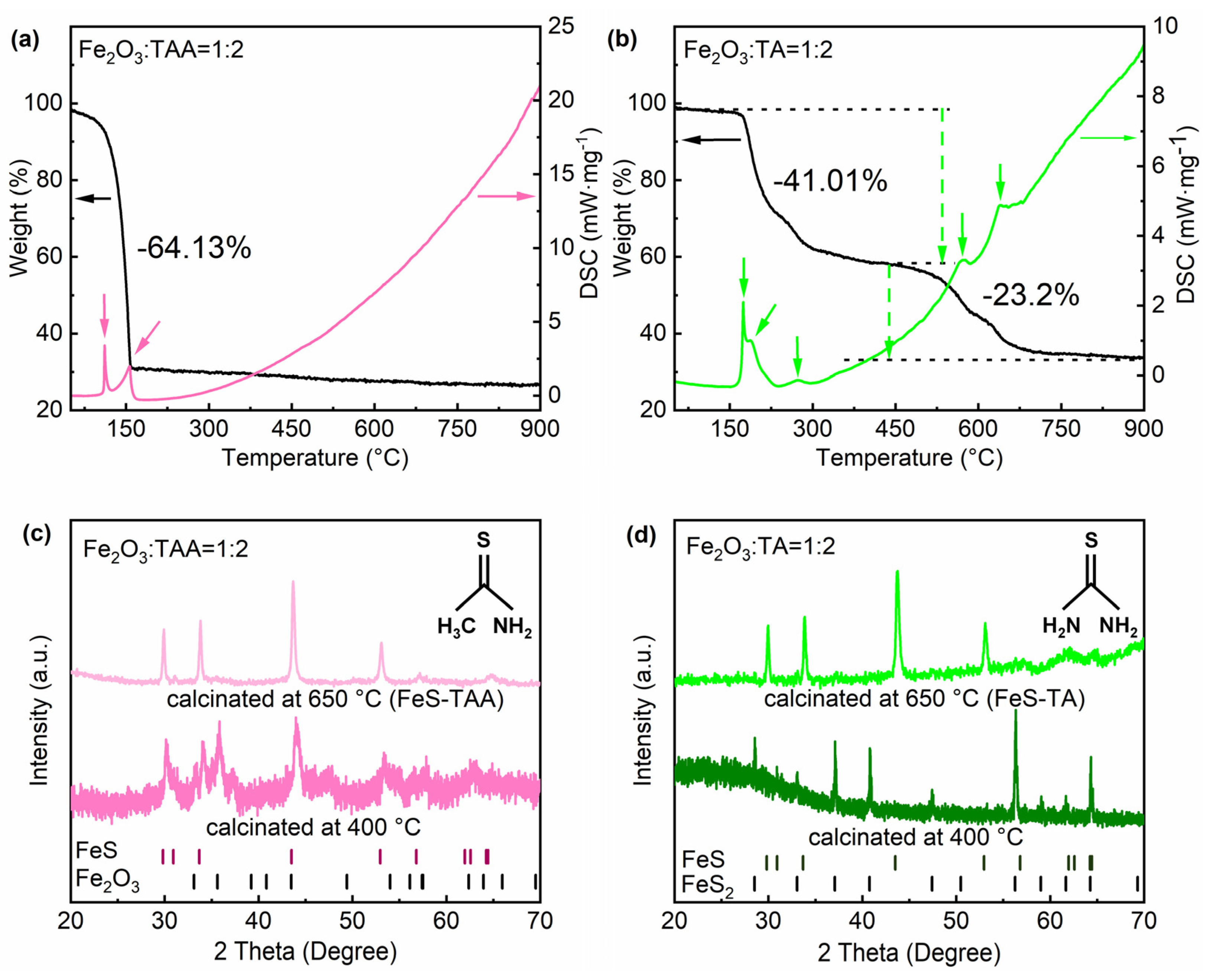
Figure 1. Analysis of the reaction pathways of F2O3 with different molecules. (a) and (b) show thermal analyses on the mixtures of Fe2O3 with TAA and TA, respectively, the pink and green straight arrows correspond to the phase transition positions, the green dashed arrow corresponds to the quality loss. (c,d) are XRD patterns of products after the calcinations of the corresponding mixtures. Molecular structures of TAA and TA are also inset in (c,d).
Energies 18 04623 g001
Figure 2. The XRD patterns of surface products of crucibles for the reactions between Fe2O3 and TA at 400 °C (a). The Raman spectra of FeS-TA and FeS-TAA (b) and the magnification of their low-shift region (c). The pink curve corresponds to the FeS-TAA sample, The green curve corresponds to the FeS-TA sample.
Energies 18 04623 g002
Figure 3. Microstructure characterizations of FeS-TAA and FeS-TA. The SEM images of two samples, respectively (a) and (b); the TEM images with lattice fringes inset (c) and (d); (e) and (f) are EDS spectra of FeS-TAA and FeS-TA, respectively.
Energies 18 04623 g003
Figure 4. XPS spectra of FeS-TAA and FeS-TA: Fe 2p region (a), S 2p region (b), C 1s region (c), and N 1s region (d). The N 1s signals of FeS-TAA are enlarged by five times for comparison.
Energies 18 04623 g004
Figure 5. Electrochemical performance of FeS-TAA and FeS-TA: Cycling performance of two samples (a); galvanostatic charge/discharge profiles of FeS-TAA (b) and FeS-TA (c); the differential capacity analysis (dQ/dV) as a function of voltage for FeS-TAA (d) and FeS-TA (e); the cyclic voltammetry of FeS-TAA (f) and FeS-TA (g); EIS spectra of two samples with the equivalent circuit inset (h), and in the low frequency region plots the relationship between the square root of frequency (ω−1/2) and Z’ (i). The pink corresponds to FeS-TAA sample, the green corresponds to FeS-TA sample, the arrow represents the Y-axis corresponding to the curve.
Energies 18 04623 g005
Figure 6. The discharge (lithiation) schematic illustration of FeS (a), ex situ XRD patterns with FeS-TAA (b), and FeS-TA (c). The peaks at the # position correspond to the peaks of the Cu foil, with I, II, and III representing the three stages of the discharge process. The blue dashed lines are used to delineate the intervals.
Energies 18 04623 g006
Figure 7. XRD patterns on products from reactions of TAA with NiO (a) and Co3O4 (b) at 400 °C. Dark blue is XRD samples synthesized from Co3O4 and TA, bright blue is XRD samples synthesized from Co3O4 and TA.
Energies 18 04623 g007
Table 1. Elemental analysis of FeS-TAA and FeS-TA by EDS spectra.
ElementFeS-TAAFeS-TA
at.%sigmaat.%sigma
C40.00.739.70.5
N0019.80.4
O4.20.23.70.1
S28.60.219.10.1
Fe27.20.217.60.1
Table 2. EIS fitted parameters with equivalent circuits.
SampleRsRctCPE1-TCPE1-PW1
FeS-TAA4.3724042.4232 × 10−50.773638.591
FeS-TA1.583265.72.3502 × 10−50.785143.249
Disclaimer/Publisher’s Note: The statements, opinions and data contained in all publications are solely those of the individual author(s) and contributor(s) and not of MDPI and/or the editor(s). MDPI and/or the editor(s) disclaim responsibility for any injury to people or property resulting from any ideas, methods, instructions or products referred to in the content.