Article

Biocomposite Fabrication from Enzymatically Treated Nanocellulosic Fibers and Recycled Polylactic Acid

1
Institute national de la recherche scientifique-Eau Terre Environnement, Québec, QC G1K 9A9, Canada
2
Department of Civil Engineering, Lassonde School of Engineering, York University, North York, Toronto, ON M3J 1P3, Canada
3
Laboratoire de Matériaux, Optimisation et Energie pour la Durabilité, Ecole Nationale d’Ingénieurs de Tunis, Université Tunis El Manar, BP 37, 1002, Le Bélvédère, Tunis 1002, Tunisia
4
Ozymes Inc., Trois-Rivières, Quebec, QC G9A 0A9, Canada
5
Département de génie civil et de génie des Eaux, Université Laval, Québec, QC G1V 0A6, Canada
6
Gesterra Inc., 330, rue J.-Aurèle-Roux, Victoriaville, QC G6T 0N5 Canada
*
Author to whom correspondence should be addressed.
Energies 2020, 13(4), 1003; https://doi.org/10.3390/en13041003
Submission received: 6 February 2020 / Revised: 19 February 2020 / Accepted: 20 February 2020 / Published: 24 February 2020
(This article belongs to the Special Issue Biomaterials and Biofuels: Small Environmental Footprint)

Abstract

:
Recycled polylactic acid (PLAr) was reinforced with treated nanocellulosic hemp fibers for biocomposite fabrication. Cellulosic fibers were extracted from hemp fibers chemically and treated enzymatically. Treated nanocellulosic fibers (NCF) were analyzed by Fourier-transform infrared spectroscopy, X-ray diffraction, and scanning electron microscopy. Biocomposite fabrication was done with PLAr and three concentrations of treated NCF (0.1%, 0.25%, and 1% (v/v)) and then studied for thermal stability and mechanical properties. Increased thermal stability was observed with increasing NCF concentrations. The highest value for Young’s modulus was for PLAr + 0.25% (v/v) NCF (250.28 ± 5.47 MPa), which was significantly increased compared to PLAr (p = 0.022). There was a significant decrease in the tensile stress at break point for PLAr + 0.25% (v/v) NCF and PLAr + 1% (v/v) NCF as compared to control (p = 0.006 and 0.002, respectively). No significant difference was observed between treatments for tensile stress at yield.

Graphical Abstract

1. Introduction

Polylactic acid (PLA) is one of the most commonly fabricated bioplastics in the last decade. Its mechanical properties (such as a high Young’s modulus), renewability, relatively low cost, and biodegradability make PLA ideal for its use as a biopolymer in the fabrication of biomaterials and decomposable packaging materials and in the automotive industry [1]. However, its use is limited due to its sensitivity to moisture and low impact resistance. Additionally, these bioplastics have several disadvantages, such as low crystallization ability, thermal degradation, longer residence time in the extruder and shredding process, which decreases its mechanical and physical properties after several recycling cycles [2]. This can be addressed by the addition of nanosized reinforcing fillers to the PLA matrix to form nanocomposite materials with improved physical, mechanical, and thermal properties [3]. Natural fibers can be an attractive candidate as reinforcement of biocomposites, as in addition to having favorable mechanical properties such as high tensile strength and stiffness, they are low-cost, nonabrasive, lightweight, nontoxic, and biodegradable.
Cellulosic fibers are one of the most abundant biopolymers found in nature and can be obtained from renewable and biodegradable resources such as wood, cotton, kenaf, flax, sisal, and hemp. These fibers can substitute commonly used reinforcements, such as glass and carbon fibers, for composite materials. A number of studies have reported the use of nanocellulosic fibers (NCFs) as reinforcement of biocomposite for enhanced mechanical proprieties, specifically Young’s modulus and tensile strength at the breakpoint [4].
Despite their advantages, the use of NCFs has been challenging in their application as reinforcement, due to their poor compatibility with biocomposite materials, poor interfacial adhesion between the NCFs and the matrix, susceptibility to thermal degradation, and low moisture resistance [5].
Studies have reported that modification of the fiber structure by removal of hemicellulose, pectin, and lignin can improve the interfacial adhesion between NCFs and matrix and consequently the mechanical properties of the fibers [6,7]. Pretreatment of the fibers can activate the hydroxyl groups present on the fiber surface, thus increasing the activity between the fibers and the matrix. Physical and chemical methods are the most commonly used pretreatment techniques for modification of the fiber surface structure. Physical methods such as thermotreatment, corona treatment, plasma treatment, etc. modify the surface structure and improve interfacial adhesion by increasing the mechanical bonding between the fibers and the matrix. Fiber structure modification by chemical pretreatment can cause changes in surface tension and polarity. Chemical pretreatment methods such as alkali treatment, acetylation, peroxide treatment etc. can activate hydroxyl groups or introduce new groups that can interact with the matrix, thus improving fiber strength, fitness, and fiber–matrix adhesion [8]. In recent years, enzymatic pretreatment has been garnering interest as an environment-friendly alternative for surface modification of fibers. Treatment of fibers with enzymes such as cellulase, xylanase, and pectinase was found to degrade lignin and improve mechanical properties of biocomposites fabricated with enzyme-treated fibers. This improvement is attributed to the degradation of the pectin layer between fiber bundles, which leads to improved fiber–matrix interface [9].
Enzymatic treatment of the extracted NCFs using laccase and cellulase has been also reported for the activation of hydroxyl groups, delignification, and size reduction. [10]. Laccase and cellulase—produced by Trametes versicolor and Trichoderma reesei, respectively—are known to degrade cellulosic fibers and activate their surface by removing hydroxyl groups [10,11].
In this study, two enzymes were used for the activation of the extracted NCFs. Laccase (a lignocellulolytic enzyme) was used for activation of hydroxyl groups present on the surface of NCFs. Cellulase was used for reducing the fiber size by the degradation of residual hemicellulose and lignin that cover the fibers and subsequently to increase the crystallinity of the NCFs. Thus, it can decrease the water sensitivity of the treated NCFs and enhance the mechanical properties of the fabricated biocomposite.

2. Materials and Methods

2.1. Chemicals

All the chemicals used were of high purity and purchased from Fisher Scientific (Ottawa, ON, Canada). Pulp and paper solid waste (PPSW) (Kruger Wayagamack Inc, Trois Rivieres, QC, Canada) was used as the source for extraction of cellulosic fibers. Recycled polylactic acid (PLAr) (recycled flakes) was obtained from NatureWorks (Minnetonka, MN, USA). Powdered blueberry pomace was provided by Dr. Diarra’s Lab (Agassiz, BC, Canada).

2.2. Enzyme Production

Laccase was produced by Trametes versicolor (ATCC-20869) using blueberry pomace as substrate. The fungus was cultured on potato dextrose agar (PDA) Petri plates at 30 ± 1 °C for 5 days, and this was used as inoculum for solid-state fermentation (SSF). About 15 g of blueberry pomace (BP) was taken per flask (moisture 75%, pH 5) and autoclaved at 121 ± 1 °C, 15 psi for 20 min. The substrate was inoculated with one Petri plate disc (diameter of Petri plate = 90 mm) of Tra. versicolor per flask. Cellulase was produced by Trichoderma reesei (NRRC-207F) using hemp fibers as substrate. Tri. reesei was cultured in PDB (pH = 4.8) and incubated at 28 ± 1 °C, 150 rpm for 3 days followed by growth on potato dextrose agar (PDA) at 30 ± 1 °C for 3 days. Spores were collected using sterile distilled water, and the spore suspension was counted using a hemocytometer and maintained at 4 ± 1 °C. About 20 g of hemp fibers (moisture 75%, pH 6.5) was dispensed in each flask and autoclaved at 121 ± 1 °C, 15 psi for 20 min. The substrate was inoculated with Tri. reesei at 107–108 spores per gram and incubated at 30 ± 1 °C for 15 days.

2.3. Enzyme Extraction

For laccase extraction, the SSF medium was thoroughly mixed with 50 mM sodium phosphate buffer (pH 6.5) at a ratio of 20:1 (v/v). This mixture was incubated at 30 ± 1 °C, 150 rpm for 1 h and then centrifuged at 9000× g for 30 min at 4 ± 1 °C [12].
For cellulase extraction, the SSF medium was mixed with distilled water containing 0.1 % (v/v) Tween-80 (25 mL per gram). This mixture was incubated at 30 ± 1 °C, 200 rpm for 30 min followed by centrifugation at 9000× g for 30 min, 4 ± 1 °C [13]. The supernatant was used for the estimation of laccase and cellulase.

2.4. Enzyme Estimation

Laccase activity was determined spectrophotometrically by oxidation of 2,2-azino bis (3-ethylbenzthiazoline-6-sulfonic acid) (ABTS) in 0.1M phosphate-citrate buffer at pH = 3.5 at 420 nm and 45 °C (ε420 = 36 mMol/cm). One unit of laccase activity was defined as the amount of enzyme required to oxidize 1 µmol of ABTS per min [14].
Cellulase activity was determined by Filter paper assay (FPase) using Whatman filter paper (Whatman no. 1. 0.25 mm pore size, 1.5 cm diameter) in 50 mM sodium citrate buffer (pH 4.8) at 50 ± 1 °C for 30 min. 3,5-dinitrosalicylic acid (DNS) method was used for spectrophotometric determination of reducing sugar concentration [15]. One international unit of Fpase activity was the amount of enzyme that forms 1 mmol of glucose [16].
Enzyme activity (or unit, EU) per ml was calculated using the following Equation (1):
EU   per   mL = d A d T × Total   volme Volume   of   enzyme × 1 ε × 1 d
where dA/dT is the rate of change of absorbance, ε is the molar extinction coefficient (M−1 cm−1), d is the path length (cm).

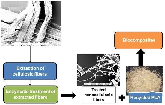
2.5. Extraction of Cellulosic Fibers

Hemp fibers were cut into small fragments of 2–3 cm and mixed with 10% (v/w) of NaOH solution for 24 h. This step was performed to remove lignin, hemicelluloses, and pectin from hemp fibers. The fibers were then washed several times with distilled water until neutral pH was achieved. The washed samples were then dried in an oven at 60 ± 1 °C for 24 h and used for the production of nanofibers.
After NaOH treatment, the nanofibers were extracted by acid hydrolysis using a 60% (v/v) solution of sulfuric acid (H2SO4) in a water bath at 80 °C for 5 h at 75 rpm. The sample was then washed several times to neutralize the fibers solution [7].

2.6. Enzymatic Treatment

Lignocellulolytic enzymes were used for activation of the extracted cellulosic fibers. This experiment was carried out using 1 g of extracted cellulosic fibers with cellulase (15 U/g) and laccase (20 U/g) in a working volume of 200 mL.
The mixture was incubated at 30 ± 1 °C, 100 rpm for 7 days [17]. The pH was maintained at 4.8 during the incubation period. After the enzymatic treatment, the sample was centrifuged at 7000× g for 15 min at room temperature. The supernatant, which is a colloidal suspension of nanocellulose, was collected for sugar analysis. (Tabka and Herpoël-Gimbert) [17]. 0.1% (v/v) of cocoamidopropyl hydroxysultaïne (CAS)—a biosurfactant—was added to the supernatant and this was sonicated for 3 min with output power at 750 W (20 kHz) to prevent agglomeration of the nanofibers. Following this, the particle size distribution analysis of cellulosic suspension was performed [18].

2.7. Fabrication of Biocomposite

PLAr films were prepared by casting the PLAr pellets in chloroform solution at a concentration of 5.0% (v/v). The PLAr/chloroform mixtures were continuously stirred at room temperature until the pellets were totally dissolved.
Biocomposite films were obtained by adding the enzyme-treated cellulosic fibers to the PLAr/chloroform mixtures at different concentrations (0.10%, 0.25%, and 1% v/v), followed by overnight stirring. The mixture was then poured into Petri dishes, and chloroform was allowed to evaporate at ambient temperature and pressure under laminar airflow. The resulting films were oven-dried for 24 h at room temperature and subjected to mechanical characterization [19].

2.8. Analysis of the Modified Fibers and the Formulated Biocomposites

Particle size was measured for hemp fibers before and after chemical extraction and enzymatic treatment using a Zetasizer Nano S90 apparatus (Malvern Instruments, Malvern, UK). Scanning electron microscopy (SEM, Carl Zeiss EVO 50) was used to study the size and the morphology of the nanofibers extracted from hemp fibers before and after chemical extraction and enzymatic treatment, respectively.
Fourier-transform infrared spectroscopy (FTIR, NICOLET IS5O) was performed to study the change in the functional groups of extracted hemp fibers at different stages of treatment. For this purpose, the surface area of the peaks for the functional groups was calculated before and after each treatment.
X-ray diffraction (XRD) was performed using a Siemens D5000 Cu lamp (ƛ = 0.154059 nm) to characterize the changes in the molecular structure of hemp fibers before and after chemical extraction and enzymatic treatment, respectively.
Thermogravimetric analysis (TGA) was performed with TA Instruments Q5000IR at 50 °C to 700 °C, 10 °C per min (N2) to evaluate the thermal effect of NCFs on manufactured biocomposite compared to PLAr.
Mechanical characterization of the novel biocomposite was carried out to evaluate the effect of cellulosic nanofibers on the properties of the biocomposite. The analysis was performed according to ASTM D256 for the tensile strength test using Instron 5565, 5 mm/min, samples type V. All the mechanical tests were carried out in five replicates, and the average and standard deviation were calculated. Analysis of variance (ANOVA) was performed, considering p-value < 0.05 as significant. A post hoc Newman-Keuls analysis was used to compare the difference in the mechanical properties between the formulations made from PLAr and varying concentrations of treated NCFs.

3. Results and Discussion

3.1. Enzyme Production by Tra. versicolor and Tri. reesei

The enzyme activities were measured after the fermentation, and the highest laccase activity was obtained after 10 days of fermentation (34.02 ± 1.95 U/gds) using blueberry pomace, which is comparable to the results reported by Chaali and Lecka [20]. Trichoderma reesei was used to produce cellulase, which was significantly higher after 12 days of fermentation (15 ± 3.1 U/gds) and comparable to the results reported by Lee and Koo [21].

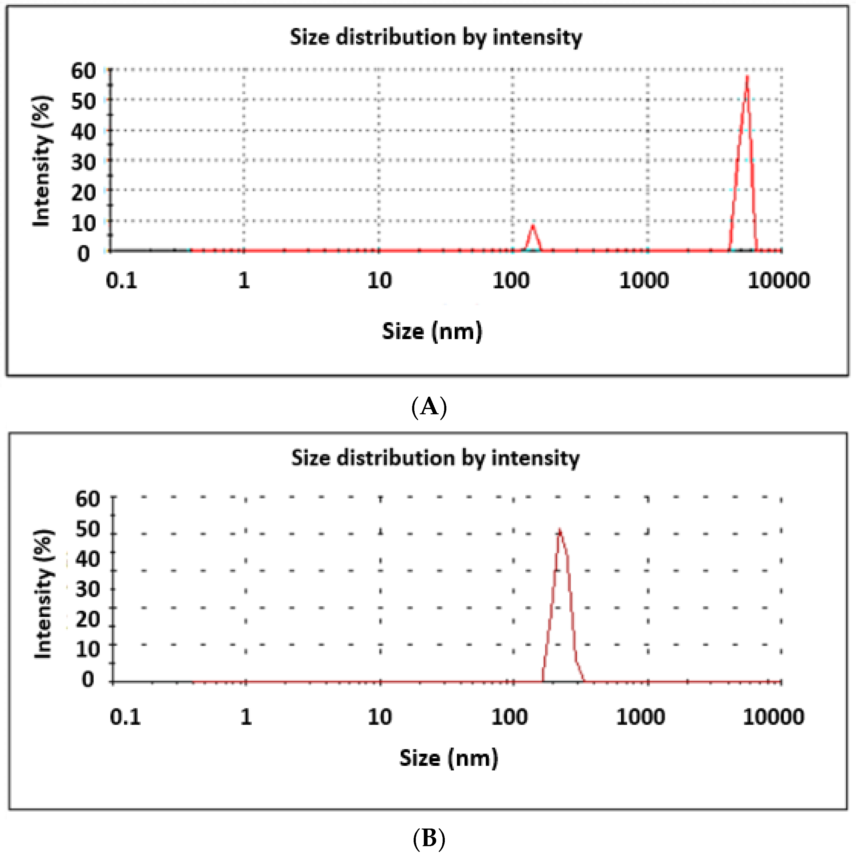
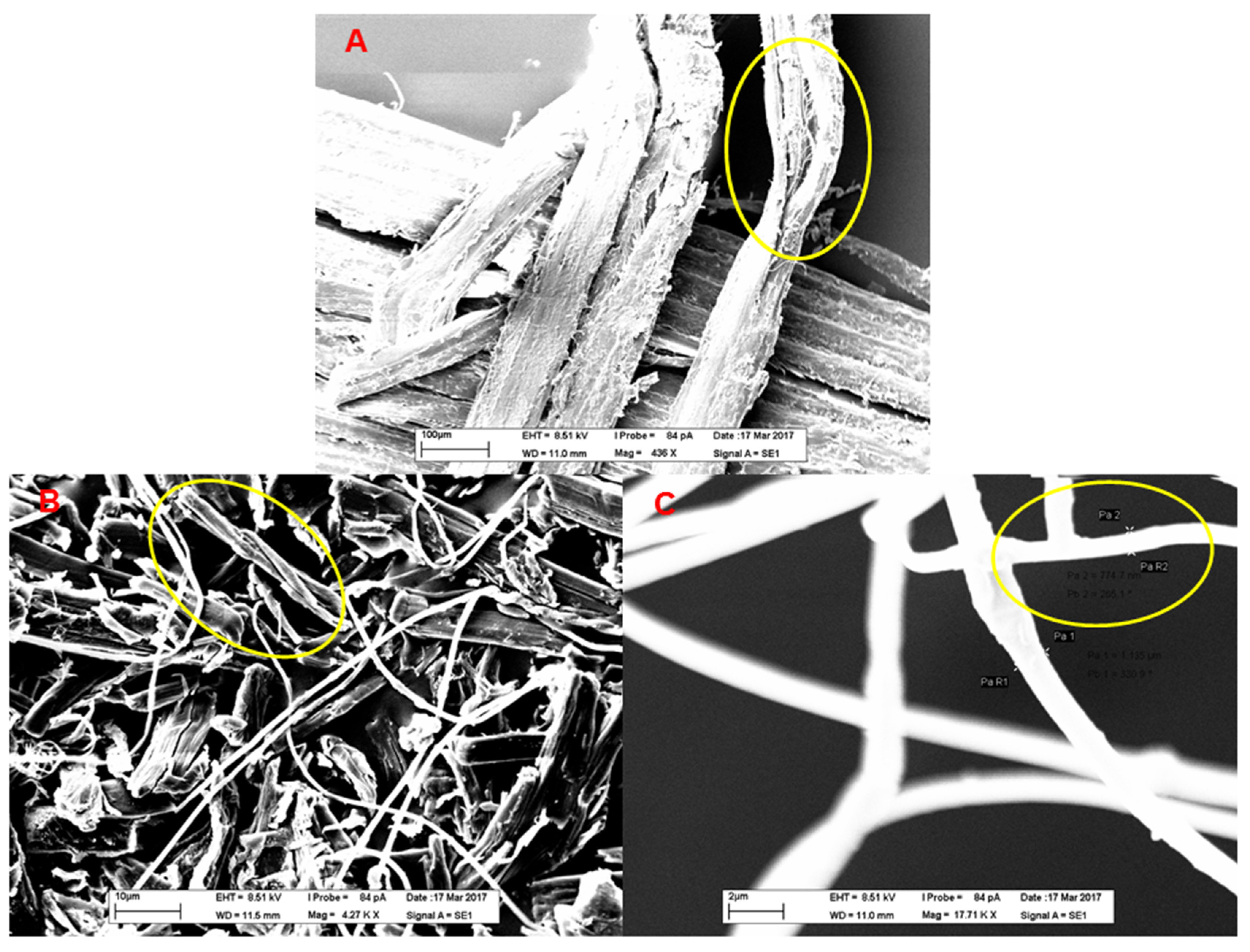
3.2. Effect of Chemical Extraction and Enzymatic Treatment of Fibers

The volumetric mean diameter was studied to determine the effect of the enzymatic treatment on the size reduction of the extracted NCFs. After chemical extraction and enzymatic treatment, the fiber size was reduced from 5024.5 ± 102.5 nm to 318.3 ± 89.09 nm with a polydispersity index (PDI) of 0.654 as shown in Figure 1, thus demonstrating the effect of the enzymatic treatment, specifically cellulase, on the size reduction of the fibers. This is due to the attack by the enzymes on the amorphous region of the fibers during treatment time by removing the coating materials such as hemicellulose, pectin, and lignin, which are present in high percentage in the cellulosic materials, and consequently, increasing the crystallinity index [22]. Figure 2 shows the SEM image of the untreated and treated hemp fibers before treatment (Figure 2A); after chemical extraction (Figure 2B); and enzymatic activation (Figure 2C).

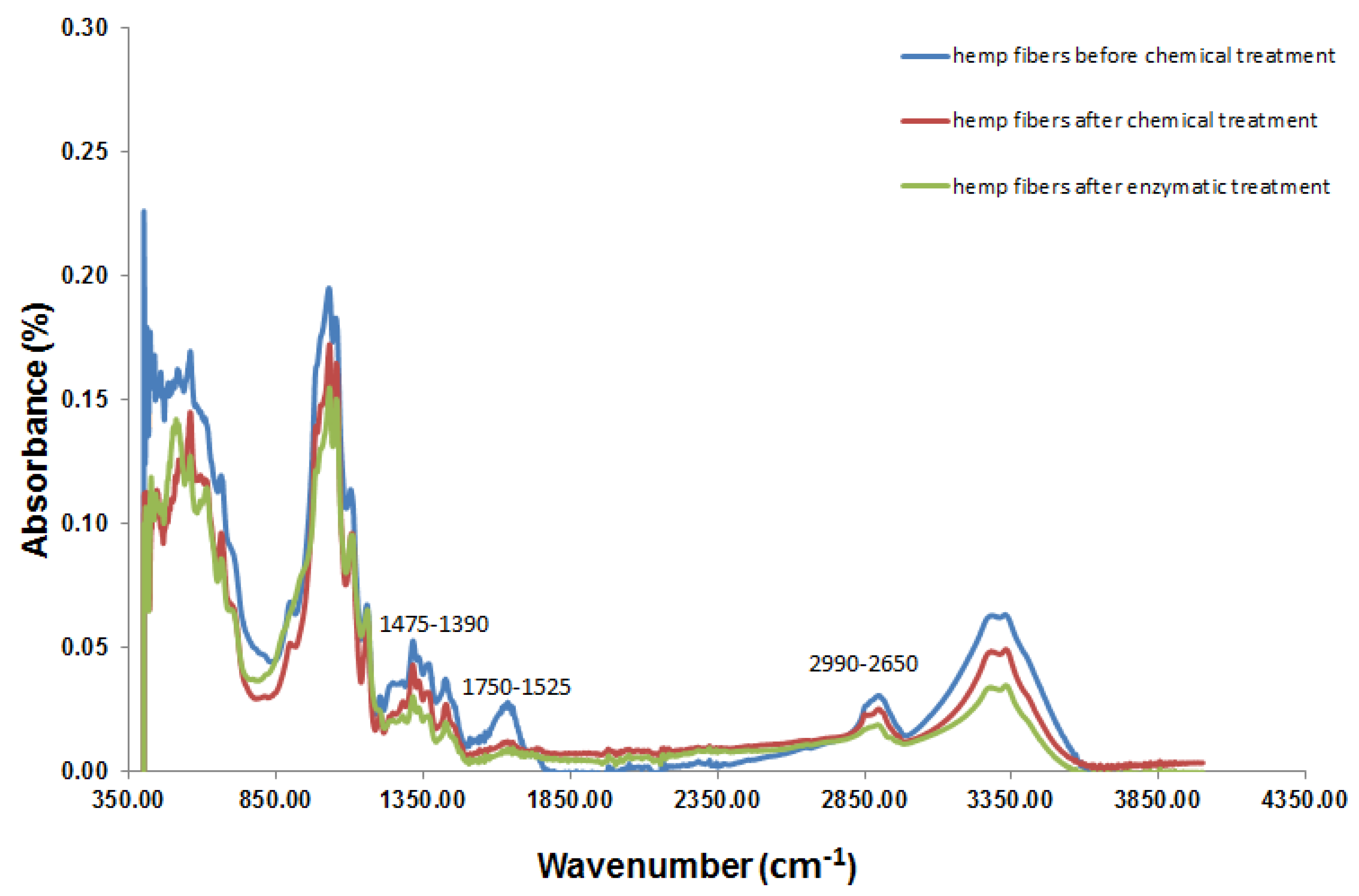
3.3. Fourier-Transform Infrared Spectroscopy (FTIR) Analysis

The FTIR spectra before and after chemical extraction and enzyme treatment are presented in Figure 3. The peaks at 1475–1390 cm−1, 1750–1525 cm−1, and 3640–3000 cm−1 were reduced, which indicates the total degradation of the lignin, hemicellulose, and cellulose respectively, after enzymatic treatment [23,24,25]. In addition, the treatment of hemp fibers with sulfuric acid (H2SO4) released around 34% (w/w) of cellulose, indicating the degradation of hemicellulose and lignin (Table 1), thus confirming the efficacy of chemical extraction of cellulosic fibers.
Enzymatic treatment with laccase and cellulase resulted in the removal of the residual hemicellulose and lignin as well as reduction of hydroxyl groups present on the surface of the nanofibers, indicating nanofiber activation (Table 1). Also, the enzymatic treatment was effective for the improvement of reactive sites between NCFs and PLAr matrix, which was confirmed by the increase in the hydrogen bonding. This was evident from the right shift in vibration frequency of carboxyl group and hydroxyl group from 1750 cm−1 to 1761 cm−1 and 3330 cm−1 to 3340 cm−1, respectively. This indicated the occurrence of hydrogen bonding between C–O and –OH.
The combined chemical and enzymatic treatments were efficient for cellulose extraction and activation of the surface of the NCFs. Further, the hydrophobicity of the NCFs increased after enzymatic treatment, as explained by Gardner et al., which could have a significant effect on the adhesion of the NCFs to PLAr [11].

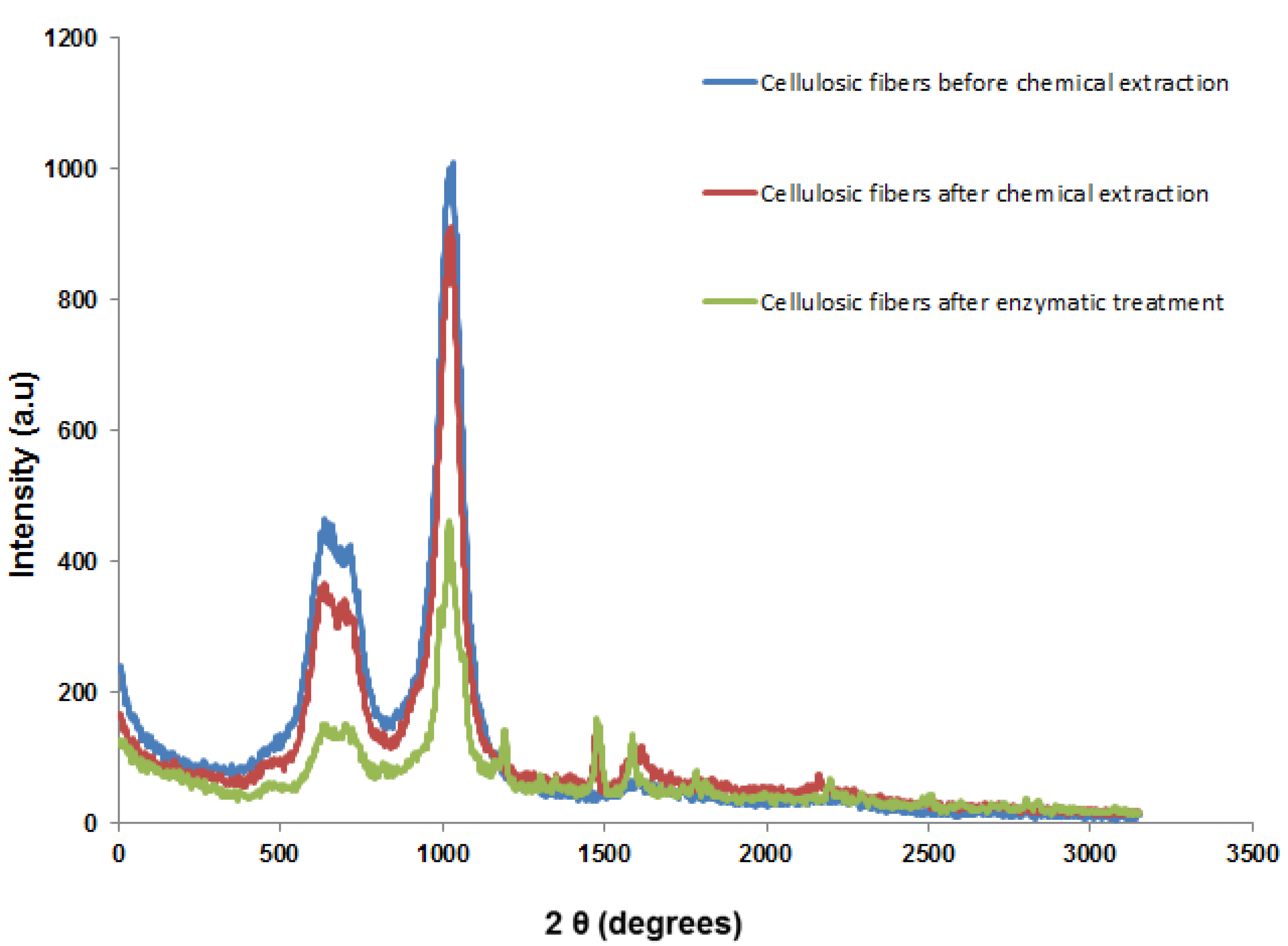
3.4. X-ray Diffraction (XRD) Analysis

The structural change in the cellulosic fibers was analyzed by XRD before and after chemical extraction and enzymatic activation, respectively (Figure 4). A remarkable change in the crystallinity peaks was observed which correspond to different diffraction planes for the cellulosic fibers before and after enzymatic treatment, respectively. In addition, the crystalline regions are presented at higher percentage after the chemical extraction which indicates the effect of chemical extraction on the cellulosic fibers as compared to raw fibers before chemical extraction. Likewise, after the enzymatic activation, the crystallinity became significantly higher compared to the fibers before enzymatic treatment. This affected the adsorption of the cellulosic fibers by increasing the hydrophobicity and prevented their swelling, which can significantly improve the mechanical properties of the fabricated biocomposite by adding crystalline NCFs at a different percentage [27].

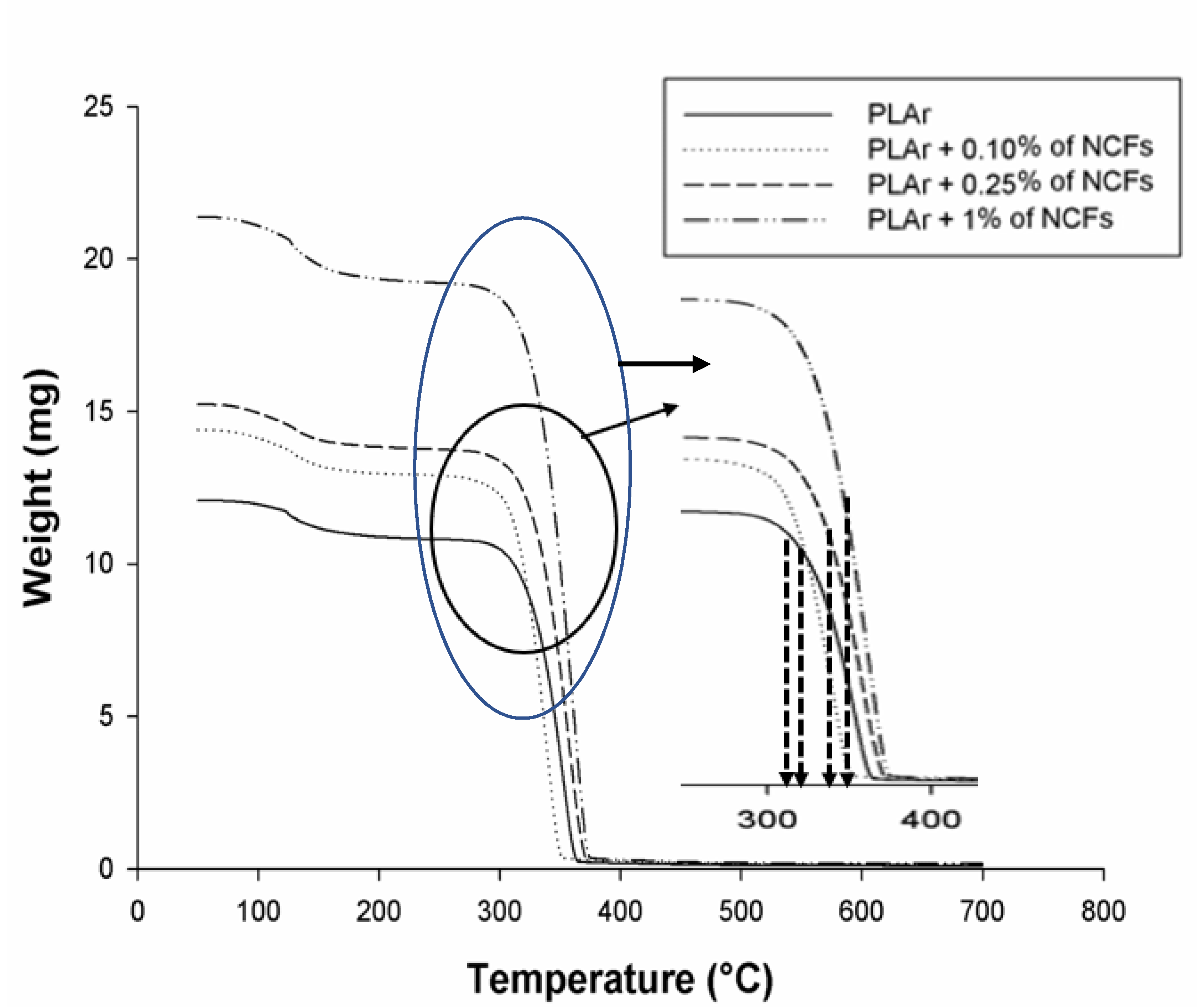
3.5. Thermogravimetric (TGA) Analysis

The thermal characterization of the PLAr (control) and the fabricated biocomposite (PLAr/NCFs) was done using TGA analysis. As presented in Figure 5, the onset of weight loss was at 311 °C, 317 °C, 331 °C, and 349 °C for PLAr, PLAr/0.1% of NCFs, PLAr/0.25% of NCFs, and PLAr/1% of NCFs, respectively. The more the relative initial weight of the sample, the more the inflection point was observed (not shown here) and thus the more was the onset temperature. Before the onset temperature, the weight of the sample remained almost the same. This range, which is also known as the initial decomposition temperature, gives an idea of the material disintegration and a measure of its thermal stability. From Figure 5, the case which showed the highest onset temperature is PLAr/1% of NCFs. This further signifies the significance of thermal stability on the overall material composite with the intrusion of nanocomposite fibers.
At 698 °C, 100% weight loss was observed for all the formulations. Therefore, the incorporation of NCFs was shown to cause a significant increase in the thermal stability of recycled PLA. The thermal stability of the fabricated biocomposite in this study was found to be 16.33% higher than the previously reported PLA bio-nanocomposite reinforced with banana fibers, short coir fibers, and nanohydroxyapatit [28,29]. The increased thermal stability due to the uniform dispersion of the NCFs on the matrix of PLAr was comparable to the higher concentration of crystalline cellulose in the treated NCFs as reported by Haafiz and Hassan [30].

3.6. Mechanical Properties of the Fabricated Biocomposites

Mechanical properties such as Young’s Modulus, tensile stress at yield (both for biocomposite stiffness), and tensile stress at breakpoint (for biocomposite malleability) were studied for the biocomposites prepared, the results for which are shown in Table 2.
The stiffness of the biocomposites was determined by Young’s modulus (Y), and a higher value of Y indicated more stiffness. The highest value of Y was obtained with PLAr + 0.25% NCFs (250.28 ± 5.47) (Table 2). The ANOVA analysis indicated that there was a significant difference in the value of Young’s modulus among the tested materials (p = 0.0049). The post hoc analysis showed that Young’s modulus for PLAr and PLAr + 0.1% NCFs were statistically comparable (p = 0.538). However, there was a significant positive effect of higher NCF concentrations on the Young’s modulus (p = 0.022 for PLAr + 0.25% NCFs, and p = 0.012 for PLAr + 0.1% NCFs).
The improvement of the Young’s Modulus of PLAr + 0.25% NCFs is an indicator of increased interfacial adhesion between the PLA matrix and NCF. This could be attributed to enzymatic pretreatment, which could improve the adhesion between fibers and PLAr matrix, possibly due to the activation of hydroxyl groups of fibers [28,31]. Previous studies have reported an improvement of Young’s Modulus, and therefore the strength of the PLA biocomposite with increasing concentrations of cellulosic fibers [32,33].
The tensile stress at breakpoint decreased significantly after the association with NCFs for all the formulations of PLAr + NCFs as compared to PLAr (Table 2). The value obtained for PLAr + 0.25% NCF and PLAr + 0.1% NCF were significantly lesser (p = 0.002 and p = 0.006 respectively), as compared to the values for the control and PLAr + 0.1% NCF, which were not significantly different statistically (p = 0.197). An increase in the percentage of treated NFCs significantly reduced the tensile stress at breakpoint (%), which could be due to the brittleness of the fabricated biocomposites as reported by [34]. In the current study, surface activation of NCFs led to the increased crystallinity of the fibers, and therefore that of the PLA biocomposite. A study by Suryanegara et al. reported that the increased brittleness of the cellulose-reinforced PLA biocomposite was due to an increase in PLA crystallinity, which in turn increased the Young’s modulus but decreased the tensile stress at break [32]. The increased brittleness could also be due to possible agglomerations formed by the higher concentrations of NCF, which decrease the tensile stress transfer between the PLA matrix and the NCF [35]. No significant difference was observed for the tensile stress at yield between the control and PLAr with different fiber concentrations (p = 0.36).

4. Conclusions

Chemical extraction followed by enzymatic treatment of cellulosic fibers were investigated using sulfuric acid and lignocellulolytic enzymes (laccase and cellulase), respectively, for improving the mechanical properties of PLAr. The interfacial adhesion between PLAr and treated NCFs was improved by the addition of nanocellulosic fibers, which affected the stiffening, malleability, and hardness of the fabricated biocomposite as confirmed by the mechanical analysis. NCFs were added to PLAr at different concentrations (0.10%, 0.25%, and 1% v/v). The formulation of PLAr + 0.25% NCF showed improved stiffness as observed by the higher value of the Young’s modulus as compared to control (p-value = 0.0049). Further, the study showed that the proposed enzymatic treatment was effective for the improvement of reactive sites between NCFs and PLAr matrix, which was confirmed by the increase in the hydrogen bonding (FTIR analysis) and the crystallinity (XRD analysis) of the treated fibers. Therefore, the use of this fabricated biocomposite can be further explored in various industries, such as packaging and automobile. The use of PLAr with natural fillers in the form of enzyme-treated nanocellulosic fibers enhanced the natural strength of the low-density plastic presenting an eco-friendly composite for its usage in automotive industry. These treated cellulosic fibers can also be used as a reinforcement material that can be correlated with lower density and higher strength properties in the packaging material. Because of high surface area, it can also strongly interact with the matrix as compared to the conventional micro reinforcement.

Author Contributions

All the authors contributed in formulating the hypothesis, problem and objectives as well as writing, reviewing and editing the manuscript. All authors have read and agreed to the published version of the manuscript.

Funding

This research was funded by the Natural Sciences and Engineering Research Council of Canada (Discovery Grant 355254 and NSERC CRD Grant).

Acknowledgments

This work was supported by the Natural Sciences and Engineering Research Council of Canada (Discovery Grant 355254 and NSERC CRD Grant), and CRIBIQ. We would like to thank Aman Kulshrestha from NatureWorks for providing PLAr samples. Likewise, the support of Ozymes Inc. is equally appreciated for valuable comments during the experimental planning from an industrial perspective. We also thank Fonds de recherche du Québec-Nature et technologies (FRQNT) for the financial assistance.

Conflicts of Interest

The authors declare no conflict of interest.

References

  1. Kashirina, A.; Yao, Y.; Liu, Y.; Leng, J. Biopolymers as bone substitutes: A review. Biomater. Sci. 2019, 7, 3961–3983. [Google Scholar] [CrossRef] [PubMed]
  2. Pillin, I.; Montrelay, N.; Bourmaud, A.; Grohens, Y. Effect of thermo-mechanical cycles on the physico-chemical properties of poly (lactic acid). Polym. Degrad. Stab. 2008, 93, 321–328. [Google Scholar] [CrossRef]
  3. Raquez, J.-M.; Habibi, Y.; Murariu, M.; Dubois, P. Polylactide (PLA)-based nanocomposites. Prog. Polym. Sci. 2013, 38, 1504–1542. [Google Scholar] [CrossRef]
  4. Faruk, O.; Bledzki, A.K.; Fink, H.-P.; Sain, M. Biocomposites reinforced with natural fibers: 2000–2010. Prog. Polym. Sci. 2012, 37, 1552–1596. [Google Scholar] [CrossRef]
  5. Abraham, E.; Deepa, B.; Pothan, L.A.; Jacob, M.; Thomas, S.; Cvelbar, U.; Anandjiwala, R. Extraction of nanocellulose fibrils from lignocellulosic fibres: A novel approach. Carbohydr. Polym. 2011, 86, 1468–1475. [Google Scholar] [CrossRef]
  6. Bledzki, A.; Reihmane, S.; Gassan, J. Properties and modification methods for vegetable fibers for natural fiber composites. J. Appl. Polym. Sci. 1996, 59, 1329–1336. [Google Scholar] [CrossRef]
  7. Yang, J.; Ching, Y.C.; Chuah, C.H. Applications of Lignocellulosic Fibers and Lignin in Bioplastics: A Review. Polymers 2019, 11, 751. [Google Scholar] [CrossRef] [Green Version]
  8. Kalia, S.; Kaith, B.; Kaur, I. Pretreatments of natural fibers and their application as reinforcing material in polymer composites—A review. Polym. Eng. Sci. 2009, 49, 1253–1272. [Google Scholar] [CrossRef]
  9. Karaduman, Y.; Gokcan, D.; Onal, L. Effect of enzymatic pretreatment on the mechanical properties of jute fiber-reinforced polyester composites. J. Compos. Mater. 2013, 47, 1293–1302. [Google Scholar] [CrossRef]
  10. Su, Y.; Xian, H.; Shi, S.; Zhang, C.; Manik, S.M.N.; Mao, J.; Liu, H. Biodegradation of lignin and nicotine with white rot fungi for the delignification and detoxification of tobacco stalk. BMC Biotechnol. 2016, 16. [Google Scholar] [CrossRef] [Green Version]
  11. Gardner, D.J.; Oporto, G.S.; Mills, R.; Samir, M.A.S.A. Adhesion and surface issues in cellulose and nanocellulose. J. Adhes. Sci. Technol. 2008, 22, 545–567. [Google Scholar] [CrossRef] [Green Version]
  12. Lonappan, L.; Rouissi, T.; Laadila, M.A.; Brar, S.K.; Hernández-Galán, L.; Verma, M.; Surampalli, R.Y. Agro-industrial produced laccase for degradation of diclofenac and identification of transformation products. ACS Sustain. Chem. Eng. 2017, 5, 5772–5781. [Google Scholar] [CrossRef]
  13. Awafo, V.A.; Chahal, D.S.; Simpson, B.K.; Lê, G.B. Production of cellulase systems by selected mutants of Trichoderma reesei in solid-state fermentation and their hydrolytic potentials. Appl. Biochem. Biotechnol. 1996, 57, 461–470. [Google Scholar] [CrossRef]
  14. Gassara, F.; Brar, S.K.; Tyagi, R.D.; Verma, M.; Surampalli, R.Y. Screening of agro-industrial wastes to produce ligninolytic enzymes by Phanerochaete chrysosporium. Biochem. Eng. J. 2010, 49, 388–394. [Google Scholar] [CrossRef]
  15. Ghose, T. Measurement of cellulase activities. Pure Appl. Chem. 1987, 59, 257–268. [Google Scholar] [CrossRef]
  16. Jung, Y.R.; Park, J.M.; Heo, S.Y.; Hong, W.K.; Lee, S.M.; Oh, B.R.; Park, S.M.; Seo, J.W.; Kim, C.H. Cellulolytic enzymes produced by a newly isolated soil fungus Penicillium sp. TG2 with potential for use in cellulosic ethanol production. Renew. Energy 2015, 76, 66–71. [Google Scholar] [CrossRef]
  17. Tabka, M.; Herpoël-Gimbert, I.; Monod, F.; Asther, M.; Sigoillot, J. Enzymatic saccharification of wheat straw for bioethanol production by a combined cellulase xylanase and feruloyl esterase treatment. Enzym. Microb. Technol. 2006, 39, 897–902. [Google Scholar] [CrossRef]
  18. Shamsabadi, M.A.; Behzad, T.; Bagheri, R. Optimization of acid hydrolysis conditions to improve cellulose nanofibers extraction from wheat straw. Fibers Polym. 2015, 16, 579–584. [Google Scholar] [CrossRef]
  19. Mwaikambo, L.Y.; Ansell, M.P. Chemical modification of hemp, sisal, jute, and kapok fibers by alkalization. J. Appl. Polym. Sci. 2002, 84, 2222–2234. [Google Scholar] [CrossRef]
  20. Chaali, M.; Lecka, J.; Suresh, G.; Salem, M.; Brar, S.K.; Hernandez-Galan, L.; Sevigny, J.; Avalos-Ramirez, A. Supplement comprising of laccase and citric acid as an alternative for antibiotics-in vitro triggers of melanin production. Eng. Life Sci. 2018, 18, 359–367. [Google Scholar] [CrossRef] [Green Version]
  21. Lee, S.-M.; Koo, Y.-M. Pilot-Scale Production of Cellulase Using Trichoderma reesei Rut C-30 Fed-Batch Mode. J. Microbiol. Biotechnol. 2001, 11, 229–233. [Google Scholar]
  22. Cao, Y.; Tan, H. Effects of cellulase on the modification of cellulose. Carbohydr. Res. 2002, 337, 1291–1296. [Google Scholar] [CrossRef]
  23. Yan, P.; Xu, Z.; Zhang, C.; Liu, X.; Xu, W.; Zhang, Z.C. Fractionation of lignin from eucalyptus bark using amine-sulfonate functionalized ionic liquids. Green Chem. 2015, 17, 4913–4920. [Google Scholar] [CrossRef]
  24. Tejado, A.; Peña, C.; Labidi, J.; Echeverria, J.M.; Mondragon, I. Physico-chemical characterization of lignins from different sources for use in phenol–formaldehyde resin synthesis. Bioresour. Technol. 2007, 98, 1655–1663. [Google Scholar] [CrossRef] [PubMed]
  25. Lionetto, F.; Del Sole, R.; Cannoletta, D.; Vasapollo, G.; Maffezzoli, A. Monitoring Wood Degradation during Weathering by Cellulose Crystallinity. Materials 2012, 5, 1910–1922. [Google Scholar] [CrossRef] [Green Version]
  26. Morán, J.I.; Alvarez, V.A.; Cyras, V.P.; Vázquez, A. Extraction of cellulose and preparation of nanocellulose from sisal fibers. Cellulose 2008, 15, 149–159. [Google Scholar] [CrossRef]
  27. Kalia, S.; Thakur, K.; Celli, A.; Kiechel, M.A.; Schauer, C.L. Surface modification of plant fibers using environment friendly methods for their application in polymer composites, textile industry and antimicrobial activities: A review. J. Environ. Chem. Eng. 2013, 1, 97–112. [Google Scholar] [CrossRef]
  28. Sajna, V.P.; Mohanty, S.; Nayak, S.K. A study on thermal degradation kinetics and flammability properties of poly (lactic acid)/banana fiber/nanoclay hybrid bionanocomposites. Polym. Compos. 2015, 38, 2067–2079. [Google Scholar] [CrossRef]
  29. Sun, Z.; Zhang, L.; Liang, D.; Xiao, W.; Lin, J. Mechanical and Thermal Properties of PLA Biocomposites Reinforced by Coir Fibers. Int. J. Polym. Sci. 2017, 2017, 2178329. [Google Scholar] [CrossRef]
  30. Haafiz, M.M.; Hassan, A.; Khalil, H.A.; Khan, I.; Inuwa, I.M.; Islam, M.S.; Hossain, M.S.; Syakir, M.I.; Fazita, M.N. Bionanocomposite based on cellulose nanowhisker from oil palm biomass-filled poly (lactic acid). Polym. Test. 2015, 48, 133–139. [Google Scholar] [CrossRef]
  31. Laadila, M.A.; Hegde, K.; Rouissi, T.; Brar, S.K.; Galvez, R.; Sorelli, L.; Cheikh, R.B.; Paiva, M.; Abokitse, K. Green synthesis of novel biocomposites from treated cellulosic fibers and recycled bio-plastic polylactic acid. J. Clean. Prod. 2017, 164, 575–586. [Google Scholar] [CrossRef]
  32. Suryanegara, L.; Nakagaito, A.N.; Yano, H. The effect of crystallization of PLA on the thermal and mechanical properties of microfibrillated cellulose-reinforced PLA composites. Compos. Sci. Technol. 2009, 69, 1187–1192. [Google Scholar] [CrossRef]
  33. Iwatake, A.; Nogi, M.; Yano, H. Cellulose nanofiber-reinforced polylactic acid. Compos. Sci. Technol. 2008, 68, 2103–2106. [Google Scholar] [CrossRef]
  34. Kasa, S.N.; Omar, M.F.; Abdullah, M.M.A.B.; Ismail, I.N.; Ting, S.S.; VAC, S.C.; VIZUREANU, P. Effect of Unmodified and Modified Nanocrystalline Cellulose Reinforced Polylactic Acid (PLA) Polymer Prepared by Solvent Casting Method. Mater. Plast. 2017, 54, 91. [Google Scholar]
  35. Robles, E.; Urruzola, I.; Labidi, J.; Serrano, L. Surface-modified nano-cellulose as reinforcement in poly (lactic acid) to conform new composites. Ind. Crop. Prod. 2015, 71, 44–53. [Google Scholar] [CrossRef]
Figure 1. Size distribution by volume (nm) after chemical extraction (A) and enzymatic treatment (B) of cellulosic fibers from hemp fibers.
Energies 13 01003 g001
Figure 2. SEM micrograph of untreated and treated hemp fibers before treatment (A); after chemical extraction (B); and enzymatic activation (C).
Energies 13 01003 g002
Figure 3. FTIR spectra of the cellulosic fibers before and after enzymatic treatment.
Energies 13 01003 g003
Figure 4. XRD spectra of the cellulosic fibers before and after enzymatic treatment.
Energies 13 01003 g004
Figure 5. TGA curves for all the biocomposite formulations.
Energies 13 01003 g005
Table 1. Percentage of cellulose, hemicellulose, and lignin after chemical and enzymatic treatment.
Wave
Number (cm1)
Functional GroupsPercentage after
Chemical Extraction
Percentage after
Enzymatic Treatment
3640–3000O–H, Cellulose [26]34.7226.29
1750–1525C=O, Hemicellulose [26]65.7518.23
1475–1390C=C, Lignin [26]71.7454.35
Table 2. Mechanical properties for all the composite formulation.
FormulationYoung’s Modulus
(MPa)
Tensile Strength at Break
(% Maximum Load: 90) (MPa)
Tensile Stress at Yield (Zero Slope) (MPa)
PLAr224.82 ± 21.60512.07 ± 37.5519.78 ± 0.06
PLAr + 0.1% NCFs227.84 ± 28.62413.11 ± 73.8618.03 ± 1.88
PLAr + 0.25% NCFs250.29 ± 5.48323.76 ± 57.4120.08 ± 0.81
PLAr + 1% NCFs248.02 ± 8.18229.58 ± 65.9118.18 ± 0.88
PLAr: recycled polylactic acid.

