Abstract
The paper presents research on the electromagnetic hazards related to the flow of electric currents throughout the bodies of employees using portable radios. The values often exceed established limits, which may lead to potential dysfunctions of the body and affect the safety of performed professional duties. The measurements of the electric field (E-field) distribution of a typical commercial portable radio, the limb contact current calculations, and the computer simulations determining a specific absorption rate (SAR) were supplemented with pilot neurophysiological tests. The assessment indicated a large spatial variability of the E-field strength around the devices. The head of the operator and the hand holding the radio were often in the electromagnetic field (EMF), which exceeded the established limits. The calculated SAR values ranged within the permitted limits. The conducted neurophysiological tests showed no effects of EMF exposure in comparison to the control group. The assessment of exposure to the EMF using a portable radio is initially performed by analyzing external measures: both computer simulations and measurements of workers’ limb currents. The SAR values exclude thermal effects; however, they do not eliminate potential neuromuscular effects. More neurophysiological tests are needed to obtain a broader picture of possible health effects.
1. Introduction
The electromagnetic field (EMF) is a common work environment factor. EMF can be produced intentionally or as a phenomenon accompanying the operation of any electrical device. Due to the large number of EMF sources in the work environment, it is necessary to have access to algorithms that allow for a quick and comprehensive assessment of employee exposure to the EMF and that enable one to take appropriate preventive measures.
Studies conducted over many years have not ruled out the negative health effects of exposure to EMF, especially concerning high-level chronic exposure, making the ability to control and limit worker exposure essential [1,2,3,4,5,6].
One of the mechanisms of the EMF influence on electrically conductive objects, such as the human body, is the induction of an electric field and electric current flow in exposed objects [4]. The flow of an electric current in the human body exposed to EMF can have both a direct effect (a so-called induced current) and an indirect EMF effect (a so-called contact current). The contact current is generated as a result of human-touching objects with a potential different from the potential of the human body. Contact with passive conductive objects in EMF can raise currents within the body, which can result in either shock and burns or localized heating [4,7,8].
Both induced and contact currents can disrupt bodily functions by interfering with endogenous electrophysiological currents. This can lead to the stimulation of the central nervous system, muscles, and/or the peripheral nerves. This phenomenon can, for example, cause muscle tremors, auditory and visual sensations, eye-hand coordination disorders, vertigo, a metallic taste, and an increase of concentration problems [1,4,6]. The electrostimulation of electrosensitive tissues dominates during exposure to the EMF at a frequency below 100 kHz, but this phenomenon can also occur for the EMF in the 5–20 MHz range [1,9]. These currents can also cause thermal effects, i.e., an increase in the temperature of tissues inside or on the surface of the body, which can lead to thermal damage. Thermal effects can occur when one is exposed to EMF with frequencies above 100 kHz. Above 10 MHz, the thermal effects become the dominant phenomenon [1,5,10]. The flowing currents reach the highest density in places where there is a narrowing of the cross-section of the human body (e.g., ankles, knees, wrists, and neck), due to the higher conductivity of bone than muscle tissue. These are the most vulnerable places, where the effects of the current flow, such as changes caused by the resistive overheating of tissue, are the earliest. Therefore, the primary concern is the exposure level assessment of the intensity of the current flowing in the limbs, especially when the worker touches components near the source of the EMF (hand current) [10,11]. The effects described above may lead to disturbances in the functioning of the body, which may affect the safety of performing professional duties and, in the case of chronic exposure, may cause potential health effects [8].
The measures of the maximum level of human exposure to EMF are the values of the thermal effects or the density of induced electric currents in the exposed organism that are not in excess, as determined on the basis of the results of biomedical research. Exposure is allowed under the conditions that there is no dangerous increase in temperature of the body or that the risk of stimulating the tissues with induced currents does not exist. For this reason, in European Directive 2013/35/EU, the international guidelines provided by the International Commission on Nonionizing Radiation Protection (ICNIRP) and in the standard from the Institute of Electrical and Electronics Engineers (IEEE), two types of values which describe the permissible conditions of exposure to EMF are defined:
- the so-called internal measures of EMF exposure—the current density and specific energy absorption rate (SAR), i.e., parameters determining the maximum permissible level of exposure to EMF (named in Directive 2013/35/EU as exposure limit values (ELVs), ICNIRP, IEEE-basic restriction)
- the so-called external measures of EMF exposure—the strength of the electric and magnetic field (E-field and H-field), i.e., exposure parameters that can be measured directly at the workplace (Directive 2013/35/EU-action levels (ALs), ICNIRP-reference level, IEEE-maximum permissible exposure) [1,2,3,12].
This paper presents studies of electromagnetic hazards related to the flow of electric currents in the limbs of workers staying in EMF while using portable radio communication units (known as radiophones) and touching the elements of these devices. The results of the performed measurements indicate that the current values in the limb often exceed the exposure limit values established by labour law in European Union-Directive 2013/35/EU several folds [12]. Therefore, in order to comprehensively assess the exposure of radiophone users to EMF with frequencies lower than 110 MHz, an algorithm was developed to correlate the parameters of both external and internal measures and link them to the possibility of neuromuscular changes.
To determine the degree and cause of potential neurological dysfunction, a portable device for electromyography (EMG) and electrical stimulation was used. This tool allows one to conduct simple and noninvasive tests on the neuromuscular function (muscle weakness, neuromuscular transmission, and conduction disorders in sensory and motor fibers of peripheral nerves) in the limbs of radiophones users [13,14]. Another essential factor that affected our choice of apparatus was the fact that its work was not interrupted by exposure to a strong EMF, as well as the fact that it was a portable device easily used during exercises in military training grounds.
This work aims to analyze workers’ exposure to EMF emitted by radiophones and to evaluate the indirect effects of EMF, in particular the relationship between limb contact currents and potential changes in the neuromuscular system function.
2. Materials and Methods
The exposure of uniformed services employees using radiophones to EMF was assessed according to the algorithm presented in Figure 1. In the stages 1–3, the algorithm complies with the guidelines on worker exposure assessment contained in Directive 2013/35/EU [12].
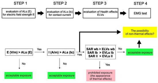
Figure 1.
The flowchart of the EMF exposure assessment algorithm (ALs—action levels, ELVs—exposure limit values, EMG—electromyography, SAR—specific absorption rate).
2.1. EMF Spatial Distribution near the Radiophones
When assessing the limb currents of a worker standing some distance away from the EMF source, the E-field component is dominant [9,15,16]. Therefore, in the first step, the test consisted of the determination of the electric field (E-field) strength spatial distribution at a distance of 10 cm and 20 cm from the device antenna (Figure 2a). Measurements were made using the NBM-520 m (NARDA STS, Pfullingen Germany). The E-field strength was tested during a conversation by the radiophone user. The examined person held the radio in the right hand, with their arm outstretched. The measurements were performed on six commonly used professional portable radio communication units. The tests were repeated five times for each radiophone. The assessments were conducted for three radiophone powers: 0.1 W, 1 W, and 5 W. They are the typical programmable power settings used for radiophones during their operation (0.1 W and 1 W in a LOW mode, 5 W in a HIGH mode). The frequency band used by both uniformed and emergency services covers the range of 30–89 MHz. Therefore, the measurements were carried out at three frequencies: 30 MHz, 55 MHz, and 80 MHz. The criterion for assessing the worker exposure to EMF at that point was to compare the measured value of the E-field strength at a distance of 10 cm from the antenna with an E-field strength of 61 V/m. This value is defined in Directive 2013/35/EU as the action level (AL) for the electric E-field strength (ALs(E)) in the frequency range of 10 MHz to 400 MHz. In the case of obtaining a measurement of the E-field strength below 61 V/m at a distance of 10 cm from the radiophone antenna, the exposure of the radiophone user to EMF was considered acceptable. If the ALs(E) value was found to exceed 61 V/m, we proceeded to the second step.
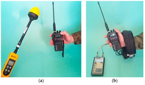
Figure 2.
The examples of measurements: (a) electric field strength distribution near the radiophone; (b) limb contact current.
2.2. Limb Contact Current Measurements
In the second step, the tested parameter was the limb contact current, which was measured with an induction (clamp-on) meter HI-4416 (ETS-Lindgren, Cedar Park, TX, USA). The clamp-on probe was placed on the forearm of the radiophone user (Figure 2b). The limb contact current test was carried out during a conversation by the radiophone user. The measurements were performed for the same radiophone operating parameters as for the EMF distribution analysis, with radiophone powers of 0.1 W, 1 W, and 5 W and frequencies of 30 MHz, 55 MHz, and 80 MHz. The examined person was in a standing, upright position, wearing typical workers’ footwear. At this step of the research, the reference value was a limb contact current value of 40 mA. This value is defined in Directive 2013/35/EU as the AL for contact current (ALs(Ic)) in the frequency range of 10 MHz to 110 MHz. Obtaining a limb contact current below 40 mA in the second step of the study meant that the exposure to EMF was acceptable. When the ALs(Ic) value was exceeded, the tests entered the third stage. All protocols concerning the volunteers was approved by the local ethics committee.
2.3. Specific Absorption Rate (SAR) Numerical Analysis
In the third step, the test involved determining the internal measures of EMF known as Exposure Limit Values (ELVs) with the use of a computer simulation method. In this test, the criterion of the exposure assessment was the SAR parameter values defined in Directive 2013/35/EU as ELVs in the frequency range of 10 MHz to 400 MHz [12]. SAR is a measure of the rate at which energy is absorbed per unit mass by a human body when exposed to a radio frequency (RF) EMF, expressed in watts per kilogram (W/kg). It is usually averaged either over the whole body or over a small sample volume (typically 10 g of tissue). [1]. The ELV limits for the assessment of workers’ exposure to EMF, as defined in Directive 2013/35/EU, are presented in Table 1.

Table 1.
The health effect ELVs for exposure to an electromagnetic field from 100 kHz to 6 GHz according to Directive 2013/35/EU [12].
For this paper, a numerical model of a 5 W radiophone operating in the LB2 band was built. The radio model consisted of a body and a rod antenna, both made of a perfect electric conductor. The dimensions of body, length, and antenna diameter are close to those of commonly used handheld radios. A small gap has been left between the antenna and body where the discrete 50 Ω impedance port has been connected. Due to the small dimensions of the antenna in comparison to the wavelength, its electric length was extended with a lumped inductance in series with the antenna. The antenna impedance was matched with an LC circuit. The lumped inductance value and LC elements were calculated numerically by way of successive approximations. Under the free space condition (without the human body model), an excellent match was reached (S11 = −75.8). The radiophone model was placed in the hand of the operator’s body model at a distance of 10 cm from the head, which corresponded to one of the variants of use (Figure 3). A posable HUGO model with a resolution of 2 mm delivered with the CST Studio Suite software (Dassault Systèmes, Vélizy-Villacoublay, France) was used as the model of the operator’s body. 29 tissues and organs were distinguished in the model. Taking into account the frequency of antenna matching, the permittivity and electrical conductivity of the tissues were selected according to the data provided by Andreuccetti et al. [17]. For the lungs, because the SAR was also averaged in time, the average permittivity and electrical conductivity of inflated and deflated lungs were taken. All calculations were performed with CST Studio Suite software. The time-domain solver was based on the Finite Integration Technique (FIT). With the exception of the base, the perfectly matched layers at the boundary conditions were used at a distance of a quarter of the wavelength. The total SAR (SARwb) averaged over the whole body and maximum local SAR averaged over any 10 g of tissue in the head and trunk volumes (SARht) and limb volumes (SARl) were calculated according to the standard [18]. Four exposure cases differing in terms of skin impedance (permittivity and electric conductivity of dry and wet skin) and the ground (the human body model placed on the grounded and ungrounded base) were analyzed. Bearing in mind the need to verify the models, the current in the hand holding the radiophone was additionally calculated. The currents were calculated on the planes of the wrists.
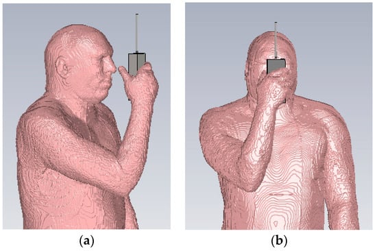
Figure 3.
The computer model of a human using a radiophone: (a) side view; (b) front view.
2.4. Neurophysiological Tests
In the final stage of the study, we introduced, as a pilot phase, a set of simple and noninvasive neurophysiological tests based on the electromyography (EMG) technique. As a diagnostic procedure, EMG allows for the assessment of the electrical activity of muscles and the nerve cells that control them (motor neurons) in order to evaluate and record the electrical activity produced by skeletal muscles. The results of such an examination can reveal nerve dysfunction, muscle dysfunction, or problems with nerve-to-muscle signal transmission. Tests were performed on 27 male volunteers with the anthropometric properties presented in Table 2. They were divided into two groups: (1) people occupationally exposed to EMF (EMF) and (2) the control group (CON), which included people not exposed to EMF (working in a safe zone). The tests were performed using the Nu-Tek® Maxi Plus 2 Model: LE3100 device (Shenzhen Dongdixin Technology Co., Ltd., Shenzhen, China). Two modules were used for the tests. The EMG module was used to measure the strength, contraction velocity, stability of contraction, and relaxation, whereas the STIM module was applied to stimulate the sensory nerve in order to determine the threshold of electrical stimulation causing muscle contractions. The tests were done using adhesive surface electrodes (Figure 4).

Table 2.
Basic anthropometric properties of volunteers.
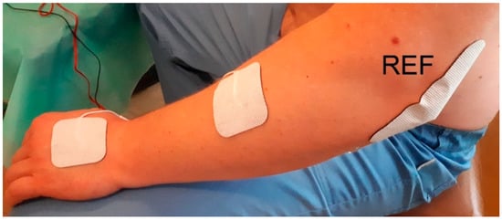
Figure 4.
The electrode placement during neurophysiological tests (REF-reference electrode).
In the first test, a subject performed a contraction (by raising his hand) to the highest level achievable and kept it at the maximum level for a period of 5 s (WORK phase), before relaxing the muscles for another 5 s (REST phase). In one trial, five contractions were performed, separated by periods of rest. The measurement was repeated three times, which was sufficient to determine significant statistical data and did not cause excessive fatigue in the subject. In the second part of the experiment, in order to determine the threshold of electrical stimulation, the value of the stimulating current was increased until unambiguous finger tremor occurred.
3. Results
3.1. Electromagnetic Field Spatial Distribution near the Radiophones
The results of the measurements of the electric field (E-field) strength at a distance of 10 cm from the radiophone antenna are presented in Figure 5a. The measurements were performed for three radiophone powers, 0.1 W, 1 W, and 5 W, for frequencies of 30 MHz, 55 MHz, and 80 MHz. The results for the radiated power of 0.1 W did not indicate the occurrence of an E-field strength exceeding the established limit of 61 V/m. In the case of the output power of 1 W in the frequency range up to 55 MHz, the E-field strength slightly exceeded 61 V/m. With a power of 5 W, the E-field strength significantly exceeded 61 V/m over the entire radio frequency range.
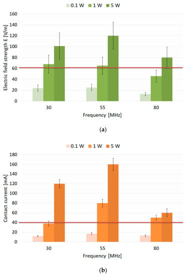
Figure 5.
The results of the measurements of: (a) the electric field strength at a distance of 10 cm from the radiophone antenna; (b) the limb contact currents in the wrist of the hand holding the radiophone. The data are expressed as mean values ± standard error of the mean (SEM).
3.2. Limb Contact Current Measurements
The measurement results of the limb contact current, taken in the wrist of the hand holding the radiophone, are shown in Figure 5b. The result for a radiated power of 0.1 W did not indicate that the limb contact current exceeded the limit of 40 mA. In the case of an output power of 1 W in the frequency range up to 55 MHz, the value of the limb contact current was almost twice the limit of 40 mA. With a power of 5 W, the value of the limb contact current exceeded the limit of 40 mA, even up to four times, over the entire radiophone operating frequency range.
3.3. SAR Numerical Analysis
The results of the numerical analysis of the human body exposed to EMF produced by an LB2 band radio powered with 5 W are shown in Table 3. The conditions of the exposure were previously described in the Materials and Methods. The obtained results for the current calculations were consistent with the results of the limb contact current measurements performed by the authors as part of routine professional practice. SARwb is equivalent to the exposure limit value (ELV) related to the whole-body heat stress expressed as the averaged SAR in the body, as defined in Directive 2013/35/EU [12]. SARwb averaged over the whole body, irrespective of the exposure case, was below 0.017 W/kg, which was no more than 5% of the ELV. With regard to the cases of exposure differing in terms of skin impedance (permittivity and electric conductivity of dry and wet skin) and the ground (the human body model placed on the grounded and ungrounded base), 10% of SARwb variability was observed. The local SARht is averaged over any 10 g of tissue in the head and trunk volumes, and is equivalent to the ELV related to the localized heat stress in the head and trunk expressed as the localized SAR in the body, as defined in Directive 2013/35/EU [2]. The maximum SARht, equaling 0.29 W/kg, was found in the trunk volume of the model (wet skin, ungrounded base) and was much lower than the appropriate ELV level (10 W/kg). Bearing in mind the cases of exposure, 6% SARht variability was observed in the head volume and slightly above 10% SARht in the trunk volume. SARl is equivalent to the ELV related to the localized heat stress in the limbs expressed as the localized SAR in the limbs, as defined in Directive 2013/35/EU [12]. The maximum SARl, equaling 9.03 W/kg, was found in the right-hand volume of the model in the wrist area (wet skin, ungrounded base) and was comparable to the appropriate ELV level (20 W/kg). Bearing in mind the cases of exposure, the variability of SARl exceeded 10%. The numerical calculation of the E-field strength rms (root-mean-square) value distribution near the radiophone and the hotspot of SARl has been illustrated in the Figure 6.

Table 3.
The results of the SAR and limb currents numerical analysis when compared to the occupational exposure limit values (ELVs) and action levels (ALs) according to Directive 2013/35/EU (Ic—wrist current rms value in mA; SARwb—total SAR averaged over the whole body in W/kg; SARht—local SAR averaged over any 10 g of tissue in the head and trunk volumes in W/kg; SARl—local SAR averaged over any 10 g of tissue in the limb volumes in W/kg).
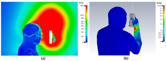
Figure 6.
The electric field strength rms (root-mean-square) value distribution: (a) on the sagittal plane of the body model; (b) SAR hotspot location.
3.4. Neurophysiological Tests
In the first part of the tests, the electrical activity of the muscle during rest and forceful contraction was measured. The results were represented with selected EMG parameters: (1) Work Average—the average value of the EMG signal from all working periods of a given session, measured in microvolts [µV]; (2) Rest Average—the mean EMG signal value from all rest periods, measured in microvolts [µV]; (3) Onset Average—the average time a muscle took to achieve 75% of the average work value from all periods of work in a given session, measured in seconds [s]; (4) Rest Average—the average muscle relaxation time, measured in seconds [s]. The analysis of the parameters of the EMG response did not indicate any differences between the control and EMF exposure groups. Additionally, the obtained values were within the normal range (Table 4).

Table 4.
The results of the selected EMG parameters for the control group (CON) and the group exposed to EMF (EMF; using handheld and portable devices of wireless communication). Parameters described in text. The data are expressed as mean values ± SEM.
As the second part of the neurological tests, we determined the threshold current (I) for muscle stimulation. The results show no significant differences between the control group (CON)—11.97 ± 0.3 mA and the group exposed to EMF (EMF)—11.67 ± 0.53 mA (Figure 7).
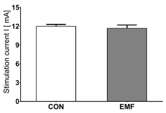
Figure 7.
The threshold stimulation in the control group (CON) and EMF-exposed group (EMF). The data are expressed as mean values ± SEM.
4. Discussion
The initial assessment of the radiophone users’ exposure to EMF was performed with the measurements of the E-field strength distributions in the vicinity of the radiophone, as well as with the measurements of the limb contact current within the users’ bodies. The measurement results indicate a large spatial variability of the E-field strength around the devices. It is important to notice that part of the radiophone operator’s body is in the danger zone (according to Polish Labor Law provisions) when the device is operating in the transmission mode at transmitter output powers of 1 W and 3 W and frequencies of 30 MHz and 55 MHz [19]. The value of the measured E-field strength exceeded 61 V/m, the value established as the limit ALs(E) in Directive 2013/35/EU [12].
Exceeded safe values applied especially to the area of the head and the hand holding the portable radio, where the E-field strength ranged from 65 to 120 V/m for 10 cm from the antenna. For these values of the E-field strength, the limb contact currents Ic ranged between 80 and 160 mA. That is 2–4 times more than the limit (40 mA), according to the ALs(Ic) defined in Directive 2013/35/EU [12].
The specific absorption rate (SAR) parameter was also included in order to assess the exposure. The measurement of this parameter is used to protect people from the adverse thermal effects of EMF exposure. Therefore, appropriate limits have been established in Polish and EU legislation, including the three-element SAR assessment [12,13,19,20,21]. First, the SAR is averaged for the whole body weight during any 6 min. Second, the SAR is averaged over any 10 g of dense tissue in the head and trunk; and third, by analogy, it is averaged in the limbs at the same time [1,22]. The SAR values obtained in the computer simulation do not indicate the possibility of exceeding the established limits. Though the results exclude the occurrence of thermal effects, they do not eliminate potential neuromuscular effects, which may be indicated by the limb contact currents exceeding the limits. Therefore, to make the study fully comprehensive, the exposure assessment was supplemented with simple neurophysiological tests that examined the condition of the neuromuscular system of the upper extremities. These tests would answer the question of whether exceeding the limits for limb contact currents by 2–4-fold may result in nonthermal effects in the neuromuscular system of the hand.
In the part of the study that included the pilot neurophysiological tests, we were fortunate to collect 27 volunteers who, despite being employed in the same workplace, could be divided into two groups: people not exposed to EMF, constituting the control group, and people exposed to EMF (radio users) as the exposed group. People in both experimental groups did not differ significantly in age or BMI. A comparison of electromyographic parameters and the threshold of muscle stimulation did not show any differences between the control group and the group exposed to EMF. However, this is not sufficient evidence to explicitly exclude the occurrence of potential health effects. What is worth mentioning is that all volunteers were young and athletic, so that their bodies could perfectly cope with possible neurological disorders (by regeneration and compensation). This is a serious general problem in studying the effects of EMF exposure on neurophysiological functions. Most of the studies published so far have been conducted on young people, mostly men. However, the neurophysiological parameters change with age. Furthermore, the effects of EMF may be differentiated by age and gender [23,24,25,26]. Lifestyle is also important, for instance smoking. With the current state of knowledge, we cannot unequivocally rule out the possibility of neurological effects at a later age [27,28]. Another possible reason for the lack of differences between the control and exposed groups may be hardware limitations. The device used in our study is an advanced portable muscle assessment and muscle rehabilitation unit suitable for clinical applications and for home use; however, we suspect that this measurement method may not be sensitive enough. Possible neurological changes may be beyond the detection range or the ability of the device. On the other hand, we emphasize once again that when choosing the method, we were primarily guided by the lack of invasiveness of the used tests, the speed and simplicity of the measurements, and the lack of susceptibility to strong, external EMF accompanying the measurements. All this leads to the need for further experiments and more measurements, as well as testing new neurophysiological parameters that will allow for more information on possible changes in the neuromuscular system to be obtained.
5. Conclusions
Our paper describes for the first time the attempt to comprehensively assess the effects of exposure radiotelephone users to EMF, taking into account external (E-field distribution near the radiotelephones) and internal measures, both measured (limb contact current) and calculated (SAR parameter), and an assessment of potential health effects through the use neurophysiological tests. The second achievement of our studies was the evaluation of all parameters under realistic conditions of EMF exposure during the use of radiophone, as well as the evaluation of the neuromuscular system function in the control group and the EMF-exposed group when providing similar anthropometric parameters.
The exposure assessment for the use of the radiophone was initially performed by analyzing external measures. The limit values of the external measures were established for a primary field with a uniform spatial distribution to protect workers against induced currents of excessive density and against excessive thermal effects of exposure. In the work environment, there may be cases of high levels of field exposure that deviate significantly from this uniform primary field model. In such cases, it is necessary to analyze in more detail the level of workers’ safety, considering how realistic conditions affect, among others, induced and contact currents flowing through the body of workers. For their evaluation, numerical simulations and numerical phantoms of the human body or measurements of currents flowing through the body of the worker were used. Both constituted a complementary method of exposure assessment. These parameters characterize the exposure conditions and the risk level much more comprehensively, and they are applicable in cases where a preliminary and simplified assessment of external measures indicates a possibility of excessive EMF exposure in the workplace. The addition of regular neurophysiological tests as part of the periodic medical examinations of workers exposed to EMF would perfectly complement the assessment of workers’ exposure.
Author Contributions
Conceptualization, J.K. and J.W.; methodology, J.K. and J.W.; investigation, J.K., J.W., J.S. and R.P.; writing—original draft preparation, J.K., J.W., and J.S.; writing—review and editing, J.K. and J.W.; visualization, J.K. and J.W.; project administration, J.K. and J.W. All authors have read and agreed to the published version of the manuscript.
Funding
This research received no external funding.
Acknowledgments
The authors would like to thank the headquarter and the soldiers of the 21st Podhale Rifles Highland Brigade for their participation and engagement in the study. We would also like to acknowledge Dawid Gawęda, from The Department of Neurology, The Provincial Polyclinical Hospital in Toruń for guidance during the running of this project.
Conflicts of Interest
The authors declare no conflict of interest.
References
- International Commission on Non-Ionizing Radiation Protection (ICNIRP). Guidelines for limiting exposure to Electromagnetic Fields (100 kHz to 300 GHz). Health Phys. 2020, 118, 483–524. [Google Scholar] [CrossRef] [PubMed]
- IEEE Std C95.6-2002. Standard for Safety Levels with Respect to Human Exposure to Frequency Electromagnetic Fields, 0 Hz to 3 kHz; Institute of Electrical and Electronics Engineers: New York, NY, USA, 2002. [Google Scholar]
- IEEE Std C95.1-2005. Standard for Safety Levels with Respect to Human Exposure to Radio Frequency Electromagnetic Fields, 3 kHz to 300 GHz; Institute of Electrical and Electronics Engineers: New York, NY, USA, 2005. [Google Scholar]
- Reilly, J.P. Applied Bioelectricity: From Electrical Stimulation to Electropathology; Springer Science & Business Media: New York, NY, USA, 2012. [Google Scholar]
- WHO—World Health Organization. Extremely Low Frequency Fields; Environmental Health Criteria, Volume 238; World Health Organization: Geneva, Switzerland, 2007. [Google Scholar]
- WHO—World Health Organization. Electromagnetic Fields (300 Hz–300 GHz); Environmental Health Criteria, Volume 137; World Health Organization: Geneva, Switzerland, 1993. [Google Scholar]
- Kavet, R.; Tell, R.A.; Olsen, R.G. Radiofrequency contact currents: Sensory responses and dosimetry. Radiat. Prot. Dosim. 2014, 162, 268–279. [Google Scholar] [CrossRef] [PubMed]
- Fish, R.M.; Geddes, L.A. Conduction of Electrical Current to and Through the Human Body: A Review. Eplasty 2009, 9, e44. [Google Scholar] [PubMed]
- Gryz, K.; Karpowicz, J. Principles for electromagnetic hazards assessment related to induced and contact currents. Podstawy I Metod. Oceny Srodowiska Pr. 2008, 4, 137–171. [Google Scholar]
- Scientific Committee on Emerging and Newly Identified Health Risks (SCENIHR). Potential Health Effects of Exposure to Electromagnetic Fields (EMF); European Commission: Brussels, Belgium, 2015; pp. 1–288. [Google Scholar] [CrossRef]
- Tell, R.A.; Tell, C.A. Perspectives on setting limits for RF contact currents: A commentary. Biomed. Eng. Online 2018, 17. [Google Scholar] [CrossRef] [PubMed]
- Directive 2013/35/EU of the European Parliament and of the Council of 26 June 2013 on the minimum health and safety requirements regarding the exposure of workers to the risks arising from physical agents (electromagnetic fields) (20th individual Directive within the meaning of Article 16(1) of Directive 89/391/EEC) and repealing Directive 2004/40/EC. Off. J. Eur. Union 2013, L-179, 1–21.
- Vigotsky, A.D.; Halperin, I.; Lehman, G.J.; Trajano, G.S.; Vieira, T.M. Interpreting Signal Amplitudes in Surface Electromyography Studies in Sport and Rehabilitation Sciences. Front. Physiol. 2018, 8. [Google Scholar] [CrossRef] [PubMed]
- Criswell, E. Cram’s Introduction to Surface Electromyography; Jones & Bartlett Publishers: Sudbury, MA, USA, 2010. [Google Scholar]
- Karpowicz, J.; Zradziński, P.; Kieliszek, J.; Gryz, K.; Sobiech, J.; Leszko, W. An In Situ and In Silico Evaluation of Biophysical Effects of 27 MHz Electromagnetic Whole Body Humans Exposure Expressed by the Limb Current. BioMed Res. Int. 2017, 2017, 1–9. [Google Scholar] [CrossRef] [PubMed]
- Wilén, J.; Mild, K.H.; Paulsson, L.-E.; Anger, G. Induced current measurements in whole body exposure condition to radio frequency electric fields. Bioelectromagnetics 2001, 22, 560–567. [Google Scholar] [CrossRef] [PubMed]
- Andreuccetti, D.; Fossi, R.; Petrucci, C. An Internet resource for the calculation of the dielectric properties of body tissues in the frequency range 10 Hz–100 GHz. IFAC-CNR, Florence (Italy). 1997. Available online: http://niremf.ifac.cnr.it/tissprop/ (accessed on 30 September 2020).
- IEC/IEEE 62704-1:2017. Determining the Peak Spatial-Average Specific Absorption Rate (SAR) in the Human Body from Wireless Communications Devices, 30 MHz to 6 GHz—Part 1: General Requirements for Using the Finite Difference Time-Domain (FDTD) Method for SAR Calculations; International Electrotechnical Commission: Geneva, Switzerland, 2017. [Google Scholar]
- Regulation of the Minister of Family, Labour and Social Policy of 29th June 2016 on health and safety at work exposed to the electromagnetic field, Dz. U., 2016-06-30, (Text No. 950): 0867-3411. In Polish.
- Dimbylow, P.J. Induced current densities from low-frequency magnetic fields in a 2 mm resolution, anatomically realistic model of the body. Phys. Med. Biol. 1998, 43, 221. [Google Scholar] [CrossRef] [PubMed]
- Zradziński, P.; Karpowicz, J.; Gryz, K. Electromagnetic Energy Absorption in a Head Approaching a Radiofrequency Identification (RFID) Reader Operating at 13.56 MHz in Users of Hearing Implants Versus Non-Users. Sensors 2019, 19, 3724. [Google Scholar] [CrossRef] [PubMed]
- Hansson Mild, K.; Alanko, T.; Decat, G.; Falsaperla, R.; Gryz, K.; Hietanen, M.; Karpowicz, J.; Rossi, P.; Sandström, M. Exposure of workers to electromagnetic fields. A review of open questions on exposure assessment techniques. Int. J. Occup. Saf. Ergon. 2009, 15, 3–33. [Google Scholar] [CrossRef] [PubMed]
- Vecchio, F.; Babiloni, C.; Ferreri, F.; Buffo, P.; Cibelli, G.; Curcio, G.; van Dijkman, S.; Melgari, J.-M.; Giambattistelli, F.; Rossini, P.M. Mobile phone emission modulates inter-hemispheric functional coupling of EEG alpha rhythms in elderly compared to young subjects. Clin. Neurophysiol. 2010, 121, 163–171. [Google Scholar] [CrossRef] [PubMed]
- Croft, R.J.; Leung, S.; McKenzie, R.J.; Loughran, S.P.; Iskra, S.; Hamblin, D.L.; Cooper, N.R. Effects of 2G and 3G mobile phones on human alpha rhythms: Resting EEG in adolescents, young adults, and the elderly. Bioelectromagnetics 2010, 31, 434–444. [Google Scholar] [CrossRef] [PubMed]
- Vecchio, F.; Tombini, M.; Buffo, P.; Assenza, G.; Pellegrino, G.; Benvenga, A.; Babiloni, C.; Rossini, P.M. Mobile phone emission increases inter-hemispheric functional coupling of electroencephalographic alpha rhythms in epileptic patients. Int. J. Psychophysiol. 2012, 84, 164–171. [Google Scholar] [CrossRef] [PubMed]
- Loughran, S.P.; McKenzie, R.J.; Jackson, M.L.; Howard, M.E.; Croft, R.J. Individual differences in the effects of mobile phone exposure on human sleep: Rethinking the problem. Bioelectromagnetics 2012, 33, 86–93. [Google Scholar] [CrossRef] [PubMed]
- Orendáčová, J.; Orendáč, M.; Mojžiš, M.; Labun, J.; Martončíková, M.; Saganová, K.; Lievajová, K.; Blaško, J.; Abdiová, H.; Gálik, J.; et al. Effects of short-duration electromagnetic radiation on early postnatal neurogenesis in rats: Fos and NADPH-d histochemical studies. Acta Histochem. 2011, 113, 723–728. [Google Scholar] [CrossRef] [PubMed]
- Pritchard, C.; Silk, A.; Hansen, L. Are rises in Electro-Magnetic Field in the human environment, interacting with multiple environmental pollutions, the tipping point for increases in neurological deaths in the Western World? Med. Hypotheses 2019, 127, 76–83. [Google Scholar] [CrossRef] [PubMed]
Publisher’s Note: MDPI stays neutral with regard to jurisdictional claims in published maps and institutional affiliations. |
© 2020 by the authors. Licensee MDPI, Basel, Switzerland. This article is an open access article distributed under the terms and conditions of the Creative Commons Attribution (CC BY) license (http://creativecommons.org/licenses/by/4.0/).