Next Article in Journal
Evaluation of the Mandibular Function, After Nonsurgical Treatment of Unilateral Subcondylar Fracture: A 1-Year Follow-Up Study
Previous Article in Journal
Early Posterior Vault Distraction Osteogenesis for the Treatment of Syndromic Craniosynostosis
 
 
Craniomaxillofacial Trauma & Reconstruction is published by MDPI from Volume 18 Issue 1 (2025). Previous articles were published by another publisher in Open Access under a CC-BY (or CC-BY-NC-ND) licence, and they are hosted by MDPI on mdpi.com as a courtesy and upon agreement with Sage.
Article

Correlation of Third Molar Status with Incidence of Condylar and Angle Fractures

by
Suresh Menon
*,
Veerendra Kumar
,
Srihari V.
and
Yogitha Priyadarshini
Department of Oral and Maxillofacial Surgery, Vydehi Institute of Dental Sciences, Bangalore 560066, Karnataka, India
*
Author to whom correspondence should be addressed.
Craniomaxillofac. Trauma Reconstr. 2016, 9(3), 224-228; https://doi.org/10.1055/s-0036-1584400
Submission received: 23 July 2015 / Revised: 20 February 2016 / Accepted: 20 February 2016 / Published: 1 July 2016

Abstract

:
The mandibular angle and condylar regions are most prone to fractures and this has been attributed to the presence/absence or the position of the third molars. This retrospective study was undertaken to analyze the correlation between the third molars and incidence of condylar and angle fractures in 104 patients treated for these fractures during the period from June 2009 to December 2013. Clinical and radiographic records of these patients were studied to look for the presence and position of third molars and their relation to incidence of condylar or angle fractures. There was a definite positive relation to impacted third molars and increased incidence of angle fractures. The condylar fractures were more commonly seen when the third molars were fully erupted or missing. Third molar impactions predispose to angle fractures and missing or fully erupted third molars predispose to condylar fractures.

In spite of being the strongest bone in the maxillofacial region, the mandible is ironically one of the most commonly fractured bones due to its prominence in the face and the weakening of the corticocancellous framework due to the presence of teeth. Fractures in the mandible are most often seen in the angle region, condylar region, and the parasymphysis. The angle region is quite vulnerable due to the fact that it forms the junction between the ramus and the body and is influenced, to a great extent, by the masticatory sling of muscles attached to the medial and lateral aspects. The incidence of mandibular angle fractures is approximately 30% [1]. One of the primary factors influencing angle fractures is the presence of third molars which tend to weaken the area, predisposing it to fracture.

Materials and Method

Study Design and Sample

A retrospective cohort study was conducted on 104 patients managed in this institution from June 2009 to December 2013 based on clinical and radiographic records of mandibular angle and condylar fractures. This study analyzed the presence of third molars in these cases and their eruptive status after reviewing the radiographic records from the archive section.
The variables included the presence, position, and depth of the third molars and the presence or absence of fractures of the condylar region and angle.

Statistical Analysis
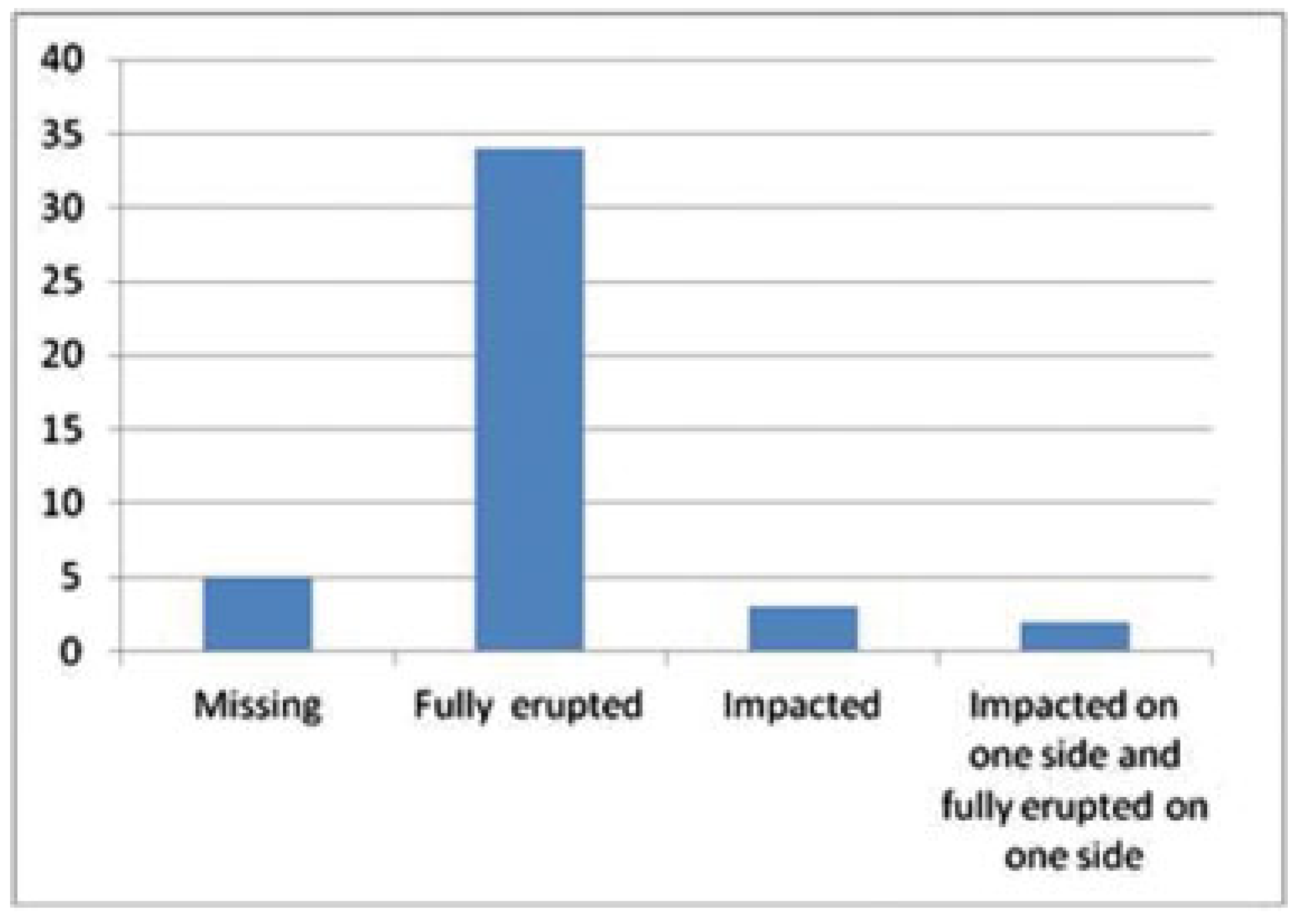
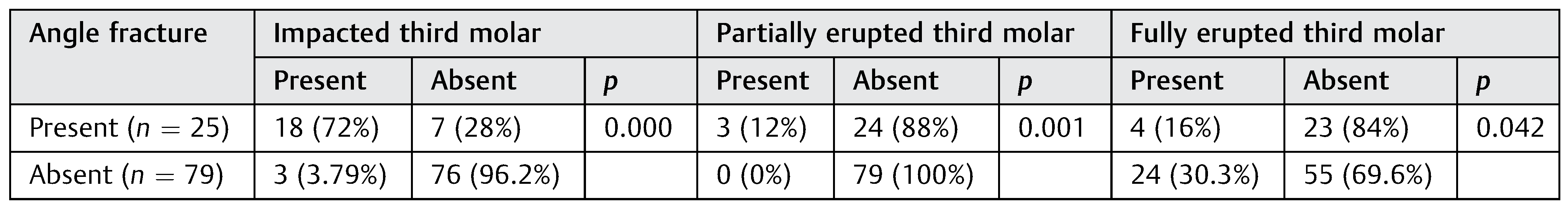
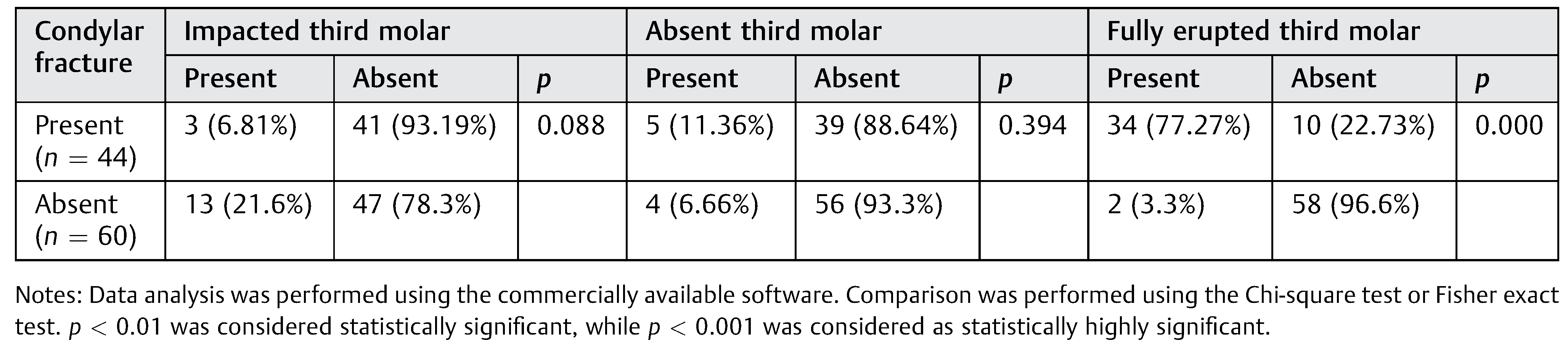
Data analysis was performed using the commercially available software. Comparison was performed using the Chisquare test or Fisher exact test. p < 0.01 was considered statistically significant, while p < 0.001 was considered as statistically highly significant.

Results

The 104 cases of mandibular fractures included 97 males and 7 females in the age range of 11 to 70 years with a mean age of 30.62. The maximum numbers of patients (53) were in the third decade of life, 22 patients were from the fourth decade, 14 from the fifth decade, 6 from the second decade, 4 each from the sixth and eighth decades, and 1 from the seventh decade of life (Figure 1).
There were 25 angle fractures and 44 condylar fractures in this series. Among the angle fractures, 7 were isolated fractures and 15 were concomitant parasymphysis fractures including 1 case of bilateral angle with parasymphysis fracture, 1 case with condylar fracture, 1 with ramus and parasymphysis, and 1 with zygomatic arch fracture.
Among the 44 condylar fractures, there were 13 isolated fractures, 6 on the left and 7 on the right side; 1 fracture also involved the angle; 4 involved the body; 14 involved the parasymphysis; 8 were bilateral cases; and 4 were associated with midface fractures.

Angle Fracture

The number of angle fractures in the series was 25 out of 104 (24.038%). There were 11 fractures on the left and 12 fractures on the right and a single bilateral angle fracture. Any third molar in Class 2 or 3 or position C (Pell & Gregory classification) or angulated were considered impacted. Eighteen of these fractures (72%) showed the presence of an impacted third molar in the side involved; 10 mesioangularly inclined, 4 class 3, 2 class 2, and 2 vertically impacted position C. In three cases (12%), the third molar was partially erupted in a vertical inclination. A third molar inclined normally but present below the occlusal levels of the other molars was designated as partially erupted. Thus, 84% of the angle fractures had an impacted or partially erupted third molar which was statistically significant (Table 1). Only in four cases (16%) were the third molars fully erupted (Figure 2). All cases were fixed by miniplates and on two occasions, the third molar had to be extracted during the surgical management because of mobility of the tooth due to their involvement in the fracture line.
The “p” value for angle fractures in the presence of impacted third molars was 0.000 which is statistically highly significant. The “p” value for partially erupted third molars in angle fractures was 0.001 which is statistically significant. Angle fractures with fully erupted third molars showed a “p” value of 0.042 which is statistically insignificant.

Condylar Fracture

Out of the 44 cases of condylar fractures which comprised 44.32% of total fractures, 24 of the condylar fractures involved the right side, 10 involved the left side, and 10 were bilaterally involved. While open reduction and rigid internal fixation were done for 37 cases, the small condylar stump was removed due to symptoms like limited mouth opening, pain, or interference in lateral movements in 7 cases. On evaluation of the status of the third molar, in 34 cases, the tooth was fully erupted (77.27%), the tooth was absent in 5 cases (11.36%) and only in 3 cases was there an impacted third molar present (6.81%) (Figure 3). Thus, in 88.63% of the cases, condylar fractures occurred when the third molar was not impacted or was missing (Table 2). In two cases of bilateral condylar fractures, there was a fully erupted third molar on one side and an impacted third molar on the other side.
The “p” value for condylar fractures in the presence of impacted third molars was 0.088 which is statistically insignificant. The “p” value for partially erupted third molars in condylar fractures was 0.394 which is also statistically insignificant. Condylar fractures with fully erupted third molars showed a “p” value of 0.000 which is statistically highly significant.

Discussion

The location of a fracture depends on various factors. These include the site, force, and direction of impact. A large force acting on a small area results in a fracture at the point of impact. But when the force is distributed, the fracture occurs at the weakest point. Therefore, it would be prudent to believe that the risk of angle fracture incidence is not related to any single factor, whether it be the vector of force, amount of force, musculature of the face, architecture of the mandible, or the presence or absence of a third molar [2].
Anatomically, the mandibular angle is at a transition zone from the dentate body to the lateral flare of the ramus, thus increasing the risk of fracture [3]. Moore has also suggested that a change in the direction of the grain of bone, which occurs where the vertical ascending ramus and the horizontal body meet, tends to weaken the angle region of the mandible and increases its susceptibility to fracture [4].
In a study of the three-dimensional biomechanical properties and the intrinsic strengths and weaknesses of the mandible by Tams et al., [5] the mandibular angle was identified as having the greatest amount of positive bending movement, resulting in tension at the alveolus and compression at the inferior border.
The presence of third molars has been suggested to contribute to an increased mandibular fragility because the mandible loses part of its bone structure to harbor tissues that do not contribute to its strength [6].
A computational method for mechanical tests by creating virtual elements with finite dimensions and physical properties based on aeronautical engineering can be adapted to real structures, to recreate load applications and present the distribution of stresses and deformation. In one such study by Bezerra et al., [7] the maximum stresses were located at the symphysis, in the retromolar area, and both condyles on the three experimental models. The presence of the third molars resulted in a difference in the stress distribution. Bezerra et al. also found that it was noticeable that the impact of force on the chin resulted in a concentration of stress on the external oblique ridge, and when the third molar was present, this concentration extended to the alveolar process. The comparative analysis showed a stress concentration on the vestibular aspect of the mandibular angle when the third molar was present, and on the condylar neck when it was absent.
Meisami et al. [6] suggest one such model where mandibular strength is derived from maintenance of cortical, not medullary, bone integrity. As such, superficially positioned M3s disrupt the cortical integrity of the external oblique ridge, producing a point of weakness in the mandible and making it susceptible to fracture. This is in contrast to our results.
An explanation to this was given by Huelke and Harger [8]. Once a force is applied to an anterior mandibular region, the energy dispersion will occur along the body toward the condyles, causing stress on the lateral aspect of the angle and condyle. The force seeks out the weakest point in the arch and causes extreme bending and tensile failure at that point. Third molars can be related to the fragility of the angle, as their presence significantly alters its biomechanics.
The increased stress on the external oblique ridge has also been shown by Szucs et al. [9]. The mandibular angle region was also shown to have the greatest amount of shear force and a small amount of torsion, resulting in the proximal segment being lingually and caudally displaced and the distal fragment being buccally and cranially displaced [10].
The close relation of third molars and angle fractures has been extensively studied by various authors and the positive correlation endorsed by them [11,12,13]. The presence of third molars has been suggested to contribute to an increased mandibular fragility because the mandible loses part of its bone structure to harbor tissues that do not contribute to its strength [14,15]. Reviewing the presence of third molars in angle fractures, Ed Ellis III [16] showed more than 90% of the cases with third molars involving the fracture and approximately 42.2% of these being impacted. This is in sharp contrast to our study where an overwhelming 70% were impacted.
In a multicentric retrospective cohort study on mandible fracture patients, Halmos et al. [17] used the third molar and its position as predictor variables. They showed a 2.8-fold increased risk for angle fractures in the presence of the third molar. However, in contrast to our results, they showed that deep impactions were not associated with an increased risk for fracture. Another retrospective cohort study by Tevepaugh and Dodson [18] showed a very small number of angle fractures in the absence of third molars. They surmised that patients with fractured mandibles and mandibular third molars are 3.8 times more likely to have an angle fracture than patients without mandibular third molars.
In addition to the presence of third molars, their position also is correlated to the incidence of angle fractures. The first positive relation between angle fractures and position of the third molar was shown by Reitzik et al. [19] in an animal study. In an in vitro study on monkey mandibles, they showed that mandibles with unerupted third molars fractured when a force of 15.8 2.5 kg was used while those mandibles that had a fully erupted third molar fractured at a force of 26.4 4.2 kg. Meisami et al. [6] have tried to explain the increased susceptibility of angle being fractured in impacted or partially erupted third molar conditions. According to them, when a tooth is completely in occlusion, the widest portion of the tooth is in the mouth and the external oblique ridge remains intact. On the other hand, when the tooth is completely impacted, the widest portion of the tooth is generally found below the external oblique ridge. However, when the tooth is partially impacted, the tension line is disrupted, weakening the mandibular angle and making it more susceptible to fracture. In studying the relationship between the presence of third molars and angle fractures in 1210 patients, a direct relation was established between the presence of the third molar and prevalence of angle fractures [20]. They also showed an increased risk of angle fracture with deeper impacted third molars. This is in conjunction with our results where 17 of the 24 angle fracture cases had impacted teeth and 4 had partially erupted teeth.
The direct correlation to the depth of impaction and angle fractures were also shown by Subhashraj [21] in whose study, increased incidence was noticed when the tooth was in position C (76%) and level 3 (85%). In evaluating the presence and state of eruption and its association to angle fractures, Ma’aita and Alwrikat [22] showed that the mandibular angle that contains an impacted third molar is more susceptible to fracture when exposed to an impact than an angle without a third molar. In addition to similar results, Safdar and Meechan [12] further surmised that the amount of bony space occupied by the impacted tooth directly affected the weakness of the bone in that region. Another interesting finding was the angle fractures being twice as likely to occur in dentate patients compared with edentate patients [23].
In contrast to all these studies, Lee and Dodson [24] showed no statistically significant results in correlating the third molar angulation and depth to angle fractures.
The relation of the third molar to both angle and condylar fractures has also been studied. In a retrospective review of 385 patients treated during a 3-year period, Choi et al. [25] found higher incidence of angle fractures in the presence of a third molar and higher incidence of condylar fractures where third molars were absent or not fully erupted. In a study relating the condylar and angle fractures with an unerupted lower third molar, Inaoka et al. [26] concluded that the absence of an impacted third molar increases the risk of condylar fracture and decreases the prevalence of angle fractures. This was akin to our results. The direct relation to depth of the third molar to incidence of condylar and angle fractures was further proved by Duan and Zhang [27]. They showed increased incidence of condylar fractures with fully erupted third molars and increased incidence of angle fractures with impacted third molars. Impacted teeth disrupt the cortical bridge of the superior border, weakening the mandibular angle. Less force and muscle tension is required to cause an angle fracture. Similar conclusions were also drawn by other surgeons [28,29,30,31].
Our retrospective study is similar in results and incidence to all these studies. In view of the significant relation of an impacted third molar to angle fractures, it would definitely be prudent to prophylactically remove the impacted teeth in people who pursue contact sports or are vulnerable to injuries due to their profession, provided the third molars are not erupted in the oral cavity.
In view of this overwhelming evidence supporting the vulnerability of mandible to angle fractures in the presence of impacted third molars, Schwimmer et al. [32] suggested that early removal of impacted teeth may decrease the risk of sustaining a mandible fracture, specifically the angle, especially in sportspersons during contact sports. However, the risk of sustaining fracture during the socket healing period does exist and it is suggested that contact sports be avoided at least 6 months after the surgical removal of the tooth.

Conclusion

This retrospective study on relation of third molars to incidence of angle and condylar fractures shows overwhelming evidence of a direct relation of impacted third molars to increased incidence of angle fractures. The absence of impacted third molars is directly related to shift in fracture incidence from angle to the condylar region.

References

  1. Ellis, E.I.I.I.; Moos, K.F.; el-Attar, A. Ten years of mandibular fractures: an analysis of 2,137 cases. Oral Surg Oral Med Oral Pathol 1985, 59, 120–129. [Google Scholar] [CrossRef]
  2. Schubert, W.; Kobienia, B.J.; Pollock, R.A. Cross-sectional area of the mandible. Discussion J Oral Maxillofac Surg 1997, 55, 689–692, discussion 693. [Google Scholar] [CrossRef]
  3. Halazonetis, J.A. The ‘weak’ regions of the mandible. Br J Oral Surg 1968, 6, 37–48. [Google Scholar] [CrossRef] [PubMed]
  4. Moore, J.R. Principles of Oral Surgery, 2nd ed; Manchester: Manchester University Press, 1976. [Google Scholar]
  5. Tams, J.; van Loon, J.P.; Rozema, F.R.; Otten, E.; Bos, R.R. A threedimensional study of loads across the fracture for different fracture sites of the mandible. Br J Oral Maxillofac Surg 1996, 34, 400–405. [Google Scholar] [CrossRef]
  6. Meisami, T.; Sojat, A.; Sàndor, G.K.; Lawrence, H.P.; Clokie, C.M. Impacted third molars and risk of angle fracture. Int J Oral Maxillofac Surg 2002, 31, 140–144. [Google Scholar] [CrossRef] [PubMed]
  7. Bezerra, T.P.; Silva Junior, F.I.; Scarparo, H.C.; Costa, F.W.; Studart-Soares, E.C. Do erupted third molars weaken the mandibular angle after trauma to the chin region? A 3D finite element study. Int J Oral Maxillofac Surg 2013, 42, 474–480. [Google Scholar] [CrossRef]
  8. Huelke, D.F.; Harger, J.H. Maxillofacial injuries: their nature and mechanisms of production. J Oral Surg 1969, 27, 451–460. [Google Scholar] [PubMed]
  9. Szucs, A.; Bujtár, P.; Sándor, G.K.; Barabás, J. Finite element analysis of the human mandible to assess the effect of removing an impacted third molar. J Can Dent Assoc 2010, 76, a72. [Google Scholar]
  10. Wolujewicz, M.A. Fractures of the mandible involving impacted 3rd molar tooth: an analysis of 47 cases. Br J Surg 1980, 18, 125–131. [Google Scholar]
  11. Tevepaugh, D.B.; Dodson, T.B. Are mandibular third molars a risk factor for angle fractures? A retrospective cohort study. J Oral Maxillofac Surg 1995, 53, 646–649, discussion 649–650. [Google Scholar] [CrossRef]
  12. Safdar, N.; Meechan, J.G. Relationship between fractures of the mandibular angle and the presence and state of eruption of the lower third molar. Oral Surg Oral Med Oral Pathol Oral Radiol Endod 1995, 79, 680–684. [Google Scholar] [CrossRef] [PubMed]
  13. Tankersly, K.; Abubaker, A.O.; Laskin, D.M. The relationship between presence of mandibular third molars and mandibular angle fractures. J Dent Res 1995, 74, 80. [Google Scholar]
  14. Iida, S.; Hassfeld, S.; Reuther, T.; Nomura, K.; Mühling, J. Relationship between the risk of mandibular angle fractures and the status of incompletely erupted mandibular third molars. J Craniomaxillofac Surg 2005, 33, 158–163. [Google Scholar] [CrossRef]
  15. Ugboko, V.I.; Oginni, F.O.; Owotade, F.J. An investigation into the relationship between mandibular third molars and angle fractures in Nigerians. Br J Oral Maxillofac Surg 2000, 38, 427–429. [Google Scholar] [CrossRef]
  16. Ellis, E., III. A prospective study of 3 treatment methods for isolated fractures of the mandibular angle. J Oral Maxillofac Surg 2010, 68, 2743–2754. [Google Scholar] [CrossRef]
  17. Halmos, D.R.; Ellis, E.I.I.I.; Dodson, T.B. Mandibular third molars and angle fractures. J Oral Maxillofac Surg 2004, 62, 1076–1081. [Google Scholar] [CrossRef]
  18. Tevepaugh, D.B.; Dodson, T.B. Are mandibular third molars a risk factor for angle fractures? A retrospective cohort study. J Oral Maxillofac Surg 1995, 53, 646–649, discussion 649–650. [Google Scholar] [CrossRef] [PubMed]
  19. Reitzik, M.; Lownie, J.F.; Cleaton-Jones, P.; Austin, J. Experimental fractures of monkey mandibles. Int J Oral Surg 1978, 7, 100–103. [Google Scholar] [CrossRef]
  20. Fuselier, J.C.; Ellis, E.E.I.I.I.; Dodson, T.B. Do mandibular third molars alter the risk of angle fracture? J Oral Maxillofac Surg 2002, 60, 514–518. [Google Scholar] [CrossRef]
  21. Subhashraj, K. A study on the impact of mandibular third molars on angle fractures. J Oral Maxillofac Surg 2009, 67, 968–972. [Google Scholar] [CrossRef]
  22. Ma’aita, J.; Alwrikat, A. Is the mandibular third molar a risk factor for mandibular angle fracture? Oral Surg Oral Med Oral Pathol Oral Radiol Endod 2000, 89, 143–146. [Google Scholar] [CrossRef] [PubMed]
  23. Amaratunga, N.A. A comparative study of the clinical aspects of edentulous and dentulous mandibular fractures. J Oral Maxillofac Surg 1988, 46, 3–5. [Google Scholar] [CrossRef]
  24. Lee, J.T.; Dodson, T.B. The effect of mandibular third molar presence and position on the risk of an angle fracture. J Oral Maxillofac Surg 2000, 58, 394–398, discussion 399. [Google Scholar] [CrossRef]
  25. Choi, B.J.; Park, S.; Lee, D.W.; Ohe, J.Y.; Kwon, Y.D. Effect of lower third molars on the incidence of mandibular angle and condylar fractures. J Craniofac Surg 2011, 22, 1521–1525. [Google Scholar] [CrossRef]
  26. Inaoka, S.D.; Carneiro, S.C.; Vasconcelos, B.C.; Leal, J.; Porto, G.G. Relationship between mandibular fracture and impacted lower third molar. Med Oral Patol Oral Cir Bucal 2009, 14, E349–E354. [Google Scholar]
  27. Duan, D.H.; Zhang, Y. Does the presence of mandibular third molars increase the risk of angle fracture and simultaneously decrease the risk of condylar fracture? Int J Oral Maxillofac Surg 2008, 37, 25–28. [Google Scholar] [CrossRef]
  28. Iida, S.; Nomura, K.; Okura, M.; Kogo, M. Influence of the incompletely erupted lower third molar on mandibular angle and condylar fractures. J Trauma 2004, 57, 613–617. [Google Scholar] [CrossRef]
  29. Zhu, S.J.; Choi, B.H.; Kim, H.J.; et al. Relationship between the presence of unerupted mandibular third molars and fractures of the mandibular condyle. Int J Oral Maxillofac Surg 2005, 34, 382–385. [Google Scholar] [CrossRef]
  30. Thangavelu, A.; Yoganandha, R.; Vaidhyanathan, A. Impact of impacted mandibular third molars in mandibular angle and condylar fractures. Int J Oral Maxillofac Surg 2010, 39, 136–139. [Google Scholar] [CrossRef]
  31. Patil, P.M. Unerupted lower third molars and their influence on fractures of the mandibular angle and condyle. Br J Oral Maxillofac Surg 2012, 50, 443–446. [Google Scholar] [CrossRef]
  32. Schwimmer, A.; Stern, R.; Kritchman, D. Impacted third molars: a contributing factor in mandibular fractures in contact sports. Am J Sports Med 1983, 11, 262–266. [Google Scholar] [CrossRef] [PubMed]
Figure 1. Age incidence.
Cmtr 09 i3f224 g001
Figure 2. Incidence of third molar position in angle fractures.
Cmtr 09 i3f224 g002
Figure 3. Incidence of molar position in condylar fractures.
Cmtr 09 i3f224 g003
Table 1. Relationship of the angle fracture to the status of the third molar.
Cmtr 09 i3f224 i001
Table 2. Relationship of the condylar fracture to the status of the third molar.
Cmtr 09 i3f224 i002

