Next Article in Journal
Management of the Temporomandibular Joint After Ablative Surgery
Previous Article in Journal
Thermoplastic Vestibuloplasty: A Novel Technique for Treatment of Lip and Cheek Adhesion
 
 
Craniomaxillofacial Trauma & Reconstruction is published by MDPI from Volume 18 Issue 1 (2025). Previous articles were published by another publisher in Open Access under a CC-BY (or CC-BY-NC-ND) licence, and they are hosted by MDPI on mdpi.com as a courtesy and upon agreement with Sage.
Article

Frontal Sinus Obliteration with Iliac Crest Bone Grafts—Review of 8 Cases

by
Marcelo Monnazzi
*,
Marisa Gabrielli
,
Valfrido Pereira-Filho
,
Eduardo Hochuli-Vieira
,
Henrique de Oliveira
and
Mario Gabrielli
Department of Diagnosis and Oral and Maxillofacial Surgery, Dental School of Araraquara (UNESP), Sao Paulo, Brazil
*
Author to whom correspondence should be addressed.
Craniomaxillofac. Trauma Reconstr. 2014, 7(4), 263-270; https://doi.org/10.1055/s-0034-1382776
Submission received: 27 January 2011 / Revised: 4 March 2013 / Accepted: 4 March 2013 / Published: 12 June 2014

Abstract

:
This study evaluated postoperative results of 8 cases of frontal sinus fractures treated by frontal sinus obliteration with autogenous bone from the anterior iliac crest. Patients and methods: The medical charts of patients sequentially treated for frontal sinus fractures by obliteration with autogenous cancellous iliac crest bone in the Oral and Maxillofacial Surgery Division of this institution were reviewed. From those, eight had complete records and adequately described long-term follow-up. All were operated by the same surgical team. Those patients were recalled and independently evaluated by 2 examiners. Radiographs and/or CT scans were available for this evaluation. Associated fractures and complications were noted. The average postoperative follow-up was 7 years, ranging from 3 to 16 years. The main complication was infection. Four patients (50%) had uneventful long-term follow-ups and four (50%) experienced complications requiring reoperation. Based on the studied sample studied the authors conclude that the obliteration with autogenous bone presented a high percentage of complications in this series.

Fractures involving the frontal sinus may result in long-term complications and sequelae related to the sinus and intracranial, orbital and nasal structures. Incidence varies from 2% to 15% of all facial fractures. Also, those are the cranial fractures most frequently associated with facial fractures. Thus, treatment is directed to prevent infectious complications and esthetic deformity of the frontal area and upper facial third [1,2,3,4].
The goals of treatment of frontal sinus fractures are well established: isolation of intracranial contents, correction of cerebral spine fluid leakage, prevention of infection and sequelae, restoration of functional integrity of involved structures whenever possible and restoration of facial contour and esthetics [4,5].
Presently, several articles describe reconstruction of frontal sinus fractures in cases where the sinus would traditionally be obliterated or cranialized [6]. Although reconstruction of the sinus should be the aim whenever possible [7,8], frontal sinus obliteration is used to treat fractures presenting with involvement of the posterior wall that do not require neurosurgical intervention; when damage to the drainage system occurs; in anterior wall comminutions, chronic infection and other nonmalignant conditions [6,9,10,11,12,13,14,15].
The success of sinus obliteration is related to meticulous extirpation of the frontal sinus mucosa, internal wall decortication, permanent occlusion of the nasofrontal ducts, isolation of the grafted environment and its capacity to harbor undesired pathogens [16,17,18,19,20,21,22,23]. Abdominal fat is considered the gold standard for obliteration procedures since it was described in the 1950s [24,25] and it has been extensively used. Infection is the main cause of failure [25,26]. There have been several reports on various materials used to obliterate the frontal sinus, such as bone, lyophilized cartilage, fibrin sponge, polytetrafluorethylene-carbon fiber, calcium sulfate, calcium phosphate cement, methylmethacrylate, oxidized cellulose, hidroxyapatite, bioactive glass. Even platelet-rich plasma has been used to fill the frontal sinus, aiming to enhance the bone and soft tissue healing. Those materials have presented varying degrees of success for frontal sinus obliteration [27,28,29,30,31,32,33,34,35,36,37,38].
This article reviews patients who sustained frontal sinus fractures treated by obliteration of the sinus with autogenous cancellous bone.

Patients and Methods

Eight patients treated on the Oral and Maxillofacial Surgery Division of this institution, were retrospectively evaluated. All had a minimum of three years of follow-up, as well as complete medical records regarding the trauma and treatment. Ethics committee approval was not requested for this study because it was a review of patients records treated with an existing treatment option and within the indications of that technique.
Those patients received obliteration of the frontal sinus with cancellous anterior iliac crest bone grafts, together with treatment of associated facial fractures. Indication for sinus obliteration was damage to the drainage system associated with fracture of the posterior wall without need for neurosurgical intervention. The anterior wall was tipically comminuted.
Obliteration of the frontal sinus followed the principles described by Weber et al, 1999 [35]. The sinus mucosa was thoroughly extirpated and the inner walls decorticated by drilling with carbide spherical bur, sometimes methylene blue was used to confirm the total debridement. The drainage orifices were obliterated with a wedge of cortical bone between two layers of free temporalis fascia. Fibrin glue was not used as it would probably be in more recent cases. The sinus was obliterated with cancellous bone and the anterior wall reconstructed (Figure 1). All were operated by the same surgeons, it was not used any kind of visual magnification such as microscope (because of it is not available at the hospitals). Demographics, etiology, associated fractures and complications were noted.
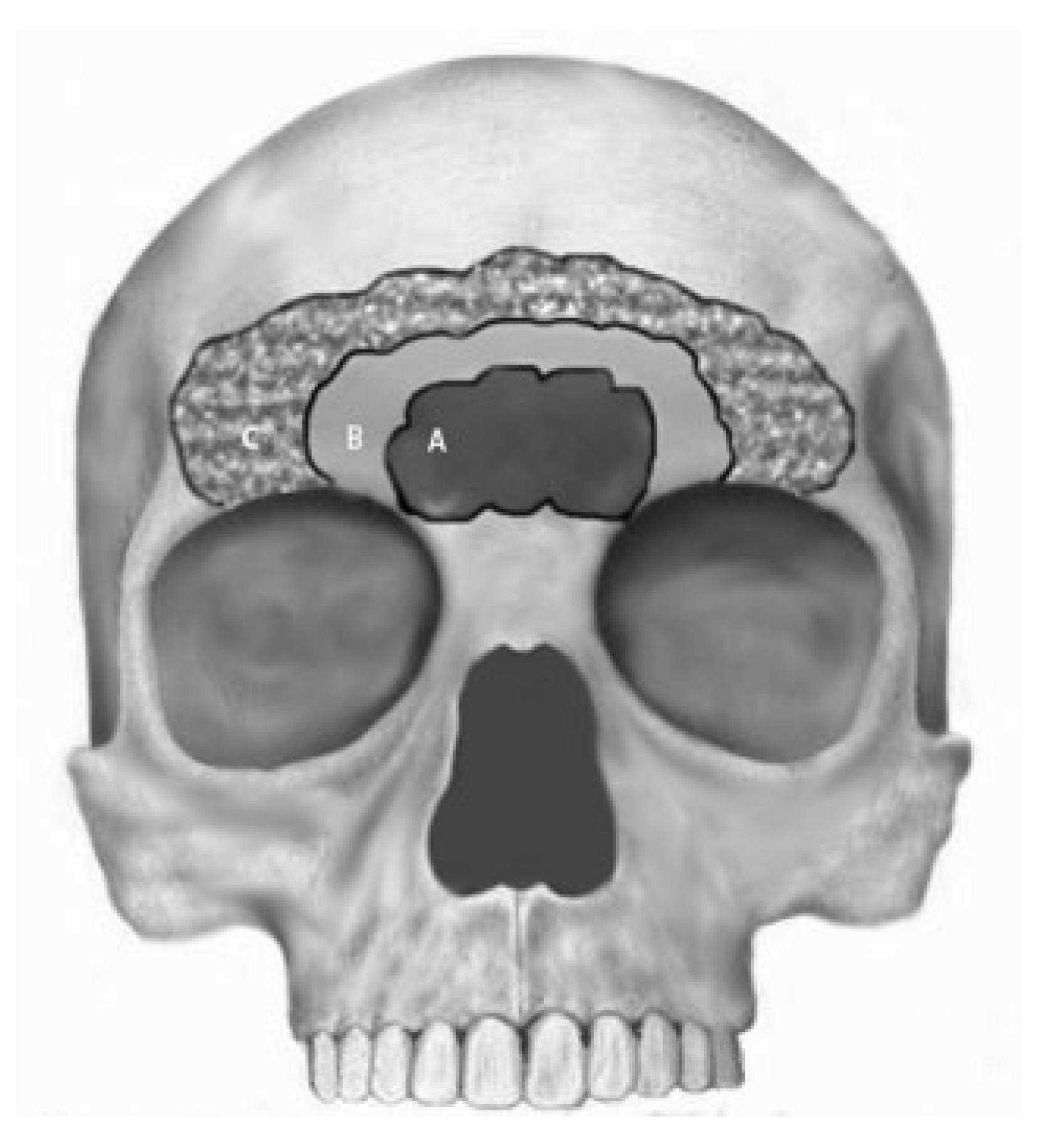
Despite the inexistence of a frontal sinus size classification, the authors classified the size clinically, as small sinus for those that presented the lateral extent going from medial orbital walls one side to other side; as medium sinus those that had the lateral extent going from the half of the orbital cavity one side to other and large sinus those ones that laterally occupied the entire frontal bone going from the lateral orbital wall one side to other (Figure 2).

Results

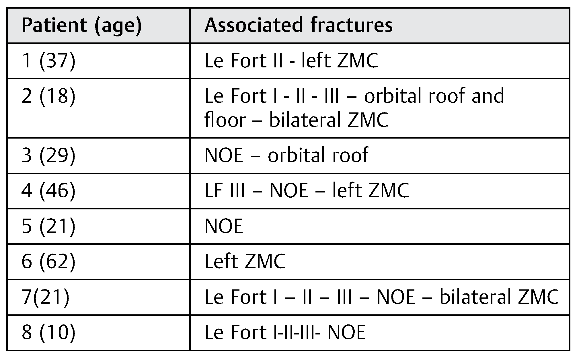
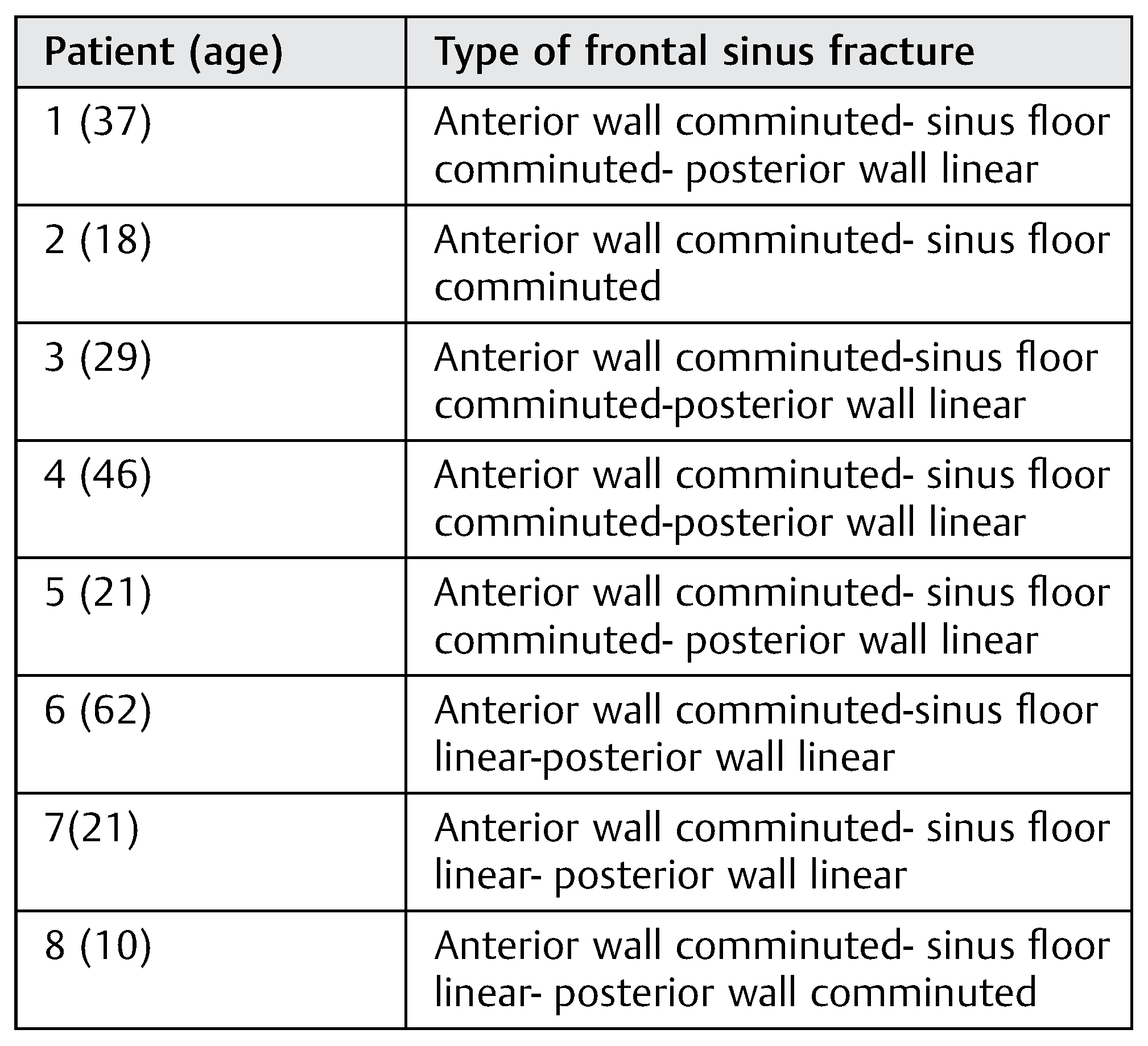
All patients were males, the etiology in 7 patients was motor vehicle accident and the other suffered a work accident caused by truck tire explosion. Age ranged from 10 to 62 years (mean = 30.5 years). The mean follow-up time was 7 years, ranging from 3 to 16 years. Type of frontal sinus fracture is shown in Table 1. Associated fractures are listed in Table 2. Complications were mild frontal asymmetry in one patient; infection in three patients, mucocele in two patients and pain on palpation of the frontal area in one patient (Table 3).
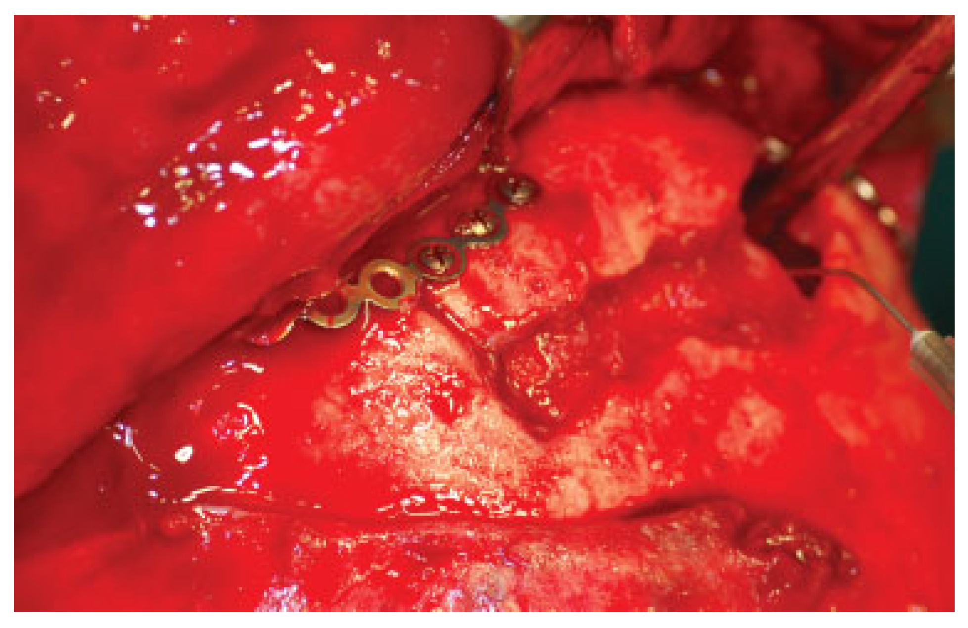
Patient 1 presented with pain and swelling of the frontal region after 2 months postoperatively. Needle aspiration recovered pus in the right side; the culture data did not reveal any kind of growing bacteria. Upon reoperation, most of his frontal sinus was filled by bone, while on the right side there was an area with infected bone chips and granulation tissue (Figure 3). The infected tissue was curetted and drainage to the area which was not obliterated was reestablished to the nose by maintaining a drain for 2 weeks. Empiric intravenous antibiotics were used in the perioperative period and an oral regimen maintained for 14 days after discharge, the postoperative follow-up was daily at the first 7 days and weekly for the next 2 months, and over this time the patient presented no signs of sinus obstruction. After 3 years he presents no signs or symptoms of infection, although he complains of some tenderness on palpation, over the area where a titanium mesh was used for contour, symptoms that may be an initial recurrent mucocele, however until the date evaluated there was no tomographic signs of such alterations.
Patient 3 presented periorbital swelling within 6 months after frontal medium to large sinus obliteration and was initially treated with antibiotics. After 10 months postoperatively a new infectious complication was present and was treated by surgical intervention for debridement, cleaning and new bone obliteration. After that he had another procedure to remove fixation materials, because of fistulation close to the inner cantal tendons. Finally, after one more year, he developed chronic fistulae bilaterally close to the medial palpebral ligaments and at the base of the nose. He was again reoperated and, as in the previous patient, infected bone was debrided and whatever was left of the frontal sinus was treated by reestablishing the drainage to the nose. He then had no more signs or symptoms of infection, but complains of periorbital pain and also pain related to the bicoronal flap area. During the reoperations the authors perform cultures two times, one negative and other positive (Sthaphylococcus sp).
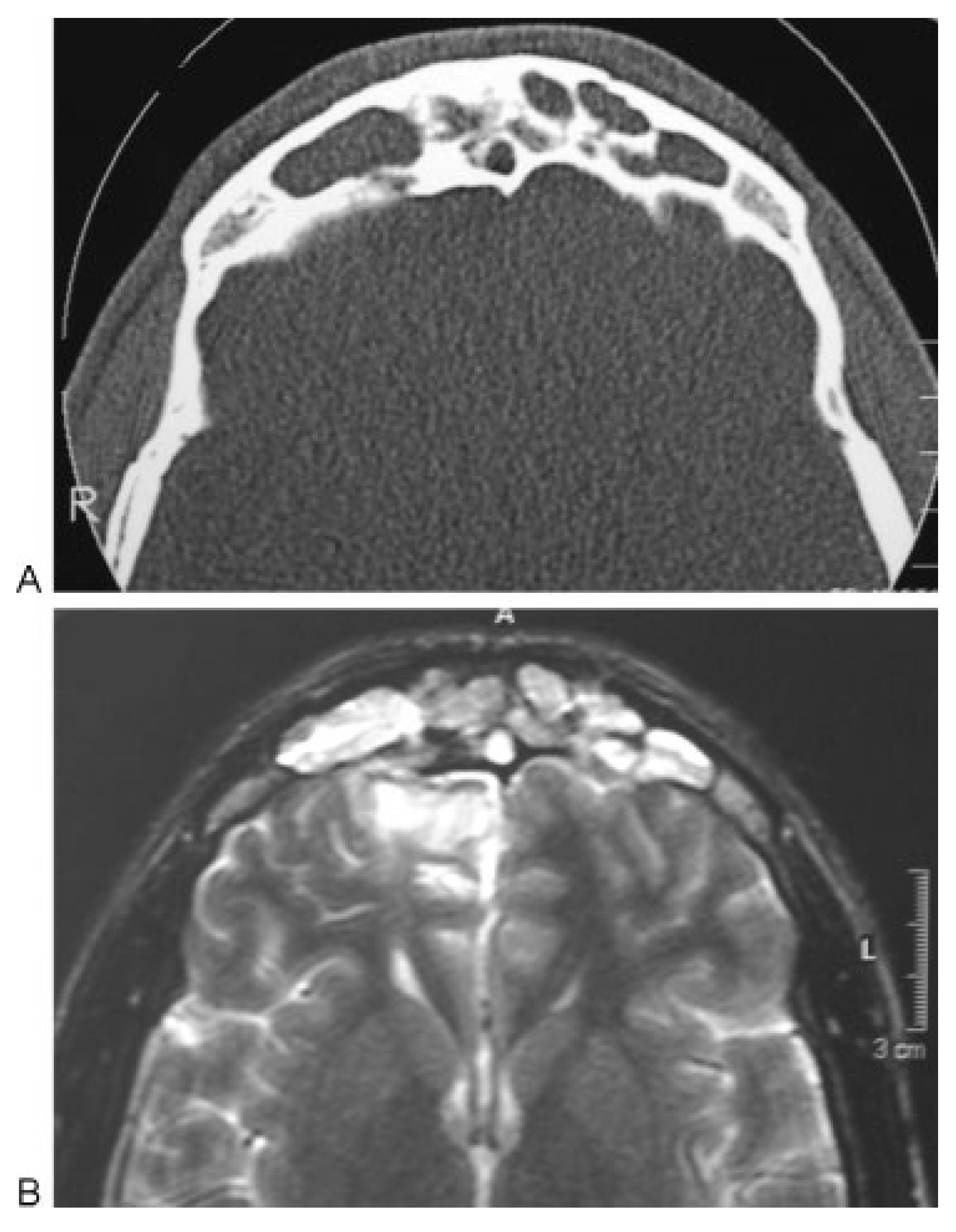
Patient five presented a large sinus. He had uncomplicated initial follow-up. After 6 months, the frontal sinus was filled by bone on CT. After 3 ½ years he had some swelling and redness at the frontal area, managed with antibiotics. After 4 years postoperatively he presented another infectious complication and TC and RNM showed the presence of multiple mucoceles within the sinus (Figure 4A,B). Cranialization of the sinus was proposed. The patient was a heavy smoker and had a very severe bronchial spasm at the induction of general anesthesia, which was very difficult to resolve. The anesthesiologist suspended the procedure and the patient was referred to a pneumologist for preoperative management, but did not accept further surgery. He was oriented about the risks of not being treated and has been followed since then remaining asymptomatic after nine years, although the sinus situation was not changed.
Patient number 8 suffered multiple facial fractures when he was 10 years old. He had a small fractured frontal sinus, which was treated by reconstruction of the anterior wall. After two years he presented with a purulent infection of the sinus. In this case, there was spontaneous obliteration of the sinus drainage, forming a cavity filled with mucosa, which led to infection and resorption of the anterior wall. He was treated by debridement, obliteration of the sinus with iliac crest cancellous bone and reconstruction of the anterior wall with split-thickness calvarial grafts. In his current follow-up he presented with a mucocele invading the right orbital cavity (Figure 5A,B) and will be treated surgically. His only symptom is mild diplopia when very tired or after drinking alcohol. Since he has referred such symptom before, it is not clear if it is related to interference by the mucocele with extrinsic ocular movements, but the patient presents mild proptosis and inferior dystopia.
Thus, four of the patients (50%) presented infections and/ or mucoceles and required secondary surgical treatment. In 2 of those patients, the graft failed, but the sinus was partially obliterated. In two patients the graft completely failed and there was mucocele formation (Table 4).

Discussion

As described by McRae et al, 2008 [16], the most frequent cause for frontal fractures in this sample were motor vehicle accidents. All patients suffered high impact trauma and had significant facial injuries. Frontal sinus fractures correspond from 5 to 12% of all facial fractures [22]. When they are present, associated intracranial, ophthalmologic and other maxillofacial injuries are frequent because of the amount of force required to fracture the frontal bone.
Fractures of the frontal sinus that involve the sinus floor and drainage apparatus or the posterior wall were thought to preclude treating the fracture with sinus reconstructive procedures. More recently clinical evidence was presented, suggesting that many such fractures can be treated by reconstruction of the sinus, provided that the patient is properly followed. Also, when reconstruction fails, more aggressive treatment can still be instituted [7,8,38]. Even dislocated fractures of the posterior wall, which were traditionally an indication for cranialization, can be treated by sinus preservation, if CSF leak is non-existent or resolved. The degree of dislocation does not correlate to the presence of CSF leak [39]. High resolution computed tomography of the frontal region in multiple planes is essential for the prediction of the frontal injury degree, associated injuries, and the type of procedure required [23,24,25].
Traditionally, fractures with involvement of the drainage apparatus were obliterated and those with fractures of the posterior wall were cranialized. Either one of those techniques has actually been used in both instances [1,22,23,25]. There has been much discussion about the ideal material for frontal sinus obliteration [27,28,29,30,31,32,33,34,35,36,37]. Success seems to depend on several details and not exclusively on the type of grafting material.
The use of autologous tissues always presents some degree of donor site morbidity and they may not be available on the required amount. Potential risk of transmission of diseases or reaction to foreign proteins has reduced the use of homogenous or heterogeneous materials. Alloplasts frequently do not do well for frontal sinus obliteration in the clinical setting due to high rates of infection and/or extrusion. When used without reconstruction of the anterior table with bone or titanium mesh they may lead to long-term defects of contour [1,17,19,22,23,26,27,28,29,34,36]
Autologous fat and cancellous bone are frequently used to obliterate the frontal sinus when treating frontal sinus fractures. Successful obliteration requires meticulous extirpation of the sinus mucosa and obliteration of the drainage system. Sealing of the drainage apparatus can be obtained with bone and fascia [32]. Fibrin glue can be additionally used to cover the area. Some failures reported on this study could be result of the lack of complete removal of mucosal instead of the bone graft infection itself.
The appearance of fat and other materials, as well as fibrosis, can pose a challenge when trying to identify suppurative complications [40], even with imaging examination. It is much easier to do that when the sinus is preserved. Particularly in trauma cases where there is also a lot of fixation material in the region, the origin of recurrent chronic fistulae, especially if not directly over the frontal area, as occurred with patient number 3, can be difficult to identify.
Complications observed in this series are the same as previously observed for obliteration procedures [8,21,39]. Reversal of the obliteration is sometimes possible [41] and that was done by open surgery in patients 1 and 3, where part of the grafted bone was resorbed and infection was present. Small frontal sinuses did usually well after obliteration with autologous bone with less complications. Still, patient number 8 had a small sinus but developed a mucocele after several years postoperatively. On the other hand, extensively pneumatized and large frontal sinuses presented many complications requiring reoperation. This may be due to the difficulty of completely removing the mucosa of large sinuses in the acute severe trauma cases. Also, it is not infrequent to find, on the postoperative CT, small empty areas not perceived clinically in large sinuses obliterated with bone. Possibly the large size of the bone graft used on those cases may difficult revascularization, facilitating resorption, necrosis and infection, as well as ingrowth of respiratory mucosa, due to the delayed graft substitution.

Conclusions

Based on this sample, the results suggest that bone obliteration should be avoided due to its high incidence of complications, particularly for large sinus.

Commentary

Frontal Sinus Obliteration with Iliac Crest Particulate Bone Graft
  • Eduardo D. Rodriguez, MD, DDS
  • Professor and Chair
  • NYU Department of Plastic Surgery
  • The Institute of Reconstructive Plastic Surgery
  • New York University School of Medicine
  • New York, New York
  • Paul N. Manson, MD
  • Professor of Surgery
  • Plastic and Reconstructive Surgery
  • R Adams Cowley Shock Trauma Center
  • University of Maryland Distinguished Service
  • Professor Johns Hopkins School of Medicine
  • Baltimore, Maryland
The authors report postoperative results in 8 cases following frontal sinus fractures treated by obliteration with autogenous bone from the anterior iliac crest a single surgical team’s experience. The average postoperative follow-up was 7 years. Radiographs and/or CT scans were used to evaluate results and complications. Four patients (50%) had uneventful long-term follow-ups and four (50%) experienced complications, mainly infection requiring reoperation. The authors conclude that obliteration with autogenous bone has a high percentage of complications. Smaller sinuses have a more favorable outcome with fewer complications. Large, extensively pneumatized sinuses have a high risk of complications and frequently need reoperation.
This is a small series, but one with good follow up. The study lacks statistical power, making it difficult to make conclusive statements with positive correlations. Based on their review, the failure of the bone graft obliteration procedure could in part be due to patient selection (i.e., frontal sinus volume). Our experience is that patients with large frontal sinus volumes are better suited for cranialization techniques, where the mucosal & sinus debridement is more complete, limiting the possibility of infection or mucocoele development. Infection and mucocoele are two distinct processes, and represent differing etiologies for the failure of the surgical procedure. One cannot make a final diagnosis of which pathology is primarily responsible without microbiological confirmation of the type of infection and histological confirmation of mucous membrane in the pathological specimen from the secondary debridement. It is our experience that pathologists do not routinely look for or comment on mucous membrane unless specifically asked to do this Our long term experience with frontal sinus infections did not reveal any routine speciation of bacteria following culture. A more likely conclusion may be that large frontal sinuses, defined by a certain volume, should not be obliterated but rather cranialized, however the data are not present in our series either to support this conclusion with certainty [42,43].
What is needed now is for several authors to quantify the difference between large and small frontal sinuses, so that successful outcome can be related to the frontal sinus volume for a particular treatment procedure. We are slowly learning the right questions to ask, and a multi-institutional study is needed to accumulate enough patients to achieve statistical significance.
Patient 1 presented with pain and swelling of the frontal region 2 months postoperatively. Needle aspiration confirmed pus in the right side. Reoperation included debridement of infected granulation tissue followed by establishing drainage to the nose with a drain for 2 weeks. Intravenous antibiotics were used in the peri-operative period and an oral regimen continued for 14 days after discharge. Three years after the reoperation he presented without signs or symptoms of infection, although he complained of tenderness to palpation over the area of titanium mesh. Was this mucus from retained mucosa, or regrowth of mucosal lining, or was the problem infection around retained metallic material?
In our experience, simple drainage procedures have for infection not been effective and more thorough debridement is required.
Quantitative patient follow up data related to success or failure of the primary procedure, and further characterized with respect to sinus volume, will eventually shed more light on those frontal sinus procedures which were not effective or successful in primary treatment of particularly over-sized sinuses.
In Patient 3, periorbital swelling within 6 months followed frontal sinus obliteration, and was treated with antibiotics. After 10 months a new infection presented which was treated surgically via re-obliteration. Thereafter he developed several episodes of cutaneous fistulae requiring further debridement and eventual re-establishment of drainage to the nose. The patient does not have signs of infection currently, but has persistent periorbital and scalp pain, and is at long term risk for further problems. This failure does not appear to be the result of the bone graft, but rather, could be from incomplete removal of mucosal lining remnants. Rodriguez, et al, have proposed a vascularized bone graft obliteration for infected sinuses with a one stage reconstruction and have encouraging results [44,45].
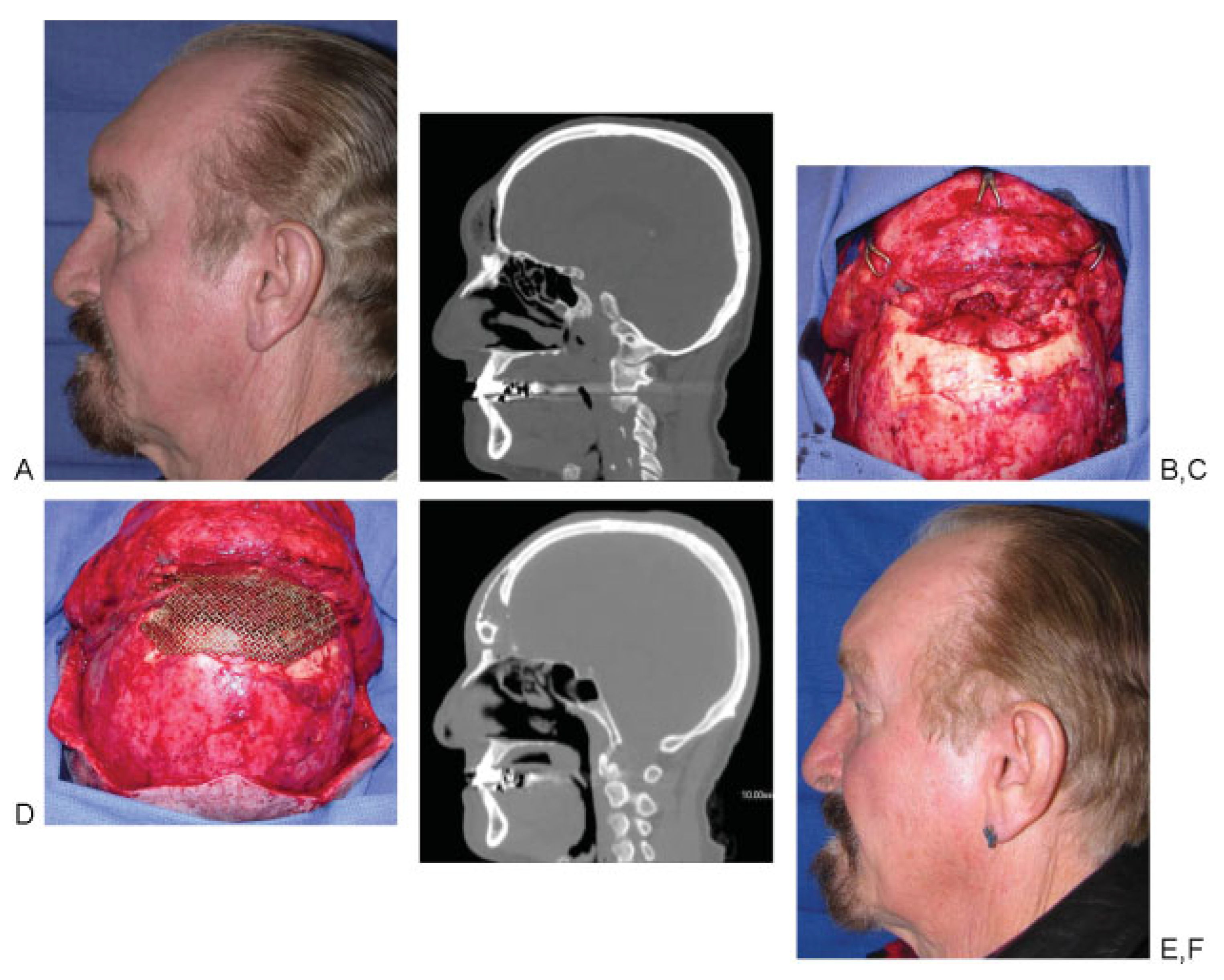
The success of sinus obliteration is entirely dependent on meticulous removal of the sinus mucosa and obliteration of the drainage system and the frontal sinus cavity.44 Characteristically, non-vascularized bone grafts are used in the initial treatment of frontal sinus fractures. It could be that large sinuses are better treated with cranialization techniques, and free vascularized tissue transfers (Figure 6) [42,44,45].
The authors are to be commended for their follow up, and for accurately reporting their series, documenting these problems in the therapy of challenging patients with larger frontal sinuses.

References

  1. Sataloff, R.T.; Sariego, J.; Myers, D.L.; Richter, H.J. Surgical management of the frontal sinus. Neurosurgery 1984, 15, 593–596. [Google Scholar] [CrossRef] [PubMed]
  2. Haug, R.H. Management of fractures of the frontal bone and sinus. In Principles of Oral and Maxillofacial Surgery; Peterson, L.J., Indresano, A.T., Marciani, R.D., et al., Eds.; JB Lippincott: Philadelphia, PA, USA, 1992. [Google Scholar]
  3. Haug, R.H.; Adams, J.M.; Conforti, P.J.; Likavec, M.J. Cranial fractures associated with facial fractures: a review of mechanism, type, and severity of injury. J Oral Maxillofac Surg 1994, 52, 729–733. [Google Scholar]
  4. Stevens, M.; Kline, S.N. Management of frontal sinus fractures. J Craniomaxillofac Trauma 1995, 1, 29–37. [Google Scholar] [PubMed]
  5. Ioannides, C.; Freihofer, H.P.M.; Bruaset, I. Trauma of the upper third of the face. Management and follow-up. J Maxillofac Surg 1984, 12, 255–261. [Google Scholar] [CrossRef] [PubMed]
  6. Godin, D.A.; Miller, R.H. Frontal sinus fractures. J La State Med Soc 1998, 150, 50–55. [Google Scholar]
  7. Carter, K.B., Jr.; Poetker, D.M.; Rhee, J.S. Sinus preservation management for frontal sinus fractures in the endoscopic sinus surgery era: a systematic review. Craniomaxillofac Trauma Reconstr 2010, 3, 141–149. [Google Scholar]
  8. Gabrielli, M.F.R.; Gabrielli, M.A.C.; Hochuli-Vieira, E.; Pereira-Fillho, V.A. Immediate reconstruction of frontal sinus fractures: review of 26 cases. J Oral Maxillofac Surg 2004, 62, 582–586. [Google Scholar]
  9. Bell, R.B.; Dierks, E.J.; Brar, P.; Potter, J.K.; Potter, B.E. A protocol for the management of frontal sinus fractures emphasizing sinus preservation. J Oral Maxillofac Surg 2007, 65, 825–839. [Google Scholar]
  10. Wolfe, S.A.; Johnson, P. Frontal sinus injuries: primary care and management of late complications. Plast Reconstr Surg 1988, 82, 781–791. [Google Scholar] [CrossRef]
  11. Heller, E.M.; Jacobs, J.B.; Holliday, R.A. Evaluation of the frontonasal duct in frontal sinus fractures. Head Neck 1989, 11, 46–50. [Google Scholar]
  12. Helmy, E.S.; Koh, M.L.; Bays, R.A. Management of frontal sinus fractures. Review of the literature and clinical update. Oral Surg Oral Med Oral Pathol 1990, 69, 137–148. [Google Scholar]
  13. Rohrich, R.J.; Hollier, L.H. Management of frontal sinus fractures. Changing concepts. Clin Plast Surg 1992, 19, 219–232. [Google Scholar] [PubMed]
  14. Vrieus, J. Fractures of the frontal sinus: A rationale of treatment. Br J Plast Surg 1993, 46, 208–214. [Google Scholar]
  15. Ioannides, C.; Freihofer, H.P. Fractures of the frontal sinus: classification and its implications for surgical treatment. Am J Otolaryngol 1999, 20, 273–280. [Google Scholar] [CrossRef] [PubMed]
  16. Luce, E.A. Frontal sinus fractures: guidelines to management. Plast Reconstr Surg 1987, 80, 500–510. [Google Scholar] [PubMed]
  17. McRae, M.; Momeni, R.; Narayan, D. Frontal sinus fractures: a review of trends, diagnosis, treatment, and outcomes at a level 1 trauma center in Connecticut. Conn Med 2008, 72, 133–138. [Google Scholar]
  18. Manolidis, S.; Hollier, L.H., Jr. Management of frontal sinus fractures. Plast Reconstr Surg 2007, 120 (Suppl. 2), 32S–48S. [Google Scholar]
  19. Bluebond-Langner, R.; Jackowe, D.; Rodriguez, E.D. Simultaneous obliteration and treatment of infected frontal sinus fractures: novel use of the fibula flap. J Craniofac Surg 2007, 18, 680–683. [Google Scholar] [CrossRef]
  20. Suonpää, J.; Sipilä, J.; Aitasalo, K.; Antila, J.; Wide, K. Operative treatment of frontal sinusitis. Acta Otolaryngol Suppl 1997, 529, 181–183. [Google Scholar] [CrossRef]
  21. Murphy, J.; Jones, N.S. Frontal sinus obliteration. J Laryngol Otol 2004, 118, 637–639. [Google Scholar] [CrossRef]
  22. Gossman, D.G.; Archer, S.M.; Arosarena, O. Management of frontal sinus fractures: a review of 96 cases. Laryngoscope 2006, 116, 1357–1362. [Google Scholar] [PubMed]
  23. Yavuzer, R.; Sari, A.; Kelly, C.P.; Tuncer, S.; Latifoglu, O.; Celevi, M.C.; Jackson, I.T. Management of frontal sinus fractures. Plast Reconstr Surg 2005, 6, 79–95. [Google Scholar]
  24. Bergara, A.R. The obliteration of the sinus in the treatment of chronic frontal sinusitis. Trans Second Pan Am Congr Otorhinolaryng Bronchosophagol 1950, 35. [Google Scholar]
  25. Tato, J.M.; Sibbald, D.W.; Bergaglio, O.E. Surgical treatment of the frontal sinus by the external route. Laryngoscope 1954, 64, 504–521. [Google Scholar]
  26. Haug, R.H.; Likavec, M.J. Frontal sinus reconstruction. Atlas of Oral Maxillofacial Surg Clin N Am 1994, 2, 65–83. [Google Scholar]
  27. Catalano, P.J.; Lawson, W.; Som, P.; Biller, H.F. Radiographic evaluation and diagnosis of the failed frontal osteoplastic flap with fat obliteration. Otolaryngol Head Neck Surg 1991, 104, 225–234. [Google Scholar]
  28. Fattahi, T.; Johnson, C.; Steinberg, B. Comparison of 2 preferred methods used for frontal sinus obliteration. J Oral Maxillofac Surg 2005, 63, 487–491. [Google Scholar]
  29. Taghizadeh, F.; Krömer, A.; Laedrach, K. Evaluation of hydroxyapatite cement for frontal sinus obliteration after mucocele resection. Arch Facial Plast Surg 2006, 8, 416–422. [Google Scholar]
  30. D’Addario, M.; Haug, R.H.; Talwar, R.M. Biomaterials for use in frontal sinus obliteration. J Long Term Eff Med Implants 2004, 14, 455–465. [Google Scholar]
  31. Petruzzelli, G.J.; Stankiewicz, J.A. Frontal sinus obliteration with hydroxyapatite cement. Laryngoscope 2002, 112, 32–36. [Google Scholar]
  32. Rohrich, R.J.; Mickel, T.J. Frontal sinus obliteration: in search of the ideal autogenous material. Plast Reconstr Surg 1995, 95, 580–585. [Google Scholar] [PubMed]
  33. Hochuli Vieira, E.; Real Gabrielli, M.F.; Garcia, I.R., Jr.; Cabrini Gabrielli, M.A. Frontal sinus obliteration with heterogeneous corticocancellous bone versus spontaneous osteoneogenesis in monkeys (Cebus apella), histologic analysis. J Oral Maxillofac Surg 2003, 61, 214–221. [Google Scholar]
  34. Peltola, M.J.; Aitasalo, K.M.; Suonpää, J.T.; Yli-Urpo, A.; Laippala, P.J.; Forsback, A.P. Frontal sinus and skull bone defect obliteration with three synthetic bioactive materials. A comparative study. J Biomed Mater Res B Appl Biomater 2003, 66, 364–372. [Google Scholar] [PubMed]
  35. Weber, R.; Draf, W.; Kahle, G.; Kind, M. Obliteration of the frontal sinus—state of the art and reflections on new materials. Rhinology 1999, 37, 1–15. [Google Scholar]
  36. Elahi, M.M.; Vanduzer, S.; Spears, J.; Gibson, J.; Mitra, A. Frontal sinus obliteration with beta-tricalcium phosphate. J Craniofac Surg 2004, 15, 967–970. [Google Scholar] [PubMed]
  37. Acosta-Feria, M.; Infante-Cossío, P.; Hernández-Guisado, J.M.; et al. [Frontal sinus obliteration using tibial bone graft and platelet-rich plasma for the treatment of chronic osteomyelitis]. Neurocirugia (Astur) 2006, 17, 351–356, discussion 356. [Google Scholar]
  38. Mendonça-Caridad, J.J.; Juiz-Lopez, P.; Rubio-Rodriguez, J.P. Frontal sinus obliteration and craniofacial reconstruction with platelet rich plasma in a patient with fibrous dysplasia. Int J Oral Maxillofac Surg 2006, 35, 88–91. [Google Scholar]
  39. Chen, K.T.; Chen, C.T.; Mardini, S.; Tsay, P.K.; Chen, Y.R. Frontal sinus fractures: a treatment algorithm and assessment of outcomes based on 78 clinical cases. Plast Reconstr Surg 2006, 118, 457–468. [Google Scholar]
  40. Weber, R.; Draf, W.; Keerl, R.; et al. Magnetic resonance imaging following fat obliteration of the frontal sinus. Neuroradiology 2002, 44, 52–58. [Google Scholar]
  41. Kanowitz, S.J.; Batra, P.S.; Citardi, M.J. Comprehensive management of failed frontal sinus obliteration. Am J Rhinol 2008, 22, 263–270. [Google Scholar]
  42. Rodriguez, E.D.; Stanwix, M.G.; Nam, A.J.; et al. Twenty-six-year experience treating frontal sinus fractures: a novel algorithm based on anatomical fracture pattern and failure of conventional techniques. Plast Reconstr Surg 2008, 122, 1850–1866. [Google Scholar] [PubMed]
  43. Manson, P.N.; Stanwix, M.G.; Yaremchuk, M.J.; Nam, A.J.; Hui-Chou, H.; Rodriguez, E.D. Frontobasal fractures: anatomical classification and clinical significance. Plast Reconstr Surg 2009, 124, 2096–2106. [Google Scholar] [PubMed]
  44. Rodriguez, E.D.; Stanwix, M.G.; Nam, A.J.; St Hilaire, H.; Simmons, O.P.; Manson, P.N. Definitive treatment of persistent frontal sinus infections: elimination of dead space and sinonasal communication. Plast Reconstr Surg 2009, 123, 957–967. [Google Scholar]
  45. Bluebond-Langner, R.; Jackowe, D.; Rodriguez, E.D. Simultaneous obliteration and treatment of infected frontal sinus fractures: novel use of the fibula flap. J Craniofac Surg 2007, 18, 680–683. [Google Scholar] [PubMed]
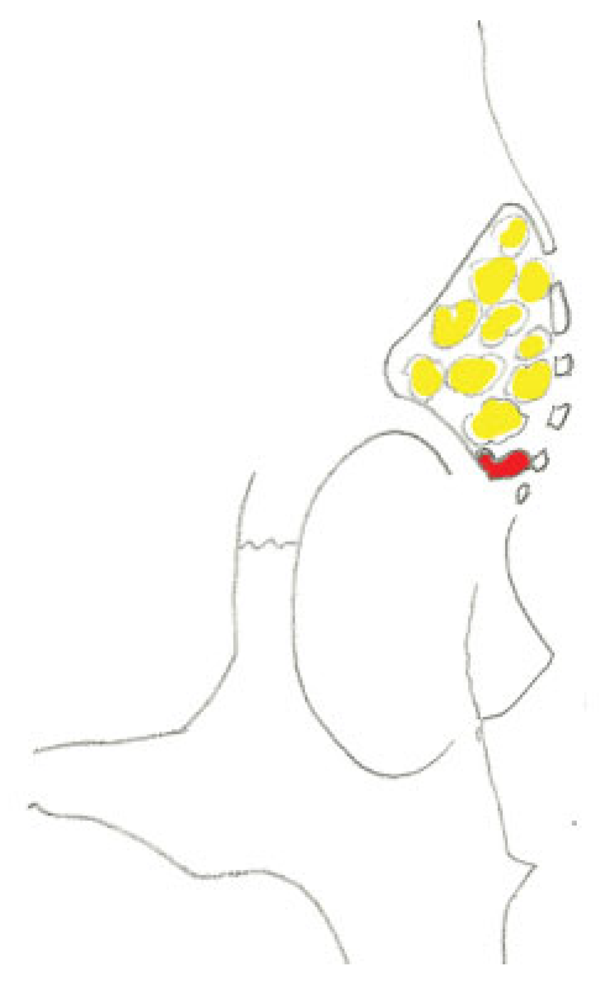
Figure 1. Sketch of the frontal sinus obliteration in a lateral view, note the spongiosa graft (yellow) and the fascia (red) on the sinus floor.
Cmtr 07 00055 g001
Figure 2. Sketch of the frontal sinus size classification adopted by the authors. (A) Small sinus, (B) medium sinus, (C) large sinus.
Cmtr 07 00055 g002
Figure 3. Frontal sinus partially obliterated by bone.
Cmtr 07 00055 g003
Figure 4. (A) Mucocele formation after obliteration on CT. (B) Mucocele formation after obliteration on NMR.
Cmtr 07 00055 g004
Figure 5. (A) Mucocele invading right orbit, axial view. (B) Mucocele invading right orbit, coronal view.
Cmtr 07 00055 g005
Figure 6. (A) A patient with forehead swelling from a mucocoele 20 years after a remote Lefort II and Frontal Sinus Fracture. (B) Lateral CT demonstrating the mucocoele. (C) The mucocoele was removed, the residual sinus cranialized. (D) A free fibula reconstruction was performed to obliterate the entire cranialized space with vascularized tissue. A titanium mesh grid was placed over the vascularized reconstruction for proper contour. (E) A post operative lateral CT demonstrated the free fibula reconstruction to obliterate “dead space” in situ. (F) A late image demonstrating retention of good contour, appearance, and absence of infection.
Cmtr 07 00055 g006
Table 1. Age and associated fractures.
Cmtr 07 00055 t001
Table 2. Type of frontal sinus fracture.
Cmtr 07 00055 t002
Table 3. Complications and current status.
Cmtr 07 00055 t002
Table 4. Probable cause of the complications.
Cmtr 07 00055 t004