Next Article in Journal
Management of Carotid Artery Trauma
Previous Article in Journal
Nasofrontal Outflow Tract Visibility in Computed Tomography Imaging of Frontal Sinus Fractures
 
 
Craniomaxillofacial Trauma & Reconstruction is published by MDPI from Volume 18 Issue 1 (2025). Previous articles were published by another publisher in Open Access under a CC-BY (or CC-BY-NC-ND) licence, and they are hosted by MDPI on mdpi.com as a courtesy and upon agreement with Sage.
Review

Transconjunctival Inferior Orbitotomy: Indications, Surgical Technique, and Complications

by
Brett W. Davies
1,
Eric M. Hink
1 and
Vikram D. Durairaj
1,2,3,*
1
Oculofacial Plastic and Orbital Surgery, Department of Ophthalmology, University of Colorado School of Medicine, Aurora, CO, USA
2
Department of Otolaryngology-Head and Neck Surgery, University of Colorado School of Medicine, Aurora, CO, USA
3
Texas Oculoplastic Consultants, Austin, TX, USA
*
Author to whom correspondence should be addressed.
Craniomaxillofac. Trauma Reconstr. 2014, 7(3), 169-174; https://doi.org/10.1055/s-0034-1374063
Submission received: 4 August 2013 / Revised: 7 August 2013 / Accepted: 7 August 2013 / Published: 22 May 2014

Abstract

:
Surgical access to the inferior orbit can be accomplished through either a transcutaneous or transconjunctival incision. The preferred approach should provide adequate surgical exposure with the fewest adverse effects. The purpose of this article is to review the literature on the transconjunctival incision and to discuss the indications and complications of the approach. The authors also discuss their preferred technique and provide a step-by-step instruction. The transconjunctival approach provides good surgical access with a low incidence of complications and a better aesthetic outcome than transcutaneous approaches.

Surgical access to the orbit must balance adequate exposure with patient expectations of aesthetic outcome. Minimally invasive surgical techniques are no longer considered a luxury and surgical outcome is often judged in part by the size and location of a visible scar. Considerable research and effort has been placed into gaining surgical access to the structures of the upper face through incisions that are well camouflaged. Perhaps nowhere does this hold more true than the periorbital area, where poor incision placement can have both functional and aesthetic consequences. Most authors believe that the transconjunctival lower eyelid incision is the best surgical approach to the inferior orbit, allowing excellent surgical exposure with the benefit of a very low complication rate and a well-hidden scar [1,2,3]. The purpose of this article is to review existing literature comparing the transconjunctival to transcutaneous approaches to the inferior orbit, as well as to review the indications, techniques, and complications of the transconjunctival approach.
The transconjunctival incision was first described for aesthetic blepharoplasty in 1924 by Bourget [4]. Its use became more popular for repair of orbital wall fractures in the 1970s [5,6]. Surgeons using the incision found that it provided excellent access for fracture reduction while avoiding a visible scar and complications associated with cutaneous incisions [7]. These findings led the next generation of surgeons to look critically at the transconjunctival incision, comparing it to the previously favored cutaneous incisions such as the subciliary and subtarsal approaches [8,9]. The subciliary approach was first described by Converse in 1944, and involves a cutaneousincision 1 to 2 mm below the lash line [1]. Whereas the subtarsal incision, also popularized by Converse, is placed in a natural skin line of the lower eyelid below the level of the tarsus [2].
Numerous studies have compared the transconjunctival to cutaneous incisions of the lower eyelid in an effort to evaluate the incidence of complications associated with each approach, notably scar formation and lower eyelid malposition. Almost all of the published research in this area is retrospective in nature and evaluated in the setting of trauma. Most studies have found that the cutaneous incision results in a higher incidence of eyelid malposition [1,10,11,12,13,14] or scar formation [15,16] in comparison to the transconjunctival approach. Fewer studies have found no statistical difference in either eyelid malposition or scar formation [17,18]. Even these studies found a higher incidence of complications from the transcutaneous incisions, but they did not have enough power to find a statistically significant difference.
Unfortunately, there is a paucity of randomized, prospective studies evaluating lower eyelid incisions [2,19], and only one prospective study comparing cutaneous versus transconjunctival incisions [2]. A prospective study by Holtmann et al. found a high rate of eyelid malposition (42%) in the subciliary group compared with the transconjunctival group (0%) [2]. As a consequence of these results, the majority of surgeons authoring the above studies concluded that the transconjunctival incision is the preferred approach to the inferior orbit.
There have been several meta-analyses reviewing eyelid malposition associated with lower eyelid incisions. Ridgway et al. performed a retrospective review and a meta-analysis comparing subciliary, subtarsal, and transconjunctival approaches with respect to lower lid malposition [11]. Their meta-analysis included the data from all retrospective studies from 1980 to 2005. A total of 2086 patients from 17 studies were included in the analysis with 11.4% of patients undergoing a subtarsal incision, 22.5% a subciliary incision, and 66.1% a transconjunctival incision. In this group of 2086 patients, the overall incidence of eyelid malposition was 5.1%, with ectropion occurring more frequently than entropion (4.7 vs. 0.5%, respectively). The subciliary incision was associated with the highest rates of ectropion (14%) versus the subtarsal (3.8%) or the transconjunctival incision (1.5%). Entropion was encountered more in the transconjunctival approach (0.7%) versus the subtarsal (0%) and subciliary approaches (0.2%). The majority of patients with eyelid malposition were managed conservatively with taping and massaging. Lower lid edema was seen in both subtarsal (2.5%) and subciliary (3.6%) approaches but not observed in the transconjunctival approach. A noticeable scar was more frequent with the subtarsal approach (3.4%) compared with the subciliary (0.9%) and the transconjunctival approach (0%).
Kothari et al. recently performed a meta-analysis on incisions used for orbital floor exploration in the setting of trauma [20]. The data set excluded review articles and case series of less than 10 patients. They found 31 articles that met inclusion criteria published between 1975 and 2011, representing 4,668 incisions in 4,406 patients. Their analysis showed that transconjunctival incisions had the lowest rate of complications. Subciliary incisions had the highest rate of complications with ectropion in 12.9% of patients, and 11% of patients requiring surgical correction. Subtarsal incisions had the lowest rate of surgical correction but a high rate of visible scar formation at 5.6%. In fact, a paper they reviewed by Feldman et al. showed that over 50% of patients were left with a “noticeable” or “extremely noticeable” scar [21]. Kothari et al. concluded that the transconjunctival incision had the fewest complications and most aesthetically pleasing results but had the steepest learning curve [20]. They found that scar formation and ectropion became more common complications when the transconjunctival incision was combined with a lateral canthotomy, although this is often not necessary [22].

Indications

The utility of the transconjunctival incision has been well documented in the literature, and new indications for its use in the surgical treatment of orbital pathology are published each year. The majority of studies discuss the transconjunctival incision in the setting of trauma, most commonly orbital floor and zygomaticomaxillary complex (ZMC) fractures [1,2,12,16,23,24]. The incision can be combined with a transcaruncular approach to treat combined orbital floor and medial wall fractures [24,25,26], and with a lateral canthotomy for wide exposure of the lateral rim and wall [15,24]. The approach has also been used to gain access to the orbit for incisional biopsy of optic nerve tumors [27], incisional and excisional biopsy of inferior orbital tumors [28,29], as well as drainage of inferior orbital abscesses and inflammatory debris [30]. Figure 1 shows an example of the transconjunctival approach to remove an inferior orbital cyst. Orbital decompression for treatment of thyroid eye disease [31,32,33] and fibrous dysplasia [34] have been described through a transconjunctival incision as well. The transconjunctival approach has been well described for repair of eyelid malpositions such as entropion [14], ectropion [35], and eyelid retraction from conditions such as thyroid eye disease and postblepharoplasty syndrome [36,37]. In addition to these orbital disorders, the transconjunctival approach has been used for access to surrounding structures including its use for dacryocystorhinostomy [38], total maxillectomy [39], and for neurosurgical access to the anterior skull base [40].
Beyond these functional indications, aesthetic surgeons use the transconjunctival incision as the primary starting point for lower eyelid blepharoplasties. The incision provides quick and reliable access to the lower lid fat pads [9,17,41,42]. From this incision, the fat can be judiciously excised or repositioned in a pre or subperiosteal plane [43]. The transconjunctival incision has also been utilized for midface lifts. Seitz et al. examined their results from patients who underwent transconjunctival deep plane midface lift [44]. Of 124 cases, no significant complications were reported, and only one patient needed reexcision of redundant lower eyelid skin. The combination of adequate exposure with an imperceptible scar makes the incision a very attractive approach to both the discerning patient and surgeon alike.

Technique

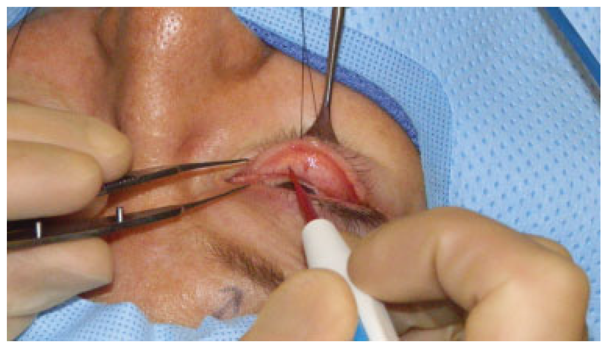
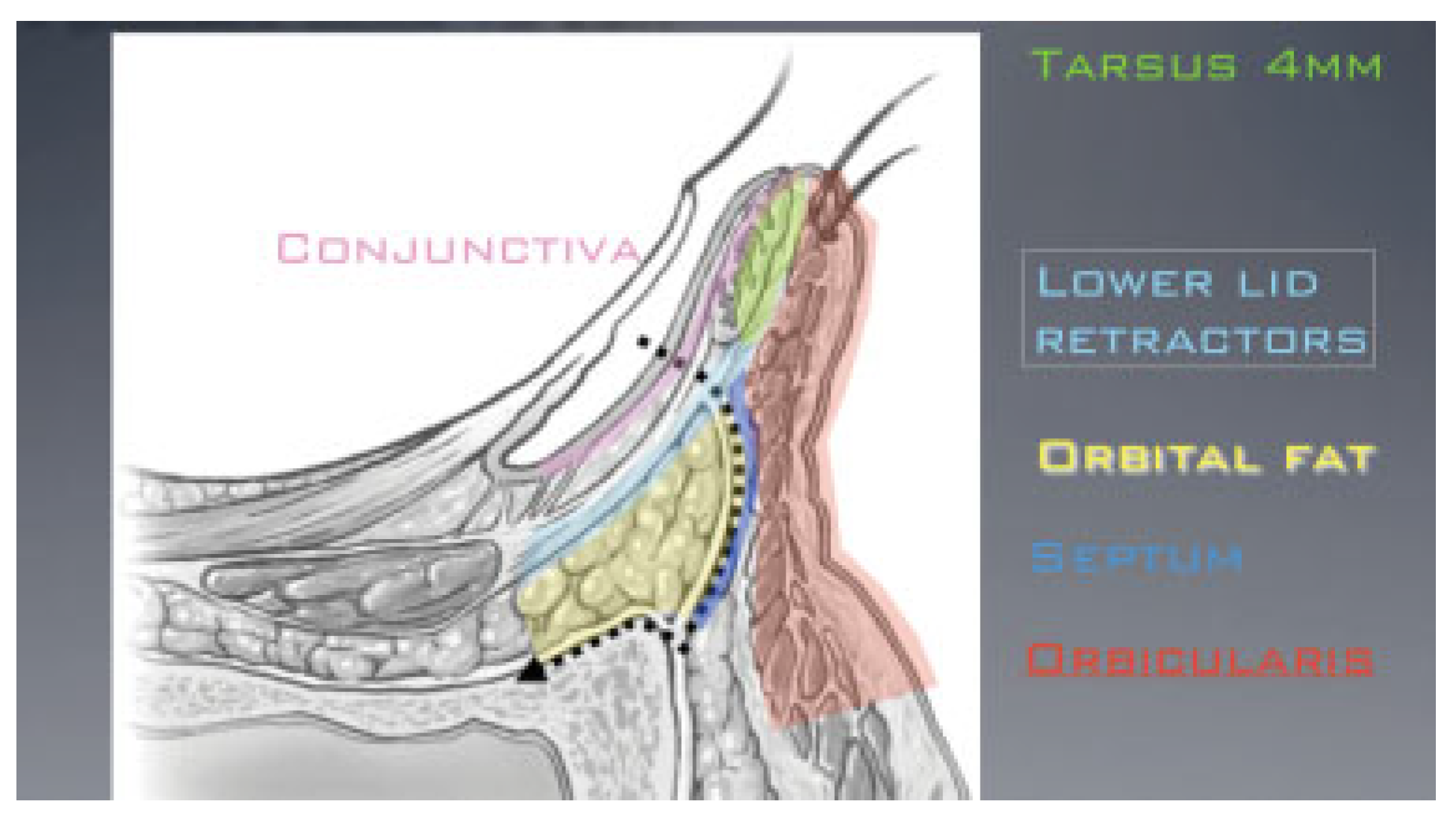
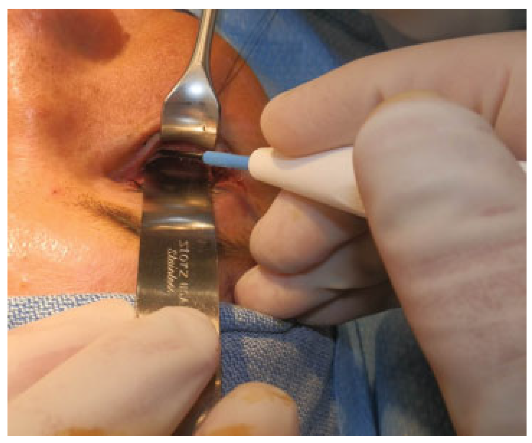
The senior author (V.D.D.) has used the transconjunctival approach over 1000 times for lower eyelid surgery and access to the inferior orbit. His preferred surgical technique for exposure to the orbit is described. After infiltration of the lower eyelid with 1% lidocaine with epinephrine 1:100,000, a 5–0 silk suture passed through the eyelid margin, specifically the gray line. The eyelid is everted over a Desmarres retractor (Robbins Instruments Inc., Chatham, NJ) and the conjunctival incision is made with a scalpel, disposable high-temperature cautery, or monopolar electrocautery needle 3 mm inferior to the tarsal border (Figure 2). The dissection is carried through the insertion of the lower lid retractors, and then inferiorly in an avascular plane toward the orbital rim (Figure 3).
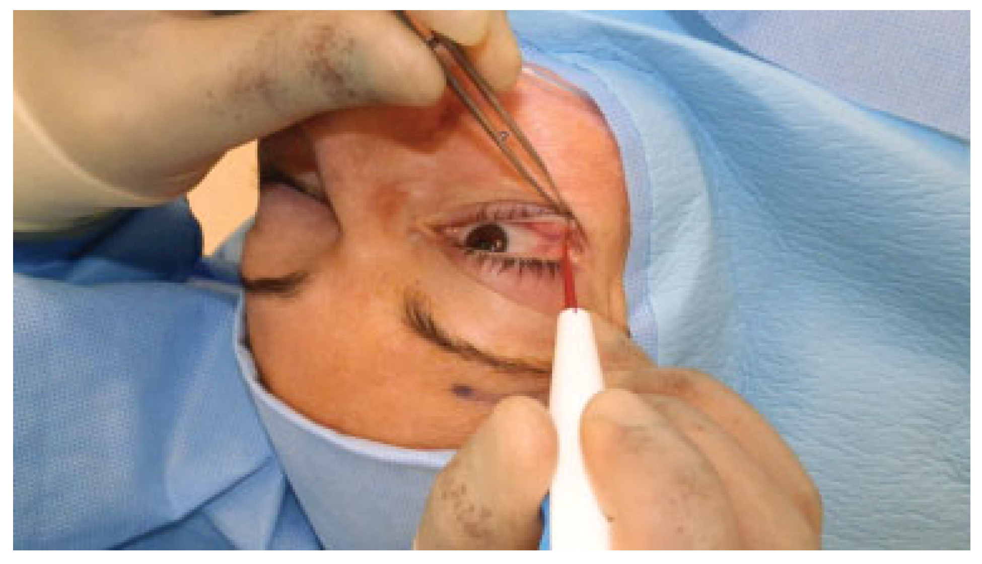
The dissection can proceed in either a preseptal or postseptal plane. The preseptal approach (preferred) avoids the orbital fat manipulation and obscuration of the surgical view from prolapsed orbital fat. Conversely, the postseptal plane provides a more direct approach to the orbital fat pads, and is particularly useful in lower eyelid blepharoplasty [45]. Once the dissection has reached the level of the orbital rim, a malleable retractor is used to retract the orbital tissues, and the electrocautery needle is used to make an incision in the periosteum along the extent of the inferior orbital rim (Figure 4). Access to the subperiosteal space along the orbital floor or the face of the zygoma is easily accomplished through careful elevation of the periosteum, typically with a periosteal elevator.
Periosteum is not closed unless plate fixation of the orbital rim is performed. When closure is indicated, we use interrupted 5–0 polyglactin suture. The conjunctiva is typically closed with one buried 7–0 polyglactin suture in the central location. In our opinion, closure of the conjunctiva reestablishes the normal anatomic position of the lower eyelid retractors. We have rarely observed both subtle elevation of the lower eyelid and disinsertion of lower lid retractors resulting in obscuration of vision in down gaze when the incision is not closed. Care is taken not to incorporate the orbital septum in the closure as this may lead to postoperative entropion. A Frost suture is not routinely placed.
While the need for lateral canthotomy when using a transconjunctival approach varies with studies [22,24,26,46,47,48], in our hands it provides much improved surgical access when necessary with minimal morbidity. This approach first starts with a canthotomy. A 15 blade is used to make a skin incision at the lateral canthus. The size of the skin incision is based on the procedure performed. For access to the orbital floor, the incision is kept short (5 mm). When more exposure of the lateral rim is needed, including access to the frontozygomatic suture, the incision is lengthened as necessary with care to place it in a natural skin crease. Dissection is then carried down to, but not through, the periosteum using the electrocautery needle. The canthotomy is then completed with tenotomy scissors, and the inferior cantholysis is completed with the electrocautery needle while holding upward traction on the lower eyelid (Figure 5). The transconjunctival incision is then made as previously described. Typical closure of the lateral canthotomy involves fixating the tarsus to the superior crus of the lateral canthal tendon and then to the inner aspect of the periosteum of the lateral orbital wall using 5–0 polyglactin suture, and layered closure of the orbicularis and skin with absorbable suture. This combination has proven to provide excellent surgical access to 270 degrees of the orbit including the inferior rim, floor, lateral rim, and lateral wall of the orbit (Figure 6) [24,49,50].
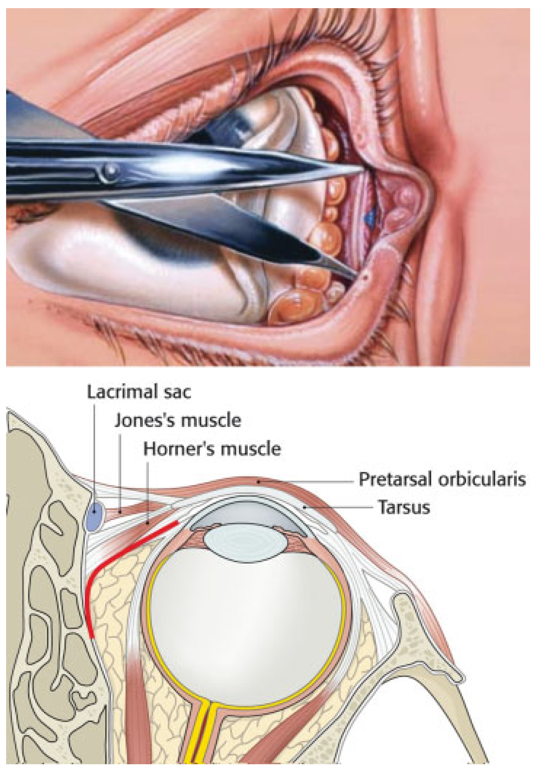
When access to the medial orbit is necessary, the transconjunctival incision can be combined with a transcaruncular incision [24,25]. This combination allows wide access to the orbital floor and medial wall. For this approach, an incision is placed through the caruncle using Westcott scissors. The incision is carried superiorly and inferiorly into the conjunctiva for approximately 15 mm in each direction. Blunt dissection with tenotomy scissors is then carried down to the posterior lacrimal crest in a plane between the orbital septum and Horner muscle (Figure 7) [51]. Once periosteum is identified, it is opened with the electrocautery needle and elevated to widely expose the medial orbital wall.

Complications

Although uncommon, complications from the transconjunctival incision exist. Improper dissection can cause canalicular injury or a buttonhole full-thickness laceration of the lower eyelid. Early transient complications can include edema, epiphora, chemosis, trichiasis, and diplopia [24,52]. Later complications include conjunctival granuloma, ectropion, entropion, intractable chemosis, lower lid retraction, and lower lid malposition secondary to postoperative scar and excessive retraction (Figure 8) [53,54,55]. The percentage of all complications across the literature is approximately 10%, with most of the complications resolving with observation [3,52]. The most common complication requiring intervention is entropion, with an average reported incidence of 0.5% [53] (studies range from 0 to 4%) [8,10,22,24,52,53]. The incidence of complications likely increases with the length of the surgical wound. Ridgway et al. reported the complication rate of 4% (2 of 45 cases) when combined with lateral canthotomy and inferior cantholysis in addition to subperiosteal exposure along the body of the zygoma for ZMC fracture repair. They also noted that neither Frost sutures nor tarsorrhaphy were used in these patients, which seemed to be protective in other cases [53,56].
Other potential downside to the transconjunctival incision include steeper learning curve and longer operative time to exposure compared with cutaneous approaches [2,16,55,57]. We disagree that increased operative time is associated with the transconjunctival approach. Typically, the transconjunctival approach combined with a lateral canthotomy takes less than 5 minutes for exposure and equal time for closure. The transconjunctival incision is relatively contraindicated in cases of unstable or high risk of globe injury, persistent chemosis, acute or chronic conjunctival disease, and preexisting lid lacerations exposing bone [52,57].
The surgeon performing the transconjunctival incision is advised to pay close attention to the anatomic structures and dissection planes of the lower lid. An incision too high can result in injury to the tarsus [15], whereas an incision too low can damage the inferior oblique [55]. A preseptal plan of dissection has been described as having lower rates of entropion and retraction [46,58], but the authors have not found this to be the case in our experience. When combined with a lateral canthotomy, there is increased risk of noticeable scar, lid malposition, and lateral canthal rounding [15,59]. Again, with good surgical planning and technique, visible scar and lid malposition can be minimized. When combined with a transcaruncular incision, the complication profile includes injury to the globe, eyelid, lacrimal system, and inferior oblique [1,55,60]. Overall, the transcaruncular extension is considered well tolerated [46,61,62].

Conclusion

In summary, the transconjunctival approach provides excellent surgical access to the inferior orbit and is associated with a low incidence of complications and a better aesthetic outcome than transcutaneous approaches. It can be easily combined with a lateral canthotomy or transcaruncular incision for wide access to the orbital floor, lateral orbital wall, and anterior zygoma. We believe the literature supports our perspective that the transconjunctival incision is the gold standard surgical approach for access to the inferior orbit.

References

  1. Wray, R.C.; Holtmann, B.; Ribaudo, J.M.; Keiter, J.; Weeks, P.M. A comparison of conjunctival and subciliary incisions for orbital fractures. Br J Plast Surg 1977, 30, 142–145. [Google Scholar] [CrossRef] [PubMed]
  2. Holtmann, B.; Wray, R.C.; Little, A.G. A randomized comparison of four incisions for orbital fractures. Plast Reconstr Surg 1981, 67, 731–737. [Google Scholar] [CrossRef] [PubMed]
  3. Kushner, G.M. Surgical approaches to the infraorbital rim and orbital floor: The case for the transconjunctival approach. J Oral Maxillofac Surg 2006, 64, 108–110. [Google Scholar] [CrossRef]
  4. Bourget, J. Les hernies graisseuses de l’orbite. Notre traitement chirurgical. Bull Acad Nat Med 1924, 92, 1227–1232. [Google Scholar]
  5. Tessier, P. The conjunctival approach to the orbital floor and maxilla in congenital malformation and trauma. J Maxillofac Surg 1973, 1, 3–8. [Google Scholar] [CrossRef] [PubMed]
  6. Tenzel, R.R.; Miller, G.R. Orbital blow-out fracture repair, conjunctival approach. Am J Ophthalmol 1971, 71, 1141–1142. [Google Scholar] [CrossRef]
  7. Rohrich, R.J.; Janis, J.E.; Adams, W.P., Jr. Subciliary versus subtarsal approaches to orbitozygomatic fractures. Plast Reconstr Surg 2003, 111, 1708–1714. [Google Scholar] [CrossRef] [PubMed]
  8. Patel, P.C.; Sobota, B.T.; Patel, N.M.; Greene, J.S.; Millman, B. Comparison of transconjunctival versus subciliary approaches for orbital fractures: A review of 60 cases. J Craniomaxillofac Trauma 1998, 4, 17–21. [Google Scholar] [PubMed]
  9. Jacono, A.A.; Moskowitz, B. Transconjunctival versus transcutaneous approach in upper and lower blepharoplasty. Facial Plast Surg 2001, 17, 21–28. [Google Scholar] [CrossRef]
  10. Appling, W.D.; Patrinely, J.R.; Salzer, T.A. Transconjunctival approach vs subciliary skin-muscle flap approach for orbital fracture repair. Arch Otolaryngol Head Neck Surg 1993, 119, 1000–1007. [Google Scholar] [CrossRef]
  11. Ridgway, E.B.; Chen, C.; Colakoglu, S.; Gautam, S.; Lee, B.T. The incidence of lower eyelid malposition after facial fracture repair: A retrospective study and meta-analysis comparing subtarsal, subciliary, and transconjunctival incisions. Plast Reconstr Surg 2009, 124, 1578–1586. [Google Scholar] [CrossRef] [PubMed]
  12. Raschke, G.F.; Rieger, U.M.; Bader, R.D.; Schaefer, O.; Guentsch, A.; Schultze-Mosgau, S. Transconjunctival versus subciliary approach for orbital fracture repair—An anthropometric evaluation of 221 cases. Clin Oral Investig 2013, 17, 933–942. [Google Scholar] [CrossRef] [PubMed]
  13. Salgarelli, A.C.; Bellini, P.; Landini, B.; Multinu, A.; Consolo, U. A comparative study of different approaches in the treatment of orbital trauma: An experience based on 274 cases. Oral Maxillofac Surg 2010, 14, 23–27. [Google Scholar] [CrossRef] [PubMed]
  14. Ben Simon, G.J.; Molina, M.; Schwarcz, R.M.; McCann, J.D.; Goldberg, R.A. External (subciliary) vs internal (transconjunctival) involutional entropion repair. Am J Ophthalmol 2005, 139, 482–487. [Google Scholar] [CrossRef]
  15. De Riu, G.; Meloni, S.M.; Gobbi, R.; Soma, D.; Baj, A.; Tullio, A. Subciliary versus swinging eyelid approach to the orbital floor. J Craniomaxillofac Surg 2008, 36, 439–442. [Google Scholar] [CrossRef]
  16. Subramanian, B.; Krishnamurthy, S.; Suresh Kumar, P.; Saravanan, B.; Padhmanabhan, M. Comparison of various approaches for exposure of infraorbital rim fractures of zygoma. J Maxillofac Oral Surg 2009, 8, 99–102. [Google Scholar] [CrossRef] [PubMed]
  17. Netscher, D.T.; Patrinely, J.R.; Peltier, M.; Polsen, C.; Thornby, J. Transconjunctival versus transcutaneous lower eyelid blepharoplasty: A prospective study. Plast Reconstr Surg 1995, 96, 1053–1060. [Google Scholar] [CrossRef]
  18. Nowinski, D.; Messo, E.; Hedlund, A. Treatment of orbital fractures: Evaluation of surgical techniques and materials for reconstruction. J Craniofac Surg 2010, 21, 1033–1037. [Google Scholar] [CrossRef]
  19. Lacy, M.F.; Pospisil, O.A. Lower blepharoplasty post-orbicularis approach to the orbit—A prospective study. Br J Oral Maxillofac Surg 1987, 25, 398–401. [Google Scholar] [CrossRef]
  20. Kothari, N.A.; Avashia, Y.J.; Lemelman, B.T.; Mir, H.S.; Thaller, S.R. Incisions for orbital floor exploration. J Craniofac Surg 2012, 23 (7, Suppl. 1), 1985–1989. [Google Scholar] [CrossRef]
  21. Feldman, E.M.; Bruner, T.W.; Sharabi, S.E.; Koshy, J.C.; Hollier, L.H., Jr. The subtarsal incision: Where should it be placed? J Oral Maxillofac Surg 2011, 69, 2419–2423. [Google Scholar] [CrossRef] [PubMed]
  22. Zingg, M.; Chowdhury, K.; Lädrach, K.; Vuillemin, T.; Sutter, F.; Raveh, J. Treatment of 813 zygoma-lateral orbital complex fractures. New aspects. Arch Otolaryngol Head Neck Surg 1991, 117, 611–620, discussion 621–622. [Google Scholar] [CrossRef] [PubMed]
  23. Santosh, B.S.; Giraddi, G. Transconjunctival preseptal approach for orbital floor and infraorbital rim fracture. J Maxillofac Oral Surg 2011, 10, 301–305. [Google Scholar] [CrossRef] [PubMed]
  24. Kim, D.W.; Choi, S.R.; Park, S.H.; Koo, S.H. Versatile use of extended transconjunctival approach for orbital reconstruction. Ann Plast Surg 2009, 62, 374–380. [Google Scholar] [CrossRef]
  25. Lee, C.S.; Yoon, J.S.; Lee, S.Y. Combined transconjunctival and transcaruncular approach for repair of large medial orbital wall fractures. Arch Ophthalmol 2009, 127, 291–296. [Google Scholar] [CrossRef]
  26. Shorr, N.; Baylis, H.I.; Goldberg, R.A.; Perry, J.D. Transcaruncular approach to the medial orbit and orbital apex. Ophthalmology 2000, 107, 1459–1463. [Google Scholar] [CrossRef]
  27. Khong, J.J.; McNab, A.A. Medial transconjunctival intrinsic optic nerve biopsy: Surgical technique and indications. Orbit 2012, 31, 227–232. [Google Scholar] [CrossRef]
  28. Carter, J.B. Transconjunctival resection of an orbital dermoid tumour. J Maxillofac Surg 1985, 13, 239–242. [Google Scholar] [CrossRef]
  29. Cho, K.J.; Paik, J.S.; Yang, S.W. Surgical outcomes of transconjunctival anterior orbitotomy for intraconal orbital cavernous hemangioma. Korean J Ophthalmol 2010, 24, 274–278. [Google Scholar] [CrossRef]
  30. Schick, U.; Hassler, W. Neurosurgical management of orbital inflammations and infections. Acta Neurochir (Wien) 2004, 146, 571–580, discussion 580. [Google Scholar] [CrossRef]
  31. Perry, J.D.; Kadakia, A.; Foster, J.A. Transcaruncular orbital decompression for dysthyroid optic neuropathy. Ophthal Plast Reconstr Surg 2003, 19, 353–358. [Google Scholar] [CrossRef] [PubMed]
  32. Barkhuysen, R.; Nielsen, C.C.; Klevering, B.J.; Van Damme, P.A. The transconjunctival approach with lateral canthal extension for three-wall orbital decompression in thyroid orbitopathy. J Craniomaxillofac Surg 2009, 37, 127–131. [Google Scholar] [CrossRef] [PubMed]
  33. O’Malley, M.R.; Meyer, D.R. Transconjunctival fat removal combined with conservative medial wall/floor orbital decompression for Graves orbitopathy. Ophthal Plast Reconstr Surg 2009, 25, 206–210. [Google Scholar] [CrossRef] [PubMed]
  34. Anastassov, Y.C.; Anastassov, G.E.; Lumeran, H.S. Craniofacial fibrous dysplasia, conservative management: Review of the literature and case report. Folia Med (Plovdiv) 2004, 46, 56–61. [Google Scholar]
  35. Jacono, A.A.; Stong, B.C. Combined transconjunctival release and midface-lift for postblepharoplasty ectropion repair. Arch Facial Plast Surg 2010, 12, 206–208. [Google Scholar] [CrossRef]
  36. Shorr, N. Madame Butterfly procedure with hard palate graft: Management of postblepharoplasty round eye and scleral show. Facial Plast Surg 1994, 10, 90–118. [Google Scholar] [CrossRef]
  37. Edelstein, C.; Balch, K.; Shorr, N.; Goldberg, R.A. The transeyelid subperiosteal midface-lift in the unhappy postblepharoplasty patient. Semin Ophthalmol 1998, 13, 107–114. [Google Scholar] [CrossRef]
  38. Kaynak-Hekimhan, P.; Yilmaz, O.F. Transconjunctival dacryocystorhinostomy: Scarless surgery without endoscope and laser assistance. Ophthal Plast Reconstr Surg 2011, 27, 206–210. [Google Scholar] [CrossRef]
  39. Goyal, A.; Tyagi, I.; Jain, S.; Syal, R.; Singh, A.P.; Kapila, R. Transconjunctival incision for total maxillectomy—An alternative for subciliary incision. Br J Oral Maxillofac Surg 2011, 49, 442–446. [Google Scholar] [CrossRef]
  40. Raza, S.M.; Quinones-Hinojosa, A.; Lim, M.; Boahene, K.D. The transconjunctival transorbital approach: A keyhole approach to the midline anterior skull base. World Neurosurg 2013, 80, 864–871. [Google Scholar] [CrossRef]
  41. Weinberg, D.A.; Baylis, H.I. Transconjunctival lower eyelid blepharoplasty. Dermatol Surg 1995, 21, 407–410. [Google Scholar] [CrossRef] [PubMed]
  42. Dodenhoff, T.G. Transconjunctival blepharoplasty: Further applications and adjuncts. Aesthetic Plast Surg 1995, 19, 511–517. [Google Scholar] [CrossRef]
  43. Yoo, D.B.; Peng, G.L.; Massry, G.G. Transconjunctival lower blepharoplasty with fat repositioning: A retrospective comparison of transposing fat to the subperiosteal vs supraperiosteal planes. JAMA Facial Plast Surg 2013, 15, 176–181. [Google Scholar] [CrossRef] [PubMed]
  44. Seitz, I.A.; Llorente, O.; Few, J.W. The transconjunctival deep-plane midface lift: A 9-year experience working under the muscle. Aesthet Surg J 2012, 32, 692–699. [Google Scholar] [CrossRef] [PubMed]
  45. Zarem, H.A.; Resnick, J.I. Expanded applications for transconjunctival lower lid blepharoplasty. Plast Reconstr Surg 1991, 88, 215–220, discussion 221. [Google Scholar] [CrossRef]
  46. Baumann, A.; Ewers, R. Transcaruncular approach for reconstruction of medial orbital wall fracture. Int J Oral Maxillofac Surg 2000, 29, 264–267. [Google Scholar] [CrossRef]
  47. Converse, J.M.; Firmin, F.; Wood-Smith, D.; Friedland, J.A. The conjunctival approach in orbital fractures. Plast Reconstr Surg 1973, 52, 656–657. [Google Scholar] [CrossRef]
  48. McCord, C.D., Jr.; Moses, J.L. Exposure of the inferior orbit with fornix incision and lateral canthotomy. Ophthalmic Surg 1979, 10, 53–63. [Google Scholar]
  49. Martin, M.; Rodriguez, E.; Grant, M.P.; et al. Three-point fixation of orbitozygomatic fracture without skin incisions: The lateral transconjunctival approach to the lateral orbit. Plast Reconstr Surg 2005, 116, S18–S19. [Google Scholar] [CrossRef]
  50. Chang, E.L.; Hatton, M.P.; Bernardino, C.R.; Rubin, P.A. Simplified repair of zygomatic fractures through a transconjunctival approach. Ophthalmology 2005, 112, 1302–1309. [Google Scholar] [CrossRef]
  51. Edgin, W.A.; Morgan-Marshall, A.; Fitzsimmons, T.D. Transcaruncular approach to medial orbital wall fractures. J Oral Maxillofac Surg 2007, 65, 2345–2349. [Google Scholar] [CrossRef] [PubMed]
  52. Novelli, G.; Ferrari, L.; Sozzi, D.; Mazzoleni, F.; Bozzetti, A. Transconjunctival approach in orbital traumatology: A review of 56 cases. J Craniomaxillofac Surg 2011, 39, 266–270. [Google Scholar] [CrossRef] [PubMed]
  53. Ridgway, E.B.; Chen, C.; Lee, B.T. Acquired entropion associated with the transconjunctival incision for facial fracture management. J Craniofac Surg 2009, 20, 1412–1415. [Google Scholar] [CrossRef]
  54. Westfall, C.T.; Shore, J.W.; Nunery, W.R.; Hawes, M.J.; Yaremchuk, M.J. Operative complications of the transconjunctival inferior fornix approach. Ophthalmology 1991, 98, 1525–1528. [Google Scholar] [CrossRef]
  55. Mullins, J.B.; Holds, J.B.; Branham, G.H.; Thomas, J.R. Complications of the transconjunctival approach. A review of 400 cases. Arch Otolaryngol Head Neck Surg 1997, 123, 385–388. [Google Scholar] [CrossRef]
  56. Habal, M.B. Transconjunctival approach to the orbit. J Craniofac Surg 2009, 20, 1416. [Google Scholar] [CrossRef]
  57. Werther, J.R. Cutaneous approaches to the lower lid and orbit. J Oral Maxillofac Surg 1998, 56, 60–65. [Google Scholar] [CrossRef] [PubMed]
  58. Lorenz, H.P.; Longaker, M.T.; Kawamoto, H.K., Jr. Primary and secondary orbit surgery: The transconjunctival approach. Plast Reconstr Surg 1999, 103, 1124–1128. [Google Scholar] [CrossRef]
  59. Schmäl, F.; Basel, T.; Grenzebach, U.H.; Thiede, O.; Stoll, W. Preseptal transconjunctival approach for orbital floor fracture repair: Ophthalmologic results in 209 patients. Acta Otolaryngol 2006, 126, 381–389. [Google Scholar] [CrossRef]
  60. Garcia, G.H.; Goldberg, R.A.; Shorr, N. The transcaruncular approach in repair of orbital fractures: A retrospective study. J Craniomaxillofac Trauma 1998, 4, 7–12. [Google Scholar]
  61. Martou, G.; Antonyshyn, O.M. Advances in surgical approaches to the upper facial skeleton. Curr Opin Otolaryngol Head Neck Surg 2011, 19, 242–247. [Google Scholar] [CrossRef] [PubMed]
  62. Graham, S.M.; Thomas, R.D.; Carter, K.D.; Nerad, J.A. The transcaruncular approach to the medial orbital wall. Laryngoscope 2002, 112, 986–989. [Google Scholar] [CrossRef] [PubMed]
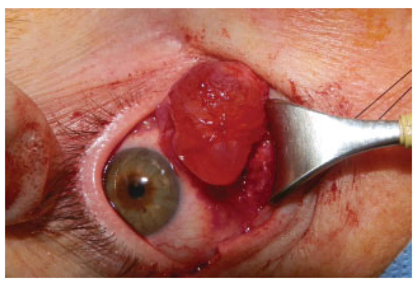
Figure 1. Inferior intraorbital cyst is easily accessed and removed through a transconjunctival approach.
Cmtr 07 00041 g001
Figure 2. The transconjunctival incision is placed 1 to 3 mm inferior to the tarsal border.
Cmtr 07 00041 g002
Figure 3. The dissection is carried through the insertion of the lower lid retractors, and then inferiorly towards the orbital rim.
Cmtr 07 00041 g003
Figure 4. Electrocautery needle is used to make an incision through the periosteum at the orbital rim.
Cmtr 07 00041 g004
Figure 5. Inferior cantholysis. Note the upward vector of pull with the non-dominant hand.
Cmtr 07 00041 g005
Figure 6. Transconjunctival incision can be combined with transcaruncular and lateral canthotomy incisions for wide exposure.
Cmtr 07 00041 g006
Figure 7. Blunt dissection with tenotomy scissors carried down to the posterior lacrimal crest in a plane between the orbital septum and.
Cmtr 07 00041 g007
Figure 8. Left lower lid retraction after transconjunctival lower lid blepharoplasty.
Cmtr 07 00041 g008