Next Article in Journal
Rapid Prototyping Technology in Orbital Floor Reconstruction: Application in Three Patients
Previous Article in Journal
Treatment Effects of Dexmedetomidine and Ketamine on Postoperative Analgesia After Cleft Palate Repair
 
 
Craniomaxillofacial Trauma & Reconstruction is published by MDPI from Volume 18 Issue 1 (2025). Previous articles were published by another publisher in Open Access under a CC-BY (or CC-BY-NC-ND) licence, and they are hosted by MDPI on mdpi.com as a courtesy and upon agreement with Sage.
Case Report

Two Stage Enucleation and Deflation of a Large Unicystic Ameloblastoma with Mural Invasion in Mandible

by
Ryo Sasaki
1,2,3,*,
Yorikatsu Watanabe
1,3,
Tomohiro Ando
2 and
Tanetaka Akizuki
1
1
Department of Plastic and Reconstructive Surgery, Tokyo Metropolitan Police Hospital, Japan
2
Department of Oral and Maxillofacial Surgery, Tokyo Women’s Medical University, School of Medicine, Japan
3
Institute of Advanced Biomedical Engineering and Science, Tokyo Women’s Medical University, Tokyo, Japan
*
Author to whom correspondence should be addressed.
Craniomaxillofac. Trauma Reconstr. 2014, 7(2), 139-142; https://doi.org/10.1055/s-0033-1364197
Submission received: 16 February 2013 / Revised: 17 February 2013 / Accepted: 17 February 2013 / Published: 13 January 2014

Abstract

:
A treatment for strategy of unicystic ameloblastoma (UA) should be decided by its pathology type including luminal or mural type. Luminal type of UA can be treated only by enucleation alone, but UA with mural invasion should be treated aggressively like conventional ameloblastomas. However, it is difficult to diagnose the subtype of UA by an initial biopsy. There is a possibility that the lesion is an ordinary cyst or keratocystic odontogenic tumor, leading to a possible overtreatment. Therefore, this study performed the enucleation of the cyst wall and deflation at first, and the pathological finding confirmed mural invasion into the cystic wall, leading to the second surgery. The second surgery enucleated scar tissue, bone curettage, and deflation, and was able to ameloblastomacontribute to the reduction of the recurrence rate by removing tumor nest in scar tissue or new bone, enhancing new bone formation, and shrinking the mandibular expanding by fenestration. In this study, a large UA with mural invasion including condyle was treated by “two-stage enucleation and deflation” in a 20-year-old patient.

Although ameloblastoma is known to be odontogenic in origin and benign in nature, inadequate treatments give a high percentage of local recurrence rate with possible malignant developments. Thus, radical incision was performed in general [1,2]. Although unicystic ameloblastomas are considered to be a less aggressive form of ameloblastoma, a treatment with a simple enucleation has been suggested to be adequate for the majority of these cases [3,4,5]. Ackermann et al [6] have reported that unicystic ameloblastoma is classified histologically into three groups: Group 1, cyst lined with a variable and often nondescript epithelium; Group 2, cyst showing the intraluminal plexiform proliferation of epithelium; Group 3, cyst with the invasion of epithelium into the cyst wall in either follicular or plexiform pattern. While Group 1 and Group 2 lesions may be treated by enucleation, Group 3 lesions should be treated aggressively like conventional ameloblastomas (Figure 1) [6]. However, it is difficult to diagnose the subtype of unicystic ameloblastoma by an initial biopsy, and there is a possibility that the lesion is an ordinary cyst, leading to a possible overtreatment in that case [7]. Moreover, the resection of the mandible including condyle gives serious complications including dysfunction and deformity. To avoid these complications, a large unicystic ameloblastoma with mural invasion including condyle was treated by “two-stage enucleation and deflation” in a 20-year-old patient.

Case Report

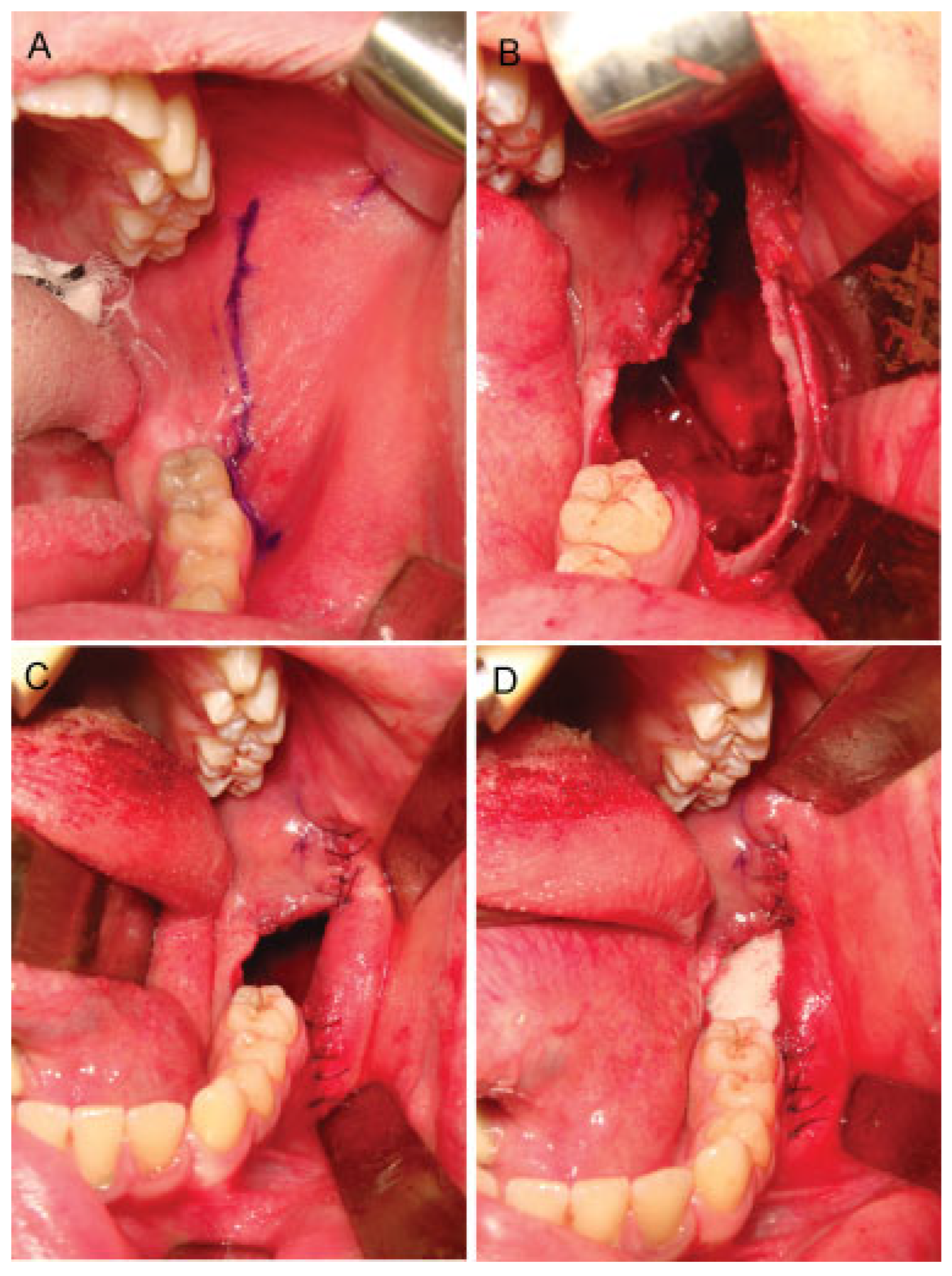
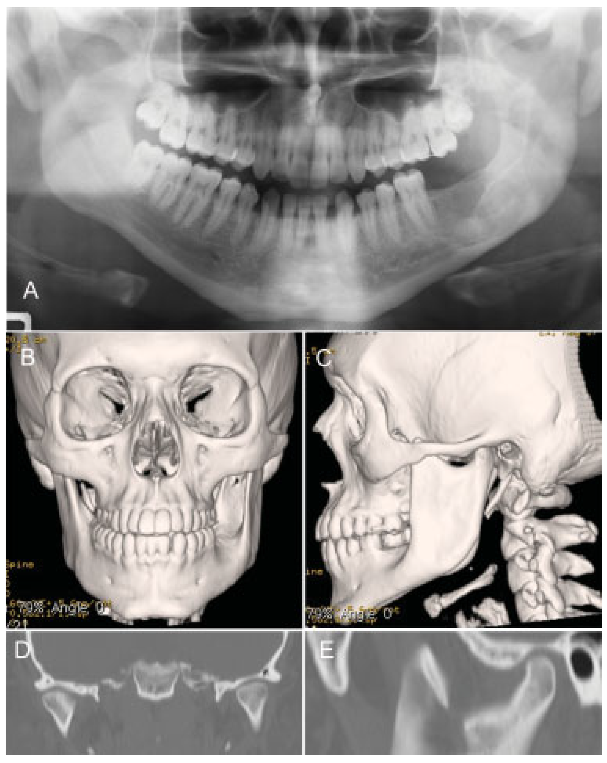
A 20-year-old male patient showing a diffuse swelling in his left face was referred to our department. Panoramic X-ray showed a large unicystic appearance of mandible ramus, coronoid process, and condyle with unerupted third molar, and computed tomography (CT) showed unicystic appearance of mandible ramus, coronoid process, and condyle with expanding buccal, lingual, and anteriorly (Figure 2). Biopsy was performed under local anesthesia. Pathological examination showed that unicystic ameloblastoma of which subtype was unknown. Enucleation and deflation were performed under general anesthesia. The front of ramus cortex was removed by osteotomy (Figure 3A). And the tumor with unerupted third molar was removed using an elevator and an endoscope, and the front of ramus mucosa and buccinator muscle were also removed (Figure 3B). Anterior and posterior of wounds were sutured using 4–0 degradable surgical sutures (Figure 3C). Gauze with an ointment was put into the cavity (Figure 3D). Fenestration was performed. At 3 days after the removal of gauze, the cavity was irrigated with water using a 10-mL syringe by the patient. Pathological findings showed the tumor was a unicystic ameloblastoma mural type. Therefore, the secondary removal operation was attempted at 3 months after the first surgery. CT and panoramic X-ray showed that visible new bone formed and expanded ramus shrank. Enucleation of scar tissue, the removal of new bone of the surface of bony cavity, and the deflation of the front of ramus mucosa were also performed. Anterior and posterior of wound were sutured. Chitin-coated gauze with an ointment was put into the cavity. Fenestration was performed. At 3 days after the removal of chitin-coated gauze, the cavity was irrigated with water using a 10-mL syringe by the patient. HE staining of removed scar tissue showed no ameloblastoma cells. Therefore, the patient was in a long-term follow-up and showed no facial asymmetry, no mental palsy, and no trismus at 2.5 years after the first surgery. CT and panoramic X-ray showed that visible new bone formed and expanded ramus shrank, and the buccal cortex of mandible was found to be symmetry (Figure 4).

Discussion

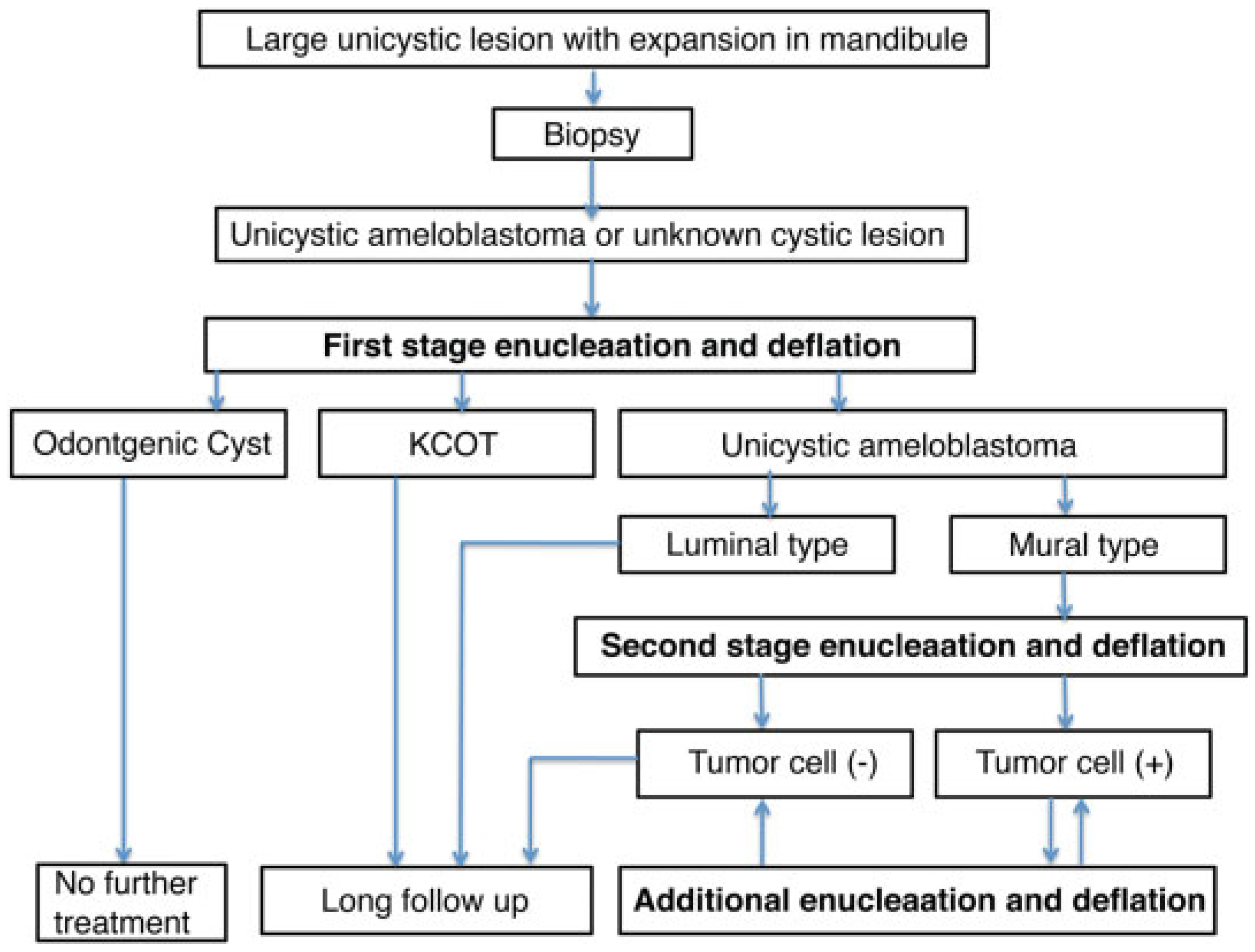
A systematic review shows that the recurrence rates of unicystic ameloblastoma were 3.6% for resection, 30.5% for enucleation alone, and 16% for enucleation followed by the application of Carnoy solution, and 18% for marsupialization with or without other treatment in the second phase [8]. However, the review has no discussion regarding recurrence rates in their pathological types including the luminal or mural type of unicystic ameloblastoma. Rosenstein et al [9] have reported that 9 of 21 tumors (43%) are recurrent after enucleation alone in unicystic ameloblastoma. Of the nine recurrent lesions, five cases (55%) showed the histologic evidence of mural invasion, suggesting that connective tissue invasion can be considered as an important microscopic sign for recurrence [9]. Therefore, a treatment strategy for unicystic ameloblastoma should be decided by its pathology type including luminal or mural type. However, it is difficult to diagnose the subtype of unicystic ameloblastoma by an initial biopsy because of widely variation in the region of the cystic wall. Therefore, this study performed the enucleation of the cyst wall at first, and the pathological finding confirmed mural invasion into the cystic wall (Ackermann Group 3), leading to the second surgery (Figure 5).
This study reported a treatment strategy as a “dredging method,” which has been reported by Kawamura et al in 1991 [10,11], for a large unicystic lesion in mandible. In their method, after the deflation and enucleation or only enucleation, enucleation is repeated by five to six times with an interval of 2 to 3 months until no tumor cell nest is identified in the pathological findings of scar tissues removed by two consecutive dredging. In ameloblastoma with a honeycombed pattern, a selective marginal resection is added. Several clinical studies also showed that “the dredging method” for mandibular ameloblastoma gives a low recurrence rate and a low complication with no dysfunction and no deformity [10,11,12,13,14]. Kawamura et al [10,11] have reported that (1) a total of 50 ameloblastoma cases in mandible, including uni- and multicystic, solid, and mixed ameloblastoma with honeycombed pattern, are treated by this dredging method with a recurrence rate of 16% in the total ameloblastoma and a recurrence rate of 15% in 33 cases of cystic ameloblastoma for a period of 2 to 16 years and (2) all cases show no malignant change and no metastasis [11]. Moreover, Sadat and Ahmed also have reported that 24 ameloblastoma cases with dredging method surgery show a recurrence rate of 12% [12]. The dredging method surgery, reported by Kawamura et al, requires (1) no tumor cell nest in the scar tissues removed by two consecutive dredging and (2) the observation of sufficient new bone formation [10,11]. On the contrary, our procedure was to remove scar and new bone until no evidence of tumor cells in scar tissue because (1) the reported dredging method was speculated to show no clear meaning to obtain two-consecutive pathological evaluation for confirming no tumor cell and (2) the secondary removal of scar was able to give sufficient new bone formation. Therefore, our procedure for treating a mural type of unicystic ameloblastoma was to remove scar and new bone until no evidence of tumor cells in scar tissue. However, long follow-up with a “wait and see” policy was necessary. When recurrence is found after the two-stage surgery, the recurrent tumor may be able to be removed by marginal resection. This idea remains in the need of justification through a larger study with better-defined criteria and long-term follow-ups.

Acknowledgments

The authors would like to thank Dr. Naoyuki Matsumoto (Department of Pathology, Nihon University, School of Dentistry) for pathological finding, and Dr. Kaori Agawa (Tokyo Metropolitan Police Hospital) for drawing the illustrations of the histological classification of unicystic ameloblastoma.

References

  1. Vayvada, H.; Mola, F.; Menderes, A.; Yilmaz, M. Surgical management of ameloblastoma in the mandible: segmental mandibulectomy and immediate reconstruction with free fibula or deep circumflex iliac artery flap (evaluation of the long-term esthetic and functional results). J Oral Maxillofac Surg 2006, 64, 1532–1539. [Google Scholar] [CrossRef] [PubMed]
  2. Zemann, W.; Feichtinger, M.; Kowatsch, E.; Kärcher, H. Extensive ameloblastoma of the jaws: surgical management and immediate reconstruction using microvascular flaps. Oral Surg Oral Med Oral Pathol Oral Radiol Endod 2007, 103, 190–196. [Google Scholar] [PubMed]
  3. Gardner, D.G.; Corio, R.L. Plexiform unicystic ameloblastoma. A variant of ameloblastoma with a low-recurrence rate after enucleation. Cancer 1984, 53, 1730–1735. [Google Scholar] [CrossRef] [PubMed]
  4. Olaitan, A.A.; Adekeye, E.O. Unicystic ameloblastoma of the mandible: a long-term follow-up. J Oral Maxillofac Surg 1997, 55, 345–348, discussion 349–350. [Google Scholar] [PubMed]
  5. Philipsen, H.P.; Reichart, P.A. Unicystic ameloblastoma. A review of 193 cases from the literature. Oral Oncol 1998, 34, 317–325. [Google Scholar] [PubMed]
  6. Ackermann, G.L.; Altini, M.; Shear, M. The unicystic ameloblastoma: a clinicopathological study of 57 cases. J Oral Pathol 1988, 17, 541–546. [Google Scholar] [CrossRef] [PubMed]
  7. Chapelle, K.A.; Stoelinga, P.J.; de Wilde, P.C.; Brouns, J.J.; Voorsmit, R.A. Rational approach to diagnosis and treatment of ameloblastomas and odontogenic keratocysts. Br J Oral Maxillofac Surg 2004, 42, 381–390. [Google Scholar] [PubMed]
  8. Lau, S.L.; Samman, N. Recurrence related to treatment modalities of unicystic ameloblastoma: a systematic review. Int J Oral Maxillofac Surg 2006, 35, 681–690. [Google Scholar] [PubMed]
  9. Rosenstein, T.; Pogrel, M.A.; Smith, R.A.; Regezi, J.A. Cystic ameloblastoma—behavior and treatment of 21 cases. J Oral Maxillofac Surg 2001, 59, 1311–1316, discussion 1316–1318. [Google Scholar] [CrossRef] [PubMed]
  10. Kawamura, M.; Inoue, N.; Kobayashi, I.; et al. Ameloblastoma treated by “Dredging method”—report of a case. Asian J Oral Maxillofac Surg 1991, 3, 89–93. [Google Scholar]
  11. Kawamura, M.; Inoue, N.; Kobayashi, I.; et al. “Dredging Method”—a new approach for the treatment of ameloblastoma. Asian J Oral Maxillofac Surg 1991, 3, 81–88. [Google Scholar]
  12. Sadat, S.M.A.; Ahmed, M. “Dredging Method”—a conservative surgical approach for the treatment of ameloblastoma of jaw. J Bangladesh Coll Phys Surg 2011, 29, 72–77. [Google Scholar]
  13. Sadat, S.M.A.; Haider, I.A.; Bhuiyan, R.A.; et al. Treatment of ameloblastoma by “Dredging Method”. Int J Oral Maxillofac Surg 2007, 36, 1035. [Google Scholar] [CrossRef]
  14. Kurokawa, H.; Ishida, C.; Murata, T.; et al. A case report of unicystic ameloblastoma treated with dredging method. J Kyushu Dental Society 2000, 54, 253–257. [Google Scholar] [CrossRef]
Figure 1. Schematic illustration of the histological classification of unicystic ameloblastoma subtypes according to Ackermann et al.6 The lesions can be classified into the following three groups. (1) Group 1, cyst is lined with a variable and often nondescript epithelium. (2) Group 2, cyst shows the intraluminal plexiform proliferation of epithelium. (3) Group 3, cyst is formed to have the invasion of epithelium into the cyst wall in either a follicular or a plexiform pattern.
Cmtr 07 00034 g001
Figure 2. A 20-year-old male patient with a large unicystic appearance of mandible ramus and condyle with buccal and lingual expansion. (A) Panoramic X-ray shows the large unicystic appearance of left mandible ramus and condyle (B and C). Three-dimensional computed tomography (CT) shows the large unicystic of mandible ramus and condyle with buccal, lingual, and anterior and posterior expansion. (D) Axial CT shows the invasions of the condylar head and coronoid, and the expansion of the coronoid. (E) Coronal CT shows the invasions of the condylar head and the expansion of the condyle.
Cmtr 07 00034 g002
Figure 3. First-stage enucleation and deflation for the case of the large unicystic appearance of mandible ramus and condyle with buccal and lingual expansion. (A) Incision of the front of the mucosa of the ramus was performed. (B) The front of ramus cortex was removed by osteotomy as large as possible. Lesion was removed using an elevator under endoscopic assistance. (C) The front of ramus mucosa with buccinator muscle was also removed as large as possible. Fenestration was performed. (D) Gauze with an ointment was put into the cavity for 3 days.
Cmtr 07 00034 g003
Figure 4. Clinical findings at 2 years and 6 months after the first-stage surgery. (A) Panoramic X-ray shows that a visible new bone formed, and expanded ramus and condyle tissues shrank. (B and C) Three-dimensional computed tomography (CT) shows that a visible new bone formed, and expanded ramus and condyle tissue shrank. (D) Coronal CT shows that bilateral condyle is symmetry with a normal structure. (E) Sagittal CT shows that the condylar head shrinks with a normal structure.
Cmtr 07 00034 g004
Figure 5. Flow chart of a treatment strategy for the large unicystic lesion of mandible angle, ramus, and extend to condyle with buccal and lingual expansion. Second enucleation was added to new bone curettage. Luminal type: Ackermann Groups 1 and 2. Mural type: Ackermann Group 3. KCOT, keratocystic odontogenic tumor.
Cmtr 07 00034 g005