Open Surgical Management of an Asymptomatic Giant Frontal Sinus Osteoma
Abstract
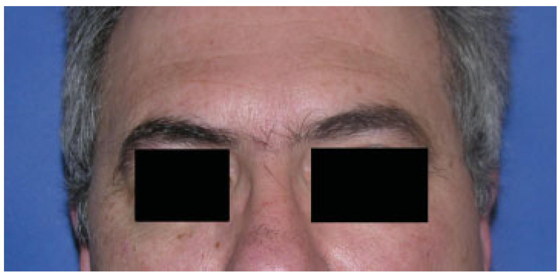
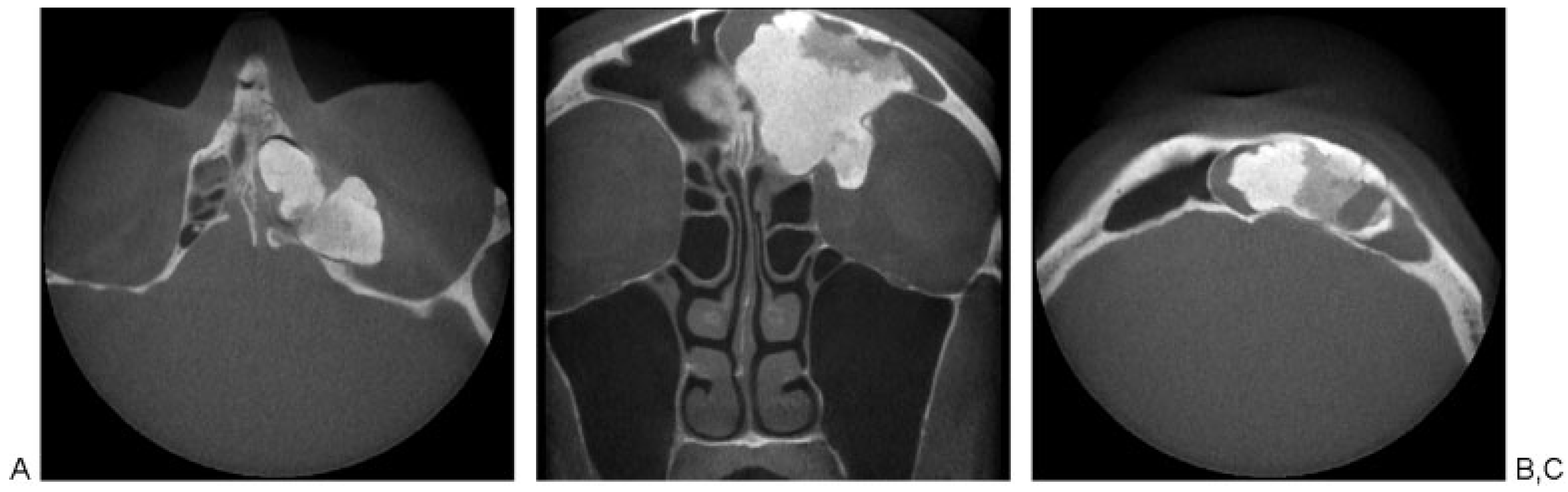
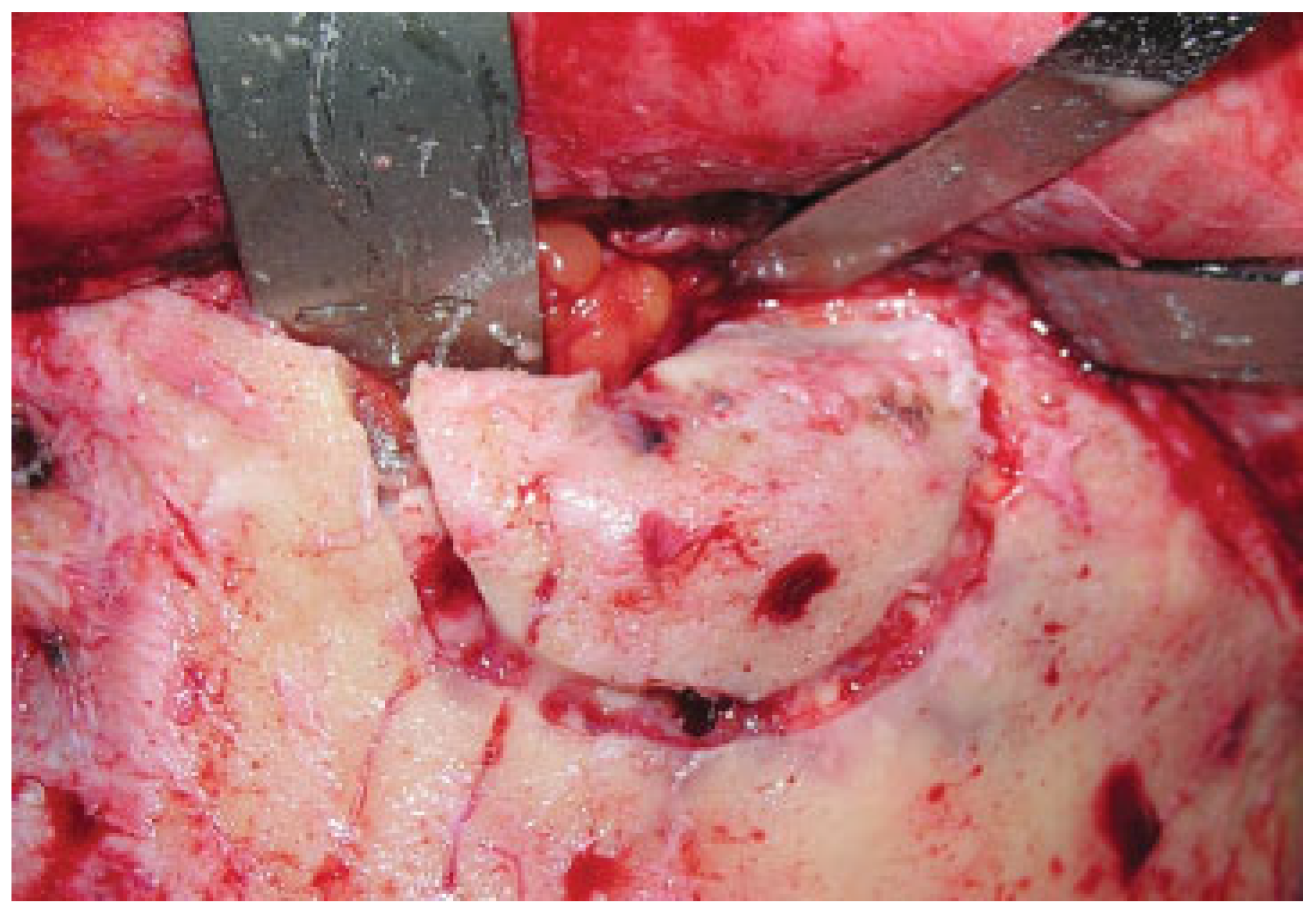
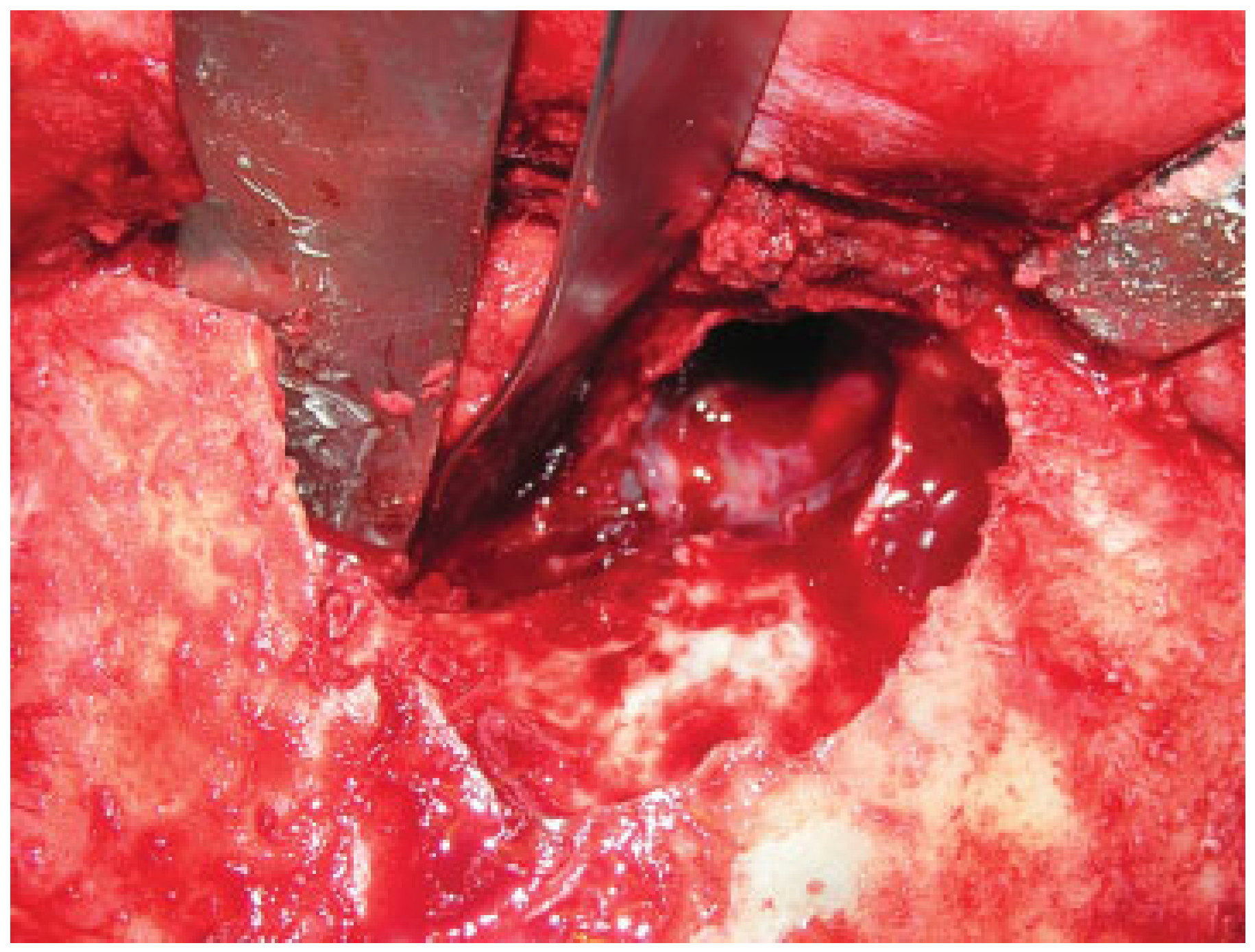
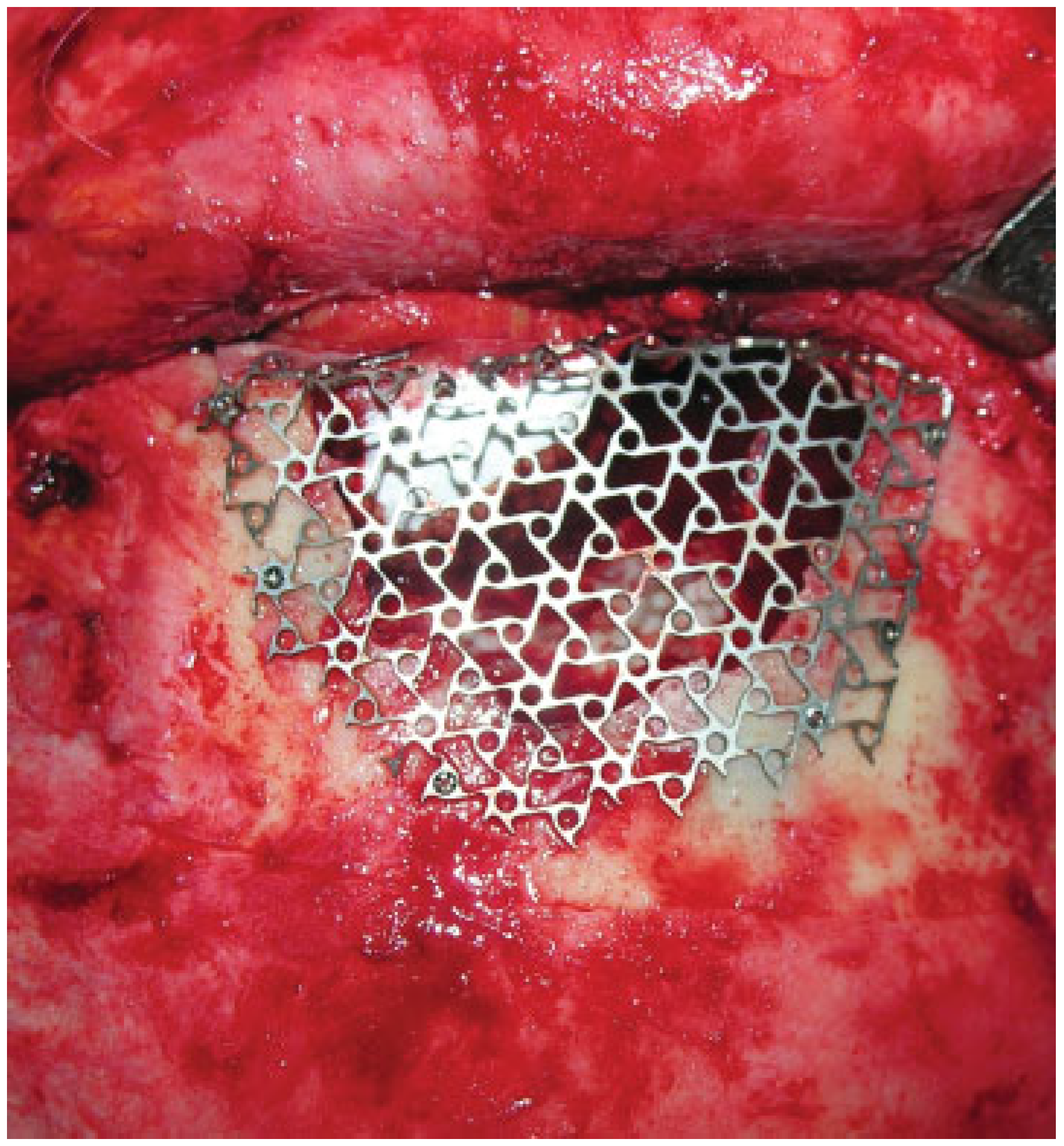
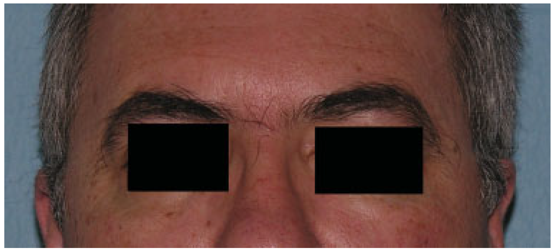
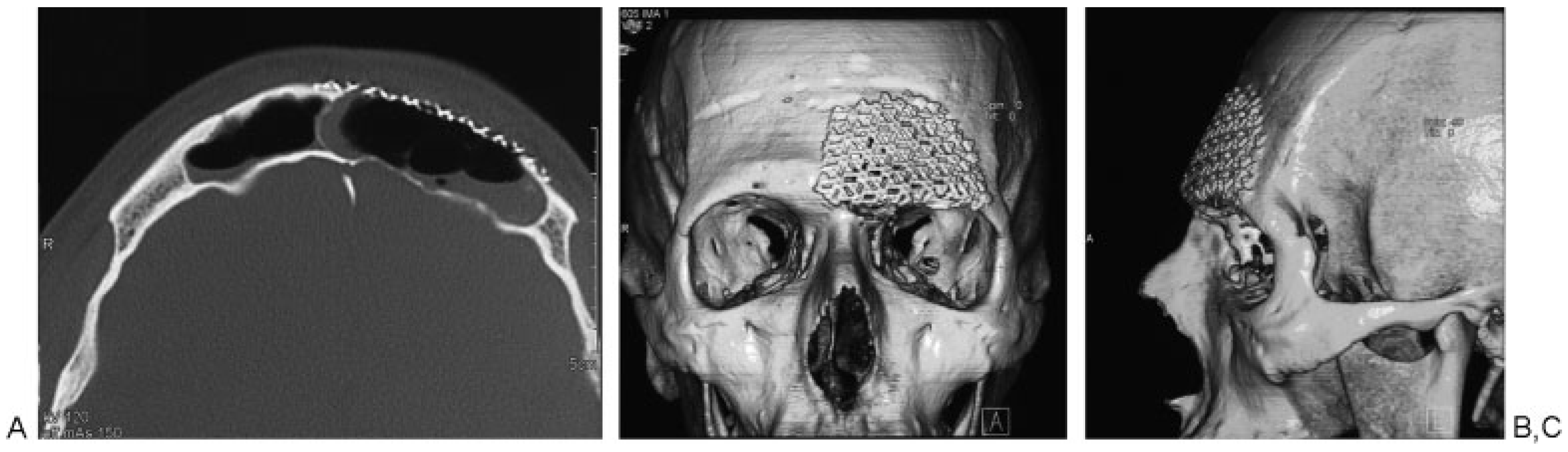
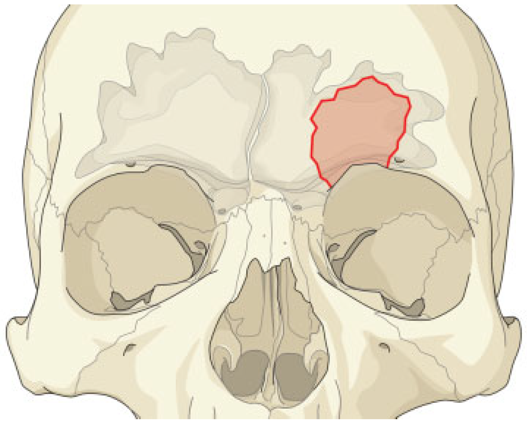
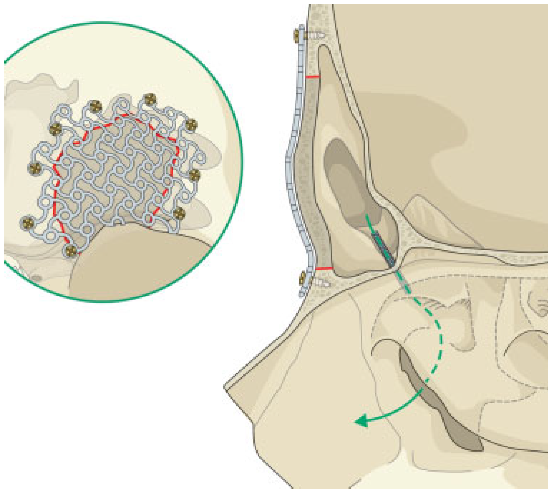
:Case Report
Discussion
References
- Boffano, P.; Roccia, F.; Campisi, P.; Gallesio, C. Review of 43 osteomas of the craniomaxillofacial region. J Oral Maxillofac Surg 2012, 70, 1093–1095. [Google Scholar] [PubMed]
- Gabrielli, M.F.; Gabrielli, M.A.; Hochuli-Vieira, E.; Pereira-Fillho, V.A. Immediate reconstruction of frontal sinus fractures: Review of 26 cases. J Oral Maxillofac Surg 2004, 62, 582–586. [Google Scholar] [CrossRef] [PubMed]
- Lakhani, R.S.; Shibuya, T.Y.; Mathog, R.H.; Marks, S.C.; Burgio, D.L.; Yoo, G.H. Titanium mesh repair of the severely comminuted frontal sinus fracture. Arch Otolaryngol Head Neck Surg 2001, 127, 665–669. [Google Scholar] [CrossRef] [PubMed]
- Kuttenberger, J.J.; Hardt, N. Long-term results following reconstruction of craniofacial defects with titanium micro-mesh systems. J Craniomaxillofac Surg 2001, 29, 75–81. [Google Scholar] [CrossRef] [PubMed]
- Castelnuovo, P.; Valentini, V.; Giovannetti, F.; Bignami, M.; Cassoni, A.; Iannetti, G. Osteomas of the maxillofacial district: Endoscopic surgery versus open surgery. J Craniofac Surg 2008, 19, 1446–1452. [Google Scholar] [CrossRef] [PubMed]
- Boffano, P.; Bosco, G.F.; Gerbino, G. The surgical management of oral and maxillofacial manifestations of Gardner syndrome. J Oral Maxillofac Surg 2010, 68, 2549–2554. [Google Scholar] [CrossRef] [PubMed]
- Miman, M.C.; Bayindir, T.; Akarcay, M.; Erdem, T.; Selimoglu, E. Endoscopic removal technique of a huge ethmoido-orbital osteoma. J Craniofac Surg 2009, 20, 1403–1406. [Google Scholar] [CrossRef] [PubMed]
- Manolidis, S.; Hollier, L.H., Jr. Management of frontal sinus fractures. Plast Reconstr Surg 2007, 120 (Suppl. 2), 32S–48S. [Google Scholar] [PubMed]
- Lazaridis, N.; Makos, C.; Iordanidis, S.; Zouloumis, L. The use of titanium mesh sheet in the fronto-zygomatico-orbital region. Case reports. Aust Dent J 1998, 43, 223–228. [Google Scholar] [PubMed]
- Corina, L.; Scarano, E.; Parrilla, C.; Almadori, G.; Paludetti, G. Use of titanium mesh in comminuted fractures of frontal sinus anterior wall. Acta Otorhinolaryngol Ital 2003, 23, 21–25. [Google Scholar] [PubMed]
Publisher’s Note: MDPI stays neutral with regard to jurisdictional claims in published maps and institutional affiliations. |
© 2014 by the author. The Author(s) 2014.
Share and Cite
Boffano, P.; Zavattero, E.; Roccia, F.; Ramieri, G. Open Surgical Management of an Asymptomatic Giant Frontal Sinus Osteoma. Craniomaxillofac. Trauma Reconstr. 2014, 7, 51-54. https://doi.org/10.1055/s-0033-1364200
Boffano P, Zavattero E, Roccia F, Ramieri G. Open Surgical Management of an Asymptomatic Giant Frontal Sinus Osteoma. Craniomaxillofacial Trauma & Reconstruction. 2014; 7(1):51-54. https://doi.org/10.1055/s-0033-1364200
Chicago/Turabian StyleBoffano, Paolo, Emanuele Zavattero, Fabio Roccia, and Guglielmo Ramieri. 2014. "Open Surgical Management of an Asymptomatic Giant Frontal Sinus Osteoma" Craniomaxillofacial Trauma & Reconstruction 7, no. 1: 51-54. https://doi.org/10.1055/s-0033-1364200
APA StyleBoffano, P., Zavattero, E., Roccia, F., & Ramieri, G. (2014). Open Surgical Management of an Asymptomatic Giant Frontal Sinus Osteoma. Craniomaxillofacial Trauma & Reconstruction, 7(1), 51-54. https://doi.org/10.1055/s-0033-1364200