Next Article in Journal
Utility of Stereolithographic Models in Osteocutaneous Free Flap Reconstruction of the Head and Neck
Previous Article in Journal
Endoscopic Dacryocystorhinostomy
 
 
Craniomaxillofacial Trauma & Reconstruction is published by MDPI from Volume 18 Issue 1 (2025). Previous articles were published by another publisher in Open Access under a CC-BY (or CC-BY-NC-ND) licence, and they are hosted by MDPI on mdpi.com as a courtesy and upon agreement with Sage.
Review

Optic Nerve Monitoring

by
Paul Schumann
*,
Horst Kokemüller
,
Frank Tavassol
,
Daniel Lindhorst
,
Juliana Lemound
,
Harald Essig
,
Martin Rücker
and
Nils-Claudius Gellrich
Department of Oral and Maxillofacial Surgery, Hannover Medical School, Carl-Neuberg-Strasse 1, 30625 Hannover, Germany
*
Author to whom correspondence should be addressed.
Craniomaxillofac. Trauma Reconstr. 2013, 6(2), 75-85; https://doi.org/10.1055/s-0033-1343783
Submission received: 6 November 2012 / Revised: 9 November 2012 / Accepted: 9 November 2012 / Published: 1 May 2013

Abstract

:
Orbital and anterior skull base surgery is generally performed close to the prechiasmatic visual pathway, and clear strategies for detecting and handling visual pathway damage are essential. To overcome the common problem of a missed clinical examination because of an uncooperative or unresponsive patient, flash visual evoked potentials and electroretinograms should be used. These electrophysiologic examination techniques can provide evidence of intact, pathologic, or absent conductivity of the visual pathway when clinical assessment is not feasible. Visual evoked potentials and electroretino-grams are thus essential diagnostic procedures not only for primary diagnosis but also for intraoperative evaluation. A decision for or against treatment of a visual pathway injury has to be made as fast as possible due to the enormous importance of the time elapsed with such injuries; this can be achieved additionally using multislice spiral computed tomography. The first-line conservative treatment of choice for such injuries is megadose methylprednisolone therapy. Surgery is used to decompress the orbital compartment by exposure of the intracanalicular part of the optic nerve in the case of optic canal compression. Modern craniomaxillofacial surgery requires detailed consid- eration of the diagnosis and treatment of traumatic visual pathway damage with the ultimate goal of preserving visual acuity.

Reliable optic nerve monitoring is an essential requirement for all medical disciplines dealing with problems concerning the prechiasmatic visual pathway. For any intervention, the major aim is not to compromise the visual system. The only exception is surgery for malignancies if a discontinuity of the anterior visual pathway is necessary for sufficient tumor resection. However, in craniomaxillofacial traumatology and reconstructive surgery, prevention of visual pathway damage is an important goal. In fact, vision loss is the most serious traumatic effect or complication associated with elective orbital surgery. In severely injured patients or during craniomaxillofacial reconstructions, tests of visual pathway function are generally unreliable. In such cases, neuro- ophthalmologic analyses that do not require a cooperative patient are indispensable. Nevertheless, clinical prediction of visual pathway injuries is currently insufficient. Here we describe the present state of visual pathway monitoring and highlight its clinical relevance.

Anatomical and Pathophysiologic Considerations

The optic nerve is divided into four sections of different lengths: the intraocular (1 mm), intraorbital (25 to 30 mm), intracanalicular (8 mm), and intracranial (15 mm) sections. The intraorbital section is rarely affected by direct or indirect trauma because of its S shape and because it is embedded in thick soft tissue [1]. Damage to the intracranial section is also unusual because in this area the nerve is well protected by the skull [2,3]. The weak point of the anterior visual pathway is the intracanalicular section. Here, the dura and periosteum form only one tight structure, and in contrast to the intraorbital and intracranial sections, the nerve is fixed to its bony surroundings by fibrous adhesions [4,5]. Due to the narrowness of the canal, any deformation of the pericanalicular area can be directly transmitted to the optic nerve.
A direct optic nerve trauma can be distinguished from an indirect trauma on the basis of etiology. Direct trauma is characterized by radiologically detectable compression by bony fragments, a hematoma (Figure 1), penetrating foreign bodies, or a fracture of the optic nerve canal itself. Indirect trauma does not correlate with any radiologic findings of an intraorbital, intracanalicular, or intracranial optic nerve injury and can be subclassified as an anterior or posterior type of indirect trauma. An anterior trauma concerns the intraocular part of the optic nerve with noticeable eye injuries; posterior trauma does not show any ophthalmoscopically detectable bulb damage immediately after trauma [6]. Blunt trauma of the viscerocranium can induce deformation with a reduction in the diameter of the optic nerve canal without any signs of direct or indirect fracture [7,8]. Moreover, optic nerve damage can be classified as a primary or secondary injury. Primary damage is caused immediately by the accident itself; secondary damage develops posttraumatically [9].
Extrinsic compression of the optic nerve by a retrobulbar hematoma [10], edema, or emphysema [11] impairs visual acuity by a combination of modified liquor circulation, interruption of direct axonal transport, and ischemia [12]. Moreover, a direct or indirect trauma can induce an optic nerve injury via a contusion, torsion, strain, or shear [7]. An intrinsic compression of the optic nerve develops by inter- or intraneural edema or internal hemorrhaging [2].
In summary, traumatic damage of the intraorbital or intracanalicular optic nerve is in most cases a multifactorial combination of a bony lesion, ischemia due to microvascular spasms or vessel occlusion, reactive edema, and spaceoccupying hemorrhage. Damage to the visual pathway can thus be the consequence of an extremely brief trauma or slowly progressive compression.

Clinical Appearance of a Traumatic Optic Nerve Injury

Many attempts have been made to define characteristic clinical findings that commonly correlate with optic nerve damage. These include wounds of the lateral eyebrow (97%), a reduction in visual acuity, respectively, relative or absolute afferent disorder associated with the ipsilateral trauma side in combination with functioning consensual light reaction of the affected eye (100%), and epistaxis (80%) [13,14]. Most fre- quently, frontonasoethmoidal fractures are associated with afferent disorders of the visual pathway [15]. Unfortunately, the severity of any assumed optic nerve damage cannot often be assessed by clinical examination immediately after trauma [16]. As an alternative, a score based on clinical findings (but not on functional testing of the visual pathway) was developed to estimate possible damage to the visual pathway [17]. However, this score did not yield the desired results.
Verification of visual acuity is a basic principle of any clinical examination of an injured patient. However, such routine checks are often not possible because of the state of awareness of the patient or because of his or her injuries [18]. For an orienting functional test of the visual pathway, differ- ent pupillary signs have to be assessed. Apart from pupillary shape and diameter, direct and indirect pupillary reflexes have to be evaluated. Anisocoria is not a consequence of damage to the anterior visual pathway. Even after unilateral cutting of the optic nerve, the pupils remain symmetrical, and 0.4% of patients with head injuries who are examined in emergency rooms show transient cortical loss of sight because of an occipital injury, without any pathologic changes in pupillary signs. In case of pathologic pupillary function, further examination is required. A malfunction of the pupil does not have to be initiated by damage to the optic nerve or other sections of the visual pathway. Apart from an injury to the iris sphincter itself, a pupillary malfunction can be the result of medications (especially morphines) or damage to the efferent limb of the visual pathway (e.g., bilateral paresis of the oculomotor nerve). Therefore, an evaluation of pupillary function alone may not constitute a sufficient assessment of visual acuity. A reliable test for primary diagnosis of optic nerve damage is the swinging flashlight test [19,20]. However, clinical experience suggests that this method is often not applicable during the first hours after a trauma because of massive periorbital swelling and the influence of medications.
The same problem is encountered in fundoscopy because an acute evaluation of intraocular bleeding or retinal lesions is often impossible in cases of severe skull injury. Pallor of the optic disk, which is a posttraumatic effect occurring after optic nerve damage, cannot be detected until 6 weeks after injury. Atrophy of the papilla can be recognized earlier when optic nerve damage occurs close to the bulb. Identification and differentiation of the type of optic nerve damage is very important, and testing the visual field is useful in this regard. Unfortunately, such investigations are only possible several days after a serious trauma. In addition, the cooperation of the injured patient is necessary even for exploratory screening. Pathognomonic disorders of the visual field do not exist in the case of traumatic optic nerve damage [21].
In summary, all neuro-ophthalmologic analyses are limit- ed by the severity of the injury, the patient’s state of aware- ness, and the influence of medications. Additionally, reversible optic nerve damage as a consequence of edema or perineural hematoma cannot be differentiated from irre- versible injury on the basis of clinical examination [22].

Electrophysiology of the Visual Pathway

The retina is negatively charged whereas the cornea is posi- tively charged. Therefore, the bulb represents an electrical dipole. The electrical activity of the first and second neurons of the visual pathway is measured by electroretinography; the function of the retinal ganglion cells (third neuron of the visual pathway) is not included in the electroretinogram (ERG) [23]. Variations in the ERG are caused by refracting changes in the eye as well as by pathologic changes in the retina itself. Disorders of the visual system from the refracting media to the visual cortex can be registered by cortical records of visual evoked potentials (VEPs). Thus, functional localization of visual pathway damage between retinal ganglion cells and the visual cortex is possible by combining ERGs and VEPs.

Primary Diagnosis with Visual Pathway Testing of Patients with Head Injuries

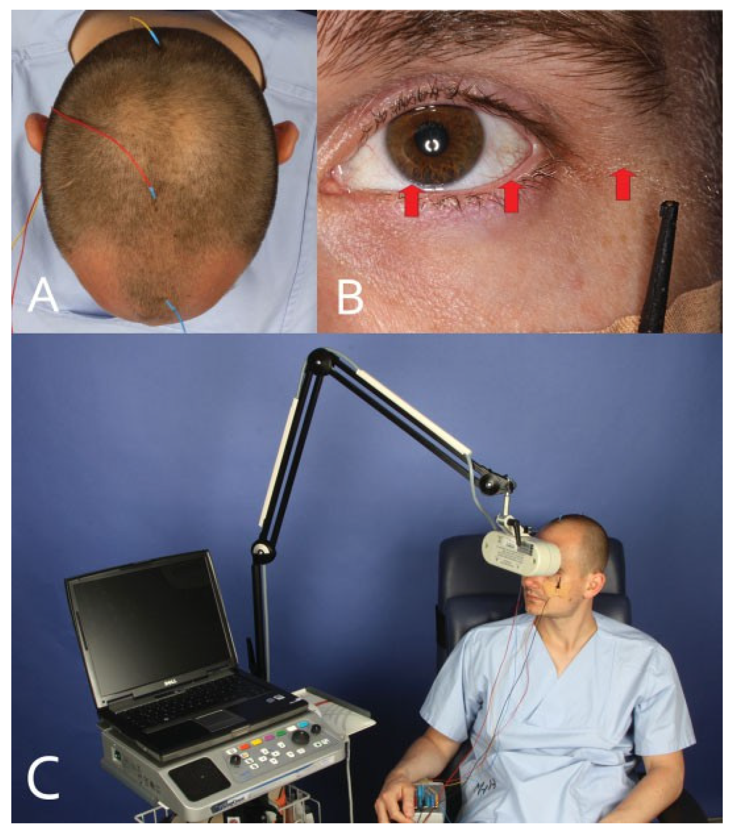
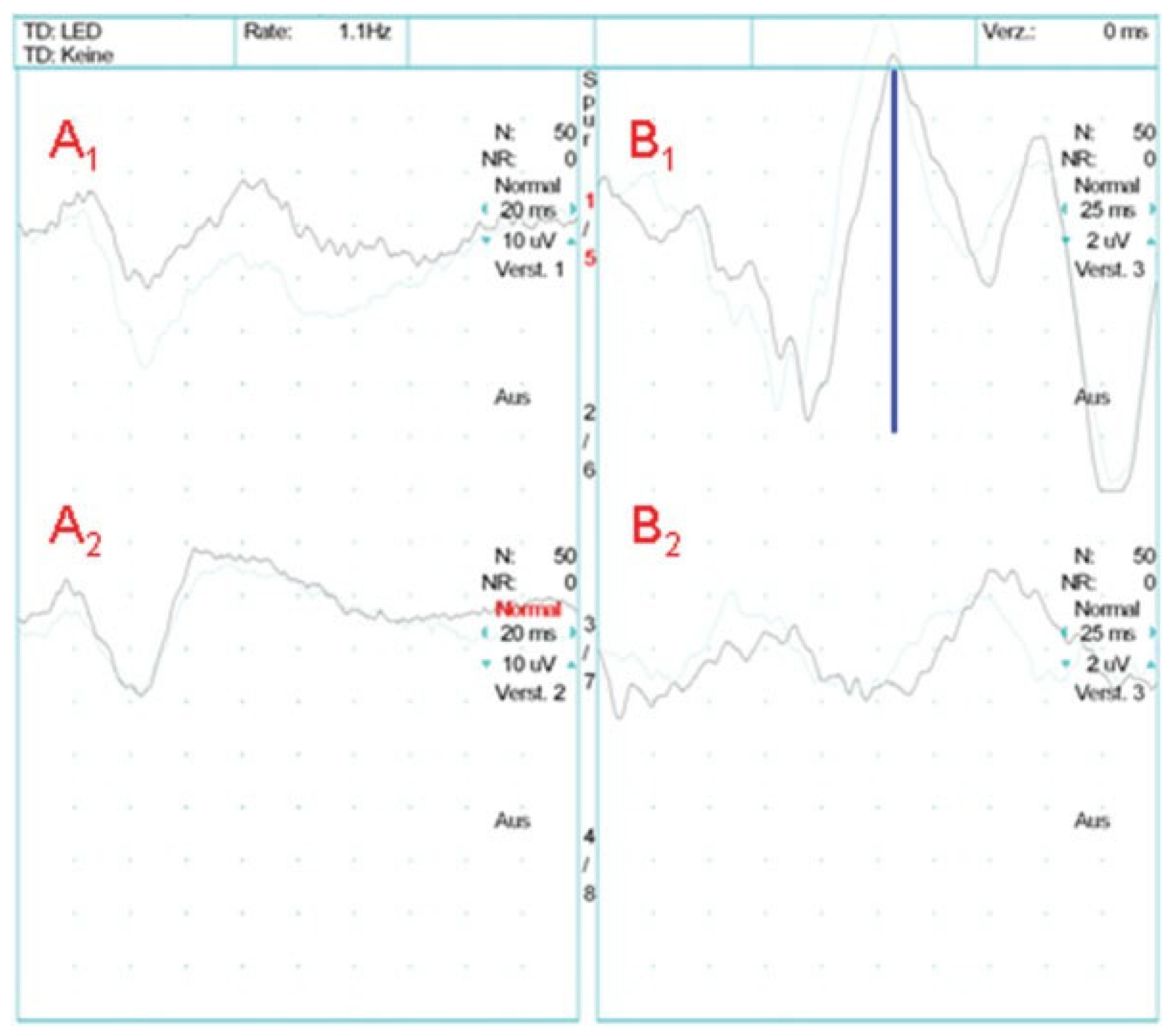
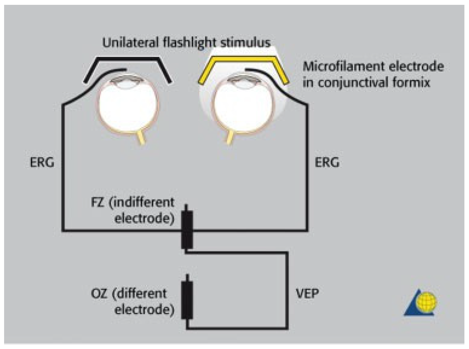
A mandatory component of any clinical primary diagnosis of patients with head injuries is an orientating analysis of the visual pathway, including pupillary response, visual acuity, visual field, and bulb motility. The swinging flashlight test is a clinical test for a relative afferent pupillary defect: light flashing into the nonaffected eye leads to a direct and consensual reaction. Afterward, the light reaction of the affected eye has to be assessed. An afferent pupillary defect is apparent when the affected eye reacts with a mydriasis. This test is a relative procedure and therefore is not applicable for bilateral optic or oculomotor nerve damage. If a clinical functional diagnosis of the visual pathway is not possible or is doubtful due to unconsciousness, morphine interference, or massive swelling, an electrophysiologic examination by means of flash VEPs and ERG is the only remaining option for objectively assessing visual pathway function in the early posttraumatic stage. In the case of intraoperative assess- ments, for example, directly after orbital reconstruction or after optic nerve decompression, electrophysiologic measurements are obtained from a narcotized patient. Accordingly, it has to be considered that VEP records can be affected strongly by several anesthetics [24,25,26]. If VEP monitoring is performed under general anesthesia, an additional postoper- ative electrophysiologic examination is beneficial if the patient is still unconscious. For such examinations, a mobile neurophysiologic measuring station is practical for performing single and parallel ERGs and VEPs; 100 single potentials of the retina (ERG) or visual cortex (VEPs) are averaged. Both ERGs and VEPs are repeated immediately to verify the results. Afterward, a diagnosis is made by assessing the amplitude and latency of the electric signals. The VEP is detected in a standard position in the midline 5 cm above the occipital protuberance against the median frontal reference point. Needle electrodes are used with connecting resistances not > 5 kΩ (Figure 2). For recording, a frequency window of 1 to 100 s−1 is chosen, and the duration of the evaluation period is 500 milliseconds. The standard limit for the VEP latency is defined as the average (arithmetic mean, 108.71 milliseconds; minimum, 69.5 milliseconds; maximum, 143 milliseconds; standard error, 2.46 milliseconds) plus 2.5 times the standard deviation (16.7 milliseconds) of a normal group: 150 milliseconds. The amplitude fluctuates within a small range in a control group. Therefore, only side-to-side differ- ences > 50% are rated as pathologic. For recording an ERG, a very thin electrode is inserted in the conjunctiva of the lower eyelid (Figure 2). Median frontal reference point is used as a reference electrode. Further conditions are in accordance with the values mentioned earlier. The involvement of the ERG is particularly important in the case of a missing or pathologic ipsilateral VEP to ensure that a signal interruption of the visual stimulus is not due to refracting changes in the eye or damage to the retina (Figure 3).
The stimulation is performed separately for each eye with a red stroboscopic light; a double stimulus (2 s−1) with a duration of 1 millisecond is used. The flashlight stimulus on each side consists of six simultaneously flashing LEDs integrated in LED goggles.
In addition to the clinical and electrophysiologic results, a spiral computed tomography (CT) scan is obtained with a scan field from the teeth of the upper jaw to the cranial border of the frontal sinus (slice thickness, 1 mm; table advance, 2 mm; increment, 1 mm). Apart from the axial images, three-dimensional reconstructions from the sella turcica to the nasal bone are necessary. Finally, a decision for or against treatment of a visual pathway injury is made by analyzing the clinical, electrophysiologic, and CT results. With regard to traumatic damage of the optic nerve, the following findings have to be considered for each side:
  • Primary signs: fracture of the optic nerve canal; fracture in the retrobulbar region; hematoma, swelling, or disconti- nuity of the optic nerve; hematoma in the posterior third of the orbit.
  • Secondary signs: shading of the sphenoidal sinus or of the posterior ethmoidal cells; air–fluid level in the maxillary sinus; epidural hematoma of the temporobasal region.
  • Concomitant injuries: lamina papyracea; frontal sinus; zygomatic bone; orbital floor; orbital roof; air collection below the frontonasal region, optic chiasm, cavernous sinus, or posterior of the big wing of the sphenoid bone; contusions; subarachnoid or subdural hemorrhage.

Traumatology

Extracranial neurologic complications after midface fractures occur in approximately 50% of all cases [27]. Damage to the optic nerve is thus the third most common cranial nerve injury after olfactory and facial nerve lesions. Traumatic optic nerve damage is normally an acute problem. Almost 2% (0.7 to 5.0%) of all closed head injuries and 20% of all frontobasal traumas are associated with damage to the visual pathway [2,28,29,30]. In most cases, the intracanalicular section is affected. The most common injuries occur in combination with frontal (72%) or frontotemporal (12%) craniocerebral injuries [6]. Bicycle and car accidents and falls are the most common causes [31,32]. Usually, patients do not present with isolated damage of one or both optic nerves but rather display complex injuries after damage to the cranio-orbital transition zone [33]. Irreversible optic nerve damage cannot be differentiated from reversible injuries through clinical examination. Therefore, the use of electrophysiologic techniques has been established. Although clinical electrophysiologic assessments such as flash VEPs and ERGs have been described for diagnosing traumatic visual pathway damage, there is no systematic use of such methods for posttraumatic acute phase assessment of patients with head injuries [34,35,36]. Most studies of electrophysiologic techniques have not combined the diagnostic aspects with the traumatic or therapeutic aspects [31,37,38]. Thus, many electro-physiologic studies refer only to checkerboard-pattern VEP examinations [39]. However, this technique requires a cooperative patient with a minimum visual acuity because of the distance of the screen from the sitting patient. Many patients with head injuries who are in the early posttraumatic stage are excluded from such examinations [40]. Flash-evoked ERG has great prognostic value in terms of recovery of visual acuity. This implies that visual acuity does not return in the case of an absent ERG [41].
In summary, the checkerboard-pattern VEP is not suitable for standard examination of head injury during the acute posttraumatic phase. In this context, flash VEP remains the only valid independent technique for assessing visual pathway functioning. It has been proven to be specific (97%) and sensitive (100%) for diagnosing afferent damage to the visual pathway [18,42]. A large study (n ¼ 128) on the early diagnosis of traumatic optic nerve damage demonstrated the value of flash VEPs for the detection of afferent visual pathway injuries in uncooperative patients (n ¼ 50). Additionally, it highlighted the importance of flash VEPs for predicting the outcome after afferent visual pathway damage [38]. Classification of VEP records as “normal”, “abnormal”, and “not reproducible” is generally sufficient. In cases of acute head trauma, VEPs have been rated as more reliable than the mostly limited neuro-ophthalmologic examination for the diagnosis of retrobulbar damage [43].
Our strategy for the early diagnosis of traumatic optic nerve damage uses an interdisciplinary approach with flash VEP and ERG examination. This approach enables determination of the appropriate therapy (surgical decompression or conservative treatment) in the acute posttraumatic phase.

Diagnostic Imaging

The aim of diagnostic imaging is to show the precise level of visual pathway damage [44]. Today, conventional radiographs are not very important for the analysis of bone injuries near the visual pathway. Ultrasonography is considered to be of value in the diagnosis of retrobulbar hematoma, especially for patients with multiple traumas. Nevertheless, ultrasonography too is not part of the standard diagnosis for traumatic injuries. However, only an individual with extensive experience is capable of providing a significant evaluation using this method [45].
Multislice spiral CT is currently the method of choice for assessing radiologic signs of traumatic optic nerve damage [46,47]. CT scans are of great importance for indicating surgery. Occasionally, only cases of direct constriction of the optic canal are classified as surgical emergencies, but subsequently, this condition can result in transethmoidal decompression of the optic nerve [48,49].
The pathomechanism of optic nerve compression in the optic canal does not require a clearly identifiable fracture. Traumatic bone deformation in the sense of a “whiplash injury” can show no fracture or only an occult fracture due to the elasticity of the bone [7,50]. Furthermore, edema can result in optic nerve compression [51]. In 31% of cases, a sphenoethmoidal hemorrhage is combined with a fracture of the optic canal [52]. In one study, 47 of 50 patients presented with a radiologic hint of anterior visual pathway damage; skull fractures were registered in 50% of the cases, and fractures of the optic canal were identified in 14% of cases [53]. The own group of patients shows that all patients with an afferent disorder have a fracture of the optic canal or a fracture of the retrobulbar orbit, but not every fracture in this location implies an afferent disorder of the visual pathway [18].
Magnetic resonance imaging (MRI) is not suitable as a standard method for primary diagnosis of optic nerve damage. MRI is more time-consuming than CT, and bony struc- tures are not sufficiently represented [54]. The importance of MRI lies in secondary evaluation of visual pathway pathologies [55,56].

Optic Nerve Monitoring with Regard to Craniofacial Reconstructions

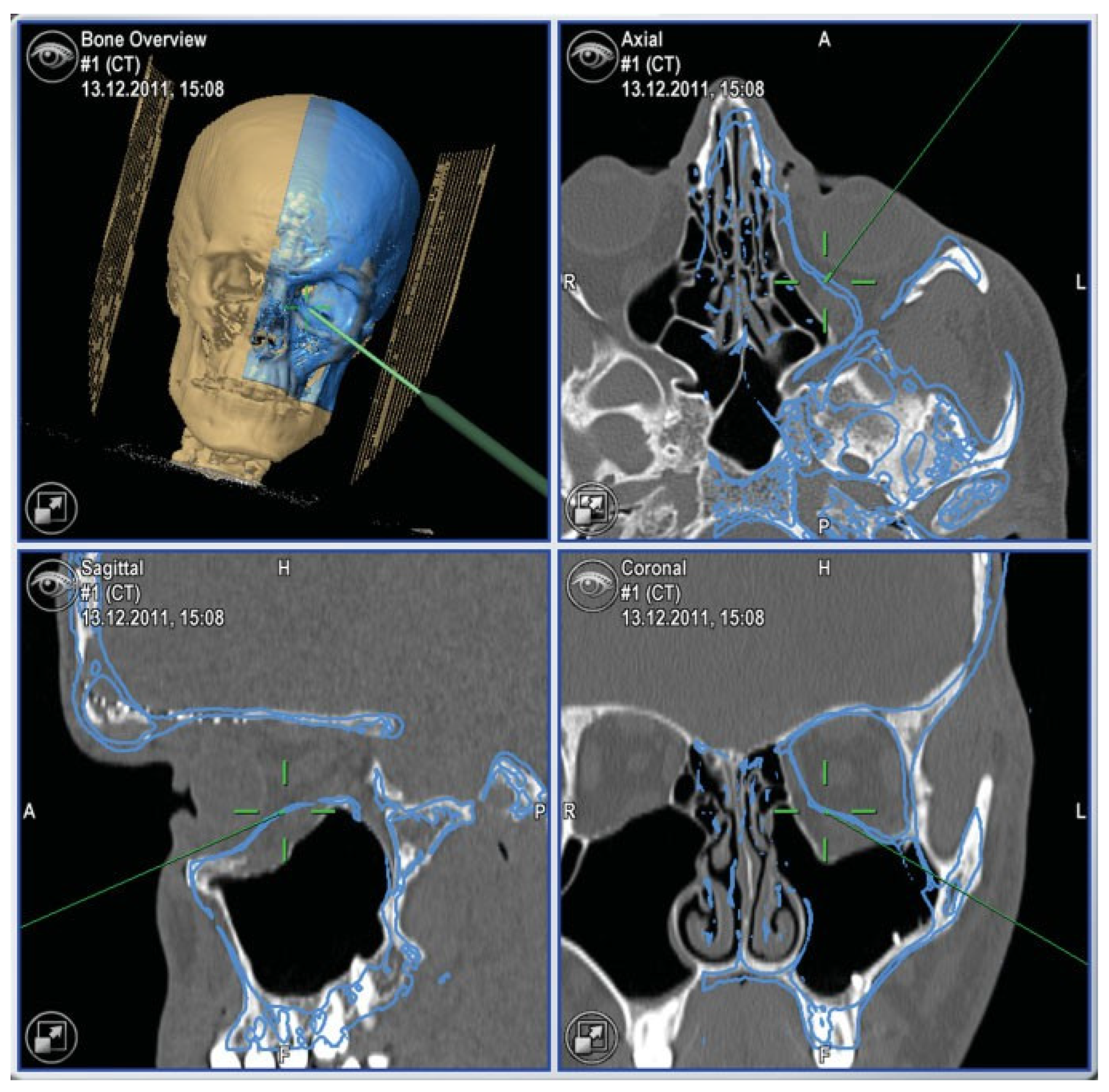
Especially in craniofacial reconstructive surgery, interven- tions associated with the midface and skull base can be accompanied by potential damage of the visual system. In principle, all the earlier-mentioned aspects of traumatic optic nerve damage also apply to craniofacial reconstructive surgery. In these mostly elective cases, optic nerve damage represents a serious complication. Particular focus is placed on orbit reconstructions that involve the posterior orbital region. Disorders of visual acuity ranging up to blindness are rare after such interventions. Nevertheless, these disorders can be reduced by using standardized procedures. The use of modern navigation systems enables exact preoperative plan- ning with virtual contouring of the selected orbital structures (Figure 4). Subsequently, these preformed orbital structures can be controlled intraoperatively with the help of three infrared cameras and can be synchronized with the surgical field (Figure 5). Thus, the risk of incorrect positioning of autogenous or allogeneic orbital transplants and implants can be reduced. The optic nerve can already be controlled preoperatively in terms of transplant or implant positioning. In particular, it can be specifically located intraoperatively. During surgery, testing of pupils is very important. To deter- mine whether light reaction is intact, an intraoperative ERG/ VEP control is recommended. Only the use of this procedure can help determine the requirement of immediate intra- operative revision.

Therapy for Traumatic Optic Nerve Damage

Apart from wait-and-see strategies, there are two therapeutic options for traumatic optic nerve damage: conservative therapy and surgical treatment [57]. Conservative therapy primarily involves treatment with glucocorticoids [49,58,59]. The effect of glucocorticoids is seen in their antiedematous, anti-inflammatory, and antioxidative activity, in addition to their prevention of vasospasm, reduction of nerve cell necrosis, and inhibition of gliofibrous scarring of the traumatized optic nerve [60,61]. The most common conservative therapeutic approach is megadose methylprednisolone therapy, which should not be initiated more than 8 hours after trauma; initially, 30 mg Urbason (Aventis Pharma GmbH, Germany) per kilogram body weight is administered intravenously, and a maintenance dose of 5.4 mg Urbason per kilogram body weight is administered for the subsequent 48 hours. This protocol is derived from a spinal cord trauma study (NASCIS) [62,63]. Often, the use of corticoids is considered a wait-and-see approach. Some authors recommend intravenous megadose corticoid administration over 12 hours; subsequently, if visual acuity is not restored, surgery is indicated [61,64]. Spoor et al. recommended termination of megadose methylprednisolone therapy when the relative afferent pupil defect, the VEP, or visual acuity was not restored within 24 to 48 hours [49]. Combinations of cortico- ids with acetazolamide and mannitol have also been used, but these protocols have not been validated [48,65].
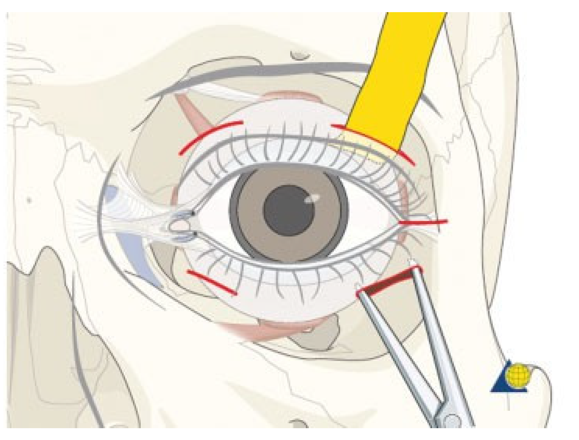
Differentiation between retrobulbar hematoma, edema, and bony causes of damage to the optic nerve is essential for the indication of surgical decompression of orbital structures. A clinically clearly identifiable protrusion of the bulb as a consequence of a retrobulbar hematoma requires an emer- gency opening of the orbital compartment if ipsilateral afferent damage of the visual pathway is proven or even suspected (Figure 6) [66,67]. This procedure is important for treating the hematoma. In addition, opening the orbital septum prevents nerve damage, because the prolapse of the orbital fat leads to decompression of the optic nerve. A restriction for this treatment is pulsating exophthalmos, which is the classical symptom of a carotid-cavernous sinus fistula [68,69]. In such cases, an angiographic diagnosis should be obtained preoperatively.
Surgical optic nerve decompression results in the mechan- ical relief of the visual pathway by uncovering the nerve from its bony surroundings, especially the optic nerve canal. Surgery may be performed with the intention of relieving absolute constriction of the optic nerve canal caused by dislocated bony fragments. Surgery may also be necessary to relieve a relative constriction resulting from increased volume in the canal or the nerve sheath due to edema or hematoma. Discussions regarding surgical treatment for traumatic optic nerve damage deal with the extent (cutting of the annulus of Zinn, additional sheath slitting) [70,71], the surgical approach [72,73], and the time point for surgery [74,75]. Slitting of the optic nerve sheath cannot be considered a standard procedure for the treatment of traumatic optic nerve damage. The requirement of transeth- moidal or transcranial extradural optic nerve decompression is assessed differently within the various disciplines dealing with head surgery [76,77]. Other surgical approaches include sublabial transsphenoidal access and microsurgical endonasal access [78,79,80].
Conclusive recommendations for surgical therapy of trau- matic optic nerve damage are very rare. A probable explanation for this is the problem of indication, because indication, in particular, is based on clinical experience, despite modern advances in high-quality imaging [81]. However, the decision for or against a conservative and/or surgical approach to therapy for visual pathway injuries should not be made only on the basis of morphological criteria (imaging). Such a decision should rather be made on the basis of combined diagnostic criteria involving shape and, in particular, function. This implies that neuro-ophthalmologic and neurophysiologic findings are important [82,83]; in the case of pathologic findings, therapy should be initiated immediately. Data on expectant therapy with postponement of optic nerve decompression and subsequent improvement of visual acuity cannot be a guideline for a successful therapeutic regime. Most probably, such individual cases are select examples of the natural variance associated with afferent optic nerve damage [10]. In contrast, optic nerve decompression is an invasive treatment that requires a careful indication despite low morbidity [84,85,86]. Therefore, preoperative CT scans of the retro-orbital section and the optic nerve canal should be mandatory [87,88,89]. Computer-assisted surgical techniques are very helpful and also reduce morbidity associated with the operation [90]. Moreover, a surgical revision of the skull base in the region of the optic nerve canal enables treatment of combined injuries of the paranasal sinuses and the dura [85]. An important part of the surgical treatment of traumatic optic nerve damage is the postoperative identification of surgical performance using CT with axial and coronary thin-layer reconstructions.
Apart from sole conservative or surgical therapy, combined therapy is also recommended. The common factor in all approaches is the lack of experimental proof of a positive therapeutic effect on the traumatized optic nerve or the peripheral visual pathway. Recommendations for therapy are generally based on findings in small numbers of patients and are often inconsistent in terms of etiology and intensity of traumatic optic nerve damage. The best type of therapy for traumatic optic nerve damage and the best time for surgical decompression to ensure the most benefit to the optic nerve remain uncertain.
A meta-analysis (n ¼ 244) revealed that compared to nontreated patients with traumatic optic nerve damage, patients who received therapy benefited from it and had improved visual acuity [91]. No significant difference was found between the different treatments (decompression, corticoids, and decompression combined with corticoids). A limiting factor for all therapies is early diagnosis of traumatic nerve damage. In the acute posttraumatic phase, early detection can be accelerated by systematic use of clinical electrophysiology techniques [92,93]. Therefore, a mobile neurophysiologic measuring station for VEP/ERG recording is necessary to verify visual acuity during shock or in the early stabilization phase for unconscious patients. This will facilitate the earliest possible initiation of therapy [35,94].
Due to the complexity of traumatic optic nerve damage in conjunction with different injury patterns, cases are never identical or even directly comparable. It is thus not justified to select one special technique for the recovery of visual acuity. Physicians should attempt to determine the precise pathomechanism of individual injuries on the basis of the actual posttraumatic clinical state to select a specific therapy. Combinations of injuries also have to be included in the decision process. In this context, the benefit of clinical electrophysiology techniques such as flash VEP and, if nec- essary, ERG has been proven [18]. Apart from definite identifi- cation of visual pathway damage, electrophysiologic examination also detects the absence of optic nerve injuries, which is an equally important result because it prevents unnecessary treatments.

Recommendations for the Treatment of Traumatic Optic Nerve Lesions

  • The integrity of the visual pathway should be tested immediately in every patient with midface or skull base trauma. If clinical examination is not reliable, VEP and ERG recordings should be performed (Figure 7). This applies also in craniofacial surgery with orbital involvement.
  • In emergency cases, a normal light reaction is a reliable parameter for an intact visual pathway. However, in primary diagnosis of patients with head injuries, pupillary function is often disturbed (e.g., by opioids, injury to the iris sphincter itself, or bilateral damage of the oculomotor nerve).
  • Classification of VEP records as “normal”, “abnormal”, or “not reproducible” is generally sufficient to grade visual pathway function. In cases of deficient neuro-ophthalmo- logic findings, the decision for or against treatment of a visual pathway injury is based on VEP records together with clinical and radiologic findings. With pathologic findings in particular, but also reproducible VEP records, immediate treatment is recommended to avoid additional secondary optic nerve damage.
  • If an afferent disorder of the visual pathway is clinically proven or cannot be excluded, or, in cases of a pathologic VEP record, megadose methylprednisolone therapy should be applied. Contraindications have to be considered.
  • Prompt surgical optic nerve decompression is indicated in cases of retrobulbar hematoma, provided pulsating exophthalmos and cerebrospinal fluid leak are ruled out.
  • Regarding conscious patients, immediate decompression of the optic canal is indicated in the case of afferent disorders with progressive loss or absence of visual acuity together with direct or indirect radiologic signs of trauma in the retrobulbar region or direct vicinity of the optic nerve canal. In these cases, return of visual acuity was not achieved if VEP records were not reproducible before decompression. Nevertheless, surgical intervention is recommended until studies consisting of large numbers of patients show that recurrence of visual acuity is impossible after finding extinct VEPs.
  • Concerning unconscious patients, decompression of the optic canal is also recommended with direct or indirect radiologic signs of trauma in the retrobulbar region or direct vicinity of the optic nerve canal. If the afferent disorder of the visual pathway cannot be clinically proven, surgical intervention is recommended in the case of a pathologic VEP record (reproducible or not).
  • In the case of optic canal decompression, postoperative evaluation of surgical outcome by using CT with axial and coronal thin-layer reconstructions is mandatory.

References

  1. Lang, J. Posterior ethmoid cells and their relation to the optic canal. HNO 1988, 36, 49–53. [Google Scholar] [PubMed]
  2. Osguthorpe, J.D.; Sofferman, R.A. Optic nerve decompression. Otolaryngol Clin North Am 1988, 21, 155–169. [Google Scholar]
  3. Wolin, M.J.; Lavin, P.J. Spontaneous visual recovery from traumatic optic neuropathy after blunt head injury. Am J Ophthalmol 1990, 109, 430–435. [Google Scholar] [CrossRef] [PubMed]
  4. Elisevich, K.V.; Ford, R.M.; Anderson, D.P.; Stratford, J.G.; Richardson, P.M. Visual abnormalities with multiple trauma. Surg Neurol 1984, 22, 565–575. [Google Scholar] [PubMed]
  5. Habal, M.B.; Maniscalco, J.E.; Rhoton, A.L., Jr. Microsurgical anatomy of the optic canal: Correlates to optic nerve exposure. J Surg Res 1977, 22, 527–533. [Google Scholar] [CrossRef]
  6. Kline, L.B.; Morawetz, R.B.; Swaid, S.N. Indirect injury of the optic nerve. Neurosurgery 1984, 14, 756–764. [Google Scholar] [CrossRef]
  7. Anderson, R.L.; Panje, W.R.; Gross, C.E. Optic nerve blindness following blunt forehead trauma. Ophthalmology 1982, 89, 445–455. [Google Scholar] [CrossRef]
  8. Rochels, R. Holographic deformation analysis of the optic canal in blunt cranial trauma. Fortschr Ophthalmol 1990, 87, 182–185. [Google Scholar]
  9. Stankiewicz, J.A. Blindness and intranasal endoscopic ethmoidec- tomy: Prevention and management. Otolaryngol Head Neck Surg 1989, 101, 320–329. [Google Scholar]
  10. McCartney, D.L.; Char, D.H. Return of vision following orbital decom- pression after 36 hours of postoperative blindness. Am J Ophthalmol 1985, 100, 602–604. [Google Scholar]
  11. Linberg, J.V. Orbital compartment syndromes following trauma. Adv Ophthalmic Plast Reconstr Surg 1987, 6, 51–62. [Google Scholar] [PubMed]
  12. Linberg, J.V. Orbital emphysema complicated by acute central retinal artery occlusion: Case report and treatment. Ann Ophthalmol 1982, 14, 747–749. [Google Scholar] [PubMed]
  13. Nayak, S.R.; Kirtane, M.V.; Ingle, M.V. Transethmoid decompression of the optic nerve in head injuries: An update. J Laryngol Otol 1991, 105, 205–206. [Google Scholar] [CrossRef] [PubMed]
  14. Nayak, S.R.; Kirtane, M.V.; Ingle, M.V. Fracture line in post head injury optic nerve damage. J Laryngol Otol 1991, 105, 203–204. [Google Scholar] [CrossRef]
  15. Raveh, J.; Vuillemin, T.; Sutter, F. Subcranial management of 395 combined frontobasal-midface fractures. Arch Otolaryngol Head Neck Surg 1988, 114, 1114–1122. [Google Scholar] [CrossRef]
  16. Cornelius, C.P.; Altenmüller, E.; Ehrenfeld, M. Flash-evoked visual potentials in patients with craniofacial fractures. Fortschr Kiefer Gesichtschir 1991, 36, 158–162. [Google Scholar]
  17. Dutton, G.N.; Al-Qurainy, I.; Stassen, L.F.; Titterington, D.M.; Moos, K.F.; El-Attar, A. Ophthalmic consequences of mid-facial trauma. Eye 1992, 6 Pt 1, 86–89. [Google Scholar] [CrossRef]
  18. Gellrich, N.C.; Zerfowski, M.; Eufinger, H.; Reinert, S.; Eysel, U.T. Interdisciplinary diagnosis and therapy of traumatic optic nerve damage. Mund Kiefer Gesichtschir 1998, 2 (Suppl. 1), S107–S112. [Google Scholar] [CrossRef]
  19. Frenkel, R.E.; Spoor, T.C. Diagnosis and management of traumatic optic neuropathies. Adv Ophthalmic Plast Reconstr Surg 1987, 6, 71–90. [Google Scholar]
  20. Joseph, E.; Zak, R.; Smith, S.; Best, W.R.; Gamelli, R.L.; Dries, D.J. Predictors of blinding or serious eye injury in blunt trauma. J Trauma 1992, 33, 19–24. [Google Scholar] [CrossRef]
  21. Hughes, B. Indirect injury of the optic nerves and chiasma. Bull Johns Hopkins Hosp 1962, 111, 98–126. [Google Scholar] [PubMed]
  22. Hager, G.; Maruniak, M.; Gerhardt, H.-J. Indications and results of operative exposure of traumatically damaged optic nerves (au- thor’s transl). Klin Monatsbl Augenheilkd 1975, 167, 515–526. [Google Scholar]
  23. Blanco, R.; Salvador, F.; Galan, A.; Gil-Gibernau, J.J. Aplasia of the optic nerve: Report of three cases. J Pediatr Ophthalmol Strabismus 1992, 29, 228–231. [Google Scholar]
  24. Sloan, T.; Sloan, H.; Rogers, J. Nitrous oxide and isoflurane are synergistic with respect to amplitude and latency effects on sensory evoked potentials. J Clin Monit Comput 2010, 24, 113–123. [Google Scholar] [CrossRef] [PubMed]
  25. Nakagawa, I.; Hidaka, S.; Okada, H.; Kubo, T.; Okamura, K.; Kato, T. Effects of sevoflurane and propofol on evoked potentials during neuro- surgical anesthesia. Masui 2006, 55, 692–698. [Google Scholar]
  26. Hamaguchi, K.; Nakagawa, I.; Hidaka, S.; Uesugi, F.; Kubo, T.; Kato, T. Effect of propofol on visual evoked potentials during neurosurgery. Masui 2005, 54, 998–1002. [Google Scholar]
  27. Bonkowsky, V.M.; Mang, W.L.; Wendl, F.; Frank, C. Neurologic compli- cations in mid-face fractures. Laryngorhinootologie 1989, 68, 539–542. [Google Scholar] [CrossRef]
  28. Gossman, M.D.; Roberts, D.M.; Barr, C.C. Ophthalmic aspects of orbital injury. A comprehensive diagnostic and management approach. Clin Plast Surg 1992, 19, 71–85. [Google Scholar] [CrossRef] [PubMed]
  29. Klopfer, J.; Tielsch, J.M.; Vitale, S.; See, L.C.; Canner, J.K. Ocular trauma in the United States. Eye injuries resulting in hospitalization, 1984 through 1987. Arch Ophthalmol 1992, 110, 838–842. [Google Scholar]
  30. Ioannides, C.; Treffers, W.; Rutten, M.; Noverraz, P. Ocular injuries associated with fractures involving the orbit. J Craniomaxillofac Surg 1988, 16, 157–159. [Google Scholar]
  31. Mahapatra, A.K.; Bhatia, R. Predictive value of visual evoked poten- tials in unilateral optic nerve injury. Surg Neurol 1989, 31, 339–342. [Google Scholar] [CrossRef] [PubMed]
  32. Shokunbi, T.; Agbeja, A. Ocular complications of head injury in children. Childs Nerv Syst 1991, 7, 147–149. [Google Scholar] [CrossRef] [PubMed]
  33. Brent, B.D.; May, D.R. Orbital apex syndrome after penetrating orbital trauma. Ann Ophthalmol 1990, 22, 267–268. [Google Scholar]
  34. Hutton, W.L.; Fuller, D.G. Factors influencing final visual results in severely injured eyes. Am J Ophthalmol 1984, 97, 715–722. [Google Scholar] [CrossRef]
  35. Jorissen, M.; Feenstra, L. Optic nerve decompression for indirect posterior optic nerve trauma. Acta Otorhinolaryngol Belg 1992, 46, 311–324. [Google Scholar]
  36. Shaked, A.; Hadani, M.; Feinsod, M.C.T. CT and VER follow-up of reversible visual loss with fracture of the optic canal. Acta Neurochir 1982, 62, 91–94. [Google Scholar] [CrossRef]
  37. Dorfman, L.J.; Gaynon, M.; Ceranski, J.; Louis, A.A.; Howard, J.E. Visual electrical evoked potentials: Evaluation of ocular injuries. Neurology 1987, 37, 123–128. [Google Scholar] [CrossRef] [PubMed]
  38. Mahapatra, A.K. Visual evoked potentials in optic nerve injury—Does it merit to be mentioned? Indian J Ophthalmol 1991, 39, 20–21. [Google Scholar]
  39. Halliday, A.M.; Halliday, E.; Kriss, A.; McDonald, W.I.; Mushin, J. The pattern-evoked potential in compression of the anterior visual pathways. Brain 1976, 99, 357–374. [Google Scholar] [CrossRef]
  40. Uren, S.M.; Stewart, P.; Crosby, P.A. Subject cooperation and the visual evoked response. Invest Ophthalmol Vis Sci 1979, 18, 648–652. [Google Scholar]
  41. Jayle, G.E.; Tassy, A.F. Prognostic value of the electroretinogram in severe recent ocular trauma. Br J Ophthalmol 1970, 54, 51–58. [Google Scholar] [PubMed]
  42. Gellrich, N.C.; Gellrich, M.M.; Zerfowski, M.; Eufinger, H.; Eysel, U.T. Clinical and experimental study of traumatic optic nerve damage. Ophthalmologe 1997, 94, 807–814. [Google Scholar]
  43. Greenberg, R.P.; Becker, D.P.; Miller, J.D.; Mayer, D.J. Evaluation of brain function in severe human head trauma with multimodality evoked potentials. Part 2, localization of brain dysfunction and correlation with posttraumatic neurological conditions. J Neurosurg 1977, 47, 163–177. [Google Scholar]
  44. Meyer, B.; François, M.; Lacau Saint-Guily, J.; Lacombe, H.; Laroche, L. Post-traumatic blindness and spontaneous decompression of optical nerve (author’s transl). Ann Otolaryngol Chir Cervicofac 1982, 99, 53–55. [Google Scholar] [PubMed]
  45. Rochels, R. Ultrasonic diagnosis of fractures of the bony orbit. Fortschr Kiefer Gesichtschir 1987, 32, 144–147. [Google Scholar] [PubMed]
  46. Chirico, P.A.; Mirvis, S.E.; Kelman, S.E.; Karesh, J.W. Orbital “blow-in” fractures: Clinical and CT features. J Comput Assist Tomogr 1989, 13, 1017–1022. [Google Scholar]
  47. Luka, B.; Brechtelsbauer, D.; Gellrich, N.C.; König, M. 2D and 3D CT reconstructions of the facial skeleton: An unnecessary option or a diagnostic pearl? Int J Oral Maxillofac Surg 1995, 24 Pt 2, 76–83. [Google Scholar]
  48. Behrens-Baumann, W.; Chilla, R. Drug and surgical therapy of traumatic optic nerve compression. Fortschr Ophthalmol 1984, 81, 87–89. [Google Scholar]
  49. Spoor, T.C.; Hartel, W.C.; Lensink, D.B.; Wilkinson, M.J. Treatment of traumatic optic neuropathy with corticosteroids. Am J Ophthalmol 1990, 110, 665–669. [Google Scholar] [CrossRef]
  50. Purvin, V. Evidence of orbital deformation in indirect optic nerve injury. Weight lifter’s optic neuropathy. J Clin Neuroophthalmol 1988, 8, 9–11. [Google Scholar]
  51. Bilyk, J.R.; Dallow, R.L.; Ojemann, R.G.; Linggood, R.M.; Shore, J.W. Man- agement of lesions at the cranioorbital junction. Int Ophthalmol Clin 1992, 32, 73–93. [Google Scholar]
  52. Manfredi, S.J.; Raji, M.R.; Sprinkle, P.M.; Weinstein, G.W.; Minardi, L.M.; Swanson, T.J. Computerized tomographic scan findings in facial fractures associated with blindness. Plast Reconstr Surg 1981, 68, 479–490. [Google Scholar] [PubMed]
  53. Mahapatra, A.K.; Tandon, D.A. Traumatic optic neuropathy in chil- dren: A prospective study. Pediatr Neurosurg 1993, 19, 34–39. [Google Scholar]
  54. Seiler, T.; Bende, T.; Schilling, A.; Wollensak, J. Magnetic resonance tomography in ophthalmology. II. Manifestations of edema of the optic nerve. Klin Monatsbl Augenheilkd 1989, 195, 72–78. [Google Scholar] [PubMed]
  55. Crowe, N.W.; Nickles, T.P.; Troost, B.T.; Elster, A.D. Intrachiasmal hemor- rhage: A cause of delayed post-traumatic blindness. Neurology 1989, 39, 863–865. [Google Scholar]
  56. Kakisu, Y.; Adachi-Usami, E.; Fujimoto, N. Pattern visually evoked cortical potential and magnetic resonance imaging in optic neuritis. J Clin Neuroophthalmol 1991, 11, 205–212. [Google Scholar] [PubMed]
  57. Lipkin, A.F.; Woodson, G.E.; Miller, R.H. Visual loss due to orbital fracture. The role of early reduction. Arch Otolaryngol Head Neck Surg 1987, 113, 81–83. [Google Scholar]
  58. Lam, B.L.; Weingeist, T.A. Corticosteroid-responsive traumatic optic neuropathy. Am J Ophthalmol 1990, 109, 99–101. [Google Scholar]
  59. Seiff, S.R. High dose corticosteroids for treatment of vision loss due to indirect injury to the optic nerve. Ophthalmic Surg 1990, 21, 389–395. [Google Scholar]
  60. Hall, E.D.; Braughler, J.M. Glucocorticoid mechanisms in acute spinal cord injury: A review and therapeutic rationale. Surg Neurol 1982, 18, 320–327. [Google Scholar]
  61. Panje, W.R.; Gross, C.E.; Anderson, R.L. Sudden blindness following facial trauma. Otolaryngol Head Neck Surg 1981, 89, 941–948. [Google Scholar] [CrossRef]
  62. Bracken, M.B.; Holford, T.R. Effects of timing of methylprednisolone or naloxone administration on recovery of segmental and long- tract neurological function in NASCIS 2. J Neurosurg 1993, 79, 500–507. [Google Scholar]
  63. Bracken, M.B.; Shepard, M.J.; Collins, W.F.; et al. A randomized, controlled trial of methylprednisolone or naloxone in the treatment of acute spinal-cord injury. Results of the Second National Acute Spinal Cord Injury Study. N Engl J Med 1990, 322, 1405–1411. [Google Scholar] [PubMed]
  64. Osguthorpe, J.D. Transethmoid decompression of the optic nerve. Otolaryngol Clin North Am 1985, 18, 125–137. [Google Scholar]
  65. Ord, R.A.; El Attar, A. Acute retrobulbar hemorrhage complicating a malar fracture. J Oral Maxillofac Surg 1982, 40, 234–236. [Google Scholar] [CrossRef] [PubMed]
  66. Dolman, P.J.; Glazer, L.C.; Harris, G.J.; Beatty, R.L.; Massaro, B.M. Mechanisms of visual loss in severe proptosis. Ophthal Plast Reconstr Surg 1991, 7, 256–260. [Google Scholar]
  67. Katz, B.; Herschler, J.; Brick, D.C. Orbital haemorrhage and prolonged blindness: A treatable posterior optic neuropathy. Br J Ophthalmol 1983, 67, 549–553. [Google Scholar] [CrossRef] [PubMed]
  68. Takenoshita, Y.; Hasuo, K.; Matsushima, T.; Oka, M. Carotid-cavernous sinus fistula accompanying facial trauma. Report of a case with a review of the literature. J Craniomaxillofac Surg 1990, 18, 41–45. [Google Scholar]
  69. Zachariades, N.; Papavassiliou, D. Traumatic carotid-cavernous sinus fistula. J Craniomaxillofac Surg 1988, 16, 385–388. [Google Scholar]
  70. Ardouin, M.; Urvoy, M.; Bourdinière, J.; Guillou, B.; Eon, J.Y. What should be one’s attitude toward trans-sinus latero-orbital decompressive surgery in optic nerve injuries? Bull Soc Ophtalmol Fr 1976, 76, 895–905. [Google Scholar]
  71. Karnik, P.P.; Maskati, B.T.; Kirtane, M.V.; Tonsekar, K.S. Optic nerve decompression in head injuries. J Laryngol Otol 1981, 95, 1135–1140. [Google Scholar] [CrossRef] [PubMed]
  72. Behrens-Baumann, W.; Miehlke, A. Rhinobasal deompression of the optic nerve following trauma (author’s transl). Klin Monatsbl Augenheilkd 1979, 175, 584–591. [Google Scholar] [PubMed]
  73. Funk, G.F.; Stanley, R.B. Jr.; Becker, T.S. Reversible visual loss due to impacted lateral orbital wall fractures. Head Neck 1989, 11, 295–300. [Google Scholar] [CrossRef] [PubMed]
  74. Kennerdell, J.S.; Amsbaugh, G.A.; Myers, E.N. Transantral-ethmoidal decompression of optic canal fracture. Arch Ophthalmol 1976, 94, 1040–1043. [Google Scholar] [CrossRef]
  75. Laroche, L.; Lacombe, H.; Meyer, B.; Saraux, H. Trans-ethmoidal- sphenoidal decompression and optic nerve injury. Bull Soc Ophtalmol Fr 1984, 84, 431–432. [Google Scholar]
  76. Girard, B.; Bouzas, E.; Lamas, G.; Topouzis, F.; Soudant, J. X-ray comput- ed tomography in surgical indication of physiological section of the optic nerve. Apropos of 15 cases. J Fr Ophtalmol 1992, 15, 93–101. [Google Scholar]
  77. Waga, S.; Kubo, Y.; Sakakura, M. Transfrontal intradural microsurgical decompression for traumatic optic nerve injury. Acta Neurochir (Wien) 1988, 91, 42–46. [Google Scholar] [CrossRef]
  78. Sofferman, R.A. An extracranial microsurgical approach to the optic nerve. J Microsurg 1979, 1, 195–202. [Google Scholar] [CrossRef]
  79. Mann, W.; Rochels, R.; Bleier, R. Microsurgical endonasal decompres- sion of the optic nerve. Fortschr Ophthalmol 1991, 88, 176–177. [Google Scholar]
  80. Takahashi, M.; Itoh, M.; Kaneko, M.; Ishii, J.; Yoshida, A. Microscopic intranasal decompression of the optic nerve. Arch Otorhinolaryngol 1989, 246, 113–116. [Google Scholar] [CrossRef]
  81. Slamovits, T.L.; Gardner, T.A. Neuroimaging in neuro-ophthalmology. Ophthalmology 1989, 96, 555–568. [Google Scholar] [CrossRef] [PubMed]
  82. Funder, J.; Hollmann, K.; Millesi, W. Results of surgically and conservatively treated cases of traumatic optic nerve lesions. Klin Monatsbl Augenheilkd 1986, 189, 421–422. [Google Scholar] [CrossRef] [PubMed]
  83. Kolenda, H.; Schröder, M.; Mühlendyck, H.; Rama, B.; Markakis, E. Transethmoidal decompression of the optic nerve in the case of craniocerebral trauma. Neurosurg Rev 1988, 11, 39–43. [Google Scholar] [CrossRef]
  84. Guyer, D.R.; Miller, N.R.; Long, D.M.; Allen, G.S. Visual function following optic canal decompression via craniotomy. J Neurosurg 1985, 62, 631–638. [Google Scholar] [CrossRef] [PubMed]
  85. Siegl, H. Decompression of the optic nerve. Laryngol Rhinol Otol (Stuttg) 1985, 64, 118–120. [Google Scholar] [CrossRef]
  86. Stoll, W.; Busse, H.; Kroll, P. Visual recovery following orbital and optic nerve decompressions. Laryngol Rhinol Otol (Stuttg) 1987, 66, 577–582. [Google Scholar]
  87. Girard, B.; Lamas, G.; Bouzas, E.; Topouzis, F.; Soudant, J. Surgical decompression of the optic nerve in intracanal injuries. Indica- tions and results. J Fr Ophtalmol 1992, 15, 83–92. [Google Scholar]
  88. Machtens, E.; Heuser, L. Basic and graduated procedures in the roentgen diagnosis of midfacial trauma in relation to degree of severity and localization. Fortschr Kiefer Gesichtschir 1991, 36, 21–25. [Google Scholar]
  89. Seiff, S.R.; Berger, M.S.; Guyon, J.; Pitts, L.H. Computed tomographic evaluation of the optic canal in sudden traumatic blindness. Am J Ophthalmol 1984, 98, 751–755. [Google Scholar] [CrossRef]
  90. Kurzeja, A.; Wenzel, M.; Korves, B.; Mösges, R. Decompression of the optic nerve after fractures of the rhino-basal skull with computer- assisted surgery. Laryngorhinootologie 1994, 73, 274–276. [Google Scholar] [CrossRef]
  91. Cook, M.W.; Levin, L.A.; Joseph, M.P.; Pinczower, E.F. Traumatic optic neuropathy. A meta-analysis. Arch Otolaryngol Head Neck Surg 1996, 122, 389–392. [Google Scholar] [PubMed]
  92. Habal, M.B.; Maniscalco, J.E. Surgical relations of the orbit and optic nerve: An anatomical study under magnification. Ann Plast Surg 1980, 4, 265–275. [Google Scholar] [CrossRef] [PubMed]
  93. Janáky, M.; Benedek, G. Visual evoked potentials during the early phase of optic nerve compression in the orbital cavity. Doc Ophthalmol 1992, 81, 197–208. [Google Scholar]
  94. Obertacke, U.; Joka, T.; Härting, F.; Nau, H.E.; Sauerwein, W. Amaurosis caused by traumatic damage to the optic nerve. Unfallchirurg 1986, 89, 132–137. [Google Scholar] [PubMed]
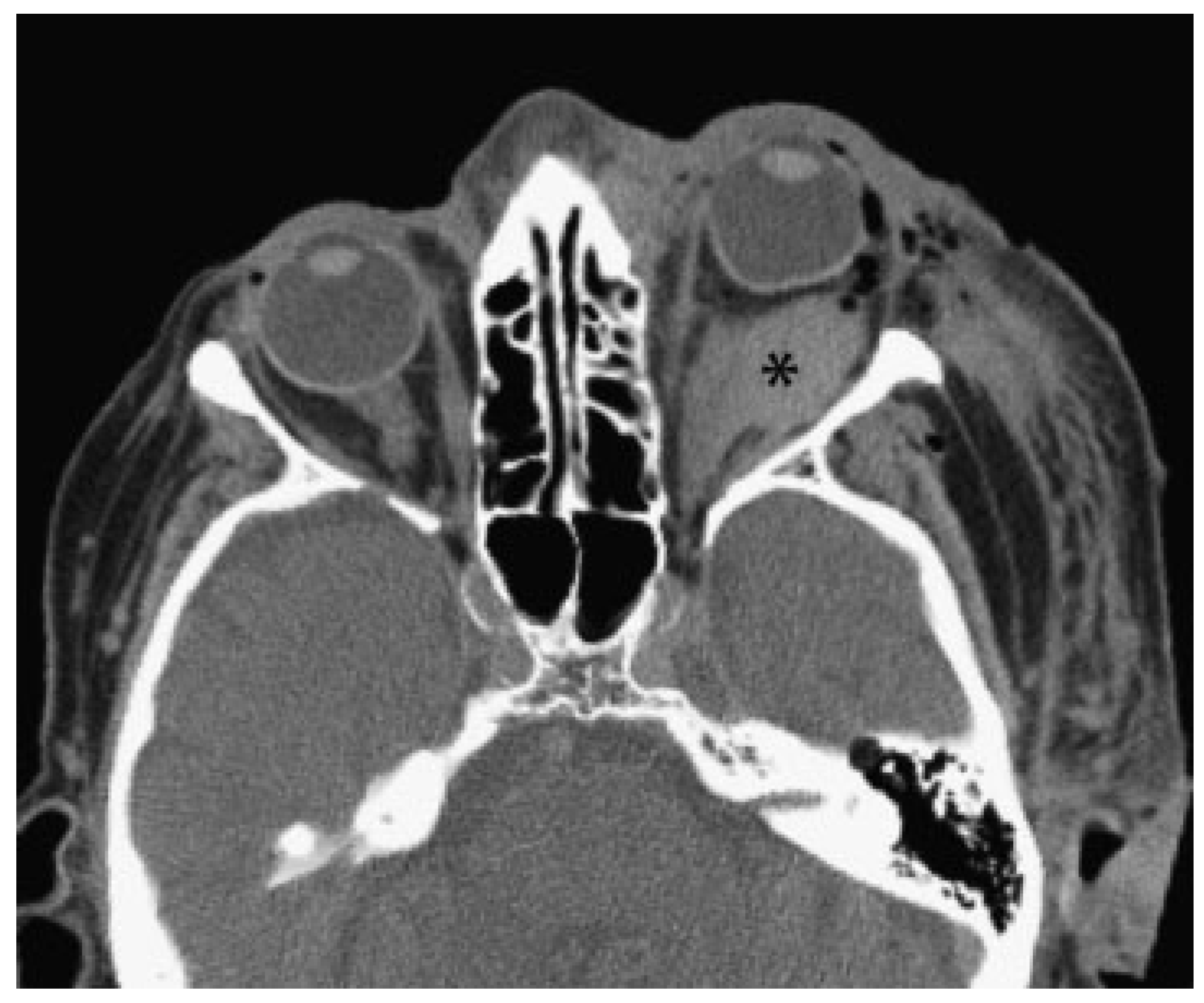
Figure 1. Axial computed tomography view showing typical radiologic signs of a retrobulbar hematoma: globe-shaped bulb, axial proptosis, and retrobulbar fluid accumulation (marked with asterisk).
Cmtr 06 00013 g001
Figure 2. Clinical images of single and parallel electroretinogram and visual evoked potential performed using a mobile neurophysiologic measuring station with a dual-channel lead: position of the needle electrodes including grounding (A) and a threadlike electrode (marked with arrows; B), overview of the examination protocol (C).
Cmtr 06 00013 g002
Figure 3. Posttraumatic ERG (A1 right eye, A2 left eye) and VEP (B1 right eye, B2 left eye). Note the physiologic VEP with a clear amplitude (blue bar) for the right eye (B1) and the pathologic VEP with a missing amplitude for the left eye (B2); ERG on both sides without pathologic findings. Abbreviations: ERG, electroretinogram; VEP, visual evoked potential.
Cmtr 06 00013 g003
Figure 4. Posttraumatic secondary reconstruction of the left orbit: preoperative planning. The unaffected right side (red) is reflected onto the affected left side (blue), creating a virtual template for reconstruction of the left orbit. The orbital floor fracture is marked with a green arrow. Abbreviation: CT, computed tomography.
Cmtr 06 00013 g004
Figure 5. Posttraumatic secondary reconstruction of the left orbit: intraoperative navigation. Intraoperative multiplanar display shows pointer-based surface matching after titanium mesh insertion for left orbital reconstruction. Correct implant position is checked by comparing the pointer tip (green line) with the virtual reconstruction template (blue segmentation). Abbreviation: CT, computed tomography.
Cmtr 06 00013 g005
Figure 6. Decompression of the orbital compartment: surgical procedure. Four incisions are placed in natural creases directly above the supra- and infraorbital rim. After dissecting the orbicularis muscle and the orbital septum (palpebral ligament), the four incisions are elongated into the orbit behind the globe securely in contact with bone. Elastic drainage tubes are inserted to release extraconal and intraconal hematomas from the orbit. Illustration reproduced by kind permission of the AO Foundation, Davos, Switzerland.
Cmtr 06 00013 g006
Figure 7. Diagrammatic representation of VEP/ERG recording. Illus- tration reproduced by kind permission of the AO Foundation, Davos, Switzerland.
Cmtr 06 00013 g007