The Importance of Accurate, Early Bony Reconstruction in Orbital Injuries with Globe Loss
Abstract
:Methods
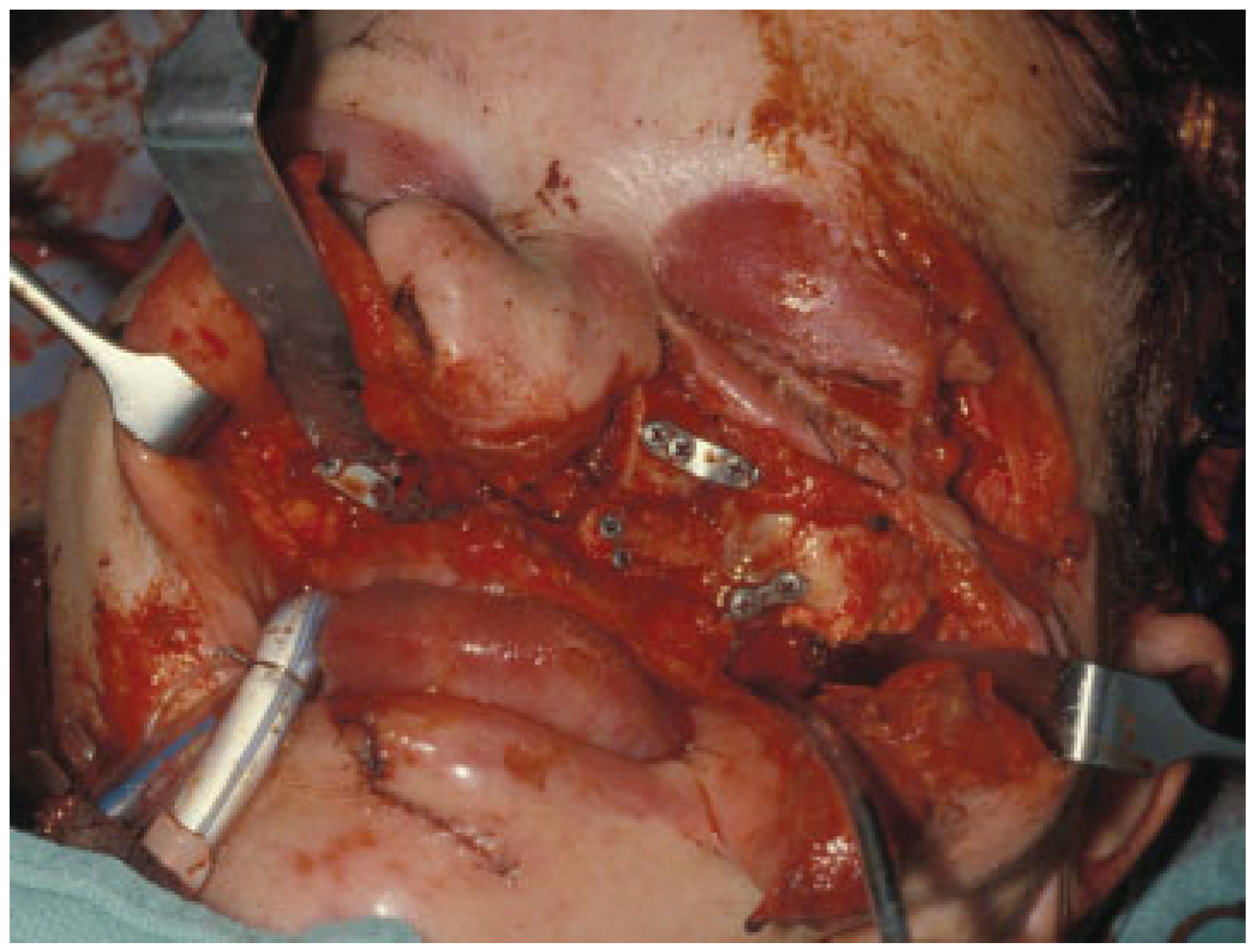
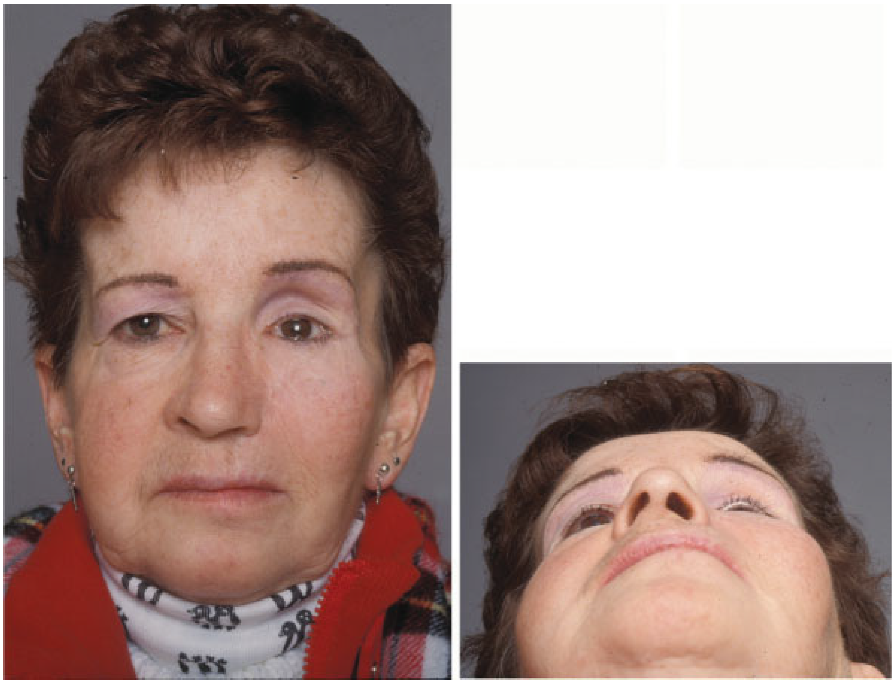
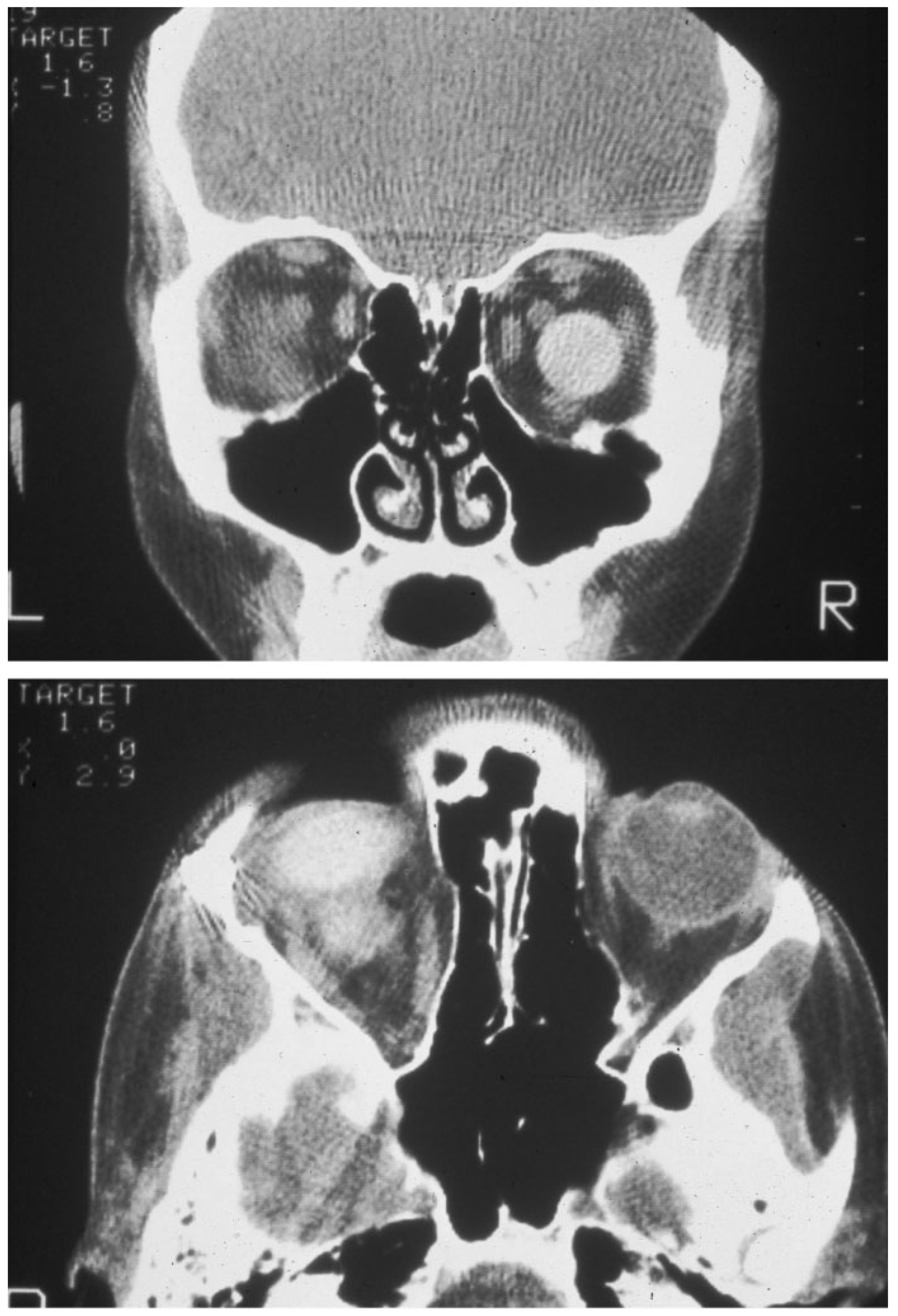
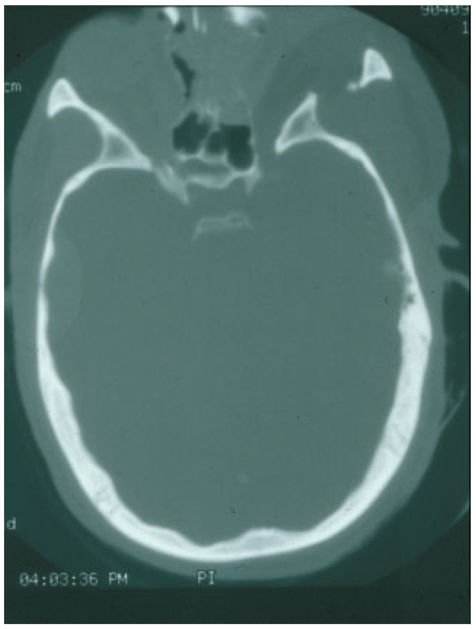
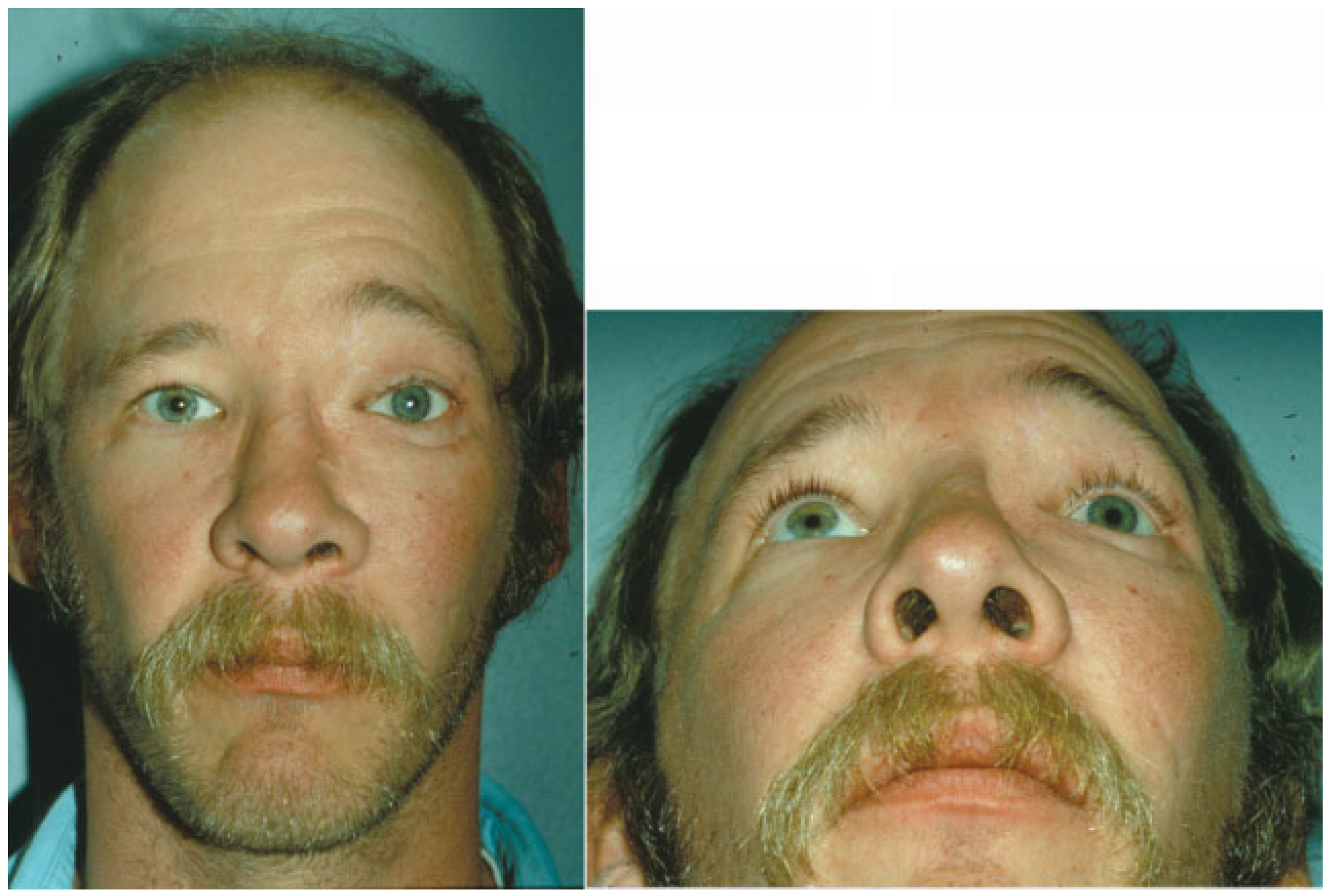
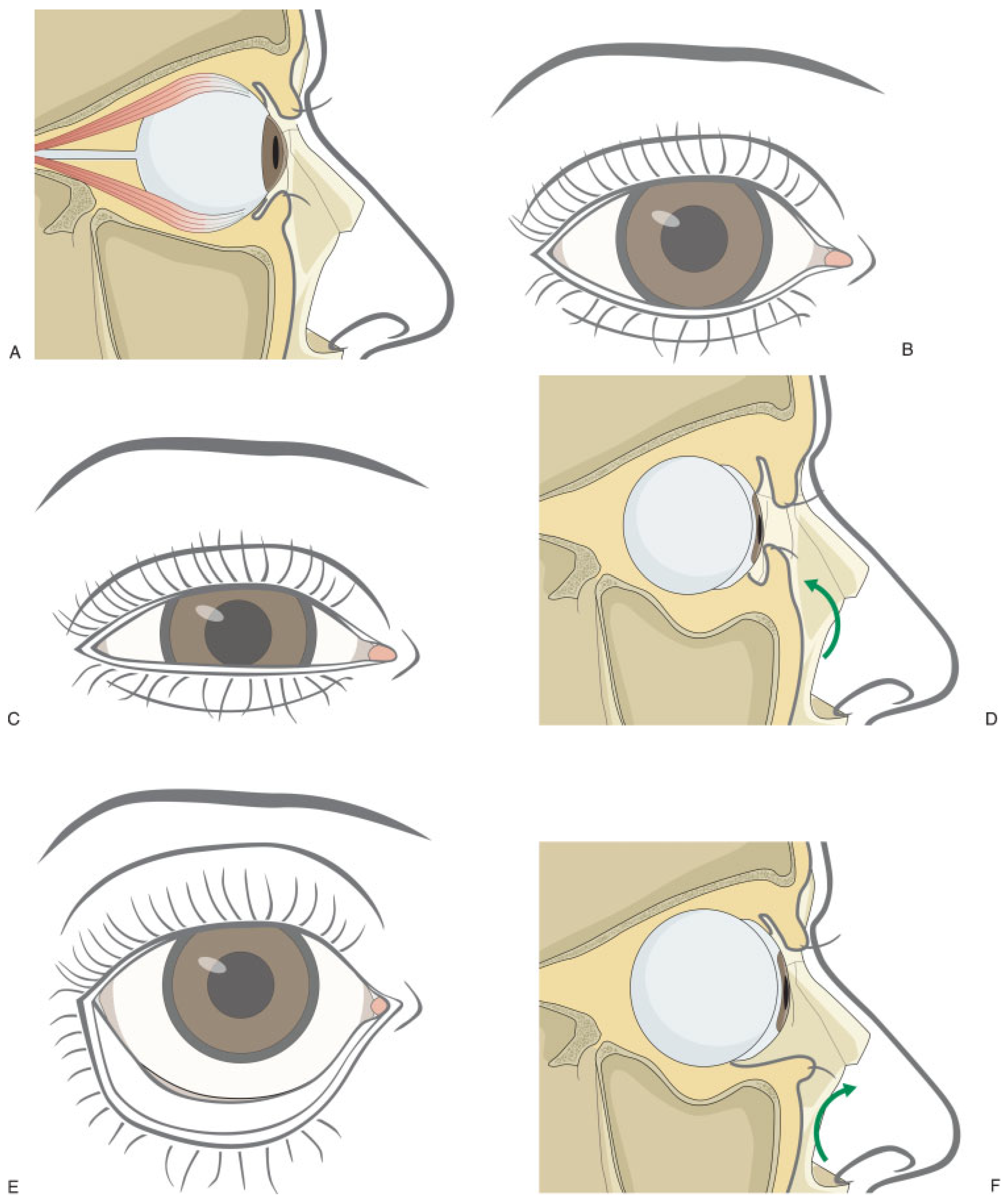
Discussion
Conclusion
References
- MacKinnon, C.A.; David, D.J.; Cooter, R.D. Blindness and severe visual impairment in facial fractures: an 11 year review. Br J Plast Surg 2002, 55, 1–7. [Google Scholar] [CrossRef]
- Nagase, D.Y.; Courtemanche, D.J.; Peters, D.A. Facial fractures—association with ocular injuries: a 13-year review of one practice in a tertiary care centre. Can J Plast Surg 2006, 14, 167–171. [Google Scholar] [CrossRef]
- Shere, J.L.; Boole, J.R.; Holtel, M.R.; Amoroso, P.J. An analysis of 3599 midfacial and 1141 orbital blowout fractures among 4426 United States Army Soldiers, 1980–2000. Otolaryngol Head Neck Surg 2004, 130, 164–170. [Google Scholar] [CrossRef] [PubMed]
- Jabaley, M.E.; Lerman, M.; Sanders, H.J. Ocular injuries in orbital fractures. A review of 119 cases. Plast Reconstr Surg 1975, 56, 410–418. [Google Scholar] [CrossRef] [PubMed]
- al-Qurainy, I.A.; Stassen, L.F.; Dutton, G.N.; Moos, K.F.; el-Attar, A. The characteristics of midfacial fractures and the association with ocular injury: a prospective study. Br J Oral Maxillofac Surg 1991, 29, 291–301. [Google Scholar]
- Girotto, J.A.; MacKenzie, E.; Fowler, C.; Redett, R.; Robertson, B.; Manson, P.N. Long-term physical impairment and functional outcomes after complex facial fractures. Plast Reconstr Surg 2001, 108, 312–327. [Google Scholar] [CrossRef] [PubMed]
- Holt, G.R.; Holt, J.E. Incidence of eye injuries in facial fractures: an analysis of 727 cases. Otolaryngol Head Neck Surg 1983, 91, 276–279. [Google Scholar] [CrossRef]
- Holt, J.E.; Holt, G.R.; Blodgett, J.M. Ocular injuries sustained during blunt facial trauma. Ophthalmology 1983, 90, 14–18. [Google Scholar] [CrossRef]
- Joseph, E.; Zak, R.; Smith, S.; Best, W.R.; Gamelli, R.L.; Dries, D.J. Predictors of blinding or serious eye injury in blunt trauma. J Trauma 1992, 33, 19–24. [Google Scholar] [CrossRef]
- Cullen, G.C.; Luce, C.M.; Shannon, G.M. Blindness following blowout orbital fractures. Ophthalmic Surg 1977, 8, 60–62. [Google Scholar] [CrossRef]
- Glassman, R.D.; Manson, P.N.; Vanderkolk, C.A.; et al. Rigid fixation of internal orbital fractures. Plast Reconstr Surg 1990, 86, 1103–1109;discussion 1110–1111. [Google Scholar] [CrossRef] [PubMed]
- Hammer, B. Orbital Fractures: Diagnosis, Operative Treatment, Secondary Corrections; Hogrefe and Huber Publications: Kirkland, WA, USA, 1995. [Google Scholar]
- Paskert, J.P.; Manson, P.N.; Iliff, N.T. Nasoethmoidal and orbital fractures. Clin Plast Surg 1988, 15, 209–223. [Google Scholar] [CrossRef]
- Chen, C.T.; Chen, Y.R.; Tung, T.C.; Lai, J.P.; Rohrich, R.J. Endoscopically assisted reconstruction of orbital medial wall fractures. Plast Reconstr Surg 1999, 103, 714–720; quiz 721. [Google Scholar] [CrossRef] [PubMed]
- Messinger, A.; Radkowski, M.A.; Greenwald, M.J.; Pensler, J.M. Orbital roof fractures in the pediatric population. Plast Reconstr Surg 1989, 84, 213–216; discussion 217–218. [Google Scholar] [CrossRef] [PubMed]
- Losee, J.E.; Afifi, A.; Jiang, S.; et al. Pediatric orbital fractures: classification, management, and early follow-up. Plast Reconstr Surg 2008, 122, 886–897. [Google Scholar] [CrossRef]
- Cole, P.; Boyd, V.; Banerji, S.; Hollier, L.H., Jr. Comprehensive management of orbital fractures. Plast Reconstr Surg 2007, 120 (Suppl. 2), 57S–63S. [Google Scholar] [CrossRef]
- Burm, J.S.; Chung, C.H.; Oh, S.J. Pure orbital blowout fracture: new concepts and importance of medial orbital blowout fracture. Plast Reconstr Surg 1999, 103, 1839–1849. [Google Scholar] [CrossRef]
- Converse, J.M.; Smith, B.; Obear, M.F.; Wood-Smith, D. Orbital blowout fractures: a ten-year survey. Plast Reconstr Surg 1967, 39, 20–36. [Google Scholar] [CrossRef]
- Patel, B.C.; Hoffmann, J. Management of complex orbital fractures. Facial Plast Surg 1998, 14, 83–104. [Google Scholar] [CrossRef]
- Miller, G.R.; Tenzel, R.R. Ocular complications of midfacial fractures. Plast Reconstr Surg 1967, 39, 37–42. [Google Scholar] [CrossRef]
- Markowitz, B.L.; Manson, P.N.; Yaremchuk, M.; Glassman, D.; Kawamoto, H. High-energy orbital dislocations: the possibility of traumatic hypertelorbitism. Plast Reconstr Surg 1991, 88, 20–28; discussion 29–30. [Google Scholar] [CrossRef] [PubMed]
- Lauer, S.A.; Rizzuto, P.R.; Goodrich, J.; Adamo, A. Orbitocraniofacial gunshot wounds: craniofacial reconstruction and preparation of the anophthalmic socket. J Craniomaxillofac Trauma 1995, 1, 21–27. [Google Scholar] [PubMed]
- Rubin, P.A.; Shore, J.W.; Yaremchuk, M.J. Complex orbital fracture repair using rigid fixation of the internal orbital skeleton. Ophthalmology 1992, 99, 553–559. [Google Scholar] [CrossRef]
- Sergott, T.J.; Vistnes, L.M. Correction of enophthalmos and superior sulcus depression in the anophthalmic orbit: a long-term follow-up. Plast Reconstr Surg 1987, 79, 331–338. [Google Scholar] [CrossRef]
- Vistnes, L.M. Mechanism of upper lid ptosis in the anophthalmic orbit. Plast Reconstr Surg 1976, 58, 539–545. [Google Scholar] [CrossRef] [PubMed]
- Fry, H.J. The enucleated eye socket. Br J Plast Surg 1968, 21, 290–293. [Google Scholar] [CrossRef]
- Nolan, W.B., III; Vistnes, L.M. Correction of lower eyelid ptosis in the anophthalmic orbit: a long-term follow-up. Plast Reconstr Surg 1983, 72, 289–293. [Google Scholar] [CrossRef]
- Clauser, L.; Sarti, E.; Dallera, V.; Galiè, M. Integrated reconstructive strategies for treating the anophthalmic orbit. J Craniomaxillofac Surg 2004, 32, 279–290. [Google Scholar] [CrossRef]
- Vistnes, L.M.; Iverson, R.E.; Laub, D.R. The anophthalmic orbit. Surgical correction of lower eyelid ptosis. Plast Reconstr Surg 1973, 52, 346–351. [Google Scholar] [CrossRef]
- Li, D.; Jie, Y.; Liu, H.; Liu, J.; Zhu, Z.; Mao, C. Reconstruction of anophthalmic orbits and contracted eye sockets with micro-vascular radial forearm free flaps. Ophthal Plast Reconstr Surg 2008, 24, 94–97. [Google Scholar] [CrossRef]
- Habal, M.B. Aesthetic considerations in the reconstruction of the anophthalmic orbit. Aesthetic Plast Surg 1987, 11, 229–239. [Google Scholar] [CrossRef] [PubMed]
- Linberg, J.V.; Anderson, R.L.; Edwards, J.J.; Panje, W.R.; Bardach, J. Preserved irradiated homolgous cartilage for orbital reconstruction. Ophthalmic Surg 1980, 11, 457–462. [Google Scholar] [PubMed]
- Smith, B.; Petrelli, R. Dermis-fat graft as a movable implant within the muscle cone. Am J Ophthalmol 1978, 85, 62–66. [Google Scholar] [CrossRef] [PubMed]
- Smith, B.; Obear, M.; Leone, C.R., Jr. The correction of enophthalmos associated with anophthalmos by glass bead implantation. Am J Ophthalmol 1967, 64, 1088–1093. [Google Scholar] [CrossRef]
- Nasr, A.M.; Jabak, M.H.; Batainah, Y. Orbital volume augmentation with subperiosteal room-temperature-vulcanized silicone implants: a clinical and histopathologic study. Ophthal Plast Reconstr Surg 1994, 10, 11–21; discussion 22–23. [Google Scholar] [CrossRef]


© 2011 by the author. The Author(s) 2011.
Share and Cite
Birgfeld, C.; Gruss, J. The Importance of Accurate, Early Bony Reconstruction in Orbital Injuries with Globe Loss. Craniomaxillofac. Trauma Reconstr. 2011, 4, 121-128. https://doi.org/10.1055/s-0031-1279673
Birgfeld C, Gruss J. The Importance of Accurate, Early Bony Reconstruction in Orbital Injuries with Globe Loss. Craniomaxillofacial Trauma & Reconstruction. 2011; 4(3):121-128. https://doi.org/10.1055/s-0031-1279673
Chicago/Turabian StyleBirgfeld, Craig, and Joseph Gruss. 2011. "The Importance of Accurate, Early Bony Reconstruction in Orbital Injuries with Globe Loss" Craniomaxillofacial Trauma & Reconstruction 4, no. 3: 121-128. https://doi.org/10.1055/s-0031-1279673
APA StyleBirgfeld, C., & Gruss, J. (2011). The Importance of Accurate, Early Bony Reconstruction in Orbital Injuries with Globe Loss. Craniomaxillofacial Trauma & Reconstruction, 4(3), 121-128. https://doi.org/10.1055/s-0031-1279673