Article

Globe Intussusception Following Orbital Trauma: Case Series and Review of Literature

by
Akruti Desai
1,
Gautam Dendukuri
2 and
Milind Naik
2,*
1
Ophthalmic Plastic Surgery Services, Shantilal Shanghvi Eye Institute, Mumbai 400037, India
2
Hariram Motumal Nasta & Renu Hariram Nasta, Ophthalmic Plastic Surgery Services, LV Prasad Eye Institute, Hyderabad 500034, India
*
Author to whom correspondence should be addressed.
Craniomaxillofac. Trauma Reconstr. 2025, 18(4), 44; https://doi.org/10.3390/cmtr18040044
Submission received: 10 September 2025 / Revised: 12 October 2025 / Accepted: 16 October 2025 / Published: 20 October 2025

Abstract

The aim of this paper is to report “Globe Intussusception” as an extreme form of globe dislocation outside the orbital pyramid, and provide a literature review. A single-center, retrospective, interventional case series of three patients is presented. A review of the English-language literature from the years 1971 to 2024 was performed using the search terms “traumatic globe dislocation”, “maxillary sinus” and “ethmoid sinus”. Three cases of globe intussusception are reported. Computed tomography imaging revealed orbital fracture, and globe prolapse into the maxillary sinus with or without involvement of ethmoid sinus. This was associated with complete intussusception of the globe through the conjunctiva, giving an “empty socket” appearance. In all three cases, fracture repair along with retrieval of the eyeball from the sinus was carried out surgically. Reduction of the intussusception, and bringing the eyeball out of the conjunctival pouch was a special additional challenge in these cases. The review of 35 cases reported in world literature till date is presented. We suggest retrieval of the intussuscepted eyeball via a 360° peritomy and suture tagging of extraocular muscles to ensure safe repositioning of globe with intact extraocular muscles.

1. Introduction

Periorbital trauma is known to cause orbital fractures and resultant enophthalmos. However, complete traumatic luxation of the globe beyond the orbital confines is an extremely rare occurrence, with approximately 35 cases reported in the literature to date. Most of these describe displacement of the eyeball into paranasal sinuses, often following high energy trauma and orbital fractures. In this series, we add three additional cases to the existing literature and introduce a novel morphological configuration observed during surgery: “intussusception of the globe”, a phenomenon wherein the globe appears to telescope through the conjunctiva in a manner reminiscent of intestinal intussusception. Recognizing this unique morphological configuration provides an important conceptual advance as it can guide surgeons towards safer strategies for repositioning the eyeball with preservation of adnexal and orbital structures.
Intussusception, classically known in gastrointestinal pathology, is a known phenomenon, where telescoping of one segment of intestine into another can lead to acute obstruction [1], providing a compelling analogy for this observed ocular phenomenon in traumatic luxation. Most published reports have highlighted early repositioning of a dislocated globe [2]. We report three such cases of traumatic globe luxation into the adjacent sinuses, each exhibiting this rare finding of “intussusception of the globe”. The challenges in retrieving the globe back into orbit in this special situation are discussed.

2. Method

This retrospective interventional case series included three patients with traumatic globe luxation into the maxillary sinus. All patients were referred to our tertiary oculoplastic service with a presumed diagnosis of anophthalmic socket. Clinical evaluation was comprised of detailed history, ocular examination and documentation of visual acuity. Computed tomography scans of the orbit in axial, coronal and sagittal planes were performed to assess orbital fractures, globe position and optic nerve integrity. All patients underwent surgical intervention for globe repositioning and orbital floor reconstruction under general anesthesia. Post-operatively, patients were followed for 12–22 months with evaluation of globe integrity, cosmesis and visual function.
A review was designed following the PICO framework to systematically identify and analyze reported cases of traumatic globe dislocation into the paranasal sinuses. The framework components were defined as follows:
  • Population (P): Patients with traumatic globe dislocation or prolapse of the eyeball into adjacent paranasal sinuses (maxillary or ethmoid), as reported in English-language literature between 1971 and 2024.
  • Intervention (I): Surgical retrieval and repositioning of the globe, with or without orbital floor and wall reconstruction.
  • Comparison (C): Different surgical approaches and reconstructive techniques (direct traction, transconjunctival, transmaxillary, Caldwell–Luc, or combined methods).
  • Outcome (O): Anatomical globe repositioning, structural integrity, and functional visual recovery post-intervention.
A systematic search was performed using the PubMed database with the keywords “traumatic globe dislocation,” “maxillary sinus,” “ethmoid sinus,” and “bull horn injury.” Only English-language case reports and series with radiological confirmation were included. Duplicate and incomplete records were excluded. The clinical parameters extracted included patient demographics, etiology of trauma, sinus involvement, time to presentation, method of globe repositioning, material used for orbital reconstruction, and post-operative visual outcome.
Due to the rarity and heterogeneity of available reports, a descriptive narrative synthesis was performed rather than a meta-analysis. Findings from the literature were compared and contextualized with the present three-case series to summarize patient demographics, sinus involvement, operative approach, outcomes, and also highlight surgical nuances in cases of traumatic globe intussusception.
Generative artificial intelligence (GenAI) has not been used in this paper to generate text, data, or graphics, or to assist in study design, data collection, analysis, or interpretation.

2.1. Case 1

A 55-year-old male patient presented to us a week after a bull horn injury to the left eye. (Figure 1) His referring ophthalmologist suspected a traumatic enucleation, and referred him for the management of his socket. Best corrected visual acuity in the right eye was 20/20. Anterior segment and fundus findings were unremarkable in the right eye. Light perception was absent on the left side. Clinical examination showed no evidence of the globe on the left side. Lower eyelid fullness was noted. Computed tomography scan of the orbit showed a large floor fracture with loss of the inferomedial orbital strut. The globe was seen in the maxillary sinus. The optic nerve was seen intact in the sagittal reconstruction images. The clinical diagnosis was orbital floor fracture with globe luxation into the maxillary sinus. The patient was taken up for globe repositioning and orbital floor fracture repair under general anesthesia. Even with meticulous retraction of the conjunctiva, the eyeball could not be visualized due to the intussusception of the globe through the conjunctiva. The orbital rim was exposed, and the bony fracture opening within the floor was enlarged using Kerrison’s punch, in order to avoid rupture of the globe during its retrieval. A 360° conjunctival peritomy had to be performed within the depths of the intussuscepted conjunctiva, and the globe was visualized. While a malleable retractor kept the globe lifted into the orbit, a cryoprobe was used to prolapse the globe out through the ballooned conjunctiva. The muscles were hooked and the globe was repositioned into the orbit. Floor fracture was repaired with a silastic sheet. The patient was followed up for 15 months following the surgical repair. The left globe continued to have no perception of light. However, anatomically the globe structure remained intact.

2.2. Case 2

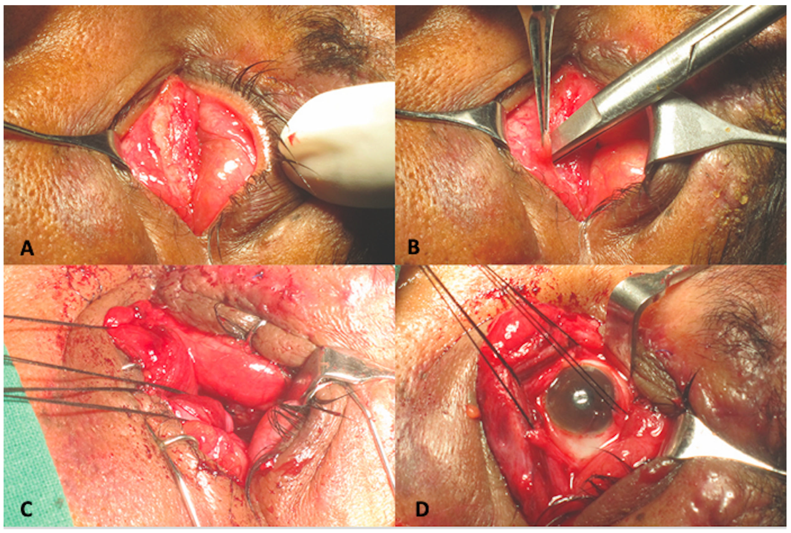
A 45-year-old male presented to us two weeks following a road traffic accident. This patient too, was referred to us as a case of anophthalmic socket. At presentation, ocular examination revealed that right eye visual acuity was 20/30. Anterior segment and fundus findings were within normal limits in the right eye. The left eye had no light perception. Upon mechanically lifting the upper eyelid, no eyeball was visible. Computed tomography of the orbit showed a large floor and medial wall fracture with luxation of the eyeball into the maxillary sinus. (Figure 2) Similar conjunctival telescoping of the globe was noted. The patient was taken up for left orbital fracture repair and retrieval of the globe. Surgically the eyeball was first approached through a trans conjunctival incision and the globe was retrieved back into the orbit. However, conjunctival telescoping required a 360° peritomy and suture tagging of all the extraocular muscles to retrieve the globe back into its normal position, but the eyeball was intact. (Figure 3) The orbital floor and medial wall were reconstructed with a titanium mesh. The patient continued to have no light perception. Last follow up was 22 months following the surgical repair and the ocular globe integrity was maintained with acceptable cosmesis.

2.3. Case 3

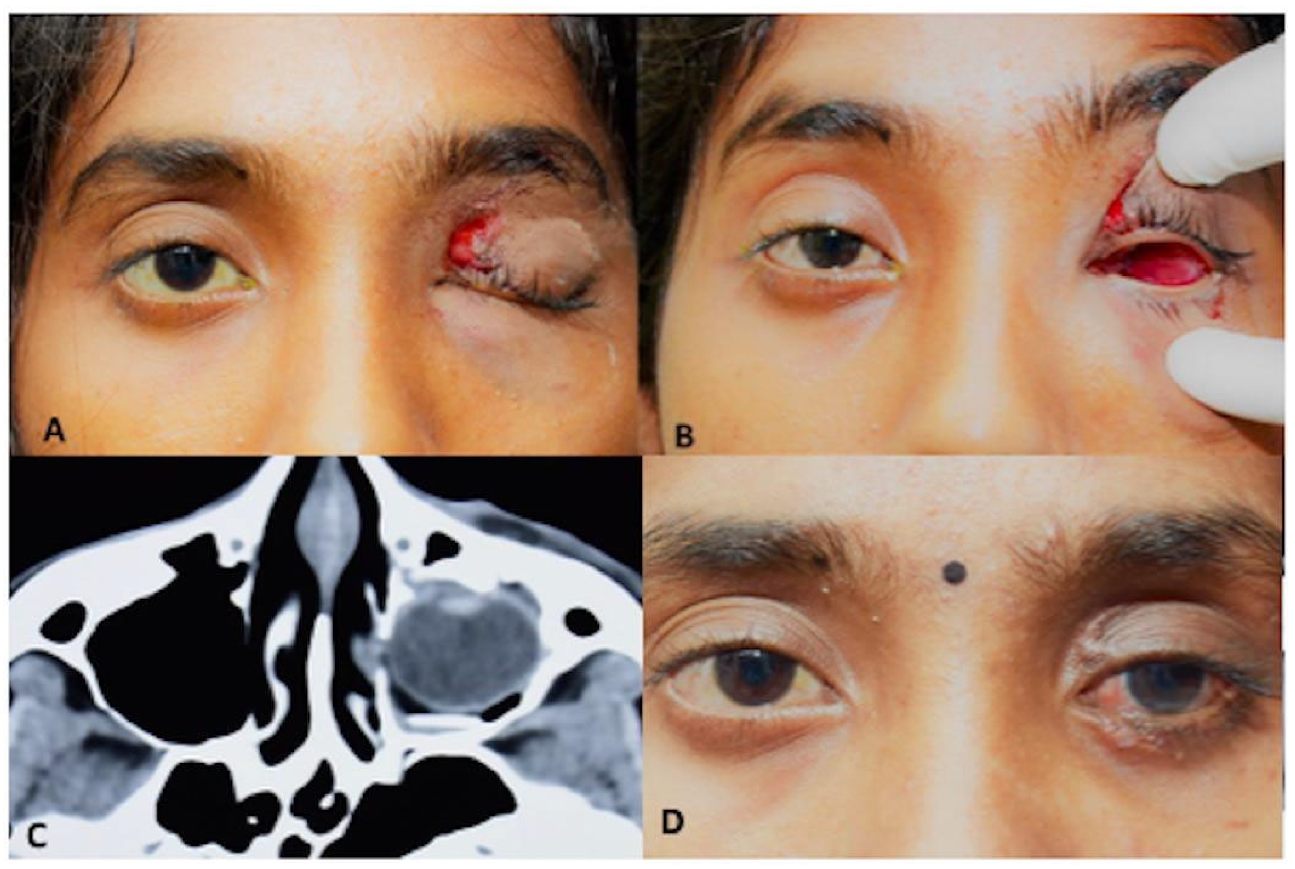
A 17-year-old woman presented with a one-day history of sudden onset painful loss of vision in the left eye following a bull horn injury. On examination, her visual acuity was 20/20 in the right eye and no perception of light in the left eye. The left orbital cavity showed no discernible globe structures. Computerized tomography of the orbit revealed orbital floor fracture with loss of inferomedial orbital strut and prolapse of the globe in the ipsilateral maxillary sinus. (Figure 4) The patient underwent surgery for left globe repositioning and fracture repair. As the eyeball had telescoped through the conjunctiva completely in the maxillary sinus, a Caldwell-Luc antrostomy was done to facilitate gentle globe reposition. The anterior wall of the maxillary sinus from the lateral end of pyriform fossa to the lateral edge of maxillary wall and from superior to insertion of canine tooth to infraorbital foramen was opened with a cutting burr and the globe gently directed to the orbit. A 360° conjunctival peritomy was done and all four recti tagged and the eyeball was retrieved back into the orbit (Figure 5). A titanium plate was fixed to repair the floor fracture. Post-operative fundus evaluation revealed a hyperemic optic disc with foveal blanching due to likely cilioretinal artery occlusion and multiple hemorrhages all over the retina (Figure 6). Although the optic nerve was intact, there was no response to any stimuli on visual evoked potential suggestive of traumatic optic neuropathy. However, the patient had an acceptable cosmetic appearance at 12 months.

3. Results

In all three cases, we were able to successfully and safely retrieve the intussuscepted globe with the described technique, reposition it within the orbital socket, and reconstruct the orbital floor and medial wall defect using a titanium mesh without globe rupture. In the third case, due to nearly complete displacement of the globe into the maxillary sinus, globe retrieval was assisted by Caldwell-Luc approach to provide better access.

4. Discussion

The term “intussusception” has not previously been applied to traumatic globe displacement and is formally proposed here to describe a distinctive morphological configuration in which the globe telescopes through an invaginated, swollen conjunctival-Tenon’s envelope, producing an apparent “empty-socket”. The word “intussusception” is derived from Latin “intus,” meaning “within” or “inside,” and “suscipere” meaning “to undertake.” These three cases differ from previously reported presentations as, despite meticulous eyelid retraction the globe remained clinically occult owing to its retraction through an invaginated, swollen conjunctival–Tenon’s sleeve. Cross-sectional imaging confirmed globe prolapse into the paranasal sinus, and intra-operative management required a controlled 360° conjunctival peritomy with suture-tagging of the recti to permit safe retrieval through a telescoped conjunctiva.
The transconjunctival approach has been specified in one case report earlier, but it did not involve a 360° peritomy and hooking of muscles [3]. Two of the three cases in our series suffered globe intussusception into the sinus following a bull horn injury. Bull horn injury can have varied presentations, from eyelid tear to globe rupture [4], and there has been only one case report with a bull horn injury leading to the globe in the maxillary sinus [5].
A review of the English-language literature from the years 1971 to 2024 was performed using the search terms: traumatic globe dislocation, maxillary sinus, ethmoid sinus and bull horn. Studies that included clinical and radiological diagnoses were included in our review. The data from previous studies is included in Table 1.
Thirty-five published case reports of traumatic dislocation of the globe into the sinuses were identified in the literature (Table 2). The age of these patients ranged from 10 to 68 years of age, and there was a clear male preponderance. More than 50% of these cases had suffered traffic accidents. Two of three cases in our series had a bull horn injury. Amongst the paranasal sinuses, traumatic globe dislocation mostly involved the maxillary sinus, but ethmoid sinus involvement has also been reported. Out of the five cases with globe luxation into the ethmoid sinus, four did not undergo any reconstruction of the medial wall, while one had a delayed reconstruction with a porous polyethylene implant.
The mechanism of traumatic globe dislocation is the same as that of orbital blow-out fracture along with variable soft tissue damage. Berkowitz et al. explained that the globe is supported within the orbit by the arrangement of the extraocular muscles, bulbar fascia, ligaments, and orbital fat [8]. It is known that even after removal of the maxilla and orbital floor, the globe does not completely sink into the maxillary sinus. A trauma forceful enough to disrupt these structures can cause globe dislocation.
It was hypothesized by Kreiner et al. that delay in treatment compromises blood supply to the optic nerve and increases the probability of irreversible vision loss due to twisting and stretching of the central retinal artery [20]. Our series could be different from the other singular case reports, where the conjunctiva had completely infolded to hide the globe. This telescoping of the conjunctiva is similar to intussusception, where a part of the intestine slides into the adjacent part. Including our three cases, we found that only 39.47% (15 out of 38) cases could recover any functional vision. Ten of these cases had been presented and surgically repositioned on the same day of trauma, one on the day after trauma, one after four days of trauma, while for three cases the interval between trauma and repair has not been documented.
Surgical management of globe dislocation is not standardized. Due to their presentation, such cases can be misdiagnosed as anophthalmic socket or auto-evisceration, and hence urgent imaging and immediate intervention are vital. Every attempt must be made to reposition the globe followed by orbital floor reconstruction. Repositioning of the globe has been reported by two techniques: (1) Direct traction by subconjunctival/sub-ciliary approach (2) Transmaxillary approach. We believe that the choice of approach should be guided by the proportion of dislocation and the intraoperative recognition of globe intussusception through the conjunctiva, an observation not previously described in the literature.
Whereas most orbital fractures are repaired within two weeks to optimize surgical planning, those with globe dislocation demand an urgent intervention with immediate reconstruction, often on the day of the injury, to restore ocular integrity and function. The urgency in such cases obviates the feasibility of patient-specific or three-dimensional printed implants [36], necessitating the use of immediately available standard materials. Various materials have been described in the literature for orbital wall reconstruction, including: titanium mesh, autogenous bone, porous polyethylene, and silicone [37].
Application of the PICO framework allowed a structured synthesis of the available literature on this rare but severe presentation of orbital trauma.
  • Population (P):
Across 35 published reports (and the current three cases), the affected cohort comprised predominantly young to middle-aged males, with a clear predominance of high-energy trauma, notably road traffic accidents (50%) and bull horn injuries. The maxillary sinus was the most commonly involved site (86%), followed by the ethmoid sinus. The rarity of this phenomenon emphasizes the need for heightened clinical suspicion in apparent “empty socket” presentations following periorbital trauma.
  • Intervention (I):
The principal intervention across studies was surgical retrieval and repositioning of the displaced globe, often accompanied by orbital wall reconstruction. Importantly, prior reports have not described the precise mechanism of dislocation into adjacent paranasal cavities; the clinical and intra-operative findings presented here provide the first detailed observations that help clarify likely pathways of displacement and inform choice of surgical approach. Our cases highlight a unique morphological variant, globe intussusception through the conjunctiva, necessitating a 360° peritomy and muscle tagging before repositioning. The addition of the Caldwell-Luc approach in one case facilitated safe retrieval from the maxillary sinus.
  • Comparison (C):
Reported approaches varied according to the extent and direction of displacement. Direct traction was used in partial dislocations, while transmaxillary and combined intraoral or subciliary routes were reserved for complete displacements. Functional visual recovery was higher when surgery was performed within 24 h of trauma. The transconjunctival approach with complete peritomy, as described in this series, offers controlled access with minimal additional trauma, though evidence from literature remains limited to anecdotal reports.
  • Outcome (O):
Among 38 collated cases (including the current three cases), only 39% achieved functional visual recovery. Early surgical intervention was the most consistent predictor of favorable outcomes. In our series, although anatomical repositioning was successful in all cases, visual recovery remained poor due to concurrent optic nerve or retinal injury. This emphasizes the need for rapid diagnosis, imaging, and urgent surgical intervention to preserve ocular integrity and optimize visual prognosis.
By integrating findings under the PICO framework, this review underlines that:
  • Early recognition and imaging are critical to differentiate true enucleation from concealed intussusception.
  • Prompt surgical retrieval, preferably within 24 h, improves anatomical and functional outcomes.
  • Choice of approach should be tailored to the type of displacement, with conjunctival intussusception warranting a full peritomy and extraocular muscle tagging.
  • Urgent reconstruction with available materials (titanium, porous polyethylene, or autogenous bone) is essential to restore orbital architecture.
Finally, this PICO-based synthesis offers a clearer evidence framework for future multicentric registries and outcome-based studies, with the goal of refining surgical strategies and optimizing both cosmetic and visual outcomes in traumatic globe dislocation.
In conclusion, three additional cases of traumatic globe luxation into the paranasal sinuses are reported and the term “globe intussusception” is formally introduced to describe the distinctive telescoping of the globe through the conjunctiva–Tenons envelope that can give the appearance of an anophthalmic socket. Clinical implications include: (1) assessment of orbital fractures by cross-sectional imaging and evaluation of the route of globe displacement are essential; (2) intra-operative recognition of conjunctival intussusception mandates a controlled 360° conjunctival peritomy with suture-tagging, cryoprobe or a bimanual technique using Caldwell–Luc access to permit safe retrieval and repositioning of the globe; and (3) urgent repositioning and timely orbital reconstruction are advised because functional visual recovery is limited; pooled data indicate that approximately 39% of patients regain useful vision. To standardize reporting and guide management, a pragmatic clinicoradiological classification is suggested for acceptance and future validation: Type I: partial displacement (globe displaced and enophthalmic but is visualized within the orbit); Type II: globe intussusception (conjunctival telescoping with an “empty socket” clinical appearance). Establishment of a prospective, multicenter registry and outcome studies is recommended to refine timing of intervention, optimize reconstructive strategies, and improve prognostic assessment of visual and cosmetic outcomes for this rare but surgically challenging injury.

Author Contributions

Conceptualization: M.N., G.D. and A.D.; Methodology: M.N., G.D. and A.D.; validation: M.N., A.D. and G.D.; formal analysis: A.D. and M.N.; investigation: M.N. and A.D.; resources: M.N. and G.D.; data curation: A.D. and M.N.; writing—original draft preparation: A.D.; writing—review and editing: M.N.; visualization: M.N. and G.D.; supervision: M.N. and G.D.; All authors have read and agreed to the published version of the manuscript.

Funding

This research received no external funding.

Institutional Review Board Statement

The study was conducted in accordance with the Declaration of Helsinki as revised in 2013. informed consent was obtained from all patients prior to undergoing the procedure. The study was conducted in accordance with the ethical principles outlined in the Declaration of Helsinki (2013). Please find below ethical approval details. LV Prasad Eye Institute. IRB number: LEC-BHR-P-02-23-1003 This study is a retrospective analysis based on intra-operative observations conducted during routing surgical procedures in accordance with standard of care. No experimental interventions were performed.

Informed Consent Statement

Informed written consent was obtained from all patients involved in the study.

Data Availability Statement

The original contributions presented in this study are included in the article. Further enquiries can be directed to the corresponding author.

Conflicts of Interest

The authors declare no conflicts of interest.

Abbreviations

The following abbreviations are used in this manuscript:
ATVAll-Terrain Vehicle
MMale
FFemale
NANot Applicable

References

  1. Lawrence, G.H.; Ulfelder, H. Intussusception; a review of experience at the Massachusetts General Hospital, 1937–1951. N. Engl. J. Med. 1952, 247, 499–502. [Google Scholar] [CrossRef] [PubMed]
  2. Haggerty, C.J.; Roman, P. Repositioning of a traumatically displaced globe with maxillary antrostomy: Review of the literature and treatment recommendations. J. Oral. Maxillofac. Surg. 2013, 71, 1915–1922. [Google Scholar] [CrossRef] [PubMed]
  3. Kim, S.; Baek, S. Traumatic dislocation of the globe into the maxillary sinus associated with extraocular muscle injury. Graefes Arch. Clin. Exp. Ophthalmol. 2005, 243, 1280–1283. [Google Scholar] [CrossRef] [PubMed]
  4. Heilbig, H.; Iseli, H.P. Traumatic rupture of the globe cause by cow horns. Eur. J. Ophthalmol. 2002, 12, 304–308. [Google Scholar] [CrossRef] [PubMed]
  5. Singh, R.I.; Thomas, R.; Alexander, T.A. An Unusual case of bull gore injury. Aust. N. Z. J. Ophthalmol. 1986, 14, 377–379. [Google Scholar] [CrossRef]
  6. Emery, J.M.; Noorden, G.K.; Sclernitzauer, D.A. Orbital floor fractures: Long-term followup of cases with and without surgical repair. Trans. Am. Acad. Ophthalmol. Otolaryngol. 1971, 75, 802–812. [Google Scholar]
  7. Stasior, O.G. The Wendell L: Hughes lecture. Complications of ophthalmic plastic surgery and their prevention. Trans. Sect. Ophthalmol. Am. Acad. Ophthalmol. Otolaryngol. 1976, 81, 543–552. [Google Scholar]
  8. Berkowitz, R.A.; Putterman, A.M.; Patel, D.B. Prolapse of the globe into the maxillary sinus after orbital floor fracture. Am. J. Ophthalmol. 1982, 91, 253–257. [Google Scholar] [CrossRef]
  9. Risco, J.M.; Stratas, B.A.; Knott, R.H. Prolapse of the globe into the ethmoid sinus. Am. J. Ophthalmol. 1984, 97, 659–660. [Google Scholar] [CrossRef]
  10. Smit, T.I.; Koorneeff, L.; Zonneveld, F.W. A total orbital floor fracture with prolapse of the globe into the maxillary sinus manifesting as post enucleation socket syndrome. Am. J. Ophthalmol. 1990, 110, 569–570. [Google Scholar] [CrossRef]
  11. Ziccardi, V.B.; Patterson, G.T.; Ramasastry, S.; Sotereanos, G.C. Management of a severe zygomatico-orbital fracture with dislocation of the globe into the antrum. J. Craniofac. Surg. 1993, 4, 95–101. [Google Scholar] [CrossRef] [PubMed]
  12. Moon, M.; Pietris, G.; Shapter, M. Dislocation of the globe into the ethmoid sinuses. Aust. N. Z. J. Ophthalmol. 1997, 25, 175–176. [Google Scholar] [CrossRef]
  13. Pelton, R.W.; Rainey, A.M.; Lee, A.G. Traumatic subluxation of the globe into the maxillary sinus. AJNR Am. J. Neuroradiol. 1998, 19, 1450–1451. [Google Scholar] [PubMed]
  14. Tung, T.C.; Chen, Y.R.; Santamaria, E.; Chen, C.T.; Lin, C.J.; Tsai, T.R. Dislocation of anatomic structures into the maxillary sinus after craniofacial trauma. Plast. Reconstr. Surg. 1998, 101, 1904–1908. [Google Scholar] [CrossRef] [PubMed]
  15. Saleh, T.; Leatherbarrow, B. Traumatic prolapse of the globe into the maxillary sinus diagnosed as traumatic enucleation of the globe. Eye 1999, 13, 678–680. [Google Scholar] [CrossRef]
  16. Tranfa, F.; Di Matteo, G.; Di Salle, F.; Strianese, D.; Bonavolontà, G. Traumatic displacement of the globe into the ethmoid sinus. Am. J. Ophthalmol. 2000, 130, 253–254. [Google Scholar] [CrossRef]
  17. Okabe, H.; Kimura, K.; Sonoda, S.; Sakamoto, T. Displacement of globe into ethmoid sinus by orbital medial wall fracture with good recovery of vision. Jpn. J. Ophthalmol. 2005, 49, 426–428. [Google Scholar] [CrossRef]
  18. Müller-Richter, U.D.; Kohlhof, J.K.; Driemel, O.; Wagener, H.; Reichert, T.E. Traumatic dislocation of the globe into the maxillary sinus. Int. J. Oral. Maxillofac. Surg. 2007, 36, 1207–1210. [Google Scholar] [CrossRef]
  19. Abrishami, M.; Aletaha, M.; Bagheri, A.; Salour, S.H.; Yazdani, S. Traumatic subluxation of the globe into the maxillary sinus. Ophthal Plast. Reconstr. Surg. 2007, 23, 156–158. [Google Scholar] [CrossRef]
  20. Kreiner, B.; Amer, R.; Sharfi, G.; Solomon, A.; Ilsar, M. Traumatic displacement of the globe into the paranasal sinuses: Case report and guidelines for treatment. J. Oral. Maxillofac. Surg. 2008, 66, 826–830. [Google Scholar] [CrossRef]
  21. Ramstead, C.; McCabe, J.; Alkahtani, M.; Leong-Sit, J.; Morhart, M. Traumatic dislocation of the globe into the maxillary sinus. Can. J. Ophthalmol. 2008, 43, 364–366. [Google Scholar] [CrossRef]
  22. Jellab, B.; Baha, A.T.; Moutaouakil, A.; Khoumiri, R.; Raji, A.; Ghannane, H.; Amkaoui, M.A.; Ait Benali, S. Management of a severe cranio-orbito-facial trauma with a dislocation of the globe into the maxillary sinus. Bull. Soc. Belge Ophtalmol. 2008, 309–310, 37–41. [Google Scholar]
  23. Akhaddar, A.; Elmostarchid, B.; Boucetta, M. Images in emergency medicine. Traumatic intraorbital stone with globe displacement into the maxillary sinus. Emerg. Med. J. 2010, 27, 828. [Google Scholar] [CrossRef]
  24. Gulati, A.; Gupta, B.; Singh, I. An unusual orbital blow-out fracture: A case report. Orbit 2011, 30, 10–12. [Google Scholar] [CrossRef]
  25. Zhang-Nunes, S.X.; Jarullazada, I.; Mancini, R. Late central visual recovery after traumatic globe displacement into the maxillary sinus. Ophthal. Plast. Reconstr. Surg. 2012, 28, 17–19. [Google Scholar] [CrossRef]
  26. Xu, B.; Xu, X.L.; Yan, J. Treatment of traumatic globe dislocated completely into the maxillary sinus. Int. J. Ophthalmol. 2013, 6, 106–107. [Google Scholar] [CrossRef]
  27. Amaral, M.B.; Nery, A.C. Traumatic globe dislocation into the paranasal sinuses: Literature review and treatment guidelines. J. Craniomaxillofac. Surg. 2016, 44, 642–647. [Google Scholar] [CrossRef] [PubMed]
  28. Wang, J.M.; Fries, F.B.; Hendrix, P.; Brinker, T.; Loukas, M.; Tubbs, R.S. Orbital Blowout Fracture with Complete Dislocation of the Globe into the Maxillary Sinus. Cureus 2017, 9, 1728. [Google Scholar] [CrossRef] [PubMed]
  29. Noman, S.A.; Shindy, M.I. Immediate surgical management of traumatic dislocation of the eye globe into the maxillary sinus: Report of a rare case and literature review. Craniomaxillofacial Trauma Reconstr. 2017, 10, 151–158. [Google Scholar] [CrossRef] [PubMed]
  30. Alam, M.S.; Noronha, O.V.; Mukherjee, B. Timing of surgery in traumatic globe dislocation. Indian J. Ophthalmol. 2017, 65, 767–770. [Google Scholar] [CrossRef]
  31. Bastos, R.M.; Taparello, C.; Tres, R.; Sawazaki, R. Orbital blowout fracture with globe displacement into the maxillary sinus: A case report and literature review. J. Oral. Maxillofac. Surg. 2021, 79, 204.e1-204.e12. [Google Scholar] [CrossRef] [PubMed]
  32. Onaran, Z.; Şimşek, G.; Konuk, O. Traumatic dislocation of the globe into the ethmoid sinus. Turk. J. Ophthalmol. 2021, 51, 308–312. [Google Scholar] [CrossRef] [PubMed]
  33. Gobeaut, M.; Merle, H.; Rocher, M.; Robert, P.Y.; Beral, L. Traumatic incarceration of the globe into the maxillary sinus: A case report. J. Fr. Ophtalmol. 2022, 45, e33–e35. [Google Scholar] [CrossRef] [PubMed]
  34. Steel, D.A.; Mortimer, T.; Bater, M.C. Severe orbital blow-out fracture: Complete displacement of the globe into the maxillary sinus. BMJ Case Rep. 2023, 16, e254055. [Google Scholar] [CrossRef]
  35. Vergez, P.; Vernhet, E.; Daien, V.; De Boutray, M. Traumatic dislocation of the globe into the maxillary sinus with early recovery of visual acuity: Case report and review of the literature on management. J. Fr. Ophtalmol. 2024, 47, 104078. [Google Scholar] [CrossRef]
  36. Bergeron, L.; Bonapace-Potvin, M.; Bergeron, F. Printing in Time for Cranio-Maxillo-Facial Trauma Surgery: Key Parameters to Factor in. Craniomaxillofac. Trauma Reconstr. 2023, 16, 121–129. [Google Scholar] [CrossRef]
  37. Burger, T.; Fan, K.; Brokmeier, J.; Thieringer, F.M.; Berg, B.I. Orbital Floor Fractures: Treatment and Diagnostics—A Survey Among Swiss, German and Austrian Maxillofacial Units. Craniomaxillofac. Trauma Reconstr. 2024, 17, NP60–NP67. [Google Scholar] [CrossRef]
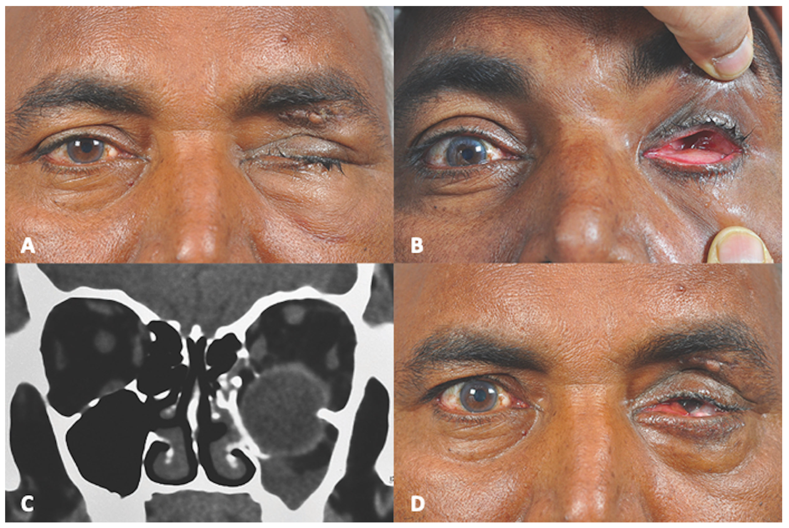
Figure 1. (A) Pre-operative Clinical Photograph showing left traumatic ptosis with sutured upper eyelid laceration. (B) Pre-operative Clinical Photograph showing absence of left globe in orbital cavity. (C) Pre-operative Computed Tomography (coronal section) showing dislocation of globe into maxillary sinus. (D) Post-operative Clinical Photograph showing retrieved globe in orbital space.
Cmtr 18 00044 g001
Figure 2. (A) Pre-operative Clinical Photograph showing left traumatic ptosis with sutured upper eyelid laceration (B) Pre-operative Clinical Photograph showing absence of left globe in orbital cavity (C) Pre-operative Computed Tomography (axial section) showing globe luxation (D) Post-operative Clinical Photograph showing globe retracted into the orbit.
Cmtr 18 00044 g002
Figure 3. (A) Intra-operative photograph with no discernable globe structure in orbit (B) Intra-operative photograph of conjunctival peritomy in depths of intussuscepted globe (C) Intra-operative clinical photograph showing tagging of extraocular muscles (D) Intra-operative clinical photograph showing globe retrieval into the orbit through conjunctival peritomy with aid of tagged extraocular muscles.
Cmtr 18 00044 g003
Figure 4. (A) Pre-operative Clinical Photograph showing left traumatic ptosis and upper eyelid laceration (B) Pre-operative Clinical Photograph showing absence of left globe in orbital cavity (C) Pre-operative Computed Tomography (axial section) showing dislocation of globe into maxillary sinus (D) Post-operative Clinical Photograph showing globe retrieved into the orbit.
Cmtr 18 00044 g004
Figure 5. (A) Intra-operative photograph showing antral window below infraorbital nerve (Black arrow) (B) Intra-operative photograph showing globe in left orbital cavity after gentle lift from antrum (C) Intra-operative photograph showing intra-oral view of the floor of the orbit with titanium mesh (Black arrow).
Cmtr 18 00044 g005
Figure 6. (A) Immediate post-operative left fundus photograph showing foveal and generalized retinal whitening with multiple pre-retinal and intra-retinal hemorrhages (B) Eight weeks post-operative left fundus photograph showing optic disc pallor with cupping and 360 degrees sclerosis of retinal vessels. Pigmentary changes seen at macula.
Cmtr 18 00044 g006
Table 1. Previously published cases of traumatic globe luxation (n = 35), into the maxillary and ethmoid sinus, arranged chronologically with authorship and year of publication, in English-language literature indexed (PUBMED) from 1971 to 2024.
Sr No.AuthorsAgeSexEtiology of TraumaInvolvement of SinusTime to PresentationGlobe RepositioningOrbital ReconstructionVisual Acuity
1Emery et al., 1971 [6]NANANAMaxillary SinusNADirect
traction
NANA
2Emery et al., 1971 [6]NANANAMaxillary SinusSame dayDirect
traction
NANA
3Stasior, 1976 [7]NAMFire hose nozzle injuryRight Maxillary SinusNATrans-maxillary manual repositioningNoRecovery 20/60
4Berkowitz et al., 1981 [8]24FPunch with fistLeft Maxillary SinusSame dayTrans-maxillary manual repositioningSilicone implantRecovery 20/20
5Risco et al., 1984 [9]40MBlunt trauma (hit with a wood block)Left ethmoid sinus5 daysDirect traction- trans-nasal repositioningNoLight perception: recurrence of globe displacement
6Singh et al., 1986 [5]32MBull horn NADirect tractionNo6/36
7Smit et al., 1990 [10]21FWindshield injuryRight Maxillary Sinus5 yearsNATeflon plateNo
8Ziccardi et al., 1993 [11]25MTraffic accidentLeft Maxillary SinusSame daySub-ciliary + Trans-maxillaryAutologous bone20/50
9Moon et al., 1997 [12]40MBlunt instrument injuryRight Ethmoid Sinus2 daysDirect TractionNoNo
10Pelton et al., 1998 [13]19MTraffic accidentLeft Maxillary SinusSame dayTrans-maxillary manual repositioningNANA
11Tung et al., 1998 [14]17MTraffic accidentRight Maxillary Sinus-Direct repositioning-sub-ciliary approachAutogenous bone graftNo
12Saleh & Leatherbarrow, 1999 [15]29MBlunt traumaLeft Maxillary Sinus2
Months
EnucleationNoNo
13Tranfa et al., 2000 [16]58MFall of a tree branchLeft Ethmoid SinusSame dayDirect TractionNoRecovery 20/100
14Kim and Baek, 2005 [3]68MTraffic accidentRight Maxillary SinusSame dayDirect traction- transconjunctivalPorous polyethyleneNo
15Okabe et al., 2005 [17]37MInjury with a piece of woodLeft Ethmoid SinusSame dayDirect TractionNo reconstruction of medial orbital wallRecovery 20/15 restricted motility
16Müller-Richter et al., 2007 [18]62MBlunt trauma by machine Right Maxillary SinusSame dayTrans-maxillary manual repositioningBalloon catheter left inside the sinusRecovery 20/20
17Abrishami et al., 2007 [19]18MTraffic accidentRight Maxillary Sinus7 daysDirect repositioning sub-ciliary approachPorous polyethyleneNo
18Kreiner et al., 2008 [20]50MWall collapse injuryLeft Maxillary SinusSame dayDirect tractionSilicone sheet No
19Ramstead et al., 2008 [21]32MStepped by a bullLeft Maxillary SinusSame dayNATitanium meshRecovery 20/200
20Jellab et al., 2008 [22]24MTraffic accidentLeft Maxillary SinusSame dayNAAutogenous boneLight perception
21Jellab et al., 2008 [22]50FTraffic accidentLeft Maxillary SinusSame dayNo repositioningNoNA
22Akhaddar et al., 2010 [23]62MTraffic accidentRight Maxillary SinusNANANANA
23Gulati et al., 2011 [24]10MBicycle accidentRight Maxillary SinusNATrans-maxillaryBalloon Catheter inside sinusRecovered 6/16
24Zhang-Nunes et al., 2012 [25]20MTraffic accidentRight Maxillary SinusSame dayNASialistic sheetRecovery 20/25
25Xu et al., 2013 [26]46FTraffic accidentRight Maxillary SinusNADirect repositionTitanium meshLight perception
26Haggerty and Roman, 2013 [2]35MTraffic accidentLeft Maxillary SinusSame dayTrans-maxillary manual repositioningTitanium meshLight perception
27Amaral and Nery, 2015 [27]44MStruck by gym weightLeft Maxillary Sinus4 daysTrans-maxillary manual repositioningTitanium meshRecovery 20/50
28Wang JM et al., 2017 [28]15MAccident riding ATVLeft Maxillary SinusNANATitanium meshNo
29Noman SA and Shindy MI, 2017 [29]24FBlunt wooden objectRight Maxillary SinusSame dayTrans-maxillary approachTitanium MeshRecover 20/40
30Alam et al., 2017 [30]45FTrauma with doorRight Maxillary Sinus3 daysDirect repositioningTitanium meshVA: counting fingers to 1 m
31Bastos et al., 2021 [31]27MPunch in the eyeRight Maxillary SinusSame dayTrans-maxillary approach14 days after repositioning of globe, reconstruction with titaniumNo VA
32Onaran et al., 2021 [32]36FBlunt trauma on door handleLeft Ethmoid SinusSame dayGentle intranasal digital
manipulation
4 months after repositioning, with porous polyethylene implantVA 20/20
33Gobeaut et al., 2022 [33]54FFall from heightRight Maxillary Sinus1 dayDirect traction and trans-maxillary approachResorbable polydiaxone osteosynthesis implant (PDS)VA 20/125
34Steel DA et al., 2023 [34]-MTraffic
Accident
Left Maxillary SinusSame dayTrans-maxillaryTitanium meshNo perception of light
35Vergez P et al., 2024 [35]67MFall from a heightLeft Maxillary SinusSame dayDirect TractionTitanium mesh20/50
Table 2. Clinical summary of 35 cases of globe intussusception into the maxillary and ethmoid sinus published in English-language literature indexed (PUBMED) from 1971 to 2024.
Total NumberAge RangeGenderEtiology of TraumaInvolvement of SinusTime to PresentationGlobe RepositioningOrbital ReconstructionVisual Acuity
3510–68
Years
Male: 25
Female: 8
Not mentioned: 2
Traffic
accident: 14
Others: 9
NA: 2
Maxillary Sinus: 30
Ethmoid Sinus: 5
Range:
Same day to 5 years
Direct reposition: 15
Transmaxillary: 12
NA: 6
No reposition: 1
Enucleation: 1
Titanium: 9
Silicone: 3
Porous poly-ethylene: 3
PDS: 1
Bone: 3
No reconstruction: 9
Others: 3, NA: 4
Functional Vision improved: 15
Finger counting 1 m: 1
Light perception: 4
Not improved: 10
NA: 5
Disclaimer/Publisher’s Note: The statements, opinions and data contained in all publications are solely those of the individual author(s) and contributor(s) and not of MDPI and/or the editor(s). MDPI and/or the editor(s) disclaim responsibility for any injury to people or property resulting from any ideas, methods, instructions or products referred to in the content.