Next Article in Journal
Factors Associated with Outcomes of Facial Reconstruction After Mohs Micrographic Surgery
Previous Article in Journal
Incidence and Characterization of Facial Lacerations in Emergency Departments in the United States
 
 
Craniomaxillofacial Trauma & Reconstruction is published by MDPI from Volume 18 Issue 1 (2025). Previous articles were published by another publisher in Open Access under a CC-BY (or CC-BY-NC-ND) licence, and they are hosted by MDPI on mdpi.com as a courtesy and upon agreement with Sage.
Article

Does Platelet-Rich Fibrin Enhance Recovery From Neurosensory Disturbance Following Mandibular Fractures? A Double-Blind, Split-Mouth Randomized Clinical Trial

by
Reza Tabrizi
1,
Hamidreza Moslemi
2,*,
Shervin Shafiei
1,
Ramtin Dastgir
3 and
Zachary S. Peacock
4
1
Department of Oral and Maxillofacial Surgery, School of Dentistry, Shahid Beheshti University of Medical Sciences, Tehran, Iran
2
Department of Oral and Maxillofacial Surgery, School of Dentistry, Babol University of Medical Sciences, Babol 47745-47176, Iran
3
Department of Oral and Maxillofacial Surgery, Case Western Reserve University, Cleveland, OH, USA
4
Department of Oral and Maxillofacial Surgery, Massachusetts General Hospital, Harvard School of Dental Medicine, Boston, MA, USA
*
Author to whom correspondence should be addressed.
Craniomaxillofac. Trauma Reconstr. 2024, 17(4), 75; https://doi.org/10.1177/19433875241257737
Submission received: 1 November 2023 / Revised: 1 December 2023 / Accepted: 1 January 2024 / Published: 27 May 2024

Abstract

Study Design: Randomized Clinical Trial. Objective: Mandibular body fractures may result in inferior alveolar nerve damage. This study examined the effectiveness of platelet-rich fibrin (PRF) application to the inferior alveolar nerve during open reduction and internal fixation (ORIF) of mandibular fractures. Methods: This was a double-blind, split-mouth randomized clinical trial. Patients with bilateral mandibular body fractures with minimal displacement (<5 mm) who underwent ORIF were assessed for enrollment. PRF was placed within the fracture site before reduction and fixation on the studied side. Fixation was performed on the contralateral side (control side) without PRF. The study and control groups were randomized using QuickCalcs software. Neurosensory disturbance (NSD) was assessed through two-point discrimination (TPD), self-reported NSD (SR-NSD), and brush directional stroke discrimination test (BDSD) at 6 and 12 months postoperatively. Results: Twenty-five subjects were enrolled. BDSB recovery was greater in the study group during all time intervals (p < 0.001). There were no differences between TPD and SR-NSD at the follow-up periods (p > 0.05). Conclusions: The results of this split-mouth randomized clinical trial indicate that PRF may enhance the recovery of a damaged inferior alveolar nerve in mandibular body fractures.

Introduction

Mandibular fractures are common maxillofacial injuries [1]. Fracture of the mandibular body of the angle can affect the inferior alveolar nerve, resulting in neurosensory disturbance (NSD) that can be permanent even with fixation [2,3,4]. Typically, a patient has altered skin and mucous membrane sensations within the mental nerve distribution. In a literature review by Thurmuller et al, the overall incidence of NSD for fractures in the mandibular angle and body region was 46%–58.5% [5]. The recovery of NSD following mandibular fractures depends on the amount of displacement, timing of repair, bone avulsion, fixation methods, and local or systemic healing factors [6,7,8].
Platelet-rich fibrin (PRF) was introduced as an adjunct for various oral and maxillofacial procedures in the early 2000s and may have nerve regenerative properties [9,10,11]. PRF and L-PRF (Leukocyte-PRF) are obtained from a second-degree concentration of plasma platelets. The main components of PRF are platelets and their accompanying growth factors, which exist in a protein matrix structure, along with cytokines and other existing leukocytes. The distinguishing factor between PRF and platelet-rich plasma (PRP) is the cohesive fibrin structure in PRF. Fibrin acts as a scaffold and agent for the slow and sustained release of various growth factors that are beneficial in applying PRF. Recently, PRF has attracted more attention because of its simplicity and speed of preparation, user-friendly nature, flexibility, and cost-effectiveness [12]. It is a fibrin gel that slowly forms after simple and quick centrifugation (about 10 minutes) of blood in vacuum tubes without anticoagulants. The clot (or biomaterial) collected is stable, flexible, strong, and adhesive. It can be cut or matched to various anatomical defects and applications.
PRF has been studied for its effects on the outcomes of third molar extraction, ridge preservation after tooth extraction, sinus augmentation, alveolar cleft repair, dental implant placement, medication-related osteonecrosis of the jaw, and closure of oroantral fistulae [9,13]. Recently, PRF enhances the recovery of IAN following various operations such as sagittal split osteotomy, nerve repositioning, and genioplasty [14,15,16].
Therefore, this study examined the effectiveness of PRF application to the inferior alveolar nerve during open reduction internal fixation (ORIF) of mandibular body fractures. The null hypothesis was that PRF does not affect the recovery of NSD following mandibular fractures. The specific aims were to assess the effect of PRF on NSD recovery outcomes and rate of recovery.

Methods

This study was a double-blind, split-mouth randomized clinical trial. Patients referred to the oral and maxillofacial surgery departments of Taleghani and Imam Hossein Hospital, Tehran, Iran, were screened for enrollment from June 2020 to June 2022. This study protocol was approved by the medical ethics committee of Shahid Beheshti University of Medical Sciences (IR.SBMU.DRC.REC.1400.095) and the Iranian Registry of Clinical Trials (IRCT20200922048799N2).
Patients who had bilateral mandibular body fractures (the region between the anterior border of the masseter muscle and the line distal to the ipsilateral canine) with minimum displacement (<5 mm, confirmed by computed tomography) and were scheduled for open reduction internal fixation (ORIF) of both fractures were eligible for enrollment. Subjects with other mandibular fractures, penetrating trauma (eg, gunshot wounds), or acute infection at the site were excluded. In addition, patients with a history of facial fractures, orthognathic surgery, nerve lateralization, IAN injury, psychiatric disorders requiring medication, underlying neuropathic disorders such as diabetes, or unwillingness to participate in the study were excluded. The authors obtained written informed consent from all subjects. Those unwilling to provide consent were excluded.

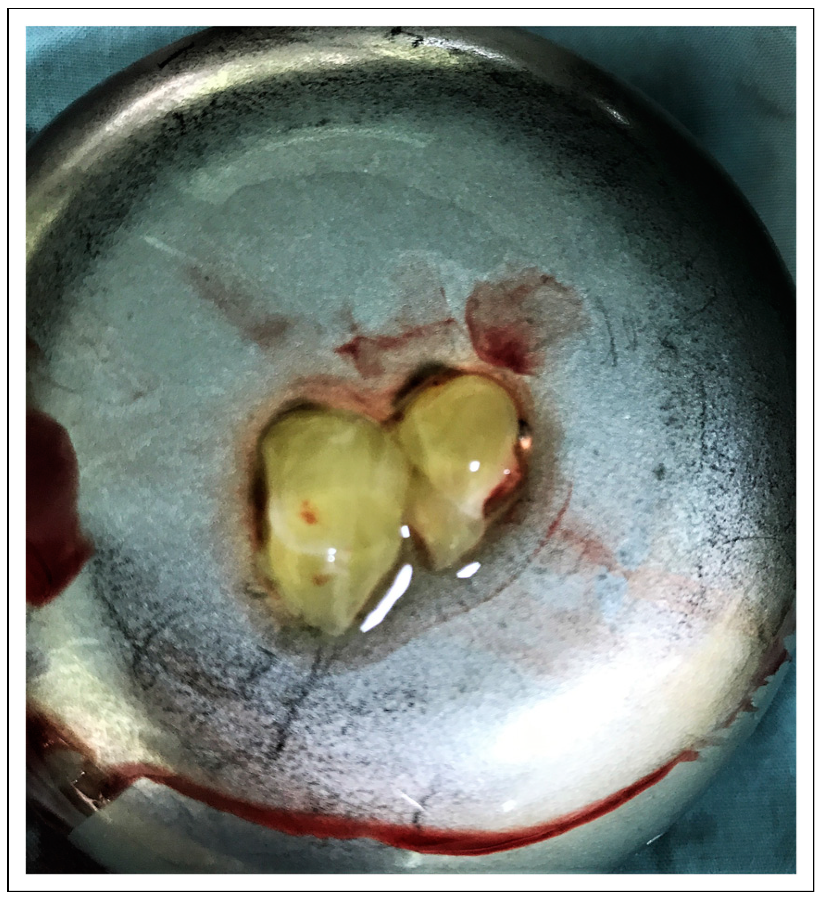
Preparation of the PRF
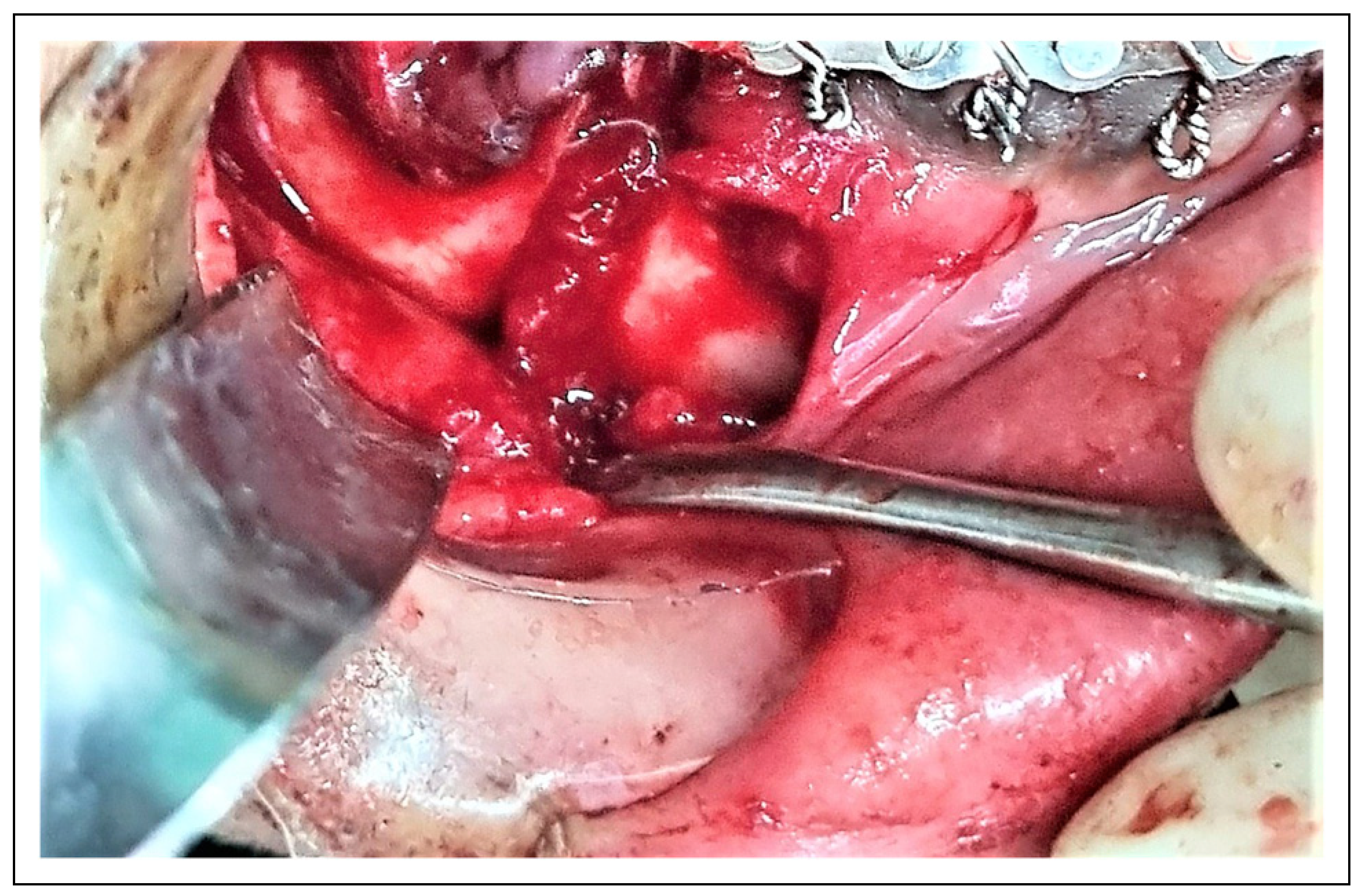
Before the operation, 20 cc of venous blood was drawn from the patient and placed in a centrifuge (IntraSpin system; Intra-Lock International, Boca Raton, FL, USA) for 12 min at 2800 revolutions per minute. After centrifugation, the contents of the tube, including L-PRF, were transferred to a special tray. The clot at the bottom of the tube was placed on the tray, covered, and allowed to sit for 5 min (Figure 1).

Surgical Procedure

Patients received 600-mg clindamycin and 8 mg of dexamethasone intravenously 30 min before the incision. All patients received general anesthesia and nasal intubation. A single surgeon (initials) served as the attending surgeon for each procedure. A hybrid arch bar (Doostan Nekooandish Parsian, Co Ltd, Tehran, Iran) was fixated on the maxilla and mandible in all patients. Local anesthetic (Lidocaine 2% with epinephrine 1/800 000, Daroopaksh, Iran) was injected at the sites of mucosal incisions for hemostasis. Bilateral mucosal incisions were made to provide sufficient access to the fracture. The mental nerve was identified and preserved. Surgeons confirmed the integrity of the inferior alveolar nerve visually. The patient was then placed in intermaxillary fixation (IMF) in an appropriate occlusion. Block randomization using QuickCalcs (GraphPad Software, Inc, La Jolla, CA) determined the side serving as the study and control group. Randomization was performed by a third-party not involved in the study. The surgeons were not masked to the treatment and control sides.
The PRF matrix was placed on the nerve on the treatment side between the proximal and distal segments of the fracture before reduction and fixation (Figure 2). Fixation was performed on the opposite side (control group) without applying PRF.
Both sides were fixated using 2 mini plates (a six-hole 1.25 mm thick miniplate with 7 mm length, 2 mm diameter screws at the inferior mandibular border and a four-hole 1.0 mm thick miniplate with 5 mm length, 2 mm diameter screws in the superior aspect of the fracture line) (MINI 2000, Doostan Nekooandish Parsian, Co Ltd, Tehran, Iran). Care was taken to avoid penetrating the IAN canal or the roots of the adjacent teeth. The mucosal incision was sutured in a single layer. A mouthwash containing .12% chlorhexidine (Iran Nazho, Tehran, Iran) was prescribed during hospitalization and after discharge. Patients were placed in guiding elastics for 2 weeks postoperatively. A postoperative panoramic radiograph was obtained from each patient. One surgical team performed all operations.

Sensory Neural Evaluation

Sensory neural assessments were performed at preoperative (T0), 6 months postoperative (T1), and 12 months postoperative (T2) by an examiner who was blinded to the study groups and surgical process. In addition, the patient was masked to the sides of treatment and control. The two-point discrimination (TPD) test was conducted. It was measured as the minimum distance between 2 distinguishable points created by 2 pinpricks on the lower lip and mental region. The brush directional stroke discrimination (BDSD) test was performed by moving a fine #2 sable brush on the lower lip and mental region, and the patient was expected to detect the anterior and posterior direction of brush movement. Self-reported neurosensory Disturbance (SR-NSD) was recorded based on the patient’s report using the Visual analog Scale (VAS), scored from 1 to 10 (Score 1 for normal sensation and score 10 for complete anesthesia).

Study Variables

The use of PRF (yes-no) was the primary predictor variable. Demographic data and injury-related factors were secondary predictors. TPD, BDSD, and SR-NSD scores were the primary outcome variables. The prevalence of infection and nonunion were secondary outcomes.
Sample Size Calculations. The sample size was estimated using the formula N=(r + 1) (Zα/2 + Z1-β)2σ2/rd2. Zα/2 was considered 1.96, Z1-β = 1.28, and r = 1 in the power study of 90% and confidence interval 95%. According to previous studies [14], a difference of .96 was considered meaningful for a primary outcome (SR-NSD). The sample size was calculated as 13 subjects per arm.

Statistical Analysis
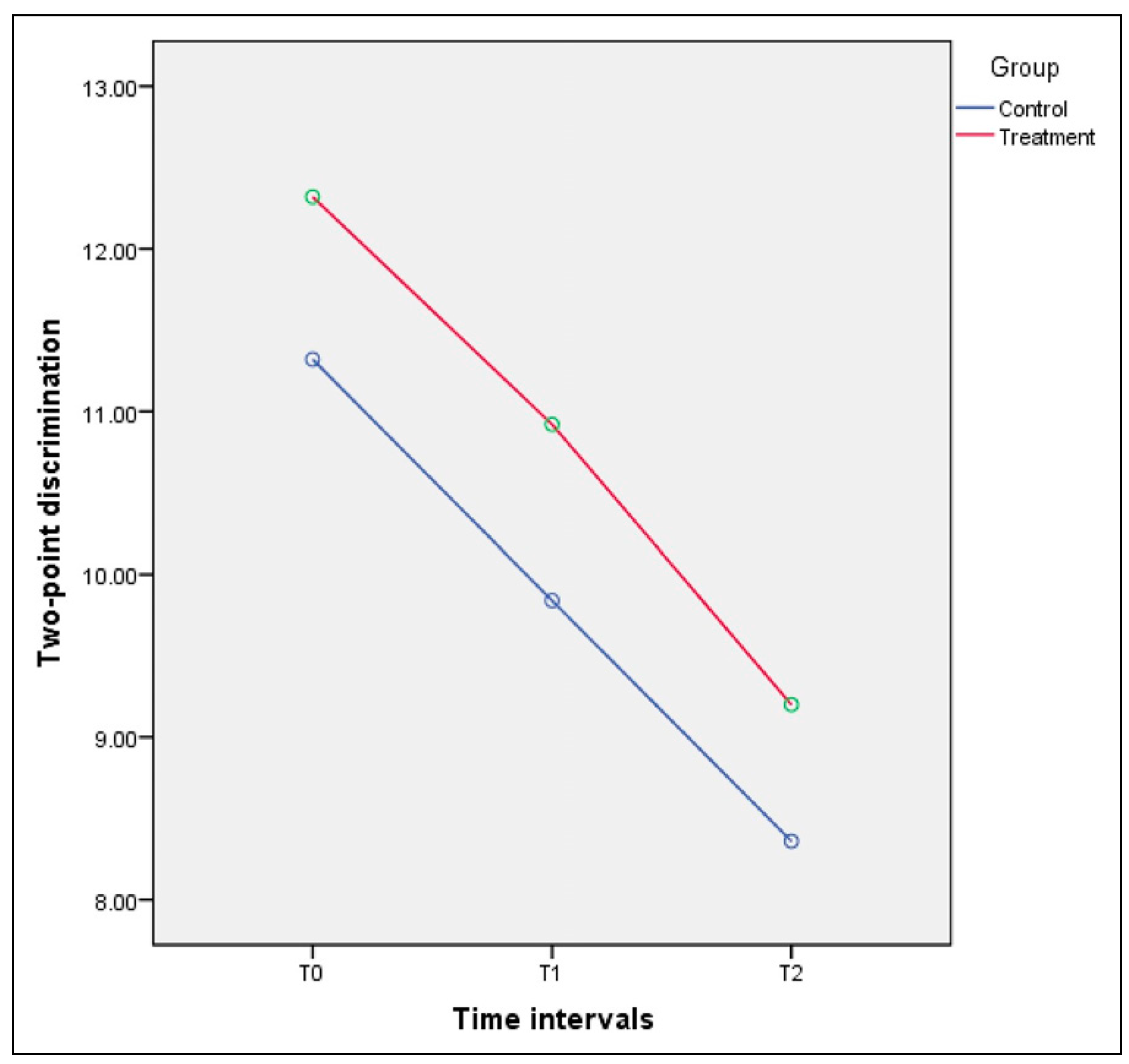
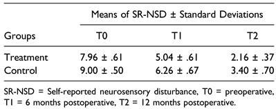
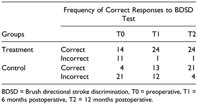
Statistical analyses were performed using the Statistical Package for the Social Sciences (SPSS) for PCs, version 22 (IBM, USA). Descriptive statistics, including gender distribution and the average age of the patients, were obtained. Repeated measure ANOVA was used to evaluate the TPD values and SR-NSD scores between the treatment and control groups during T0, T1, and T2 times. Moreover, generalized estimating equations (GEE) were used to compare the 2 groups’ ability to determine the brush’s direction in the BDSD test. The level of significance was considered at P-value <.05.

Results

Initially, 30 subjects were enrolled in the study, but 5 subjects did not return for follow-up. Therefore, 25 patients with bilateral mandibular body fractures were included in the study, of which 17 patients were male (68%). The mean age of the included patients was 34.48 ± 9.75 years. No subjects developed an infection or nonunion.
The descriptive data related to TPD, SR-NSD, and BDSD at the 3 time intervals of T0, T1, and T2 are presented in Table 1. Results from the TPD analysis using the repeated measures ANOVA showed significant improvement over time intervals in both treatment and control groups (P < .001) (Table 2, Figure 3). The SR-NSD significantly improved during the study period in both groups (P < .001) (Table 3, Figure 4). No differences were found between the groups at each time interval (P = .535).
Those in the treatment group were more likely to have a return of BDSD compared to controls (P < .001) (Table 4).

Discussion

The inferior alveolar nerve passes through a canal of the same name inside the mandible and is supported by the associated vasculature. Fractures of the body and the angle of the mandible often involve the IAN canal, resulting in disturbed sensation of the lower lip and chin [17,18,19]. The use of PRF to enhance IAN regeneration during elective procedures has been reported [14,15,16]. This study was designed to investigate the effect of PRF on the recovery of NSD following mandibular body fractures.
The results of this study demonstrated no difference in TPD and SR-NSD with PRF compared with the contralateral control. However, the subjects’ brush stroke direction was improved with PRF application. The first author of this study has previously applied PRF in sagittal split osteotomy sites, demonstrating that PRF enhanced the recovery of IAN [14]. A recent survey by Behnia et al [16] demonstrated an improvement in NSD by applying L-PRF to the mental nerve after osseous genioplasty. Khojasteh et al. [15] retrospectively compared the recovery of neurosensory impairment using PRF following lateralization of the inferior alveolar nerve at the time of dental implant placement. In the study group (n = 14), PRF was applied to the nerve and covered with a collagen membrane. NSD was assessed at 3, 6, and 12 months after surgery. Although there were no differences at 12 months, the sensation was better (TPD and static light touch) in the PRF group at 6 months. The authors concluded that PRF may accelerate nerve healing and reduce the duration of patient discomfort.
In a similar study, Tabrizi et al. [14] used a split-mouth double-blind, randomized trial to assess the effect of PRF on lower lip sensation in 21 subjects after bilateral sagittal split osteotomies (BSSO). The results of the TPD, BDSD, and SR-NSD tests were significantly better on the treatment side than on the control side.
DBSD requires proprioception provided by large A– Alpha and A-Beta myelinated fibers that are most susceptible to injury [20]. TPD assesses the density and quantity of functional sensory receptors and afferent fibers. Small, myelinated A-Delta and unmyelinated C fibers are stimulated with sharp and painful stimuli. The C fibers are least susceptible to injury, with an A-delta between the A-Alpha and C fibers in injury susceptibility [21,22]. In the current study, PRF did not accelerate the return of TPD in the treatment group, which may indicate that the regeneration of small fibers occurs at the same speed or rate with or without PRF. The recovery of the more susceptible large A-alpha fibers (involved in brush stroke direction) may be improved by PRF.
In addition, SR-NSD was not affected by PRF in this study. SR-NSD is a subjective measurement that may be less reliable than TPD or BDSD. A split-mouth design may better detect differences in these semi-objective variables. This study has several limitations. The surgeons could not be masked to the experimental or control side, but the examiners could. It is not known how much PRF remains within the fracture site adjacent to the inferior alveolar nerve after reduction. Other studies have reported a more controlled application of PRF to the mental nerve or fully exposed IAN during BSSO. PRF applied in this study would not directly affect the exposed mental nerve, which may be a factor in NSD. Additionally, direct force to the mandible may result in soft tissue contusion and nerve injury at sites away from the fracture. Lastly, the sample size was lower than our calculated sample size to detect differences in study variables and may have affected the results.

Conclusion

The outcomes of this split-mouth randomized clinical trial indicated that using PRF may enhance the recovery of the inferior alveolar nerve in mandibular body fractures, but the difference was limited.

Funding

The author(s) received no financial support for the research, authorship, and/or publication of this article.

Conflicts of Interest

The author(s) declared no potential conflicts of interest with respect to the research, authorship, and/or publication of this article.

References

  1. Abhinav, R.P.; Selvarasu, K.; Maheswari, G.U.; Taltia, A.A. The patterns and etiology of maxillofacial trauma in South India. Ann Maxillofac Surg. 2019, 9, 114–117. [Google Scholar] [CrossRef]
  2. Alladi, S.; Dhasarathan, P.; Muralidoss, H.O.R.; Murugesan, K.; Vivigdha, V. Incidence neurosensory deficit in mandibular fractures. Bioinformation. 2023, 19, 725–728. [Google Scholar] [CrossRef]
  3. Singh, R.K.; Pal, U.S.; Singh, P.; Singh, G. Role of fixation in posttraumatic nerve injury recovery in displaced mandibular angle fracture. Natl J Maxillofac Surg. 2016, 7, 29–32. [Google Scholar] [CrossRef] [PubMed]
  4. Schenkel, J.S.; Jacobsen, C.; Rostetter, C.; Grätz, K.W.; Rücker, M.; Gander, T. Inferior alveolar nerve function after open reduction and internal fixation of mandibular fractures. J Cranio-Maxillo-Fac Surg. 2016, 44, 743–748. [Google Scholar] [CrossRef]
  5. Thurmüller, P.; Dodson, T.B.; Kaban, L.B. Nerve injuries associated with facial trauma: natural history, management, and outcomes of repair. Oral Maxillofac Surg Clin. 2001, 13, 283–293. [Google Scholar]
  6. Tabrizi, R.; Pourdanesh, F.; Khoshnik, P.L.; Centenero, S.A. Does the lag time between injury and treatment play a role in recovery of inferior alveolar nerve neurosensory disturbances following mandibular body fracture? J Craniofac Surg. 2019, 30, 2128–2130. [Google Scholar] [CrossRef]
  7. Tay, A.B.; Lai, J.B.; Lye, K.W.; et al. Inferior alveolar nerve injury in trauma-induced mandible fractures. J Oral Maxillofac Surg. 2015, 73, 1328–1340. [Google Scholar] [CrossRef]
  8. Boffano, P.; Roccia, F.; Gallesio, C.; Karagozoglu, K.; Forouzanfar, T. Inferior alveolar nerve injuries associated with mandibular fractures at risk: a two-center retrospective study. Craniomaxillofacial Trauma Reconstr. 2014, 7, 280–283. [Google Scholar] [CrossRef]
  9. Canellas, J.; Medeiros, P.J.D.; Figueredo, C.; Fischer, R.G.; Ritto, F.G. Platelet-rich fibrin in oral surgical procedures: a systematic review and meta-analysis. Int J Oral Maxillofac Surg. 2019, 48, 395–414. [Google Scholar] [CrossRef]
  10. Bielecki, T.; Dohan Ehrenfest, D.M.; Everts, P.A.; Wiczkowski, A. The role of leukocytes from L-PRP/L-PRF in wound healing and immune defense: new perspectives. Curr Pharmaceut Biotechnol. 2012, 13, 1153–1162. [Google Scholar] [CrossRef]
  11. Dohan, D.M.; Choukroun, J.; Diss, A.; et al. Platelet-rich fibrin (PRF): a second-generation platelet concentrate. Part III: leucocyte activation: a new feature for platelet concentrates? Oral Surg Oral Med Oral Pathol Oral Radiol Endod. 2006, 101, e51–e55. [Google Scholar] [CrossRef] [PubMed]
  12. Zumarán, C.C.; Parra, M.V.; Olate, S.A.; Fernández, E.G.; Muñoz, F.T.; Haidar, Z.S. The 3 R’s for platelet-rich fibrin: a “super” tridimensional biomaterial for contemporary naturally-guided oro-maxillo-facial soft and hard tissue repair, reconstruction and regeneration. Materials 2018, 11, 1293. [Google Scholar] [CrossRef]
  13. Simonpieri, A.; Del Corso, M.; Vervelle, A.; et al. Current knowledge and perspectives for the use of platelet-rich plasma (PRP) and platelet-rich fibrin (PRF) in oral and maxillofacial surgery part 2: bone graft, implant and reconstructive surgery. Curr Pharmaceut Biotechnol. 2012, 13, 1231–1256. [Google Scholar] [CrossRef]
  14. Tabrizi, R.; Pourdanesh, F.; Jafari, S.; Behnia, P. Can platelet-rich fibrin accelerate neurosensory recovery following sagittal split osteotomy? A double-blind, split-mouth, randomized clinical trial. Int J Oral Maxillofac Surg. 2018, 47, 1011–1014. [Google Scholar] [CrossRef]
  15. Khojasteh, A.; Hosseinpour, S.; Nazeman, P.; Dehghan, M.M. The effect of a platelet-rich fibrin conduit on neurosensory recovery following inferior alveolar nerve lateralization: a preliminary clinical study. Int J Oral Maxillofac Surg. 2016, 45, 1303–1308. [Google Scholar] [CrossRef] [PubMed]
  16. Behnia, P.; Behnia, H.; Ghanbari, A.M.; Tabrizi, R. Does leucocyte-and platelet-rich fibrin enhance neurosensory recovery after genioplasty? A double-blind, split-mouth, randomized clinical trial. Br J Oral Maxillofac Surg 2023, 61, 534–539. [Google Scholar] [CrossRef] [PubMed]
  17. Genú, P.R.; Vasconcelos, B.C. Influence of the tooth section technique in alveolar nerve damage after surgery of impacted lower third molars. Int J Oral Maxillofac Surg. 2008, 37, 923–928. [Google Scholar] [CrossRef]
  18. Şenses, F.; Önder, M.E.; Koçyiğit, I.D.; et al. Effect of platelet-rich fibrin on peripheral nerve regeneration. J Craniofac Surg. 2016, 27, 1759–1764. [Google Scholar] [CrossRef]
  19. Smith, A.C.; Barry, S.E.; Chiong, A.Y.; et al. Inferior alveolar nerve damage following removal of mandibular third molar teeth. A prospective study using panoramic radiography. Aust Dent J. 1997, 42, 149–152. [Google Scholar] [CrossRef]
  20. Antony, P.G.; Sebastian, A.; Varghese, K.G.; et al. Neurosensory evaluation of inferior alveolar nerve after bilateral sagittal split ramus osteotomy of mandible. J Oral Biol Craniofac Res. 2017, 7, 81–88. [Google Scholar] [CrossRef]
  21. Ghali, G.; Epker, B. Clinical neurosensory testing: practical applications. J Oral Maxillofac Surg. 1989, 47, 1074–1078. [Google Scholar] [CrossRef] [PubMed]
  22. Mohajerani, S.H.; Tabeie, F.; Bemanali, M.; Tabrizi, R. Effect of low-level laser and light-emitting diode on inferior alveolar nerve recovery after sagittal split osteotomy of the mandible: a randomized clinical trial study. J Craniofac Surg. 2017, 28, e408–e411. [Google Scholar] [CrossRef] [PubMed]
Figure 1. Platelet-rich fibrin matrix.
Cmtr 17 00075 g001
Figure 2. Platelet-rich fibrin matrix placed between the proximal and distal segments of mandibular body fracture on the exposed inferior alveolar nerve.
Cmtr 17 00075 g002
Figure 3. Two-point discrimination test showing a significant improvement over time intervals in both treatment and control groups (P < .001).
Cmtr 17 00075 g003
Figure 4. Self-reported neurosensory disturbance test showing significant improvement over time intervals in both treatment and control groups (P < .001).
Cmtr 17 00075 g004
Table 1. Description of the Study Sample.
Cmtr 17 00075 i001
Table 2. Results of Two-point Discrimination Test in Treatment and Control Group.
Cmtr 17 00075 i002
Table 3. Results of Self-Reported Neurosensory Disturbance Test in Treatment and Control Group.
Cmtr 17 00075 i003
Table 4. Frequency of Correct Responses of Brush Directional Stroke Discrimination Test in Treatment and Control Group.
Cmtr 17 00075 i004

