Next Article in Journal
Corrigendum to "A Review of 13470 Head and Neck Injuries From Trampoline Jumping"
Previous Article in Journal
The Reconstruction of Mandible Defects in War Injuries: Systematic Review and Meta-Analysis
 
 
Craniomaxillofacial Trauma & Reconstruction is published by MDPI from Volume 18 Issue 1 (2025). Previous articles were published by another publisher in Open Access under a CC-BY (or CC-BY-NC-ND) licence, and they are hosted by MDPI on mdpi.com as a courtesy and upon agreement with Sage.
Article

Measuring Orbital Volume Using Open Source Software and Its Application in Orbitozygomatic Fractures

by
Taradevi P. V. Narayan
* and
Vikas Dhupar
Department of Oral and Maxillofacial Surgery, Goa Dental College and Hospital, Goa, India
*
Author to whom correspondence should be addressed.
Craniomaxillofac. Trauma Reconstr. 2024, 17(2), 169-172; https://doi.org/10.1177/19433875231163982
Submission received: 1 November 2022 / Revised: 1 December 2022 / Accepted: 1 January 2023 / Published: 16 March 2023

Abstract

:
Study Design: Technical note. Objective: Changes in orbital volume can lead to esthetic and functional complications of the orbit. In this article, the procedure to calculate the orbital volume using the open source software Aliza 3D DICOM is described. Methods: This article describes the steps to use this novel software. To validate the software, the normal orbital volume was calculated bilaterally on CT scans with normal orbital anatomy. The volumes of unilateral orbitozygomatic fracture cases were compared. Results: This open source software has easy access. The normal orbital volume calculated using this software was 24.4 cc ± 0.72. In the unilateral orbitozygomatic fracture cases, an increased orbital volume was calculated. Conclusions: This easy access, inexpensive, and convenient computer aided software can be used to calculate orbital volume facilitating treatment plan for correction of the orbit volume.

Introduction

Trauma of the orbit is a common cause for changes in bony orbital volume. Esthetic and functional impairment such as enophthalmos, diplopia, and hypoglobus occur due to volume changes [1,2]. But volumetric assessment can be tedious, expensive, and requiring additional expertise. Thus, repair is usually carried out based on fracture appearances such as site and size of defect. The benefit of evaluation of preoperative CT scans for orbital volume facilitates treatment planning of internal orbital reconstruction [3].
In order to routinely evaluate orbital volume, it is neccessary for a conveninet computer aided software to be inexpensive and easy access. One such novel open source software for orbital volume calculation is the Aliza 3D DICOM. This article aims to evaluate and validate this software for orbital volume calculations.

Technical Note

The following are the steps to calculate the orbital volume using Aliza 3D DICOM software:
  • The DICOM images of the CT scans were loaded into the software.
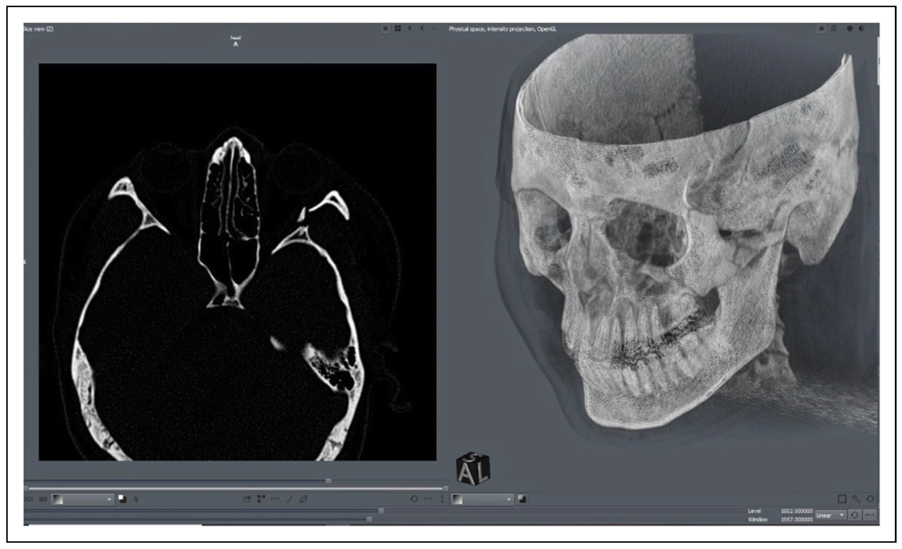
  • The interface screen displayed the cross-section of each slice in a 2D format on the left and the 3D rendering of the patient on the right. (Figure 1).
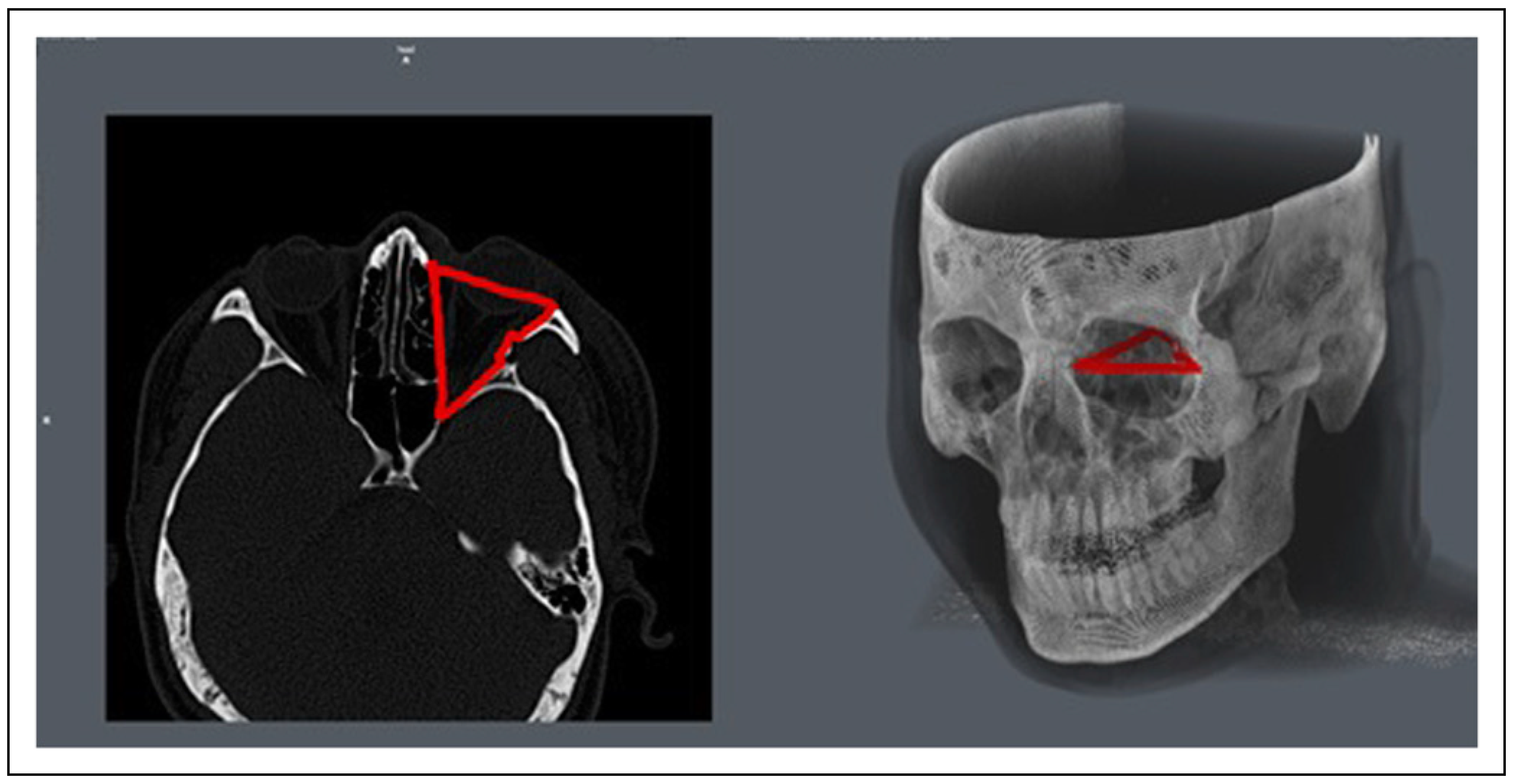
  • In each axial section image, the margins of the orbit were manually selected by using the “Draw” option of the Region of Interest (ROI) tool. (Figure 2) For volume calculation of each orbit, the marking on every axial section was added to the same ROI. The anterior boundary was determined by a line joining the medial and lateral orbital rims. The posterior boundary was defined by the initiation of the optic foramen, inferior and superior orbital fissure [4].
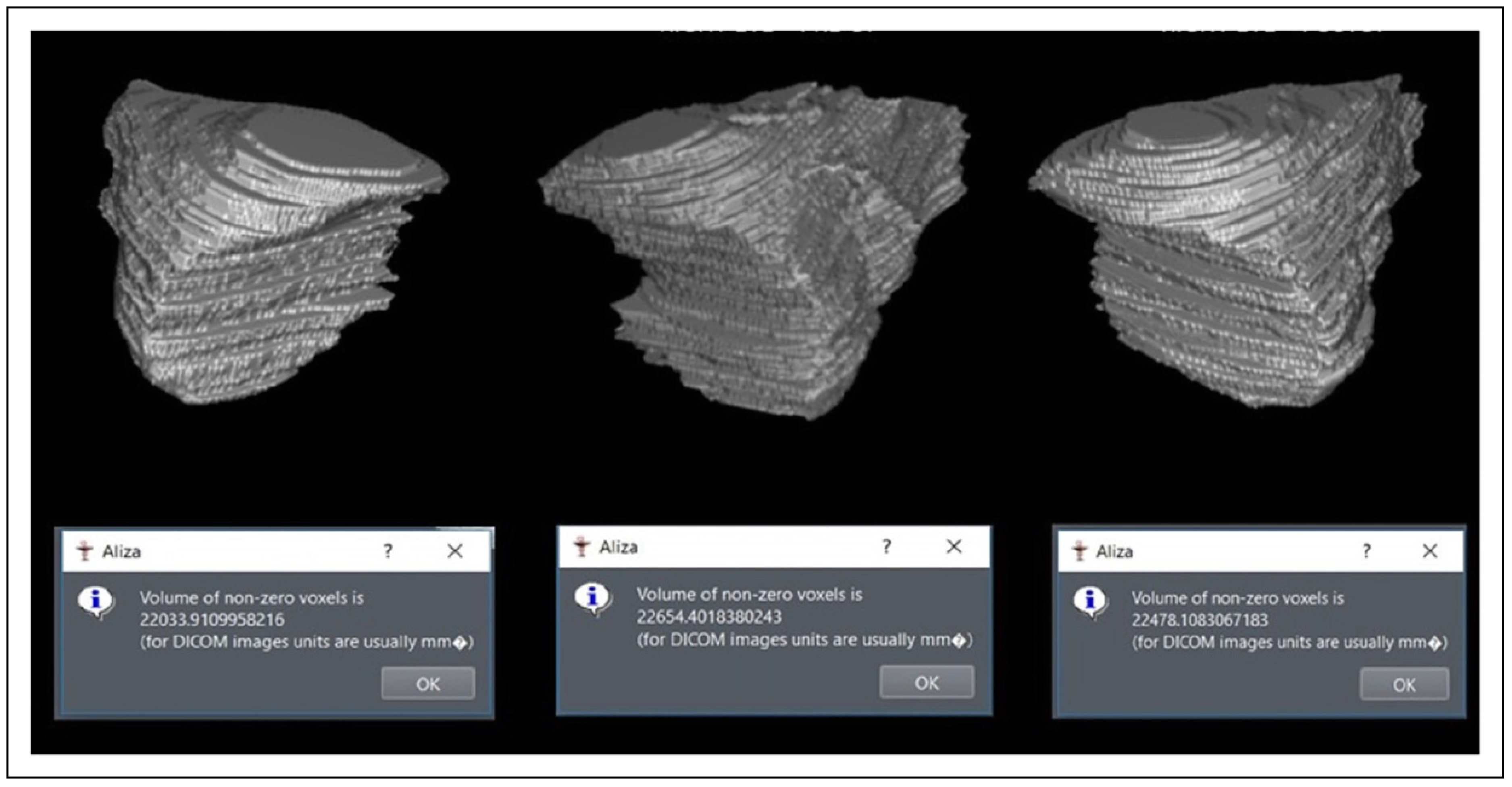
  • After completion of manual segmentation of the concerned orbit, the ROI is selected and converted to a binary image. The volume was computed automatically by the software in mm3 (Figure 3) and then converted to cubic centimeters.
The use of a pen tablet provides higher accuracy than the mouse of a computer. The Wacom Intuos Series pen tablet was used.
To validate the software, 25 middle aged, male patients of Indian origin were evaluated (Table 1). 10 patients who underwent CT scans for other medical reasons but had normal orbital anatomy were identified. Orbital volumes of bilateral orbit for each patient were calculated using the above procedure. The mean average normal orbital volume was 24.4cc ± .72. The mean average difference of the left and right orbit was .26 cc. Average time taken to calculate both orbital volumes of a single patient was 19.9 min.
The preoperative orbital volumes of 15 male patients with unilateral orbitozygomatic fracture were compared. Mean average increase of 27.2 cc was noted. Following open reduction and internal fixation of these 15 patients, the mean average post-operative orbital volume was decreased to 25.4 cc.

Discussion

Advancing CT technologies and computer software help diagnose and predict the surgical outcome which in turn improves the accuracy and quality of the surgery. There are studies [4,5,6] describing different ways of orbital volume calculation. An easy access and convenient computed aided software such as the Aliza 3D DICOM viewer helps to assess extent of damage and calculate orbital volume.
For measurement of orbital volume, segmentation techniques that have been described are manual, threshold based, atlas based, and model based [7]. Studies [7,8,9] using manual segmentation have proven to be an accurate technique wherein the margins of the orbit are marked by the user in each image slice. Manual segmentation was used with this software.
The need for orbital volume reduction is important as Ebrahimi A et al. [10] had concluded that for each 1 cc of orbital volume change, clinical and radiographic enophthalmos of .68 mm and .71 mm respectively was noted. But in order to assess if there is an increase in orbital volume, the normal orbital volume must be calculated. The volume can vary depending on the race, sex, and age of patient. In this study, middle aged male patients of Indian origin were evaluated for normal orbital volume.
After establishing the normal values, any increase or decrease in orbital volume can be detected and treatment planned accordingly. Thus, facilitating and improving quality of treatment and preventing post-operative complications associated with change in orbital volume.
The method of orbital volume calculation by this software is similar to that of commonly used paid softwares such as OssiriX and Siemens Somatom Sensation 16. This software lacks the capability to detect the missing ROI in between selected segmentations and editing within the selected ROI. More studies with larger sample size would need to be carried out for assessing the accuracy. Inspite of these limitations, this open source software is easily accessible, inexpensive, and convenient to be used on personal computers. Basic knowledge of interpretation of orbital anatomy of a CT scan is needed.

Conclusions

The Aliza 3D DICOM is an open source software and convenient to view and calculate orbital volume. Thereby, facilitating treatment plan. This procedure of volume calculation can also be used to obtain volume of frontal and maxillary sinus if needed.

Funding

The author(s) received no financial support for the research, authorship, and/or publication of this article.

Declaration of Conflicting Interests

The author(s) declared no potential conflicts of interest with respect to the research, authorship, and/or publication of this article.

References

  1. Ploder, O.; Klug, C.; Voracek, M.; et al. Evaluation of computer-based area and volume measurement from coronal computed tomography scans in isolated blowout fractures of the orbital floor. J Oral Maxillofac Surg 2002, 60, 1267–1272. Available online: https://pubmed.ncbi.nlm.nih.gov/12420258/ (accessed on 30 April 2021). [PubMed]
  2. Fan, X.; Li, J.; Zhu, J.; et al. Computer-assisted orbital volume measurement in the surgical correction of late enophthalmos caused by blowout fractures [Internet]. Ophthalmic Plast Reconstr Surg 2003, 19, 207–211. Available online: https://pubmed.ncbi.nlm.nih.gov/12918556/ (accessed on 30 April 2021). [PubMed]
  3. Ellis, E.; Reddy, L. Status of the Internal Orbit after Reduction of Zygomaticomaxillary Complex Fractures. J Oral Maxillofac Surg 2004, 62, 275–283. Available online: https://pubmed.ncbi.nlm.nih.gov/15015156/ (accessed on 30 April 2021). [PubMed]
  4. Jansen, J.; Schreurs, R.; Dubois, L.; et al. Orbital volume analysis: validation of a semi-automatic software segmentation method. Int. J. Comput. Assist. Radiol. Surg. 2016, 11, 11–18. [Google Scholar] [CrossRef] [PubMed]
  5. Osaki, T.H.; de Castro, D.K.; Yabumoto, C.; Mingkwansook, V.; Ting, E.; Nallasamy, N.; Curtin, H.; Fay, A. Comparison of Methodologies in Volumetric Orbitometry. Ophthalmic Plast Reconstr Surg. 2013, 29, 431–436. [Google Scholar] [CrossRef] [PubMed]
  6. Zhang, X.; Han, C.Y.; Dai, M.J.; et al. Application of computer-assisted surgery techniques in the management of zygomatic complex fractures. Chinese Journal of Traumatology—English Edition 2018, 21, 281–286. [Google Scholar]
  7. Sharma, R.; Muralidharan, C.G.; Roy, I.D.; et al. Radiological evaluation of sphenozygomatic suture fixation for restoration of orbital volume: A retrospective study. J Cranio-MaxilloFac Surg 2016, 44, 1903–1908. [Google Scholar] [CrossRef] [PubMed]
  8. Charteris, D.G.; Chan, C.H.; Whitehouse, R.W.; et al. Orbital volume measurement in the management of pure blowout fractures of the orbital floor. Br J Ophthalmol 1993, 77, 100–102. [Google Scholar] [PubMed]
  9. Regensburg, N.I.; Kok, P.H.B.; Zonneveld, F.W.; et al. A new and validated CT-based method for the calculation of orbital soft tissue volumes. Invest Ophthalmol Vis Sci 2008, 49, 1758–1762. [Google Scholar] [PubMed]
  10. Ebrahimi, A.; Kalantar Motamedi, M.H.; Rasouli, H.R.; et al. Enophthalmos and Orbital Volume Changes in Zygomaticomaxillary Complex Fractures: Is There a Correlation Between Them? J Oral Maxillofac Surg 2019, 77, 134.e1. [Google Scholar] [CrossRef] [PubMed]
Figure 1. Interface screen displaying the axial section and 3D view of a fracture case.
Cmtr 17 00024 g001
Figure 2. Margins of the orbit manually selected.
Cmtr 17 00024 g002
Figure 3. Computed orbital volume in cubic millimeters.
Cmtr 17 00024 g003
Table 1. Descriptive characteristics of the study and validation sample.
Cmtr 17 00024 i001

