Next Article in Journal
The Reconstruction of Mandible Defects in War Injuries: Systematic Review and Meta-Analysis
Previous Article in Journal
Objective Photoanalysis of Feminizing Frontal Cranioplasty Outcomes
 
 
Craniomaxillofacial Trauma & Reconstruction is published by MDPI from Volume 18 Issue 1 (2025). Previous articles were published by another publisher in Open Access under a CC-BY (or CC-BY-NC-ND) licence, and they are hosted by MDPI on mdpi.com as a courtesy and upon agreement with Sage.
Article

Flattening the Curve and Cutting Corners—Pearls and Pitfalls Facial Gender Affirming Surgery

by
Sven Gunther
,
Jourdan Carboy
,
Breanna Jedrzejewski
and
Jens Berli
*
Department of Plastic and Reconstructive Surgery, Oregon Health and Science University, 3181 SW Sam Jackson Park Rd, Portland, OR 97239, USA
*
Author to whom correspondence should be addressed.
Craniomaxillofac. Trauma Reconstr. 2024, 17(2), 146-159; https://doi.org/10.1177/19433875231178968
Submission received: 1 November 2022 / Revised: 1 December 2022 / Accepted: 1 January 2023 / Published: 6 June 2023

Abstract

:
Study Design: This is an experiential article based on the past 6 years experience of providing facial gender confirmation surgery (fGAS) at an academic medical center. Objective: While trainees are getting increasing exposure to aspects of facial gender affirming surgery (fGAS), the gap between trained providers and patients who can access care is currently still widening. A handful of fellowships across the country have emerged that include fGAS in their curriculum, but it will take another decade before the principles of affirming care and surgeries are systematically taught. Fortunately, the surgical principles and techniques required to perform fGAS are part of the skill set of any specialty surgeon trained in adult craniofacial trauma and esthetic facial surgery/rhinoplasty. It is the aim of this article to provide directly applicable knowledge with the goal to assist surgeons who consider offering fGAS in flattening the learning curve and hopefully contribute to increasing the quality of care provided for the transgender and gender diverse population. We hope to provide the reader with a very tangible article with the aims to 1) provide a simple systematic framework for an affirming consultation and preoperative assessment and 2) provide translatable surgical pearls and pitfalls for forehead feminization and gonial angle resection. The frontal sinus set back and gonial angle resection in our opinion are the most unique aspect to fGAS as rhinoplasty, genioplasty and other associated procedures (e.g., fat grafting) follow well established principles. We hope that the value of this article lies in the translatability of the presented principle to any practice setting without the need for VSP, special surgical instruments or technology beyond basic craniofacial tools. Methods: This is an experiential article based on the senior authors 6 year experience offering fGAS in an academic setting. The article is structured to outline both pearls and pittfalls and is supplemented by photographs and a surgical video. Results: A total of 19 pearls and pitfalls are outlined in the article. Conclusions: Facial gender affirming surgery mostly follows established craniofacial and esthetic surgery principles. Forehead feminization and gonial angle feminization are the 2 components that diverge most from established surgical techniques and this article hopefully provides guidance to shorten the learning curve of surgeons.

Introduction

The number of gender affirming surgeries (GAS) performed in the United States have experienced an exponential rise in last 5 years, and centers and providers with focus on GAS are struggling to meet the surging demand after decades of neglect in this healthcare domain [1]. While trainees more recently in some residency programs are starting to get exposure to at least some aspects of gender affirming care and surgery, the gap between trained providers and patients who can access care is currently still widening. This is particularly true for the complex aspects of GAS such as genital and facial GAS. For facial GAS (fGAS) the issue is additionally compounded by the only recent increase in insurance coverage as the evidence and expert consensus through the World Professional Association for Transgender Health (WPATH) Standard of Care, Version 8 (SOC) outlines fGAS as a medically necessary procedure. A handful of fellowships across the country have emerged that include fGAS in their curriculum, but it will take another decade before the principles of affirming care and surgeries are systematically taught. Fortunately, the surgical principles and techniques required to perform fGAS are part of the skill set of any specialty surgeon trained in adult craniofacial trauma and esthetic facial surgery/rhinoplasty. This can potentially increase access to care. However, those with no fGAS in their training would have to accept a learning curve that comes with any expansion of practice. Cadaver courses, live surgical courses, or visiting professorships are all legitimate means by which to get exposure and understand how to combine pre-existing skills in a manner to femininize an individual’s face. It is the aim of this article to provide directly applicable knowledge to those trained in adult craniofacial procedures with the goal to assist flattening the curve and hopefully contribute to increase the quality of care provided for the transgender and gender diverse population. We want to use the word count provided to us wisely and will defer to the pre-existing literature on GAS and fGAS for context and basics as we hope to provide the reader with a very tangible article with the aims to 1) provide a simple systematic framework for an affirming consultation and preoperative assessment and 2) provide translatable surgical pearls and pitfalls for forehead feminization and gonial angle resection. The frontal sinus set back and gonial angle resection in our opinion are the most unique aspect to fGAS as rhinoplasty, genioplasty, and other associated procedures (e.g., fat grafting) follow well established principles. We hope that the value of this article lies in the translatability of the presented principle to any practice setting without the need for VSP, special surgical instruments, or technology beyond basic cranio-facial tools.

Methods

This is an experiential article based on 5 years’ experience providing fGAS in the setting of an academic institution and with the support of a multi-disciplinary team including mental health professionals. These techniques have been used at our institution in resident training with understanding and execution of maneuvers across training levels of our residents. Patient consent for all photographs presented in this article has been obtained. No institutional review board approval required. Pearls are noted with P# and pitfalls with P#.

General Principles Consultation and Assessment

The youngest individual that had surgery at our institution was 18 and the oldest 78, what links the 2 experiences is that they both have desired this surgery since they got exposed to endogenous testosterone—one for 5 years and one for 64 years. P# Understanding the magnitude the surgical consult has in an individual’s life including the associated anxiety and excitement is the foundation to connect and establish trust. Verbalizing that genuine understanding can be helpful before embarking on the consultation.
“Is it the way that society responds to your face; or is it more the way that you perceive yourself that bothers you?”
P# Our faces are subject to the dualism of the human experience and mind; understanding which one of those the patient experiences most gender dysphoria from is crucial as it fundamentally affects the consultation and avoids the pitfall of a “cookie–cutter” approach to fGAS. While most individuals will be somewhere along the axis of this question it is common to hear that someone states that if they lived in an affirming environment, they would not seek to have fGAS but desire surgery to not be stigmatized and harassed in their daily lives. P# In which case, it is prudent to warn the patient that you may point out aspects of their face that should be addressed but they may not even be aware of. This may aggravate gender dysphoria and verbal consent should be obtained prior. Consider further to understand what procedure are covered in the patient’s insurance plan or ability to afford. While actual harm can be afflicted by inexperienced provider; the consultation is also an opportunity to profoundly affirm a patient. P# Using the Gender Facial Mosaic Concept: All faces have masculine and feminine traits regardless of estrogen or testosterone exposure in adolescence. Asking the patient first which aspects of their face they like, gives an opportunity to start with the perceived positive aspects of their appearance; it further gives you an opportunity to see if the patient experiences the same areas of her/their face as feminine that you deem feminine. For example, we had a patient saying they liked their chin and commenting that it looks like their mothers and aunts, even if our in our assessment the chin would benefit from fGAS. Once the patient is done outlining their positive findings it gives you the opportunity to point out other feminine aspects of the face which a patient may not even be aware of. To hear this from a surgeon can be therapeutic and improve gender dysphoria. Only now do we move on to discuss the aspect of the face the patient dislikes followed by facial measurements, functional exam, and photographs.

Facial Dimensions From Quantitative to Qualitative to Subjective

P# We find it useful to take a few objective measurements to compare outcomes by, and doing so provides an opportunity to examine the face of the patient close up. These measurements include forehead height, nasal length/projection/alar width, nostril height, chin height, and upper lip length [Figure 1 and Figure 2]. Advanced facial analysis using the plethora of published angles, anatomical landmarks, etc., are only marginally useful when confronted with an individual patient presenting for fGAS. In the absence of virtual surgical modeling that can show a patient their expected changes it can be challenging to assure that both surgeon and patient are on the same page. We have found that using the 3 dimensions of - > vertical/horizontal and sagittal is an uncomplicated way to communicate to patients. Once the plan is made this language can be used to make sure that you and the patient understand the goals of surgery. The simplicity of stating everything in the basic 3 dimensions is helpful.
“It is important that you understand the way that we think about facial surgery and what it is that I am looking at when I assess your face. In fact, it is quite simple as we have three dimensions: vertical, horizontal, and sagittal. And with the various surgeries we either increase or decrease those dimensions – be that bone, cartilage, or soft tissue. As you look in the mirror you can see that your forehead is horizontally too wide, vertically too tall and projects too far making your overall forehead appear masculine. Is that what you see as well?”
P# All domains that are objectively affected by exposure to testosterone during adolescence are included in the consultation note, even if the patient may not be fully decided at this point. For example, a dorsal hump may be extenuated to appear more masculine after forehead feminization and while the patient may not currently desire a rhinoplasty this could change down the road, therefore it is important to document this at the initial consultation to avoid insurance denial if a patient decides to address this in the future.
With increasing access to insurance coverage for fGAS we as providers together with the patient and mental health professional will have to determine what aspects of the face are eliciting gender dysphoria and which aspects are to be considered an esthetic procedure (e.g., facelift after a jawline reduction may be considered part of fGAS, whereas it may not as a stand-alone procedure).

Holistic Forehead Assessment and Surgical Planning—Identifying the Fixpoint

P# With too much focus on the sinus the surgeons run risk to under-correct the rest of the forehead. This was certainly the case in our early experience. In our opinion this is also a shortcoming of virtual surgical planning (VSP) as the bone structure surrounding the sinus changes dramatically if the forehead is seen as one unit and reduction of the frontal bone including fronto-temporal and zygomatico-frontal (ZF) sutures are performed.
While off course the frontal sinus is a key area to address it detracts from a holistic assessment of the forehead which includes horizontal width both superior and inferior, sagittal projection and lateral orbital rim hooding as well as forehead inclination and shape. P# It is worthwhile to fully ignore the frontal bossing in the radiographic and photographic assessment but rather focus on the desired shape of the whole forehead. With appropriate surgical technique (outlined below) it is almost always possible to set the sinus to back to the level of the nasal root however it is not always possible to reduce the superior part of the frontal bone to match a significant setback (e.g., a steep forehead with thin anterior cortex and a marked frontal sinus protuberance). It is the goal to reach forehead harmony where hairline, forehead shape, forehead contour, orbit shape, brow position, and nasal transition work to match facial balance and heritage. A notable common misconception is that a feminine hairline is lower; however, this is a factor of aging as the masculine hairline starts lower but recedes with aging. The planning should start with visual inspection of the forehead inclination, position of the hairline in relation to the most convex aspect of the forehead itself, followed by measurements of the anterior cortex thickness in a sagittal view. Forehead and hairline position and shape varies across different genetic pools and geographical regions. This ties in with the above-mentioned point to explore what shape is most in line with the patient’s heritage and desire, rather than merely trying to achieve an obtuse naso-frontal angle with a near round arc of a circle and a low set hairline. Figure 3 outlines various examples of distinctly different forehead types. A patient with native middle or northern American heritage may desire to retain forehead inclination with a more flattened rather than circular ascent and a distinctly lower hairline; whereas a patient with eastern African heritage may prefer a forehead that is more circular and a hairline that lies above the point of maximum convexity. Using the above principle allows us to determine where on the current forehead the fix point is from where to draw a line towards the nasofrontal junction and the superior frontal bone [Figure 4].
This fixed point is identified in the sagittal view from which the slope of the desired forehead shape can be extrapolated. If that point is posterior to the diploic space than patient expectations must be tempered to achieve the ideal forehead shape for that given patient. The patient can decide if they either prefer a forehead that is potentially steep (as the sinus gets set back)—or to expect a persistent frontal sinus convexity and more acute naso-frontal angle. Discussing the variability in the cis-female anatomy by showing photographs of famous actresses can be useful to normalize the anatomic spread (e.g., Jennifer Garners forehead or Angelina Jolie’s jawline). If the fix point lies anterior to the diploic space on the CTscan, than that point is the minimal amount of bone that needs to be reduced in that area and from where the rest of the shaping will take place. Video 1 outlines these principles and shows how this planning gets translated to the operating room. This paradigm shift in assessing and thinking about the forehead has made a tremendous difference in our practice and in setting expectations.

Jawline Assessment

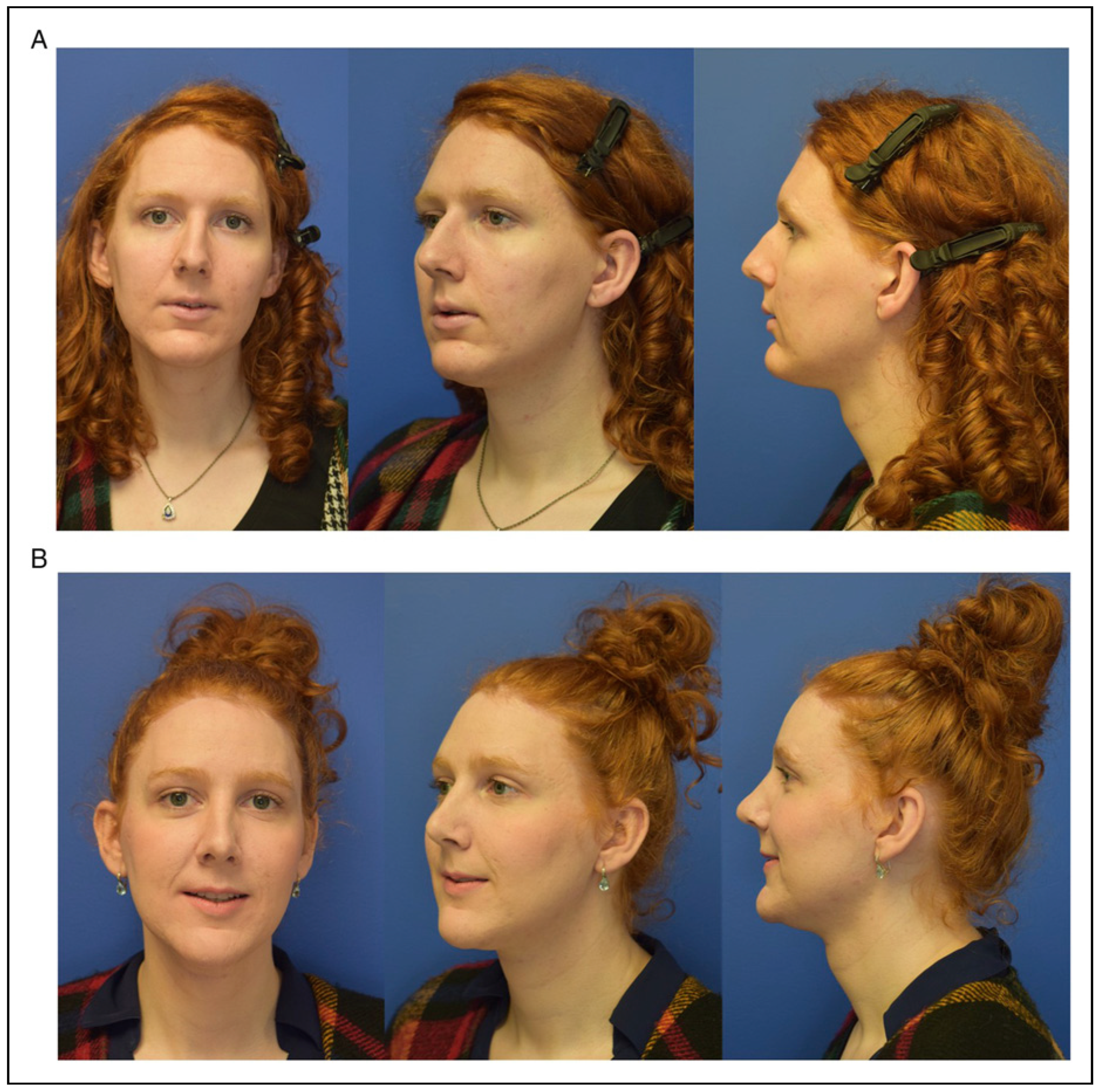
The assessment of the jawline follows the previously mentioned use of dimensions. Orthognathic surgery, while powerful and indicated in some individuals, is rarely a covered benefit. However, if malocclusion or significant discrepancies between the midface and jaw are noticeable in the assessment, a discussion around the pro/con’s should be had with the patient regardless. In social media forums, the “V-shaped” jawline is frequently cited and often desired by patients presenting for fGAS. Jawline assessment and planning is often more challenging than forehead feminization since it includes the variability of the soft tissue envelope that may have pre-existing laxity, varying thickness including glandular structures and differing response to surgical subperiosteal release. The variability of the overlying soft tissue can make surgical planning less predictable than the forehead and patients need to understand this limitation, which may include the need/desire for secondary or immediate rhytidectomy. The gonial angle is the most technically challenging aspect of jawline feminization as the access and available instrumentation for a bicortical cut are limited. It is no surprise therefore that many surgeons, including the senior author in his earlier practice, shy away or recommend against gonial angle resection in most patients. Reduction of the horizontal dimensions are still possible through osteoplasty of the gonial angle flare after full release of the masseteric sling. Botox is another avenue by which this can be achieved in patients with master hypertrophy. However, neither maneuver addresses the vertical height of the posterior mandible nor changes the angle between ramus and mandibular body. To achieve a feminine harmony of the overall jawline, the other associated planned maneuvers need to be taken into consideration. Figure 5 shows a patient where the gonial angles were not addressed but by increasing the vertical height and sagittal position of the chin the overall shape and angle were feminized. Balance and facial harmony again are key for natural and desired results. For jawline and chin countering and planning VSP holds a lot of promise to provide more predictability and protect the inferior alveolar nerve. Figure 6 shows a patient that desired gonial angle resection. However, her overall facial height compared to sagittal facial proportions would not be favorable if the gonial angle were resected.

Surgery—Technical Pearls and Pitfalls Forehead

As mentioned in the introduction we will refer to all the excellent articles existent in our literature as to the established surgical techniques. We also want to give credit to Dr Luis Capitan and the entire FacialTeam who have built on Dr Douglas Osterhout’s pioneering work and taught the senior author. This section intends to provide distinct surgical pearls that represent an inflection point in our learning curve. Means by which the structural components can be altered in the above mentioned 3 dimensions include: Osteoplasty, Ostectomy, Osteotomy and Repositioning, and autologous/allograft and alloplastic augmentation. Table 1 gives an overview of the most frequent surgical techniques and associated CPT codes.

Hairline and Access Approach

Many patients benefit from hairline lowering surgery. In evaluation the patient’s heritage should be considered, for example, a higher hairline in a person of eastern African descent may be desirable compared to a lower hairline in a person of native southern American or Indian descent. Various techniques for hairline lowering have been described with some performing immediate hair transplantation in combination with posterior bicoronal incision [2,3]. Pretrichial trichophytic incision is probably the most frequently used with extension of the incision posterior to the hairline at posterior extension of the lateral recession. P# While we used to perform the excision of excess non-hair bearing skin at the end of the surgery, we have altered our practice to mark out the expected skin excision at the time of incision. This is estimated on laxity and mobility on the skin on the forehead as well as desired advancements. A skin scalp reconstruction a distance of 2 cm of advancement is possible in most patients [4]. This saves time and allows for less blood loss at the conclusion of the forehead surgery. Areas where more skin can be excised can always be further reduced at the time of closure. We advise to only do this after sufficient experience in assessing the expected excision. P# We also routinely excise skin in the temporal region (1–2.5 cm) as this exerts direct pull on the midface and can be particularly powerful if a direct midfacial lift is performed through a Gilles approach. P# Harvesting some superficial temporal fascia can be useful for both sealing the frontal sinus or for subsequent use in a rhinoplasty. P# If midfacial or lip fat grafting is planned injecting some fat in a plane deep to the deep temporal fascia is a safe plane to provide additional volume to the temporal region. #P When exposing the forehead, it is important to clear at least 4–5 mm past the ZF and NF suture. Similarly, the temporal crest should be fully exposed.

Osteoplasty

A Stryker TPS Elite egg burr, 6 mm (Kalamazoo, Michigan) is used for this step. Free style burring of the forehead without a clear plan is ill advised. Based on the preoperative CT scan and forehead assessment the fixed point that was decided upon is marked out with a sterile pencil [Video 1 and Figure 7]. The areas that are outlined as separate subunits include:
  • Frontal sinus
  • Supraorbital rim including ZF suture
  • Temporal Crest
  • The remaining frontal bone is divided into 12 quadrants
Figure 7. Markings that are used during the osteoplasty portion of forehead feminization. Depending on the planned forehead shape the quadrant that includes the “fixed point” is reduced first from which the rest of the shaping takes place.
Cmtr 17 00022 g007
The fix point usually sits in one of the lower two rows but in some patients may be at the level of the sinus. This is where osteoplasty commences and the bone is reduced until the fixed point is reached. From there we continue unit by unit until ½ of the forehead is reduced. The frontal bone osteoplasty reduces the anterior cortex by app. 1–3 mm and at the ZF suture a reduction of 5–10 mm is common. Video 1 shows a frontal sinus set back on the left side while the right side has sufficient anterior cortex to reduce it as a type 1 (osteoplasty only).

Sinus Set Back

[Video 1 and Figure 8 and Figure 9], The key technical pearl that is rarely discussed or shown in the literature are the details of marking the anterior table of the frontal sinus. There are obvious practice difference between surgeons. In our practice and based on the experience of the FacialTeam we extend the pencil marking superior and lateral past the outline of the frontal sinus (measured at the nasal root in a coronal view) by 2-3 mm. This allows to keep a bony ledge for cortico-cancellous bone to bone contact for subsequent vascular ingrowth. The bony ledge further can be burred down 1–3 mm to sink the anterior table further assisting in camouflaging the convexity of the anterior table. Inferiorly the marking stays on the anterior table at the exact level where the convexity of the anterior table transitions to its most steep decline towards the nasofrontal junction. [Figure 8] This small detail is of utmost importance to achieve substantial and reproducible sinus setbacks. We use the Synthes Piezoelectric system (Oberdorf, Switzerland) that is held tangential superior-laterally and perpendicular inferiorly. The piezo is safe and a posterior table injury is essentially impossible if true tangential position is held. This can similarly be achieved with a conventional saw blade. We recommend using a rounded tip oscillating saw over a reciprocating sawblade as it makes injury to the posterior table unlikely. Once the anterior table is lifted off crests on the piece are reduced with a burr. P# The intersinus septum is reduced with a burr or rangeur so that both sinuses are connected. The naso-frontal outflow tract is inspected to assure patency on both sides and any sinus polyps (if present) cauterized. The inferior border is then reduced with a 4 mm egg burr held perpendicular to the plane of the nasofrontal junction and the actual set back performed until the bone is reduced to the desired level. Superior-lateral there is usually a rim of bone that is also reduced to the level of the previous frontal bone osteoplasty. Any crests are reduced to avoid interference with the anterior table bone piece. During the previous step of the operation, the anterior table is thinned to about 1 mm. We strongly advocate keeping the anterior table substantially thick to avoid potential future resorption by allowing revascularization of the cortical bone. Areas that are too thin will have a “blue hue” where the sinus begins to be visualized through the bone. Substantial cancellous bone to bone contact is desirable and hence the cut being made beyond the outline of the sinus in the superior and lateral portions. Once all the above steps are completed the bone piece is put in its location and mostly is too wide in a horizontal dimension. Using a pencil, the areas that extend are marked and the anterior table reduced by means of a burr on the back table until the dimensions fit. A Stryker TPS Elite egg burr, 4 mm (Kalamazoo, Michigan) can assist in creating a sunken in ledge as mentioned above. The combination of removing the most lateral convex portions of the table and the inferior set back allows to maximize the reduction in frontal sinus anterior table convexity. If bone gaps are present, they should be minimized inferiorly where gravity could in theory lead to extrusion of mucous cells. Superior bone gaps can be covered with a thin titanium mesh. Bone dust has been shown to be osteogenic but is ill suited for bone gaps as it can find its way into the frontal sinus. Additionally, bone chips have been shown to have fewer osteoclasts than bone dust, therefore greater osteogenic potential because of slowed resorption [5,6]. We prefer to use a 2.5 cm osteostome and with gentle taps harvest calvarial bone chips that can be placed underneath the titanium mesh or plate [6] or be wedged into the defects. Temporoparietal fascia can easily be harvested and secured to the titanium plates to provide additional coverage and camouflage through augmentation of mild residual convexities. In patients with a marked and voluminous corrugator—direct corrugator resection can be considered as previously documented in the esthetic literature. The browlift can be accomplished through various methods and opinions and practices mirror controversies in esthetic browlift. In case of a pretrichial incision endotyne should be avoided in the non-hair bearing skin as even a low-profile implant may not only be palpable but has the risk of extrusion/infection. Galeal scoring can assist in maximizing hairline advancement in select patients but may come with an increased risk of shock alopecia. During closure care must be taken to close the deep layer with galea-only bites to protect the hair follicles, decrease tension on the epidermal closure, and additionally mitigate the occurrence of folliculitis and ingrown hairs. The surgical site is closed over a 15 French Jackson-Pratt drain that is removed on postoperative day 1. P# We only use a surgical dressing for patients with jawline feminization or neck liposuction. Patients are instructed to avoid positive upper airway pressure by blowing the nose only with a gaping mouth and the same for any sneeze for 6 weeks.

Surgery—Technical Pearls Gonial Angle

The approach to the jaw and chin uses a triple incision approach as described by the facial team [7] [Figure 10]. P# At the level of the gonial angle release of masseteric attachments by means of a long bovie tip can be helpful. P# Except for the masseteric attachments it is prudent to keep the periosteal sleeve intact as it reduces swelling and risk of traction injury of the marginal mandibular nerve. In our early experience we bent an oscillating 9 × 35 × 0.64 mm sawblade. However, the cut was not predictable and the visualization poor. The piezo attachments for the Synthes Piezoelectric system (Oberdorf, Switzerland) barely reached and only effectively performed a unicortical cut. Because of these technical limitations we only performed gonial angle resections in very select few patients. The other possibility drawn from orthognathic surgery is a vertical excision of the gonial angle using a reciprocating saw. Due to fear of injury to the inferior alveolar nerve and/or retromandibular vascular structures we have not used this technique. P# To circumvent the main 2 issues of 1) visibility and 2) perpendicular angle of the sawblade we started threading a Stryker micro-reciprocating sawblade (Kalamazoo, Michigan) retrograde through a trocar with subsequent removal of the trocar. This allows for excellent visualization [Figure 11]. To avoid any thermal injuries of the soft tissue we continuously irrigate the external skin with cold irrigation fluid. This approach also would lend itself to the use of VSP including cutting guides. Because the reciprocating saw is not protected by a trocar, care must be exercised to not pull the saw blade into the soft tissues with risk of inadvertent injury to the marginal mandibular nerve. The external incision can also be used for neck liposuction. By implementing this relatively simple approach to the gonial angle our threshold for offering gonial angle resection in patients who would benefit has significantly lowered and our outcomes for jawline feminization and reproducibility thereof increased [Figure 10 and Figure 11].

Conclusions

Facial gender affirming surgery draws from principles of esthetic surgery, cranio-maxillo facial surgery including adult craniofacial trauma. These principles are combined to reduce or augment the 3 spatial dimensions of the face to give a harmonized and natural feminization of the forehead, orbit, and mandible. Principles outlined in this article constitute a systematic approach to this important surgery and can be implemented with basic craniofacial tools and a preoperative CT scan [Figure 12].

Limitations

This is an opinion and experience article and not based on outcome data. The authors approach to affirming care is specific to the geographic region and socio-cultural context in which this care is provided and can differ in other regions.

Funding

The author(s) received no financial support for the research, authorship, and/or publication of this article.

Declaration of Conflicting Interests

The author(s) declared no potential conflicts of interest with respect to the research, authorship, and/or publication of this article.

Disclosure

IRB exempt as not human research.

References

  1. Chaya, B.F.; Berman, Z.P.; Boczar, D.; et al. Current trends in facial feminization surgery: An assessment of safety and style. J Craniofac Surg. 2021, 32, 2366–2369. [Google Scholar] [CrossRef] [PubMed]
  2. Capitan, L.; Simon, D.; Bailón, C.; et al. The upper third in facial gender confirmation surgery: Forehead and hairline. J Craniofac Surg. 2019, 30, 1393–1398. [Google Scholar] [CrossRef] [PubMed]
  3. Bared, A.; Epstein, J.S. Hair transplantation techniques for the transgender patient. Facial Plast Surg Clin North Am. 2019, 27, 227–232. [Google Scholar] [PubMed]
  4. Garcia-Rodriguez, L.; Thain, L.M.; Spiegel, J.H. Scalp advancement for transgender women: Closing the gap. Laryngoscope. 2020, 130, 1431–1435. [Google Scholar] [PubMed]
  5. Street, M.; Gao, R.; Martis, W.; et al. The efficacy of local autologous bone dust: A systematic review. Spine Deform. 2017, 5, 231–237. [Google Scholar] [CrossRef] [PubMed]
  6. Ye, S.; Seo, K.B.; Park, B.H.; et al. Comparison of the osteogenic potential of bone dust and iliac bone chip. Spine J. 2013, 13, 1659–1666. [Google Scholar] [CrossRef] [PubMed]
  7. Simon, D.; Capitán, L.; Bailón, C.; et al. Facial Gender Confirmation Surgery: The Lower Jaw. Description of Surgical Techniques and Presentation of Results. Plast Reconstr Surg 2022, 149, 755e–766e. [Google Scholar] [CrossRef] [PubMed]
Figure 1. Patient pre- and post-facial feminization surgery. (A) The 3 dimensions are outlined. Red: would benefit from alteration; Green: appropriate; (B) Pre- and postoperative after forehead feminization, reductive genioplasty, jawline shaping, and gonial angle resection.
Cmtr 17 00022 g001
Figure 2. Patient pre- and post-facial feminization surgery. (A) Vertical and Horizontal dimensions outlined. Red: would benefit from alteration; Green: appropriate; (B) Pre- and postoperative after forehead feminization, rhinoplasty, midfacial and lip fatgrafting, lip lift, reductive genioplasty, and anterior jawline shaping.
Cmtr 17 00022 g002
Figure 3. Overview of different forehead shape examples based on varying heritages: (A) Eastern European (B) Asian (C) Native Northern American (D) Native Middle American (E) African American (F) Caucasian.
Cmtr 17 00022 g003
Figure 4. Planning of the amount of anterior table resection in (A) A steep forehead—if patient desires a more sloped appearance only a type 1 is performed by merely thinning the anterior table but resecting as much of the anterior cortex as possible; if patient is ok with a steeper forehead the sinus can be set back; (B) A sloped forehead in a patient desiring the forehead shape to be less sloped at the end of the surgery. Therefor the fix point is placed at the area where we are closest to the diploe. (C) A sloped forehead where the patient desires to retain a sloped forehead with sinus set back. The fix point is therefore higher on the forehead where the maximum amount of anterior cortex is removed.
Cmtr 17 00022 g004
Figure 5. Patient pre- and post-facial feminization. Note that the gonial angle has not been altered but by reducing the chin projection the overall jawline has been feminized. Patient has also undergone forehead feminization, rhinoplasty, and midfacial fatgrafting.
Cmtr 17 00022 g005
Figure 6. Pre- and postoperative photographs of gonial angle flare reduction without vertical height resection by means of gonial angle resection. Note the overall facial height dimension compared to the facial projection at the level of the gonial angle. Gonial angle resection would negatively affect the proportions and risk an over-resected appearance. Note hairline incision for concomitant forehead feminization as well as status post feminizing rhinoplasty.
Cmtr 17 00022 g006
Figure 8. Outline of frontal sinus osteotomy pattern in sagittal view leaving enough bone to create a cancellous bone ledge.
Cmtr 17 00022 g008
Figure 9. (a) Intraoperative oblique view prior to frontal sinus set back and osteoplasty. (b) Intraoperative oblique view after frontal sinus set back and osteoplasty.
Cmtr 17 00022 g009
Figure 10. Triple approach to jaw and chin feminization surgery. Note mucosal bridge between the access to the gonial angle and the chin.
Cmtr 17 00022 g010
Figure 11. View of gonial angle with transbuccal reciprocating saw in the view.
Cmtr 17 00022 g011
Figure 12. Pre- and postoperative 3D CT scan after forehead osteoplasty/frontal sinus setback and resection of the gonial angle and ostectomy of inferior border of chin and anterior jawline.
Cmtr 17 00022 g012
Table 1. Common CPT Codes Used for Facial Feminization Surgery Broken Down by Upper, Middle and Lower Third of the Face. Upper Third CPT Codes Middle Third CPT Codes Lower Third CPT Codes.
Cmtr 17 00022 i001