Next Article in Journal
Extracorporealization of the Mandibular Condyle: Effects on Viability and Function
Previous Article in Journal
Patterns and Trends of Facial Fractures at a Tertiary Care Trauma Center in India—A 13 Years Retrospective Study
 
 
Craniomaxillofacial Trauma & Reconstruction is published by MDPI from Volume 18 Issue 1 (2025). Previous articles were published by another publisher in Open Access under a CC-BY (or CC-BY-NC-ND) licence, and they are hosted by MDPI on mdpi.com as a courtesy and upon agreement with Sage.
Article

Printing in Time for Cranio-Maxillo-Facial Trauma Surgery: Key Parameters to Factor in

by
Léonard Bergeron
1,*,
Michelle Bonapace-Potvin
1 and
François Bergeron
2
1
Plastic Surgery Department, CIUSSS-du-Nord-de-l’Île-de-Montréal and Université de Montréal, 5400 Boulevard Gouin ouest, Montréal, QC H4J 1C5, Canada
2
École des Sciences de l’administration, Université TÉLUQ, Québec, QC, Canada
*
Author to whom correspondence should be addressed.
Craniomaxillofac. Trauma Reconstr. 2023, 16(2), 121-129; https://doi.org/10.1177/19433875221083231
Submission received: 1 November 2021 / Revised: 1 December 2021 / Accepted: 1 January 2022 / Published: 20 April 2022

Abstract

:
Study Design: retrospective cohort study. Objective: 3D printing is used extensively in cranio-maxillo-facial (CMF) surgery, but difficulties remain for surgeons to implement it in an acute trauma setting because critical information is often omitted from reports. Therefore, we developed an in-house printing pipeline for a variety of cranio-maxillo-facial fractures and characterized each step required to print a model in time for surgery. Methods: All consecutive patients requiring in-house 3D printed models in a level 1 trauma center for acute trauma surgery between March and November 2019 were identified and analyzed. Results: Sixteen patients requiring the printing of 25 in-house models were identified. Virtual Surgical Planning time ranged from 0h 08min to 4h 41min (mean = 1h 46min). The overall printing phase per model (pre-processing, printing, and post-processing) ranged from 2h 54min to 27h 24min (mean = 9h 19min). The overall success rate of prints was 84%. Filament cost was between $0.20 and $5.00 per model (mean = $1.56). Conclusions: This study demonstrates that in-house 3D printing can be done reliably in a relatively short period of time, therefore allowing 3D printing usage for acute facial fracture treatment. When compared to outsourcing, in-house printing shortens the process by avoiding shipping delays and by having a better control over the printing process. For time-critical prints, other time-consuming steps need to be considered, such as virtual planning, pre-processing of 3D files, post-processing of prints, and print failure rate.

Introduction

Virtual Surgical Planning (VSP) and 3D printed models are increasingly used in cranio-maxillo-facial (CMF) surgery [1]. In acute trauma, where the lead time for models must be very short, in-house 3D printing eliminates long delivery times [2,3] This may allow the use of models routinely for a wide variety of cranio-maxillo-facial fractures [2].
However, many other variables on model production must be known by the trauma surgeon before initiating the production of a 3D model and obtaining it before an acute trauma surgery. These variables remain rarely reported and documented: the success/failure rate of printing, the variability in time of the different production stages, the time necessary to start a print, precise information regarding the printing material, the time necessary to clean a model after printing, etc.
For the surgeon wishing to know if he can have a model in time for the surgery, these variables are nevertheless essential to know because they can impose a delay before the surgery. This study therefore focuses on the key variables in the 3D printing process that need to be taken into consideration to obtain a model in time for trauma surgery.
Since September 2017, we have been working on developing a process for printing in-house 3D models for acute trauma cases [2]. Three-dimensional models were used for press-fitting plates to anatomically corrected models, press-fitting orbital implants, press-fitting meshes to the desired vault contour, and fabricating occlusal splints for complex maxilla-mandibular fractures (Figure 1). Printing in-house (Figure 2) instead of outsourcing (Figure 3) allows for a quicker availability of models and has made their routine use possible for acute trauma cases.

Methodology

In this retrospective case series, all in-house 3D prints that were prepared for consecutive acute CMF trauma surgeries operated by the senior author in a University-affiliated level 1 trauma center between March 18th and November 19th, 2019 were reviewed. A case log contained detailed information on the time required for Virtual Surgical Planning (VSP), different steps involved in printing, printing material used, printing parameters, printing material costs, and unforeseen events such as print failure. Approval was granted by our institutional ethics board on December 1st 2020 (project 2021-2238).

Phases for Obtaining a 3D Printed Model

In a typical setting familiar to most CMF surgeons, after a surgeon deems that a 3D model could be beneficial for treatment, the production of an outsourced, commercially printed model is undertaken after confirmation with the manufacturer of the surgery date. This process consists of four phases (Figure 3): (1) VSP—to reconstruct the defect using a patient’s CT scan, (2) printing—the outsourced (remote) industrial printing of the 3D model, (3) shipping—the model is carried to the hospital facility, transiting by a supplier warehouse, and (4) implant contouring and sterilization—molding of plates and meshes on the printed models, followed by sterilization for surgery. Due to shipping delays, the models created via this classical commercial printing pipeline are often unavailable in time for an acute facial fracture surgery. Therefore, manufacturers almost always decline to provide models for acute cases. To circumvent the issue, an in-house printing pipeline was developed (Figure 2).
1. 
Virtual Surgical Planning
VSP involves virtually reducing fractures or replacing shattered bone with parts mirrored from the contralateral unaffected side with a 3D modeling software. The 3D model obtained by VSP will, once printed, be used as a template for custom plate and mesh bending. Planning may also involve virtually reducing the maxillary and mandibular fractures to restore a virtually acceptable occlusion, from which an occlusal splint is printed.
When printing in-house (Figure 2), simple cases such as an isolated orbital floor fracture can be planned locally with software like Autodesk MeshMixer v3.5.474 (Autodesk, San Rafael, CA, USA). More complex cases, such as extensive facial fractures or cases requiring intra-operative navigation [4,5] require advanced software functions and are planned with a remotely located biomedical engineer who has access to specialized software. Virtual reductions of fractures and mirroring of affected parts are done via videoconferencing. The .STL file of the 3D model is downloaded over the internet for in-house printing once approved by the surgeon. The outsourced printing alternative (Figure 3) requires that VSP be done exclusively with a remotely located biomedical engineer.
2. 
Printing
The printing phase can be further subdivided into 3 steps: (a) pre-processing, (b) model printing, and (c) post-processing. Details of the commercial printing phase are usually not disclosed by suppliers.
a. Pre-processing. When printing is done in-house, the .STL models are confirmed for surgery and printability using MeshMixer. The .STL files are then exported to a slicer, a software that takes the 3D model and “slices” it into printable layers for a specific printer. Printing resolution, use of supports to prevent model tipping during printing, etc. are determined during this step. This is done semi-automatically with a slicer such as Ultimaker Cura v3.6.0 – v4.3.0 (Utrecht, Utrecht, Netherlands). When printing is outsourced, all actions are executed by the manufacturer.
b. Model Printing. In-house printing for this study was done with an Ultimaker 3 printer (Utrecht, Utrecht, Netherlands) with a variety of polylactic acid (PLA) filaments from Ultimaker (Utrecht, Utrecht, Netherlands) and Materio3D (Saint-Hubert, QC, Canada), at resolutions ranging from .06 mm to .2 mm with a standard .4 mm print nozzle (Video 1). PLA is strong, does not deform during printing, and does not shrink during cooling, making it a material of choice for a model on which plates will be bent or press-fit without breaking or deforming. Water-soluble polyvinyl alcohol (PVA) (Ultimaker PVA (Utrecht, Utrecht, Netherlands) assists in model support during printing and can be used to print both simple and more complex structures.
c. Post-Processing. Once the print is completed in-house, post-processing is needed to obtain a clean model. This step involves removing the model from the printer print-bed and removing PLA or PVA supports. A visual examination of the print is then completed for quality check.
3. 
Shipping
If printing is outsourced (Figure 3), the model is first shipped to a warehouse where customs are cleared (if necessary) and quality check is performed. The model is then re-shipped to the hospital where it is received, forwarded to the operating room and eventually delivered to the surgeon. When outsourced, shipping can take several additional days after printing.
4. 
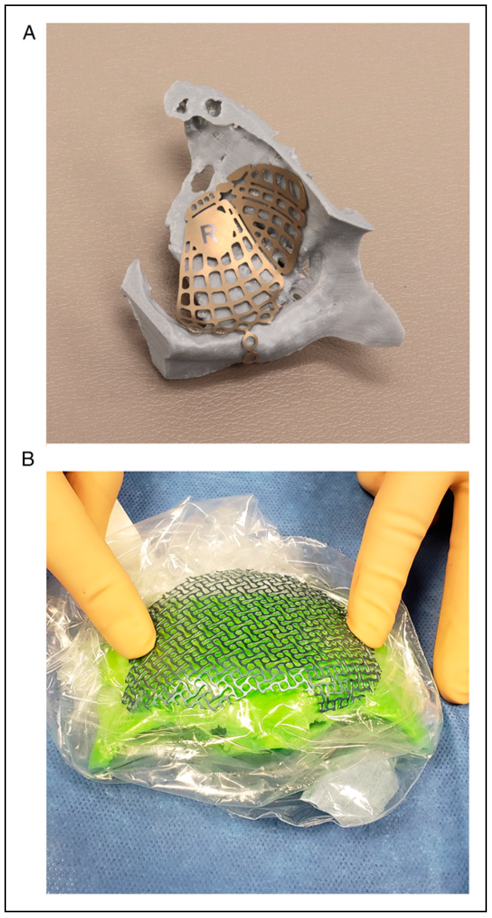
Implant Contouring and Sterilization
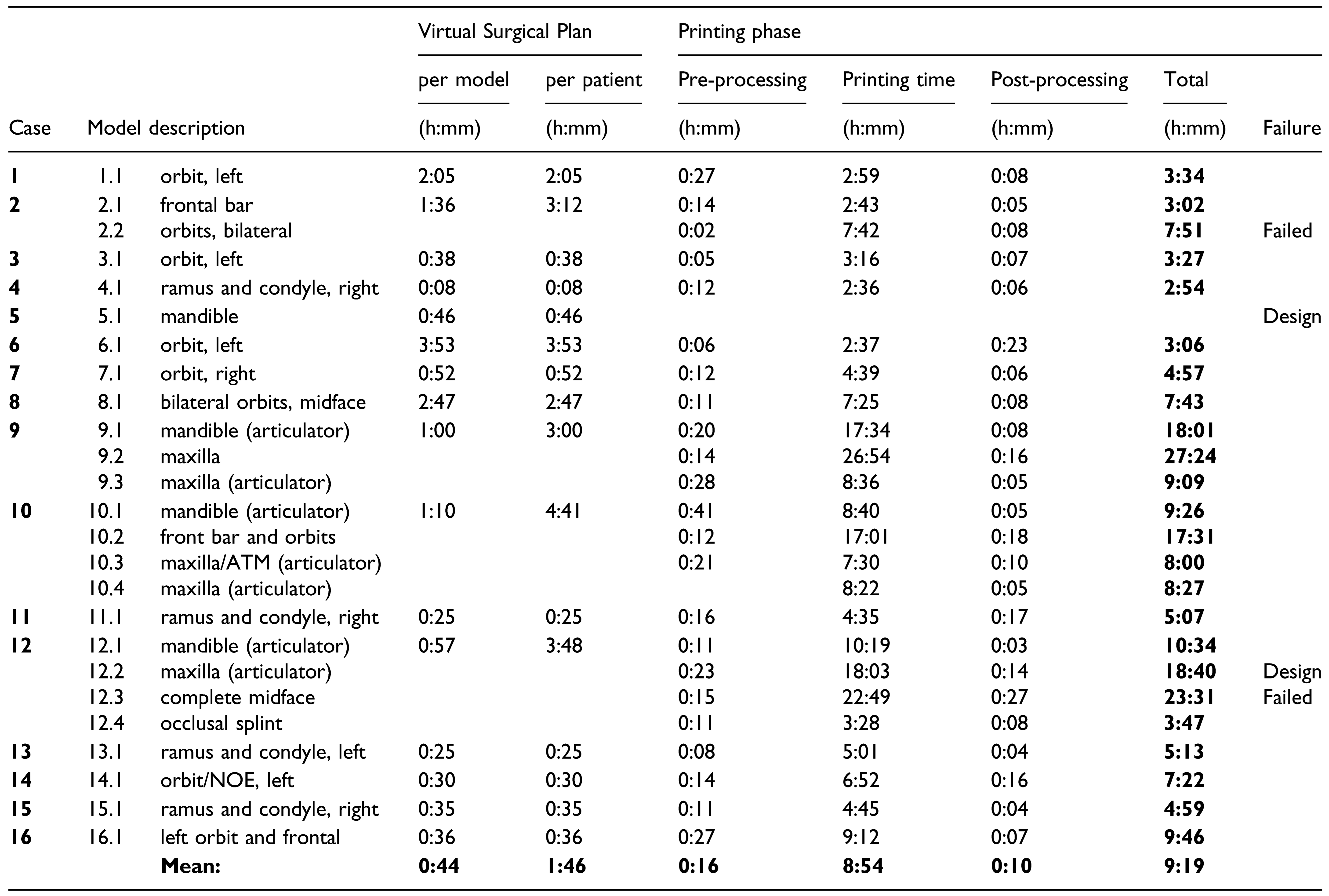
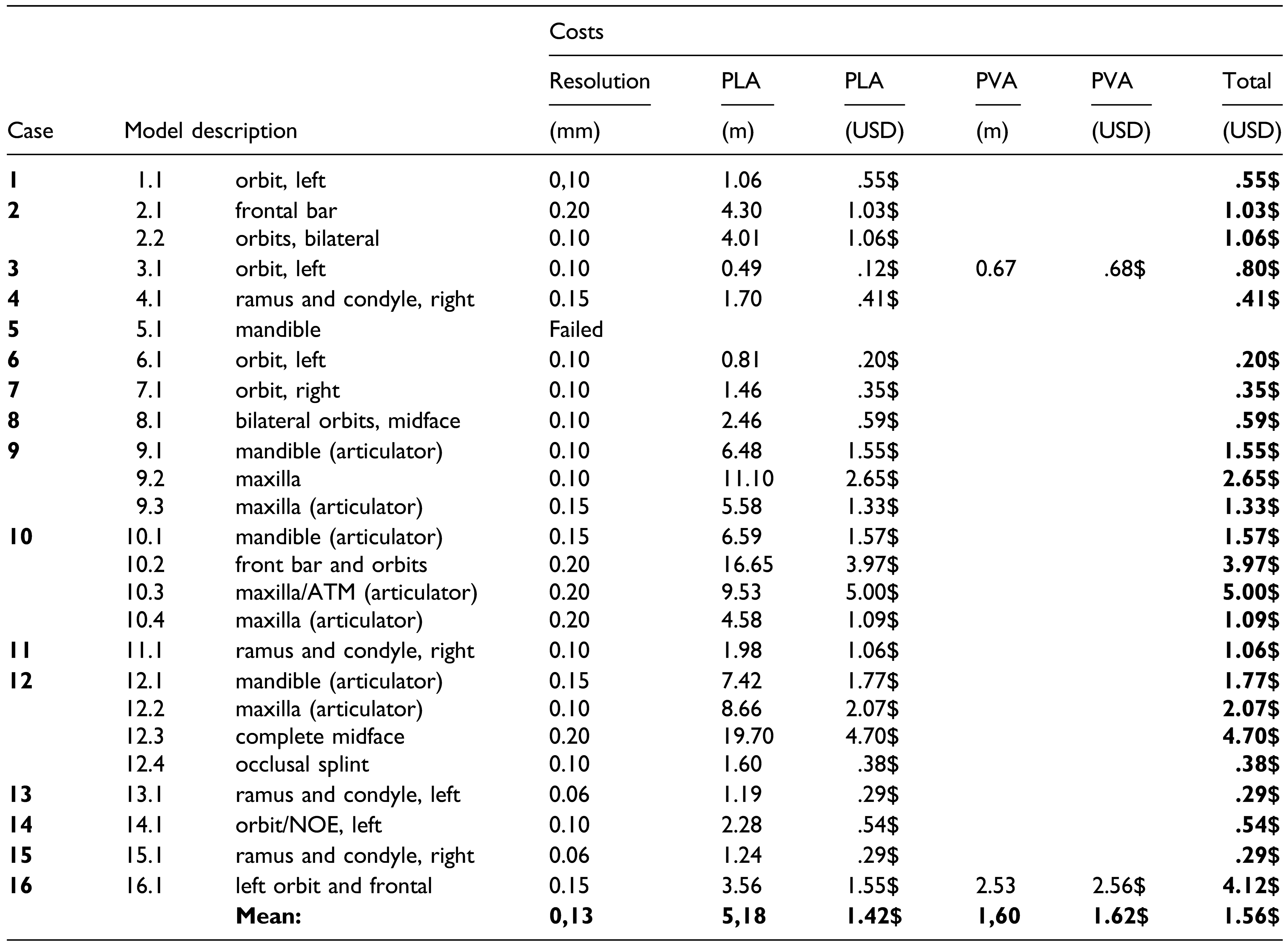
The 3D model is used to pre-bend plates or meshes before surgery, which are then sterilized. Current Canadian and American regulations prevent sterilization of in-house printed models; therefore, they are wrapped in a sterile plastic bag before being brought into the surgical field. Outsourced models, on the other hand, are usually approved for biocompatibility and sterilization, making it possible to bring them directly in the operating field. However, previously as mentioned, for acute trauma surgery specifically, it has been almost impossible to receive outsourced models in time for surgery.

Results

Twenty-five (25) consecutive models were printed for 16 consecutive patients presenting with acute facial fractures and requiring 3D models for treatment (Table 1). VSP per patient (Table 2) varied from 8min to 4h 41min (mean = 1h 46 min). The overall in-house printing phase time per model ranged from 2h 54min to 27h 24min (mean = 9h 19min). The overall success rate of prints was 84%, which includes two failed prints (8%) and two design issues (8%) noticed after printing completion.
PLA filament cost (Table 3) was between $0.12 and $5.00 per model (mean = $1.42). Dissolvable PVA supports were used for 2 models. The additional cost of PVA ranged from $0.68 to $2.58 (mean = $1.62), increasing the mean total cost of models to $1.56. In this research, all costs and reported dollar figures are in USD.

Discussion

When comparing outsourced to in-house 3D printed models, there are many aspects that must be taken into consideration: costs in equipment [3,6] printing material [6,7,8,9,10,11] and labor, case preparation time by the surgeon, elapsed time to manufacture the model [6,7,8,9,11,12] model accuracy [7,12] regulatory agency approval [3,6,13,14] surgeon’s level of expertise with 3D technologies, etc.
This paper focuses on a key issue of 3D printing for acute CMF trauma: the time to manufacture a model. Despite most aspects of 3D printing being perfected for elective reconstructions, models cannot currently be received in time for acute surgery. As a result, we elected to print the models in-house, therefore decreasing both production and shipping times. Additionally, this allowed for a better control on the printing process, possibly shortening it. For instance, the print quality and printing speed can be adjusted for completion before surgery. In this study, the print success rate was 84%, similar to the 86% reported by the printer manufacturer [15]. Of the two printer-related issues encountered in this study, one was due to filament shortage and the other was filament underextrusion from nozzle blockade. Of the design issues encountered, one was related to the pre-processing software refusing to open the model, and the other was that a printed maxilla with an integrated articulator support would not fit in the articulator because of a design error in its support.
Most importantly, printing models locally avoided shipping delays and custom clearance. We were not able to find a manufacturer that was able to ship directly to our hospital without transiting by a warehouse, adding several days of shipping delays.

Accuracy of the Model

All models printed in-house were found to be of adequate accuracy, an observation also made by other groups using in-house printing [7,9,11,12,16,17]. PLA filament printing was also found to be reliable [16] Printing issues with PLA, such as underextrusion and running out of filament, were easily detectable by simple visual inspection. Model shrinkage or warping was not observed in this study. All plates bent on a 3D model could be used without modification of shape once placed on the patient. There were no operative takebacks due to inadequate implant shape.

Printing Supply Cost

Printing supply costs to produce a “surgical grade” model was between $0.20 and $5.00 (mean = $1.56). Reports of in-house printing costs are scarce [6,11]. Comparison is often not possible because published figures do not show a human skull [18] appear to present an assembly of two materials when only one material is reported [18], and information on printing failure rates and costs of discarded material is not disclosed [11,13,18]. However, PLA printing supply costs of in-house cranial vaults by Naftulin was found to be in a similar price range [16], as well as orbital and mandibular molds by Gleissner [6].

Printer Cost

3D printer cost has decreased dramatically in the past decade [10], starting at hundreds of thousands of dollars for commercial printers down to $170 for recent hobbyist-grade resin printers (but inadequate for surgical purposes). Realistically, “prosumer” grade FFM and resin printers that are reliable and usable in a busy clinical setting currently cost between $5,000 and $10,000. Lower cost printers usually require manual assembly, require frequent manual calibration, might require custom part design and modifications, and have lower reliability, making them unsuitable for most surgeons. In addition to the printer cost, consumable parts such as printer nozzles and tubes have to be considered in the maintenance costs of the printer.

Printing Time

The printing phase time for in-house models (mean = 9h 19min) was very satisfying. Prints were all considered of adequate surgical grade quality (Figure 1) at resolutions ranging from .06 mm to .2 mm. Printing time with a filament printer depended on model size, desired accuracy and complexity, printing technology used, and resolution. It is important to note that there is a direct link between printing time, accuracy, and resolution—the higher the quality, the longer a model takes to print. In order to compare printing times, it is essential to take printing parameters into account, as well as including the pre-processing and post-processing time. This information is unfortunately often missing from published reports, precluding comparison with others. Details of printing parameters and materials used in this study could be used as a comparison point for further studies.

Printing Strategy to Decrease Time

There are printing strategies that decrease model production time. As a reference point, an in-house pilot study revealed that using an Ultimaker 3 printer, printing a complete adult skull and mandible in PLA and PVA supports can take 6 days and 7 hours at a .2 mm resolution, and 11 days 9 hours at a .1 mm resolution. This imparts excessive delays before surgery. With current printing technologies, printing only the region of interest improves speed significantly [16,19] a strategy we have used in this study. Also, we have found that printing resolutions ranging from .06 mm to .2 mm all provide satisfactory “surgical grade” models. Therefore, using the lower .2 mm resolution reduces the printing time as more plastic can be deposited with a single nozzle pass.
Emerging technologies such as resin monochromatic mSLA (masked stereolithography) printing are rapidly developing. The time required to print models with this technology is less affected by model size, amount of details, and number of simultaneously printed models, therefore making it a great candidate for rapid production of highly detailed craniofacial prints in the near future.
In this study, model print time did not contribute to the delays found between the trauma and surgery in any of our cases. Additional factor, including difficult access to the operating room in our healthcare system, unstable polytrauma patients in the ICU, patients requiring staged surgery with other trauma surgeons, and presence of excessive facial oedema contributed to surgical delays.

Labor Cost and Training

In-house printing also requires staff training and salary, which adds to the cost. Training for VSP is quite complex and takes time; therefore, working with industry clinical engineers who have access to advanced software is both productive and necessary. Simple VSP cases where only mirroring is required or where a single bone fragment needs to be reduced can be done locally. When more complex cases are undertaken, such as panfacial fractures or when surgical navigation is used [4,5], VSP is more efficient and can often only be done with industry engineers who send back 3D models electronically for in-house printing.
Training for the printing process is relatively easy, as many consumer guides exist online for widely available 3D printers. There is a learning curve for understanding how to import a VSP file into printing software and how to prepare the model for printing on a particular machine (printer calibration, model orientation, support placement, post-processing, etc.). At our institution, the process is done entirely by the surgeon and is not remunerated as such. The shortened surgical time [7] brought by using models can also decrease the surgical fees, which are often time-based, thus possibly contributing to a slower rate of adoption of this technology.
The alternative to in-house printing—industrial outsourced printing—also carries specific costs that must be taken into account, notably transaction costs [20,21]. Although not measured in this retrospective study, the transaction costs of outsourcing can be substantial. These costs refer to the expenses incurred to find a service provider, inquire about service costs and bargaining, the time required to write and sign a contract, as well as policing and enforcement of costs (quality control, delivery constraints, etc.) [22]. When the transaction costs of outsourcing are too high, such as those implied by a very short printing cycle and delivery requirements for acute trauma, the only economical alternative is to print in-house.

Regulatory Agency Approval

Another issue to consider in Canada and USA is regulatory agency approval for the usage of 3D printed models, which is quite complex. Currently, it does not seem possible to buy an off-the-shelf “biocompatible” and/or “sterilizable” filament to print models for surgery. Even though the models are not implanted, but are only used to bend material, they are considered as implantable material (and not surgical instruments) and as such must undergo complex biocompatibility testing similar to printed implantable devices. In this study, this lack of filament certification is why models were wrapped in sterile plastic bags when they were required in the surgical field.
One future development would be to directly print an implantable metal plate or mesh. Although there are some commercial offerings, implant designs and printing time need to improve to accommodate trauma surgery constraints.

Limitations

This article focuses on one main aspect of in-house printing: time to produce. Other aspects remain to be studied. They include an objective assessment of model accuracy printed with different printers and material, the steep learning curve required for VSP and printing associated to a significant level of expertise, the labor costs, the transactional costs, etc. These should be explored in the future now that the printing pipeline has been proven feasible for acute cases. As 3D software and 3D prosumer printers evolve to become more user-friendly and efficient, we expect a rapid improvement and uptake of the process, as has occurred in other areas of CMF surgery.

Conclusion

3D printing is used in all areas of CMF surgery including oncology, congenital, orthognathic, and secondary trauma reconstruction—but has been more challenging to use in the acute care setting because of the short lead time. Printing in-house circumvents shipping delays from the manufacturer. However, there are also other time-critical steps that need to be taken into account before undertaking a print: VSP time, pre-processing of models, printing, post-processing, and failure rate of prints. The process presented has allowed to reliably obtain models in time for surgery. The rapid improvements in the usability of 3D software and speed improvements in printing technology will likely contribute to further adoption of these technologies by CMF-trauma surgeons.

Author Contributions

Leonard Bergeron: (1) substantial contributions to conception and design, acquisition of data, and analysis and interpretation of data; (2) drafting and revision of the article; (3) final approval; and (4) agreement to be accountable for all aspects of the work. Michelle Bonapace-Potvin: (1) substantial contributions to conception and design, acquisition of data, and analysis and interpretation of data; (2) drafting and revision of the article; (3) final approval; and (4) agreement to be accountable for all aspects of the work. Francois Bergeron: (1) substantial contributions to analysis and interpretation of data; (2) drafting and revision of the article; (3) final approval; and (4) agreement to be accountable for all aspects of the work.

Funding

The author(s) received no financial support for the research, authorship, and/or publication of this article.

Ethics

The study was conducted in compliance with the principles of the Declaration of Helsinki and the institutional review board. Final board approval was granted on January 7th, 2021.

Conflicts of Interest

The author(s) declared no potential conflicts of interest with respect to the research, authorship, and/or publication of this article.

References

  1. Hollier, L.H., Jr. The confluence of technique and technology in craniofacial surgery. Plast. Reconstr. Surg. 2021, 147, 1027–1028. [Google Scholar] [CrossRef] [PubMed]
  2. Bergeron, L.; Bonapace-Potvin, M.; Bergeron, F. In-house 3D model printing for acute Cranio-maxillo-facial trauma surgery: process, time, and costs. Plast. Reconstr. Surg. Glob. Open 2021, 9, e3804. [Google Scholar] [CrossRef]
  3. Marschall, J.S.; Dutra, V.; Flint, R.L.; et al. In-house digital workflow for the management of acute mandible fractures. J. Oral. Maxillofac. Surg. 2019, 77, 2084.e1–2084.e9. [Google Scholar] [CrossRef]
  4. Bergeron, L.; Bouchard, S.; Bonapace-Potvin, M.; Bergeron, F. Intraoperative surgical navigation reduces the surgical time required to treat acute major facial fractures. Plast. Reconstr. Surg. 2019, 144, 923–931. [Google Scholar] [CrossRef] [PubMed]
  5. Bergeron, L.; Bouchard, S.; Bonapace-Potvin, M.; Bergeron, F. Response to Drs. Chen and Bradley’s recent letter to the editor concerning our paper on the use of navigation in the treatment of major facial fractures. Plast. Reconstr. Surg. 2020, 146, 509e–510e. [Google Scholar] [CrossRef]
  6. Gleissner, H.; Castrillon-Oberndorfer, G.; Gehrlich, S.T. Introduction of 3D printing in a german municipal hospital - practical guide for CMF surgery. Craniomaxillofacial Trauma. Reconstr. 2021. [Google Scholar] [CrossRef]
  7. Lim, C.G.; Campbell, D.I.; Clucas, D.M. Rapid prototyping technology in orbital floor reconstruction: application in three patients. Craniomaxillofacial Trauma. Reconstr. 2014, 7, 143–146. [Google Scholar] [CrossRef]
  8. Jacobs, C.A.; Lin, A.Y. A new classification of three-dimensional printing technologies: systematic review of three-dimensional printing for patient-specific craniomaxillofacial surgery. Plast. Reconstr. Surg. 2017, 139, 1211–1220. [Google Scholar] [CrossRef]
  9. He, W.; Sun, Y.; Tian, K.; Xie, X.; Wang, X.; Li, Z. Novel arch bar fabricated with a computer-aided design and three-dimensional printing: a feasibility study. J. Oral. Maxillofac. Surg. 2015, 73, 2162–2168. [Google Scholar] [CrossRef]
  10. Chae, M.P.; Rozen, W.M.; McMenamin, P.G.; Findlay, M.W.; Spychal, R.T.; Hunter-Smith, D.J. Emerging applications of bedside 3D printing in plastic surgery. Front. Surg. 2015, 2, 25. [Google Scholar] [CrossRef]
  11. Dvoracek, L.A.; Lee, J.Y.; Unadkat, J.V.; et al. Low-cost, three-dimensionally-printed, anatomical models for optimization of orbital wall reconstruction. Plast. Reconstr. Surg. 2021, 147, 162–166. [Google Scholar] [CrossRef]
  12. Rogers-Vizena, C.R.; Weinstock, P.; Livingston, K.; Prabhu, S.P. Letter to the editor concerning the article “The current role of three-dimensional printing in plastic surgery” by Kamali, P. et al. Plast. Reconstr. Surg. 2017, 139, 811e–812e. [Google Scholar] [CrossRef]
  13. Kamali, P.; Dean, D.; Skoracki, R.; et al. The current role of three-dimensional printing in plastic surgery. Plast. Reconstr. Surg. 2016, 137, 1045–1055. [Google Scholar] [CrossRef] [PubMed]
  14. Goodson, A.M.C.; Parmar, S.; Ganesh, S.; et al. Printed titanium implants in UK craniomaxillofacial surgery. Part I: access to digital planning and perceived scope for use in common procedures. Br. J. Oral. Maxillofac. Surg. 2021, 59, 312–319. [Google Scholar] [CrossRef]
  15. Lozova, L. Bringing the Ultimaker 3 to Market with Bridge Manufacturing, 2016 ed.; Ultimaker: Utrecht, The Netherlands.
  16. Naftulin, J.S.; Kimchi, E.Y.; Cash, S.S. Streamlined, Inexpensive 3D Printing of the Brain and Skull. PLoS ONE 2015, 10, e0136198. [Google Scholar] [CrossRef]
  17. Longcac, M.; Depeyre, A.; Pereira, B.; Barthelemy, I.; Pham Dang, N. Virtual surgical planning and three-dimensional printing for the treatment of comminuted zygomaticomaxillary complex fracture. J. Stomatol. Oral Maxillofac. Surg. 2021, 122, 386–390. [Google Scholar] [CrossRef]
  18. Gerstle, T.L.; Ibrahim, A.M.; Kim, P.S.; Lee, B.T.; Lin, S.J. A plastic surgery application in evolution: three-dimensional printing. Plast. Reconstr. Surg. 2014, 133, 446–451. [Google Scholar] [CrossRef] [PubMed]
  19. Msallem, B.; Beiglboeck, F.; Honigmann, P.; Jaquiery, C.; Thieringer, F. Craniofacial reconstruction by a cost-efficient template-based process using 3D printing. Plast. Reconstr. Surg. Glob. Open 2017, 5, e1582. [Google Scholar] [CrossRef]
  20. Coase, R. The problem of social cost. J. Law. Econ. 1960, 3, 1–44. [Google Scholar] [CrossRef]
  21. Williamson, O.E. The theory of the firm as governance structure: from choice to contract. J. Econ. Perspect. 2002, 16, 171–195. [Google Scholar] [CrossRef]
  22. Dahlman, C. The problem of externality. J. Law. Econ. 1979, 22, 141–162. [Google Scholar] [CrossRef]
Figure 1. 3D printed models for acute trauma surgery. A. A patient-specific reconstructed right orbit, obtained through virtual-surgical planning, is used to custom-bend an orbital implant before surgery. B. A patient-specific forehead model can be used intra-operatively to shape a mesh for forehead reconstruction.
Cmtr 16 00017 g001
Figure 2. In-house 3D printing. Virtual Surgical Planning is done in-house for simple cases. More difficult cases are done over the internet with an industrial partner, as well as when surgical navigation is done. VSP files are sent and printed locally. Shipping delays are avoided and printing problems can be readily corrected. Plates are pre-bent on the model and sterilized. In-house prints are usually not regulatory agency-approved for CMF surgery and cannot be processed for sterilization. Models are therefore wrapped in a sterile bag if they are required in the operative field.
Cmtr 16 00017 g002
Figure 3. Outsourcing of 3D model printing. Virtual Surgical Planning is done over the internet with a commercial supplier. Printing is done on industrial printers and shipped to a local warehouse where they are cleared by customs and quality checked by the manufacturer. The model is then reshipped to the hospital. Plates are then bent pre-operatively and sterilized. Commercially printed parts (regulatory agency-approved) are sterilized and brought into the operating field. Because of the delays brought by remote printing and shipping, this process can almost exclusively used for secondary trauma cases at this time.
Cmtr 16 00017 g003
Table 1. Diagnosis and model description. Twenty-five models were printed for 16 acute facial fracture cases (mean: 1.6 model per patient).
Cmtr 16 00017 i001
Table 2. Virtual Surgical Planning (VSP) and Printing phase time. The mean VSP time per model was 44min and 1h 46min per patient. In the printing phase, the mean pre-processing time (preparing the model for printing) was 16min, the mean printing time (work done by the printer) was 8h 54min, and the mean post-processing time (support removal and model cleanup) was 10min. The overall printing phase time averaged 9h 19min. The printing failure rate was 8%, and design issues occurred in 8% of cases, with an overall 84% model-printing success rate.
Cmtr 16 00017 i002
Table 3. Printing supply costs. The mean cost per model printed in PLA is 1.56$. PLA: poly-lactic acid filament for printing. PVA: polyvinyl alcohol water-soluble supports.
Cmtr 16 00017 i003

