Next Article in Journal
Single Institute Audit of Maxillofacial Trauma Cases Before and During COVID-19 Pandemic
Previous Article in Journal
Treatment of Persistent Post-Traumatic Diplopia—An Algorithmic Approach to Patient Stratification and Operative Management
 
 
Craniomaxillofacial Trauma & Reconstruction is published by MDPI from Volume 18 Issue 1 (2025). Previous articles were published by another publisher in Open Access under a CC-BY (or CC-BY-NC-ND) licence, and they are hosted by MDPI on mdpi.com as a courtesy and upon agreement with Sage.
Article

Comparison of Efficacy and Safety of Hybrid Arch Bar with Erich Arch Bar in the Management of Mandibular Fractures: A Randomized Clinical Trial

by
Hariram Sankar
,
Sachin Rai
*,
Satnam S. Jolly
and
Vidya Rattan
Unit of Oral and Maxillofacial Surgery, Oral Health Sciences Centre, Postgraduate Institute of Medical Education and Research, Sector 12, Chandigarh 160012, India
*
Author to whom correspondence should be addressed.
Craniomaxillofac. Trauma Reconstr. 2023, 16(2), 94-101; https://doi.org/10.1177/19433875221080019
Submission received: 1 November 2021 / Revised: 1 December 2021 / Accepted: 1 January 2022 / Published: 29 March 2022

Abstract

:
Study Design: A clinical randomized control trial. Objective: To compare the efficacy and safety of Hybrid arch bar (HAB) with Erich arch bar (EAB) in fracture management of the mandible. Methods: In this randomized clinical trial, 44 patients were divided into 2 groups:- Group 1, N = 23 (EAB group) and Group 2, N = 21 (HAB group). The primary outcome was time taken for the application of arch bar, while the inner and outer glove puncture, operator prick, oral hygiene, arch bar stability, complications of HAB, and cost comparison were secondary outcomes. Results: The time taken for the application of arch bar in group 2 was significantly shorter than group 1 (55.66 ± 17.869 min vs 82.04 ± 12.197 min) and the frequency of outer glove puncture was also significantly lesser for group 2 (0 punctures vs 9 punctures). Better oral hygiene was found in group 2. EAB was cost-effective than HAB (Rs 700 ± 239.79 vs Rs 1742.50 ± 257.14). The stability of the arch bar was comparable in both groups. Group 2 had associated complications of root injury in 2 out of 252 screws placed and the screw head got covered by soft tissue in 137 out of 252 screws placed. Conclusions: Thus, HAB was better than EAB with a shorter time of application, less risk of prick injury, and improved oral hygiene. Clinical trial registry name- clinical trials registry- India, URL-http://ctrl.nic.in, registration number- CTRI/2020/06/025966.

Introduction

Maxillomandibular fixation (MMF) is a commonly used method for the reduction of jaw fractures. The three main principles of MMF are to establish occlusion, provide stability, and immobilize the jaws [1]. Whether the management of jaw fracture is through open or closed reduction the application of Erich arch bar (EAB) for MMF has been the superior technique of fixation of jaws so far. Some significant disadvantages associated with EAB are increased time taken for its application and removal, wire prick injury, and glove puncture. Other underreported or unreported occurrences are the injuries related to periodontium and poor oral hygiene status [2]. MMF screws were introduced to overcome these limitations, but the reinforcement provided by these screws is significantly less than EAB and also they do not exert tension band effect [3]. They are primarily used for short-term MMF or minimally displaced fractures. Many other alternatives for EAB like Ivy loops, Leonard buttons, and plastic anchorage points are also in use. However, the versatility of EABs for both long and short-term MMF is generally undisputed because of its simplicity in use and long history of successful treatment.
Recently introduced Hybrid arch bar (HAB) is a modification of conventional EAB which is fixed to the bone with the help of screws. The screws are placed in the interradicular area below the level of marginal gingival tissue in the eyelet that extends from the arch bar without affecting the periodontium [2] One possible concern is that it may cause root injury while drilling for screws in the interradicular area. There are only few studies that have compared the efficacy of HAB with EAB. The purpose of the study was to compare the efficacy and safety of HAB with EAB in the management of jaw fractures and also evaluate various complications associated with the HAB.

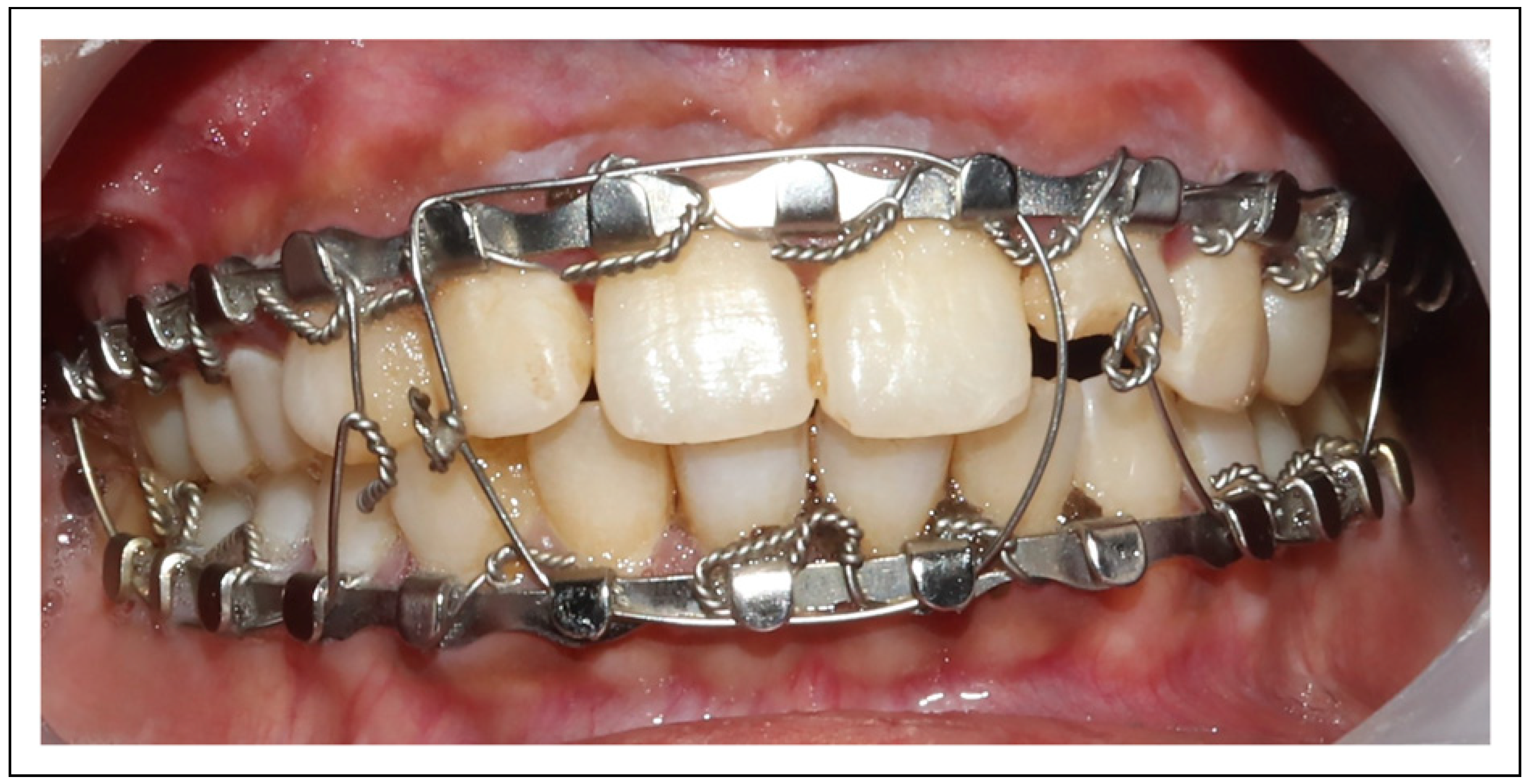
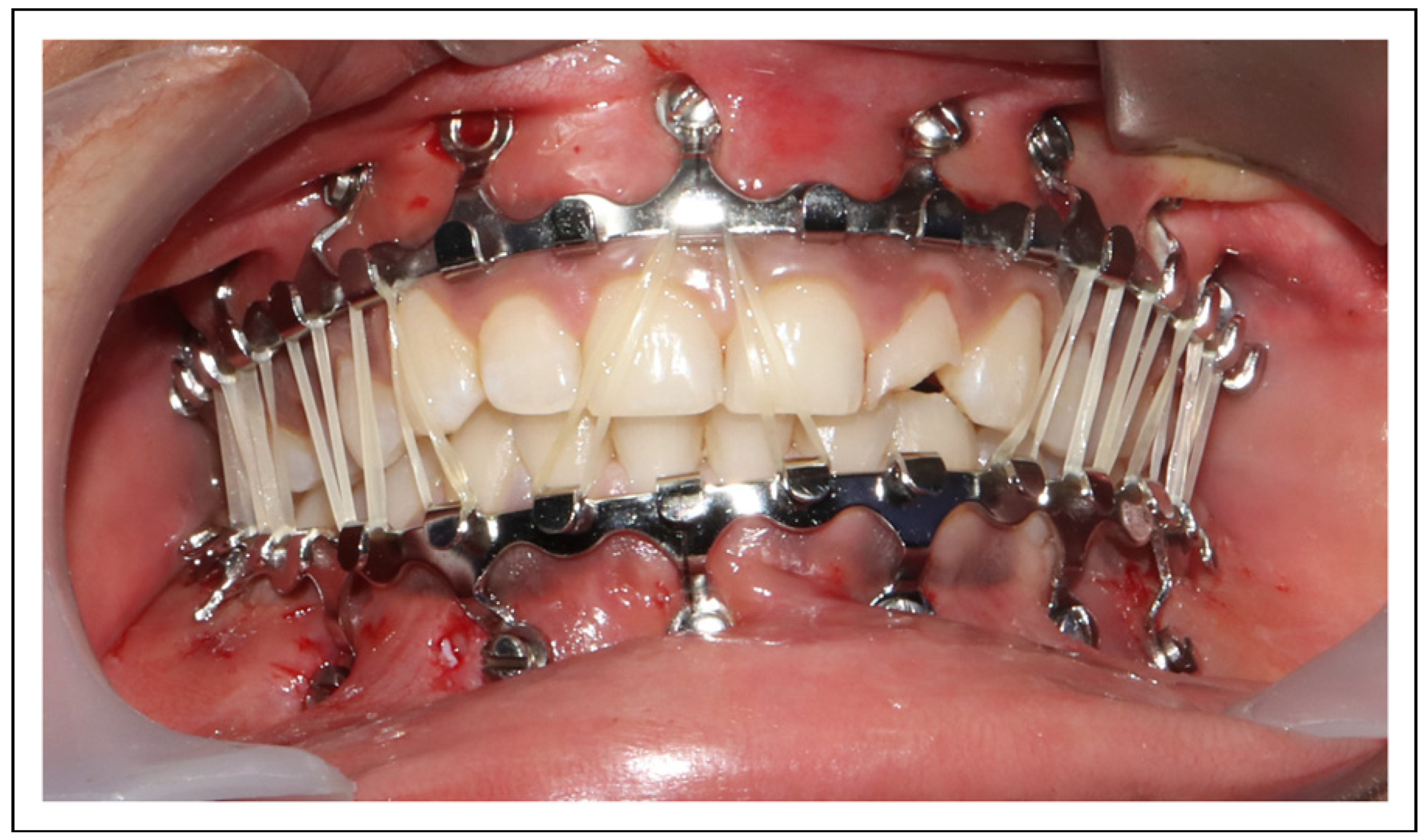
Materials and Method

This study was a prospective randomized clinical trial and was registered under clinical trial registry number of India (CTRI/2020/06/025966). The study population consisted of patients with mandible fractures treated between June 2020 and December 2020 in our institution. The present study was approved by the Institutional Ethics Committee (Approval number- INT/IEC/2019/002713). Patients were informed about the procedure, possible complications, and the materials used, and they signed a detailed written consent form. Criteria of inclusion were patients requiring reduction and immobilization of mandible fractures with the arch bar in place for a minimum of 4 weeks, patients with age between 18 and 45 years, patients with a minimum of 20 teeth, and a minimally displaced isolated single site fracture of the mandible without communition. The patients were excluded if they had less than 20 periodontally healthy teeth, Glassgow Coma Scale less than 8, pregnant females, and condylar fractures to avoid long-term immobilization.
A total of 50 patients were recruited. The selected subjects were allocated to 2 treatment groups, 25 each using computer-generated randomization. Six patients were lost to follow-up, 2 in EAB group and 4 in HAB group (Figure 1). The sequentially numbered opaque sealed envelope method was used for allocation concealment. In group 1, EAB was fixed with 26 gauge stainless steel wire (S.K.Surgicals, Pune, Maharastra, India) by a single operator (Figure 2). In group 2, HAB (S.K. Surgicals, Pune, Maharashtra, India) was placed by the same operator (Figure 3). A minimum of 6 screws were placed in each arch after assessing the root position with OPG/CBCT. The Hybrid arch bar used in our study was made of stainless steel. It was a regular non-customized type. It consists of a regular arch bar from which the arms extending to which holes for the screws were present. The HAB system was presented with 2×6 mm, 2×8 mm, and 2×10 mm screws. These were non-self-drilling screws. The arm extensions can be adjusted with the pliers according to the root position.
The primary outcome of the study was the time taken (in minutes) for the application of the arch bar. The inner and outer glove puncture, operator prick, oral hygiene, arch bar stability, complications of HAB, and cost comparison were the secondary outcomes. The glove penetration and needle stick injury to the operator during the application of the arch bar was recorded. Double gloves were used while applying the arch bars. After application of the arch bar, both the outer and inner gloves were removed and the inner glove was marked with a pen. Both the gloves were filled with tap water and compressed to check for any puncture as for the water inflation method [4]. The oral health of the patient was evaluated with a modified OHI(S) index [5] which had 3 components: debris index, bleeding index, and gingival enlargement index. In HAB, the soft tissue growth over the screw head was recorded with the screw head soft tissue coverage index. All the patients were subjected to professional cleaning of teeth before placement of the arch bars. The debris component score was as follows:
  • No debris present: 0
  • Debris present but not covering the arch bar: 1
  • Debris covering not more than half the arch bar: 2
  • Debris covering more than half of the arch bar: 3
The gingival bleeding component score and criteria were as follows;
  • No bleeding on provocation: 0
  • Light bleeding on provocation: 1
  • Moderate bleeding on provocation: 2
  • Spontaneous bleeding: 3
The gingival enlargement score and criteria were as follows;
  • No gingival enlargement: 0
  • Enlargement present but not covering the arch bar: 1
  • Enlargement covering the arch bar: 2
All these components were evaluated for the index teeth 11,14, 26, 31, 34, and 46 and were evaluated preoperatively and once weekly for 4 weeks. The scores at all these teeth regions were summated to obtain a total score. The total score was divided by the number of teeth evaluated (6) to get an oral hygiene score. The bleeding component was assessed with the help of William’s periodontal probe. The probe was made to run along the gingival margin with mild force. Grade 0 when there was no bleeding on provocation. Grade 1 when isolated bleeding spots was present (mild) on provocation. Grade 2 when there was a confluent red line along the sulcus (moderate) on provocation. Grade 3 when bleeding is spontaneous.
The screw head soft tissue coverage index score and criteria were as follows
  • No soft tissue covering the screw head: 0
  • Soft tissue covering not more than half the screw head: 1
  • Soft tissue covering more than half the screw head: 2
  • Soft tissue completely covering the screw head: 3
This index was recorded for each screw in the upper and lower arch. The total score was divided by the number of screws (12) evaluated to obtain a screw head soft tissue coverage score.
Arch bar stability was checked every week applying force with probe using following criteria,
  • Non-perceptible (no mobility): 0
  • Mild (mobility less than 1 mm/loose hardware that can be tightened): 1
  • Perceptible (mobility more than 1 mm/missing hardware or those hardware requiring removal): 2
If any screw was found loose, it was removed. Any loose wires were tightened with help of a wire twister. Root injury as a result of screw placement was evaluated every week by percussing the teeth for tenderness. If any tooth was found to be symptomatic, then cone beam computed tomography (CBCT) was taken to confirm the relation of the screw to the root.

Statistical Analysis
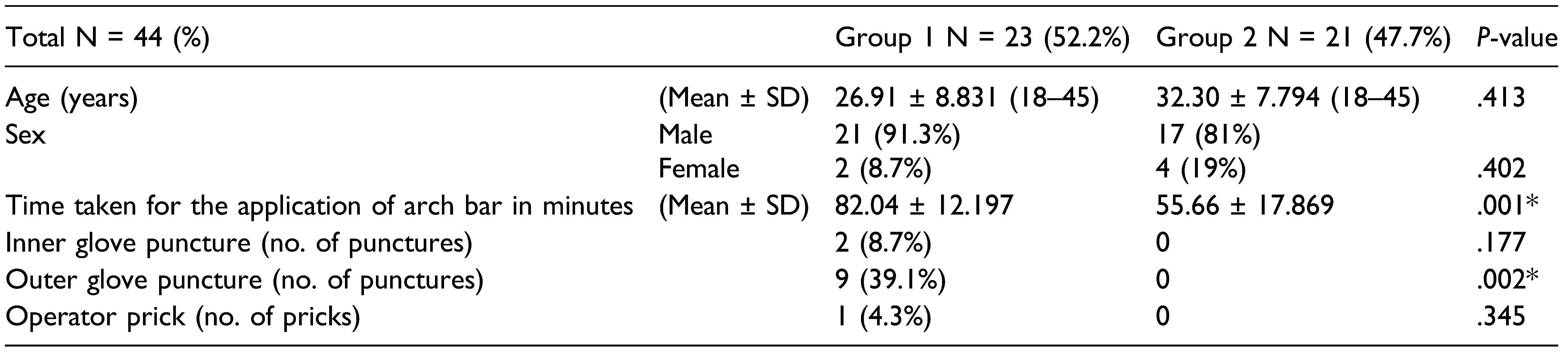
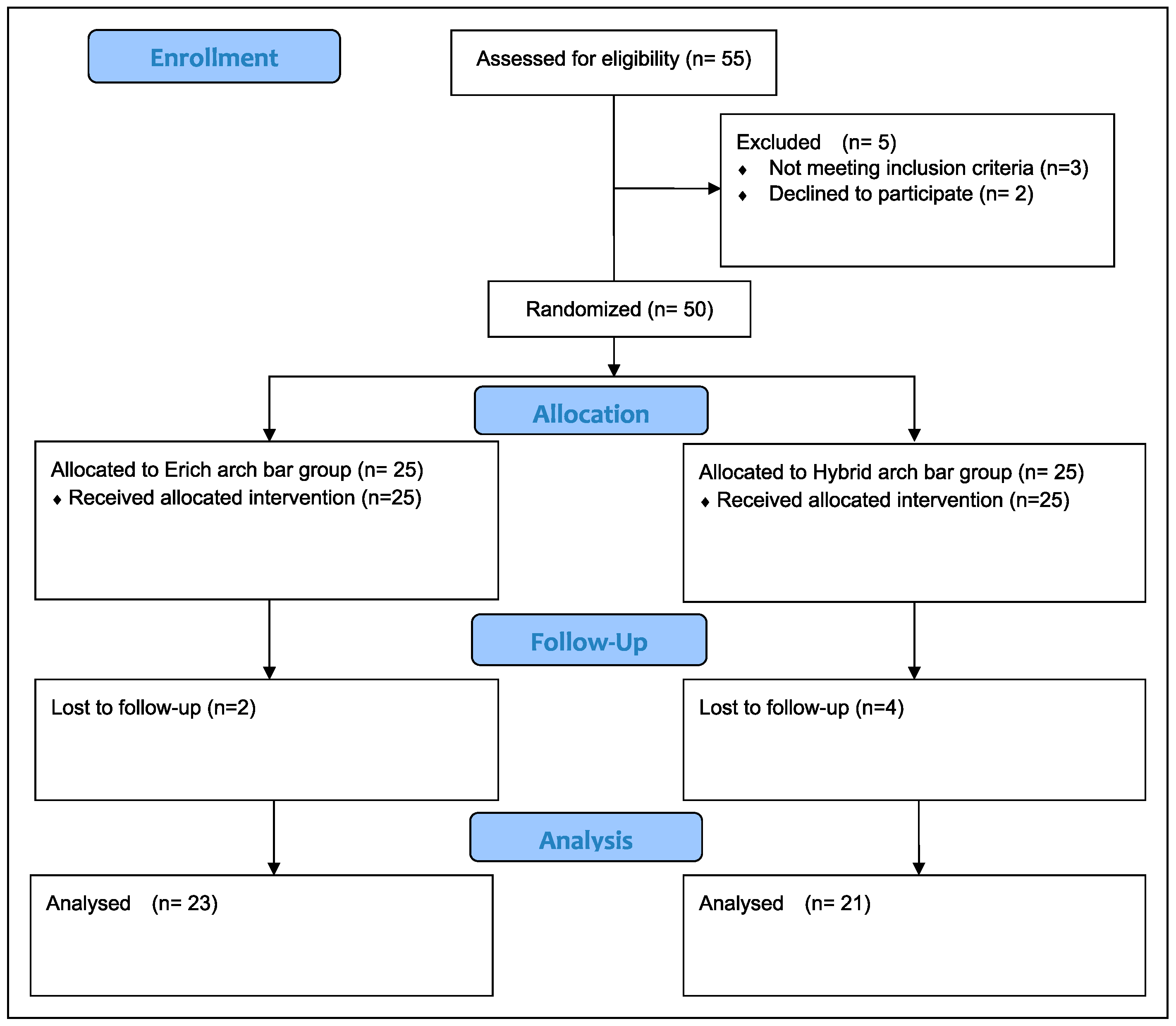
Descriptive and inferential statistical analysis was carried out by using SPSS version 18 software (IBM Corporation, SPSS Inc., Chicago, IL, USA). Results on continuous measurements were presented as Mean ± SD (min–max) and results on categorical measurements were presented in frequency (percentage). The normality of the data was assessed using the Shapiro–Wilk test. Inferential statistics like the chi-square test/ Fischer exact test and independent t-test were used to check the differences between the groups. Paired t-test was used to check difference within a group over a period of time. P-value less than .05 was considered to be significant.

Results

The per-protocol analysis was used in the present study and a total of 44 patients were evaluated at 4 weeks follow-up. Group 1 (EAB) had 23 patients and group 2 (HAB) had 21 patients. There were 21 males and 2 females in group 1 and 17 males and 4 females in group 2 (Figure 3). There were a total of 12 symphysis fractures, 18 angle fractures, and 14 body fractures of the mandible. The mean age (P = .413) and gender (P = .402) were not significantly different between the groups. All the fractures healed satisfactorily in both groups.

Time Taken for the Application of Arch Bar

The mean arch bar application time in group 1 was 82.04 ± 12.19 min and group 2 was 56.66 ± 17.86 min (Table 1). The time of application was significantly reduced in group 2 (P = .001).

Operator Prick and Glove Puncture
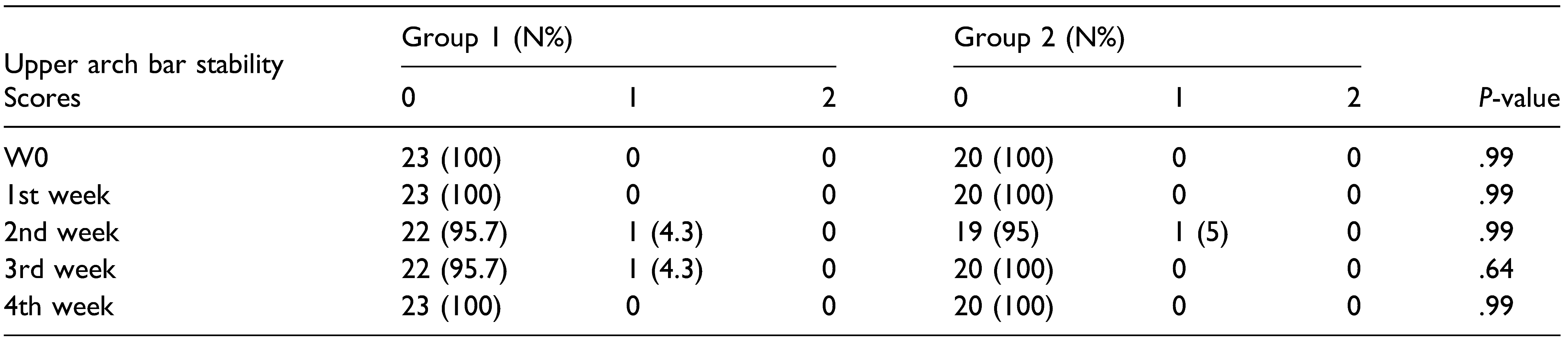
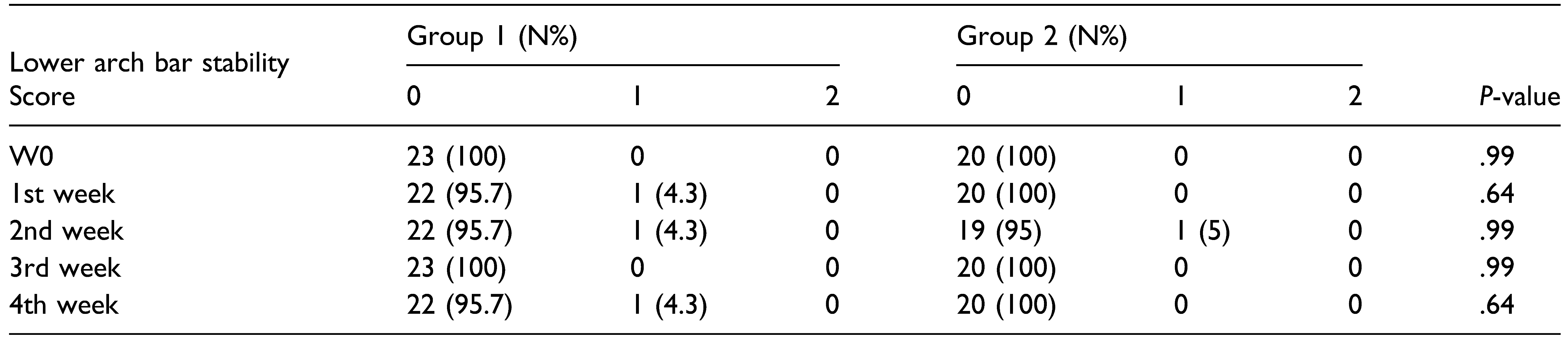
In group 1, there were 9 outer glove punctures, 2 inner glove punctures, and 1 operator prick. There were no glove puncture and operator prick in group 2 (Table 1). There was a statistically significant difference in terms of outer glove puncture between group 1 and group 2 (P = .002).

Oral Hygiene

Oral hygiene was better in group 2 than in group 1. OHI(S) index was significantly higher in group 1 than group 2 from the 2nd to the 4th week; meanwhile, OHI(S) during the 1st week was comparable between the groups (Figure 4).
Arch Bar Stability
The stability of the upper and lower arch bar was comparable between both the groups during this period of 4 weeks. One loose screw was present in group 2 and its removal did not affect the stability of the arch bar. Two loose circumdental wires were found in group 1. Both the groups showed good stability (Table 2 and Table 3).
Complications of Hybrid Arch Bar
Totally 252 screws were placed in the 21 patients in group 2. Tenderness of adjacent teeth on percussion was found in 3 screws (1.25%). Among these, root injury was confirmed with CBCT in 2 screws (.83%). An infected screw was found (.416%). There were 137 (54.36%) buried screws during the 4th week. The mucosal covering kept on increasing significantly from the 1st to the 4th week.
Cost of Arch Bar
The mean cost for EAB and stainless steel wire was Rs 700 ± 239.79 and that for HAB and screws was Rs 1742.50 ± 257.14.

Discussion

Erich arch bar, MMF screws, and direct wiring techniques, etc., are routinely used for MMF in closed reduction or prior to open reduction. Even though other wiring techniques like bridle wiring, MMF screws can be used in the management of few cases successfully, and they do not provide complete bracing effect like arch bars and often leads to posterior open bite and mild occlusal discrepancies. EAB is the gold standard for jaw immobilization due to the unparalleled reinforcement provided [6], but it has some limitations. It takes a longer time to pass wire around each tooth with an increased risk for prick injury. At the same time, HAB has a high risk root injury and it is expensive. In our study, we tried to compare standard EAB and HAB in various aspects.
All the arch bars were applied by a single operator. The mean difference in time of application of arch bar between the 2 groups was found to be around 26 mins, with HAB having the shorter time period. This was similar to the mean time difference of 20–25 min by other authors [2,3,7].
The outer and inner glove puncture was verified using the water inflation test and a self-reported operator prick was noted. There was no incidence of glove puncture or operator prick in the HAB group. In EAB group, there were 9 outer glove punctures (5 single puncture sites; 2 double puncture sites), 2 inner glove punctures, and 1 operator prick injury. Thus, there was a significantly higher outer glove puncture in the EAB group than that of HAB. In a similar study by King et al., it was found that there were significantly more glove tears or penetrations during application for the EAB group (.56 ± .91 per application) compared to the HAB group (.11 ± .32 per application) [2]. In a study conducted by Pieper et al. [8], it was found that glove perforation after double gloving in the EAB group was about 37%. In our study, the outer glove puncture was found to be 39.1%.
During the 1st week evaluation, oral hygiene status between 2 groups was found to be comparable. The oral hygiene status of HAB was found to be better statistically than EAB during the 2nd, 3rd, and 4th week follow-up. The circumdental wires in the EAB cause soft debris accumulation and were the cause of poor oral hygiene in the EAB group. Also, the removal of extension arms of HAB where screws were not placed and restriction of inferior extension of HAB not beyond the mucogingival junction provide adequate space for placement of toothbrush in the vestibule and thus helps to maintain good oral hygiene.
The stability of the arch bar in both groups was found comparable throughout all 4 weeks. A study conducted by king et al. showed that there was no loose hardware in both the HAB and EAB groups at the time of removal [2]. Quereshi et al. compared the stability of MMF between EAB and MMF screws and found that both were comparable in terms of stability [4].
Root injury and buried screws were found in the HAB group. There are only limited studies which have evaluated root injury after HAB placement. In our study, tenderness on percussion was found in 3 incisor teeth adjacent to screws out of 252 screws placed and among these 2 teeth were confirmed with CBCT for root injury. Mandibular incisor roots were injured and none of them required any intervention. Our study found that the root injury was commonly seen in the mandibular incisor area since their roots are closely placed. Therefore, this area may be avoided for screw placement to mitigate the risk of root injury. The two teeth with root injury became asymptomatic after 2 weeks. These findings were comparable with other studies conducted to evaluate the root injury in MMF screws [9]. In a systematic review conducted by Alves et al. [10] for the root injury following MMF screw placement, it was found that among 597 patients treated with 3647 IMF screws, tooth damage was among 88 patients and 18 of them were non-vital, and only 3 required extraction of the tooth. In a study conducted by Fabbroni et al. [11] among 232 MMF screws placed, 26 (11.2%) teeth had major contact and 37 (15.9) had minor contact with less than 50% of the root surface. Among these, only a single tooth required intervention with root canal treatment [12,13,14,15,16,17,18,19].
In our study, buried screw heads were found among all the 21 patients of HAB. Over 137 out of 252 screw heads were covered with soft tissue at the end of the 4th week. The amount of mucosa covering the screw head kept on increasing from 1st to 4th week. These patients required uncovering of the oral mucosa at the time of arch bar removal. But none of them reported with discomfort post screw removal. These findings in our study were supported by the study conducted by Rai et al. [6] in which among 240 patients with MMF screws mucosal covering has increased from 1st to 4th week.
The cost of EAB and HAB was compared and was found that the EAB was cheaper than the HAB (INR 700 ± 239.79 vs INR 1742.50 ± 257.14). King et al. found that the HAB (1980 USD) was expensive than the EAB together with stainless steel wire (100 USD) [2]. The factor discussed by Khelemsky et al. in their study was the reduction of operating room fees with the usage of HAB since the operating room fees decrease with shorter operating time. The mean operating time was significantly longer for the EAB than that of the HAB, thereby subsequent increase in cost [20]. But in our government-funded teaching institution, there were no charges for operating room and all these cases were done under local anesthesia and hence cost analysis based on operator room fee was not performed, which is one of our limitation.
One of the limitations of our study was using CBCT for assessing root injury only in symptomatic patients and tenderness on percussion that we used for evaluating root injury in asymptomatic patients is not as reliable as CBCT. Other limitation of our study was small sample size. Thus, future further studies with large sample size and usage of CBCT for assessing root injury in all the patients with HAB should be performed.

Conclusion

Hybrid arch bar had a shorter time of arch bar application, lesser risk of glove perforation, and improved oral hygiene when compared with standard EAB. The stability of HAB was comparable with that of EAB. Soft tissue mucosal covering did not affect the gingival health of the patients and healed spontaneously after screw removal. The HAB was costlier than EAB.

Funding

The author(s) received no financial support for the research, authorship, and/or publication of this article.

Declaration of Conflicting Interests

The author(s) declared no potential conflicts of interest with respect to the research, authorship, and/or publication of this article.

References

  1. Kendrick, D.E.; Park, C.M.; Fa, J.M.; Barber, J.S.; Indresano, A.T. Stryker SMARTLock hybrid maxillomandibular fixation system: Clinical application, complications, and radiographic findings. Plast. Reconstr. Surg. 2016, 137, 142e–150e. [Google Scholar] [CrossRef] [PubMed]
  2. King, B.J.; Christensen, B.J. Hybrid arch bars reduce placement time and glove perforations compared with Erich arch bars during the application of intermaxillary fixation: A randomized controlled trial. J. Am. Associat. Oral Maxillofac. Surg. 2019, 77, e81228. [Google Scholar] [CrossRef] [PubMed]
  3. Chao, A.H.; Hulsen, J. Bone-supported arch bars are associated with comparable outcomes to Erich arch bars in the treatment of mandibular fractures with intermaxillary fixation. J. Oral Maxillofac. Surg. 2015, 73, 306–313. [Google Scholar] [CrossRef] [PubMed]
  4. Qureshi, A.; Reddy, U.; Warad, N.; Badal, S.; Jamadar, A.; Qurishi, N. Intermaxillary fixation screws versus Erich arch bars in mandibular fractures: A comparative study and review of literature. Ann. Maxillofac. Surg. 2016, 6, 25–30. [Google Scholar] [CrossRef] [PubMed]
  5. Kumar, P.; Menon, G.; Rattan, V. Erich arch bar versus hanger plate technique for intermaxillary fixation in fracture mandible: A prospective comparative study. Natl. J. Maxillofac. Surg. 2018, 9, 33–38. [Google Scholar] [CrossRef] [PubMed]
  6. Rai, A.; Datarkar, A.; Borle, R.M. Are maxillomandibular fixation screws a better option than Erich arch bars in achieving maxillomandibular fixation? A randomized clinical study. J. Oral Maxillofac. Surg. 2011, 69, 3015–3018. [Google Scholar] [CrossRef] [PubMed]
  7. Bouloux, G.F. Does the use of hybrid arch bars for the treatment of mandibular fractures reduce the length of surgery? J Oral Maxillofac Surg 2018, 76, 2595–2597. [Google Scholar] [CrossRef] [PubMed]
  8. Pieper, S.P.; Schimmel e, S.R.; Johnson, J.A.; Harper, J.L. A prospective study of the efficacy of various gloving techniques in the application of Erich arch bars. J. Oral Maxillofac. Surg. 1995, 53, 1174–1176. [Google Scholar] [CrossRef] [PubMed]
  9. Hashemi, H.M.; Parhiz, A. Complications using intermaxillary fixation screws. J. Oral Maxillofac. Surg. 2011, 69, 1411–1414. [Google Scholar] [CrossRef] [PubMed]
  10. Alves, M.J.F.; Baratieri, C.; Araujo, M.T.S.; Souza, M.M.G.; Maia, L.C. Root damage associated with intermaxillary screws: A systematic review. Int. J. Oral Maxillofac. Surg. 2012, 41, 1445–1450. [Google Scholar] [CrossRef]
  11. Fabbroni, G.; Aabed, S.; Mizen, K.; Starr, D.G. Transalveolar screws and the incidence of dental damage: A prospective study. Int. J. Oral Maxillofac. Surg. 2004, 33, 442–446. [Google Scholar] [CrossRef]
  12. Hartwig, S.; Boettner, A.; Doll, C.; et al. Drill-related root injury caused by intraoperative intermaxillary fixation: An analysis of 1067 screw applications. Dent. Traumatol. 2017, 33, 45–50. [Google Scholar] [CrossRef]
  13. Coletti, D.P.; Salama, A.; Caccamese, J.F., Jr. Application of intermaxillary fixation screws in maxillofacial trauma. J. Oral Maxillofac. Surg. 2007, 65, 1746–1750. [Google Scholar] [CrossRef] [PubMed]
  14. Roccia, F.; Rossi, P.; Gallesio, C.; Boffano, P. Self-tapping and self-drilling screws for intermaxillary fixation in management of mandibular fractures. J. Craniofac. Surg. 2009, 20, 68–70. [Google Scholar] [CrossRef] [PubMed]
  15. Schulte-Geers, M.; Kater, W.; Seeberger, R. Root trauma and tooth loss through the application of pre-drilled transgingival fixation screws. J. Cran-Max-Fac. Surg. Off. Public. Eur. Assoc. Cran-Max-Fac. Surg. 2012, 40, e214–e217. [Google Scholar] [CrossRef] [PubMed]
  16. Nandini, G.D.; Balakrishna, R.; Rao, J. Self tapping screws v/s Erich arch bar for inter maxillary fixation: A comparative clinical study in the treatment of mandibular fractures. J. Maxillofac. Oral Surg. 2011, 10, 127–131. [Google Scholar] [CrossRef] [PubMed]
  17. West, G.H.; Alan Griggs, J.; Chandran, R.; Vincent Precheur, H.; Buchanan, W.; Caloss, R. Treatment outcomes with the use of maxillomandibular fixation screws in the management of mandible fractures. J. Oral Maxillofac. Surg. 2014, 72, 112–120. [Google Scholar] [CrossRef] [PubMed]
  18. Van den Bergh, B.; Blankestijn, J.; Van Der Ploeg, T.; Tuinzing, D.B.; Forouzanfar, T. Conservative treatment of a mandibular condyle fracture: Comparing intermaxillary fixation with screws or arch bar. A randomised clinical trial. J. Cranio-Maxillofacial Surg. 2015, 43, 671–676. [Google Scholar] [CrossRef] [PubMed]
  19. Coburn, D.G.; Kennedy, D.W.G.; Hodder, S.C. Complications with intermaxillary fixation screws in the management of fractured mandibles. Br. J. Oral Maxillofac. Surg. 2002, 40, 241–243. [Google Scholar] [CrossRef] [PubMed]
  20. Khelemsky, R.; Powers, D.; Greenberg, S.; Suresh, V.; Silver, E.; Turner, M. The hybrid arch bar is a cost-beneficial alternative in the open treatment of mandibular fractures. Craniomaxillofacial Trauma. Reconstr. 2019, 12, 128–133. [Google Scholar] [CrossRef] [PubMed]
Table 1. Demographic Data, Time Taken for the Application of Arch Bar, Outer and Inner Glove Puncture, and Operator Prick; Group 1—EAB, Group 2—HAB.
Cmtr 16 00014 i001
P < .05- Statistically significant*.
Table 2. Upper Arch Bar Stability; Group 1—EAB, Group 2—HAB.
Cmtr 16 00014 i002
P < .05- Statistically significant*.
Table 3. Lower Arch Bar Stability; Group 1—EAB, Group 2—HAB.
Cmtr 16 00014 i003
P < .05- Statistically significant*.
Figure 1. Consort flow diagram.
Cmtr 16 00014 g001
Figure 2. MMF with the Erich arch bar (EAB).
Cmtr 16 00014 g002
Figure 3. MMF with hybrid arch bar (HAB).
Cmtr 16 00014 g003
Figure 4. Comparison of modified oral hygiene index at various time intervals depicting no significant difference between the groups during the first week. Group 2 showed significant improvement in oral hygiene during the 2nd, 3rd, and 4th week. (P = .001, P = .006, and P = .001, respectively).
Cmtr 16 00014 g004

