Next Article in Journal
Comparison of Efficacy and Safety of Hybrid Arch Bar with Erich Arch Bar in the Management of Mandibular Fractures: A Randomized Clinical Trial
Previous Article in Journal
Editorial
 
 
Craniomaxillofacial Trauma & Reconstruction is published by MDPI from Volume 18 Issue 1 (2025). Previous articles were published by another publisher in Open Access under a CC-BY (or CC-BY-NC-ND) licence, and they are hosted by MDPI on mdpi.com as a courtesy and upon agreement with Sage.
Article

Treatment of Persistent Post-Traumatic Diplopia—An Algorithmic Approach to Patient Stratification and Operative Management

by
Sashank K. Reddy
1,2,*,†,
Salih Colakoglu
1,†,
Joshua S. Yoon
3,
Myan Bhoopalam
1,
Shannath L. Merbs
4,
Paul N. Manson
1 and
Michael P. Grant
3
1
Department of Plastic and Reconstructive Surgery, Johns Hopkins University School of Medicine, 601 N. Caroline Street Baltimore, MD 21287, USA
2
Department of Biomedical Engineering, Johns Hopkins University School of Medicine, Baltimore, MD, USA
3
Department of Plastic, Reconstructive, and Maxillofacial Surgery, R. Adams Cowley Shock Trauma Center, University of Maryland School of Medicine, Baltimore, MD, USA
4
Department of Ophthalmology, University of Maryland School of Medicine, Baltimore, MD, USA
*
Author to whom correspondence should be addressed.
These authors contributed equally to this work.
Craniomaxillofac. Trauma Reconstr. 2023, 16(2), 89-93; https://doi.org/10.1177/19433875221083084
Submission received: 1 November 2021 / Revised: 1 December 2021 / Accepted: 1 January 2022 / Published: 26 March 2022

Abstract

:
Study Design: Retrospective chart review of revisional orbital surgery outcomes in patients with diplopia from prior operative treatment of orbital trauma. Objective: Our study seeks to review our experience with management of persistent post-traumatic diplopia in patients with previous orbital reconstruction and present a novel patient stratification algorithm predictive of improved outcomes. Methods: A retrospective chart review was performed on adult patients at Wilmer Eye Institute at Johns Hopkins Hospital and at the University of Maryland Medical Center who underwent revisional orbital surgery for correction of diplopia for the years 2005–2020. Restrictive strabismus was determined by Lancaster red-green testing coupled with computed tomography and/or forced duction. Globe position was assessed by computed tomography. Seventeen patients requiring operative intervention according to study criteria were identified. Results: Globe malposition affected fourteen patients and restrictive strabismus affected eleven patients. In this select group, improvement in diplopia occurred in 85.7% of cases with globe malposition and in 90.1% of cases with restrictive strabismus. One patient underwent additional strabismus surgery subsequent to orbital repair. Conclusions: Post-traumatic diplopia in patients with prior orbital reconstruction can be successfully managed in appropriate patients with a high degree of success. Indications for surgical management include (1) globe malposition and (2) restrictive strabismus. High resolution computer tomography and Lancaster red-green testing discriminate these from other causes that are unlikely to benefit from orbital surgery.

Introduction

Fractures of the orbit are common accounting for one-third of all facial fractures and 40% of those involving the midface [1,2]. Of the known primary sequelae of orbital fractures, and surgical repair of orbital fractures, diplopia is among the most problematic for patients and surgeons alike. While most cases of post-traumatic diplopia resolve over weeks, with decreased swelling or with repair of the underlying fractures, a number of patients develop diplopia that lingers, with or without surgery [3,4]. Persistent diplopia has been associated with a decrease in patient-reported quality of life and is predictive of inability to return to work [5,6]. Hatt et al. reported that patients with persistent diplopia not only have physical discomfort in the eyes, neck, and back, but also develop psychosocial problems including low self-esteem and social anxiety [7].
Given the high burden imposed by persistent posttraumatic diplopia, a number of studies have examined factors associated with this condition. In primary orbital injuries the impact of fracture severity, surgical timing, surgical technique, and various implant materials have all been investigated. In these studies, delay in surgical treatment (>14 days), multiple orbital wall fractures, and larger (>one-half floor fractured) orbital fractures were all associated with increased incidence of post-operative diplopia and with generally worse outcomes [8,9,10,11,12,13,14,15,16].
Despite the enumeration of factors correlating with posttraumatic diplopia, little consensus exists on the best treatment strategy of post-operative, or secondary diplopia, with reports of improvement following orbital surgery ranging widely [17,18,19]. Lingering diplopia symptoms following orbital wall reconstruction have been reported in up to 52% of cases [9,20,21]. We hypothesize that the heterogeneity in outcomes may reflect underlying heterogeneity in diplopia etiology. To test this, we first broadly categorize patients with post-traumatic diplopia as belonging to two groups. The first group entails those with defects in orbital anatomy leading to globe malposition or globe restriction. These patients are predicted to have improvement in diplopia upon accurate restoration of pre-traumatic anatomy. The second group are those with intrinsic eye muscle pathology. These are unlikely to improve with orbital surgery but may find benefit with strabismus surgery. To test this hypothesis, we identified patients in group 1 via high resolution CT scans to diagnose orbital volume anomalies and Lancaster red-green testing coupled with CT to find those with restrictive strabismus (Figure 1). Diplopia outcomes in this select group are reported.

Methods

All patients who underwent orbital surgery for correction of diplopia at the Wilmer Eye Institute at Johns Hopkins Hospital or at the University of Maryland Medical Center between January 2015 and July 2020 by the senior author (MG) were identified. Patients with preexisting ophthalmic abnormalities, with diplopia secondary to nerve or direct muscle injury, and those lacking adequate records were excluded from the study. A retrospective chart review was performed following institutional review board approval.
Baseline characteristics including age, gender, comorbidities, and relevant past surgical history were collected. Operative records and imaging studies were reviewed to identify characteristics of the fracture and/or prior reconstruction, presence of enophthalmos or vertical dystopia, and resistance to forced duction. Consultation and follow up clinic notes were reviewed to assess outcomes and complications. Restrictive strabismus was determined by Lancaster red-green testing coupled with computed tomography and/or forced duction tests. Globe position was assessed by computed tomography with 3D reconstruction.
Non-identifiable patient data from all 3 academic institutions were tabulated and merged in Microsoft Excel (Microsoft Corp., Redmond WA, USA) and stored in a firewall protected secure system.

Results

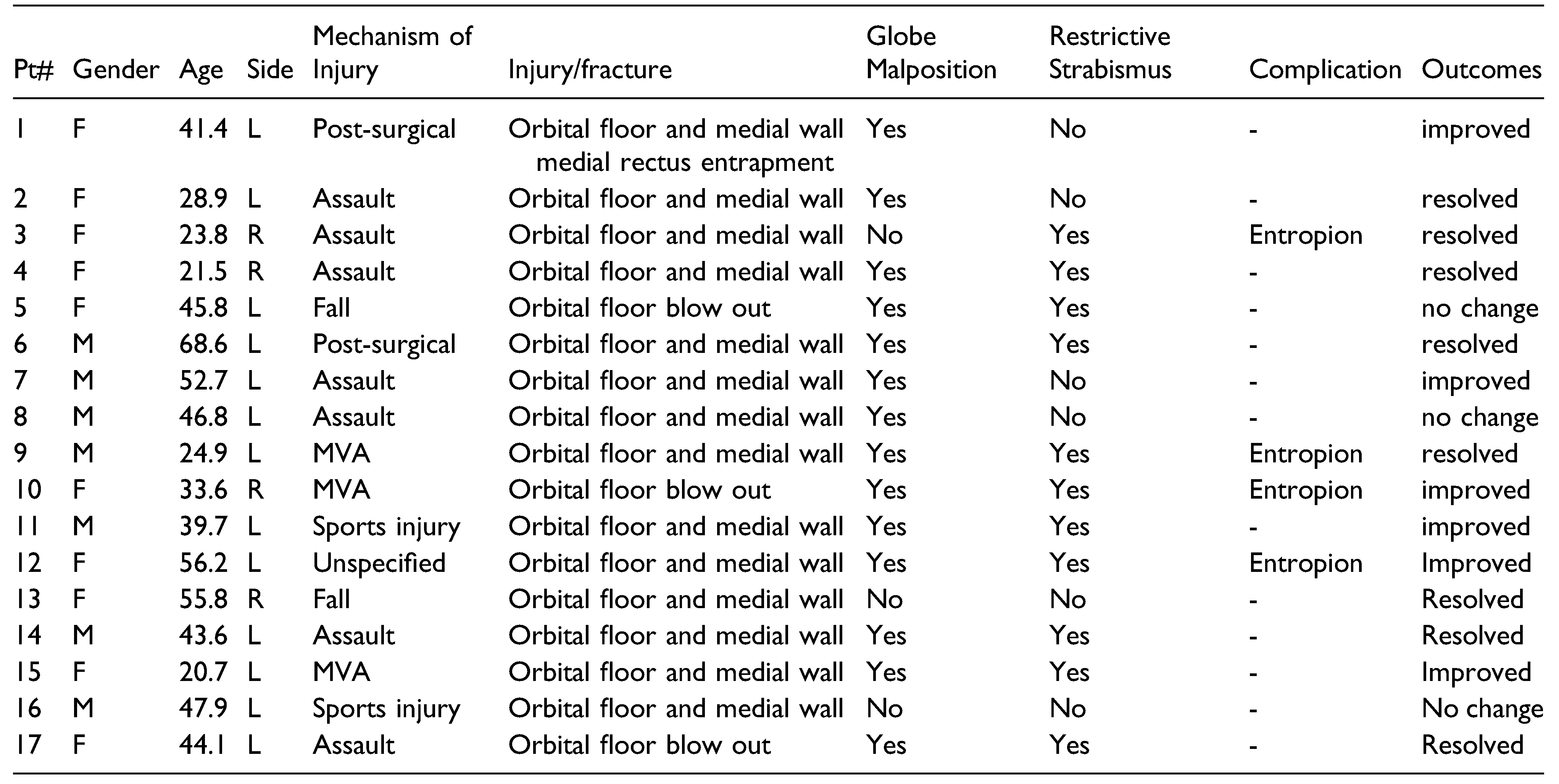
Seventeen patients with post-traumatic secondary diplopia requiring operative intervention according to study criteria were identified. The mean patient age was 40.9 years (21.0– 68.6 years) and 58.8% of patients were female. Assault (41.2%) and MVA (17.6%) were the most frequent reasons for injury. Vertical diplopia was the most common diplopia direction (76.4%) with upward gaze usually involved (64.7%). The average number of orbital surgeries prior to definitive customized implant placement for diplopia was 1.1 (range 1–3 surgeries).
Restrictive strabismus and/or globe malposition were the indications for surgical management. Globe malposition affected 14 patients (82.4%) and restrictive strabismus affected 11 patients (64.7%). Intraoperatively 3 patients were found to have restrictions in the forced duction test.
In this select group, complete resolution of diplopia occurred in 47.1% (8/17) with any improvement in diplopia occurring in 88.2% (15/17) of cases. 85.7% (12/14) of cases with globe malposition and 90.1% (10/11) of cases with restrictive strabismus showed improvement or resolution in symptoms.
Three patients had no significant clinical change in diplopia symptoms. Subsequently, one of these patients also underwent strabismus surgery with left superior oblique tenotomy with minimal improvement. Additionally, 4 cases were complicated with entropion requiring surgical treatment (Table 1).

Discussion

Post-traumatic diplopia can be challenging for patients and surgeons alike. With widely discrepant reports of diplopia improvement following orbital surgery, we wished to develop an improved treatment strategy that could yield more predictable outcomes. Our hypothesis that diplopia resulting from globe malposition or globe restriction should respond favorably to orbital surgery was borne out in this series.
The previously reported variation in improvement of diplopia with primary orbital surgery has been attributed to operative timing (with worse outcomes after 14 days), presence of periocular soft tissue entrapment, size of fracture (with worse outcomes in fractures involving more than 50% of orbital floor) and in two wall fractures involving the floor and orbital wall (8–10, 19–21). While all of these causes play a role in persistent diplopia after orbital surgery, there is no unifying principle that can guide operative management and lead to more secure outcomes.
We conjectured that diplopia resulting from globe malposition, or restriction due to disruption in orbital anatomy, would resolve with release of the restriction and accurate restoration of orbital volume. To stratify patients according to these criteria, we recommend a simple algorithm based on high resolution CT and Lancaster red-green testing (Figure 1). The first step is to identify symptomatic patients with globe malposition on CT, bearing in mind that not all patients with globe malposition will develop diplopia. However, for those who have persistent diplopia and who have globe malposition, restoration of pre-morbid orbital anatomy through the use of patient-specific implants improved diplopia in nearly all (85.7%) of cases. In several secondary cases presenting to the senior author, the initial attempt at repair had still left the patient with orbital asymmetry, necessitating removal of the initial implant and revision with a patient-specific implant to improve diplopia.
Patients with diplopia who do not have globe malposition are next evaluated for a pattern of eye movement defects through the use of Lancaster red-green testing. This special protocol conducted by neuro ophthalmologists can identify the axis of deficient movement, in any field of gaze. Following this examination, patients with an identified restriction should again have evaluation of the high resolution CT to look for concomitant entrapment of extraocular muscles, periorbita or evidence of periorbital scarring and fibrosis that may restrict movement, that correspond to the pattern elucidated in the Lancaster red-green testing. In these circumstances, orbital surgery is again recommended since liberation of these structures can enable untrammeled eye movement with resolution of diplopia in over 90% of cases in our series. Patients who have evidence of movement impairment upon Lancaster testing, but who do not have correlative evidence of orbital damage and resulting restriction are presumed to have intrinsic eye muscle damage. These patients are referred for eye muscle surgery and/or prism treatment.
There are important limitations to this treatment strategy. First, diplopia is a complex phenomenon involving both anatomic issues of eye position and movement as well as higher order processing in the central nervous system leading to a breakdown in perceptual fusion [3,18,19,22,23,24,25,26]. This interplay between eye and brain in producing diplopia is incompletely understood. Therefore, even excellent anatomic restoration may not lead to resolution of diplopia if central mechanisms cannot adjust. This may be particularly challenging in repair of longstanding diplopia in which central accommodation to the aberrant eye position may have become fixed. For these and other reasons, while our patient selection schema coupled with precise surgery can improve diplopia in greater than 85% of patients, it cannot be expected to improve all patients. A second limitation of our approach is the reliance on high resolution CT to identify anatomic causes of diplopia. While gross globe malposition is readily detected by this method, there is no strict guideline as to the degree of globe malposition that leads to diplopia, rendering judgments on operative intervention somewhat subjective. Further, subtle anomalies in orbital anatomy that correlate with globe restriction may elude radiological or surgical detection. Therefore, there is a possibility that reliance on CT could lead to over or under treatment. These caveats notwithstanding, application of our patient selection strategy should simplify preoperative decision making in post-traumatic diplopia patients and improve the safety and predictability of results for this most challenging of conditions.

Funding

The author(s) received no financial support for the research, authorship, and/or publication of this article.

Declaration of Conflicting Interests

The author(s) declared no potential conflicts of interest with respect to the research, authorship, and/or publication of this article.

References

  1. Ellis, E.; El-Attar, A.; Moos, K.F. An analysis of 2,067 cases of zygomatico-orbital fracture. J Oral Maxillofac Surg. 1985, 43, 417–428. [Google Scholar] [CrossRef] [PubMed]
  2. Gassner, R.; Tuli, T.; Hächl, O.; Rudisch, A.; Ulmer, H. Craniomaxillofacial trauma: A 10 year review of 9543 cases with 21067 injuries. J Cranio-Maxillofacial Surg. 2003, 31, 51–61. [Google Scholar] [CrossRef]
  3. Gart, M.S.; Gosain, A.K. Evidence-based medicine: Orbital floor fractures. Plast Reconstr Surg. 2014, 134, 1345e–1355e. [Google Scholar] [CrossRef]
  4. Young, S.M.; Kim, Y.D.; Kim, S.W.; et al. Conservatively treated orbital blowout fractures: Spontaneous radiologic improvement. Ophthalmology. 2018, 125, 938–944. [Google Scholar] [CrossRef]
  5. Wen Ying, W.C.; Christoff, A.; Subramanian, P.S.; Eggenberger, E.R. Diplopia and quality of life. Ophthalmology. 2011, 118, 1481. [Google Scholar] [CrossRef]
  6. Loba, P.; Nowakowska, O.; MarczykW, W.; Sokalska, K.; Broniarczyk-Loba, A. [Diplopia as a factor influencing occupational and social activities of people after orbital trauma]. Med Pr. 2012, 63, 541–546. Available online: http://www.ncbi.nlm.nih.gov/pubmed/23373322.
  7. Hatt, S.R.; Leske, D.A.; Kirgis, P.A.; Bradley, E.A.; Holmes, J.M. The effects of strabismus on quality of life in adults. Am J Ophthalmol. 2007, 144, 643–647. [Google Scholar] [CrossRef]
  8. Hawes, M.J.; Dortzbach, R.K. Surgery on orbital floor fractures: Influence of time of repair and fracture size. Ophthalmology. 1983, 90, 1066–1070. [Google Scholar] [CrossRef]
  9. Biesman, B.S.; Hornblass, A.; Lisman, R.; Kazlas, M. Diplopia after surgical repair of orbital floor fractures. Ophthalmic Plast Reconstr Surg. 1996, 12, 9–16. [Google Scholar] [CrossRef]
  10. Furuta, M.; Yago, K.; Iida, T. Correlation between ocular motility and evaluation of computed tomography in orbital blowout fracture. Am J Ophthalmol. 2006, 142, 1019–1025. [Google Scholar] [CrossRef]
  11. Lee, H.B.H.; Nunery, W.R. Orbital adherence syndrome secondary to titanium implant material. Ophthal Plast Reconstr Surg. 2009, 25, 33–36. [Google Scholar] [CrossRef] [PubMed]
  12. Damgaard, O.E.; Larsen, C.G.; Felding, U.A.; Toft, P.B.; Von Buchwald, C. Surgical timing of the orbital “blowout” fracture: A systematic review and meta-analysis. Otolaryngol Head Neck Surg. 2016, 155, 387–390. [Google Scholar] [CrossRef]
  13. Yu, D.Y.; Chen, C.H.; Tsay, P.K.; Leow, A.M.; Pan, C.H.; Chen, C.T. Surgical timing and fracture type on the outcome of diplopia after orbital fracture repair. Ann Plast Surg. 2016, 76, S91–S95. [Google Scholar] [CrossRef] [PubMed]
  14. Mehta, V.J.; Chelnis, J.G.; Chen, Q.; Mawn, L.A. Effect of time to operative intervention on motility outcomes following orbital floor fracture repair in children. Ophthal Plast Reconstr Surg. 2018, 34, 351–354. [Google Scholar] [CrossRef]
  15. Shah, H.A.; Shipchandler, T.; Vernon, D.; et al. Extra-ocular movement restriction and diplopia following orbital fracture repair. Am J Otolaryngol Head Neck Med Surg. 2018, 39, 34–36. [Google Scholar] [CrossRef]
  16. Yamanaka, Y.; Watanabe, A.; Sotozono, C.; Kinoshita, S. Impact of surgical timing of postoperative ocular motility in orbital blowout fractures. Br J Ophthalmol. 2018, 102, 398–403. [Google Scholar] [CrossRef]
  17. Loba, P.; Kozakiewicz, M.; Broniarczyk-Loba, A. Surgical management of upgaze diplopia in patients after posttraumatic orbital floor reconstruction. J Cranio-Maxillofacial Surg. 2015, 43, 976–980. [Google Scholar] [CrossRef]
  18. Kovar, D.; Voldrich, Z.; Voska, P.; Lestak, J.; Astl, J. Indications for repositioning of blow-out fractures of the orbital floor based on new objective criteria Tissue protrusion volumometry. Biomed Pap. 2017, 161, 403–406. [Google Scholar] [CrossRef]
  19. Ordon, A.J.; Kozakiewicz, M.; Wilczynski, M.; Loba, P. The influence of concomitant medial wall fracture on the results of orbital floor reconstruction. J Cranio-Maxillofacial Surg. 2018, 46, 573–577. [Google Scholar] [CrossRef]
  20. Gosse, E.M.; Ferguson, A.W.; Lymburn, E.G.; Gilmour, C.; MacEwen, C.J. Blow-out fractures: Patterns of ocular motility and effect of surgical repair. Br J Oral Maxillofac Surg. 2010, 48, 40–43. [Google Scholar] [CrossRef]
  21. Balaji, S. Residual diplopia in treated orbital bone fractures. Ann Maxillofac Surg. 2013, 3, 40. [Google Scholar] [CrossRef] [PubMed]
  22. Hara, J.; Okinaka, Y.; Takahashi, M. Orbital blowout fracture with persistent mobility deficit due to fibrosis of the inferior rectus muscle and perimuscular tissue. Ann Otol Rhinol Laryngol. 1999, 108, 1174–1176. [Google Scholar] [CrossRef]
  23. Harris, G.J.; Garcia, G.H.; Logani, S.C.; Murphy, M.L. Correlation of preoperative computed tomography and postoperative ocular motility in orbital blowout fractures. Ophthal Plast Reconstr Surg. 2000, 16, 179–187. [Google Scholar] [CrossRef] [PubMed]
  24. Jin, H.R.; Lee, H.S.; Yeon, J.Y.; Suh, M.W. Residual diplopia after repair of pure orbital blowout fracture: The importance of extraocular muscle injury. Am J Rhinol. 2007, 21, 276–280. [Google Scholar] [CrossRef] [PubMed]
  25. Xia, Q.; Wang, Z.; Yan, J. Surgical management of strabismus in patients with orbital fracture. J Craniofac Surg. 2018, 29, 1865–1869. [Google Scholar] [CrossRef]
  26. Campbell, B.C.; Shipchandler, T.Z.; Ting, J.Y.; et al. Ocular motility and diplopia measurements following orbital floor fracture repair. Am J Otolaryngol Head Neck Med Surg. 2021, 42, 102879. [Google Scholar] [CrossRef]
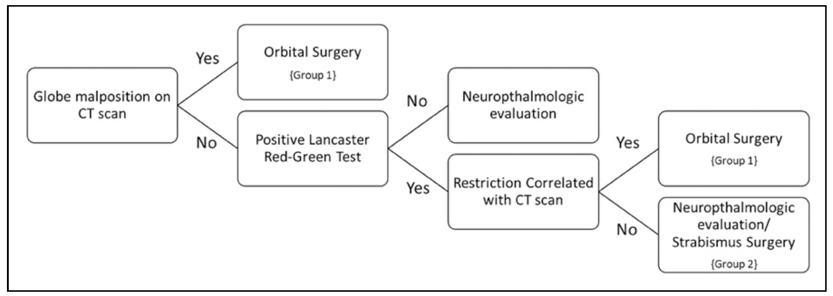
Figure 1. Persistent post-traumatic diplopia treatment algorithm.
Cmtr 16 00013 g001
Table 1. Patient and injury characteristics.
Cmtr 16 00013 i001