Next Article in Journal
Analysis of Neoclassical Canons in Adult Kenyans of Indian Descent
Previous Article in Journal
Likelihood of Craniofacial Injury and Hospitalization with Alcohol Use While Skateboarding and Scootering
 
 
Craniomaxillofacial Trauma & Reconstruction is published by MDPI from Volume 18 Issue 1 (2025). Previous articles were published by another publisher in Open Access under a CC-BY (or CC-BY-NC-ND) licence, and they are hosted by MDPI on mdpi.com as a courtesy and upon agreement with Sage.
Article

Complicated Facial Lacerations: Challenges in the Repair and Management of Complications by a Facial Trauma Team

by
Moumita De
1,*,
Sushma Sagar
2,
Aniket Dave
1,
Ruchi Pathak Kaul
3 and
Maneesh Singhal
1
1
Plastic, Reconstructive and Burns Surgery, All India Institute of Medical Sciences, New Delhi, India
2
Division of Trauma Surgery and Critical Care, JPN Apex Trauma Centre, All India Institute of Medical Sciences, New Delhi, India
3
Department of Surgery, JPN Apex Trauma Centre, All India Institute of Medical Sciences, New Delhi, India
*
Author to whom correspondence should be addressed.
Craniomaxillofac. Trauma Reconstr. 2023, 16(1), 39-54; https://doi.org/10.1177/19433875211064512
Submission received: 1 November 2021 / Revised: 1 December 2021 / Accepted: 1 January 2022 / Published: 1 February 2022

Abstract

:
Study Design: This is a descriptive study where we present our experience in managing complicated facial wounds over a period of 1 year at a level 1 trauma centre by a dedicated facial trauma team consisting of a plastic surgeon, a trauma surgeon and a Maxillofacial surgeon. Objective: Facial deformities have profound impact on the social and psychological aspect of a person’s life. Hence, management of facial wounds is very crucial. Most of the facial injuries are usually managed by emergency care physician and emergency surgeon. But certain wounds require specialised knowledge and care due to their complicated nature. The objective of this paper is to highlight those special types of wounds and the challenges they pose. It also aims to enumerate the best possible management according to each situation in a protocol-based manner, which will help in decision making by the attending emergency physician/surgeon. Methods: Facial lacerations were designated as “complicated” according to some pre-defined features and pre, intra and post operative data and photographs were collected by interviewing the concerned surgical team. The data were analysed and presented as different clinical scenarios. Results: The cases were broadly grouped under 6 scenarios according to the unique combination of difficulties faced and their specific management. The challenges faced were enumerated and the steps undertaken were also mentioned against them. Lastly, the scenarios were compared with available literature to find out the best possible management in each situation and to present them in a protocol-based manner. Conclusion: Protocol-based management of injuries to the different parts and specialised structures of the face is extremely helpful. Role of a specialised facial trauma team also should be emphasised in complicated facial injuries.

Introduction

The face is our window to the world. It represents our identity and conveys our emotions. The face is also the yardstick of pulchritude. Therefore, the importance of a good facial wound repair on the life of a patient cannot be emphasized enough. Although most facial lacerations are simple and can be managed by a primary care physician [1,2], some lacerations are complicated by their size, involvement of vital structures (eyes, lip, ear, and nose), and multiple layers (skin, muscle, neurovascular layer, and bone) or due to a functional deficit. Sometimes a seemingly straightforward laceration may turn out to be complex due to some unforeseen complications. Much has been written about the techniques and protocols of facial laceration repair; however, literature is scarce when it comes to discussing the difficulties and complications faced in the management of such wounds. This article addresses the challenges encountered during the management of complicated facial lacerations and our approach to their management at a level 1 trauma center.

Materials and Method

We evaluated the cases of complicated facial lacerations that were repaired by our facial trauma team (consisting of a plastic surgeon, a trauma surgeon and a maxillofacial surgeon) over a period of 1 year (December 2018 to November 2019) at a level 1 trauma center.

Definition

We defined complicated facial laceration as a facial laceration with at least one of the following features:
  • Large size (>10 cm);
  • involving more than 2 facial zones;
  • extensive skin avulsion or skin loss;
  • involving any specialized facial structure (eyelid, nose, lip, or ear);
  • involving neurovascular structures of the face; and
  • involving glands or ducts.

Exclusion Criteria

Wounds with facial bone fracture requiring fixation were excluded from the study.
Clinical data, pre- and post-operative clinical photographs, intra-operative photographs (whenever available) of all the included patients were collected. The concerned surgery team members were interviewed regarding the challenges faced and what was done to address them. Then in retrospect, we tried to ascertain the best possible solution for each of the challenges faced.

Results

We explored the included patients and identified the difficulties faced in their management and accordingly the cases were broadly grouped under 6 scenarios. Each scenario represented a unique combination of challenges that required tactful management. Cases included in each category had similar injuries albeit with variations concerning anatomical location, orientation and size. The thought process and protocols involved in the management of each scenario could be broadly generalized. Representative cases of each scenario are listed below.

Scenario 1: Large Wound (>10 cm)

Case details: 26-year-old male patient, presented to emergency with extensive soft tissue laceration over face involving multiple facial units and in altered sensorium. The mechanism of injury was Road Traffic Incident (twowheeler rider, without a helmet). The patient had minor bruises and lacerations over elbows, forearms and right leg and foot.
On evaluation in the ER, the patient was diagnosed with head injury (left frontal bone fracture with multiple hemorrhagic contusion, managed conservatively by the Neurosurgery team) with facial injury (nasal bone fracture, large laceration extending from left of the temporal hairline, across the face to the opposite side angle of mouth) (Figure1A).

Challenges Faced and Steps Taken

  • Involvement of multiple units: Involvement of the forehead, brow, eyelid, nose, and cheek entailed that each part was addressed according to the protocol prescribed for that unit. Knowledge of the subunits of face and reconstruction of each part was of paramount importance.
The use of anchor sutures at specific landmark points helped in proper alignment of the laceration, for example, at the eyebrow, at medial canthus, at the root of the nose, and at the nasolabial fold area.
After thorough lavage, layered closure was done. Muscles were sutured with absorbable interrupted sutures. Subcuticular absorbable (4-0 vicryl) and non-absorbable skin sutures (4-0 prolene) were used (Figure 1B).
2.
Underlying head injury: The fracture of the left frontal bone was managed conservatively by simple reduction alone. There was no underlying dural breach. However, the presence of head injury and altered sensorium precluded any thorough clinical examination of the facial nerve function.
3.
Nasal bone fracture: Compound fracture of nasal bone was present but it was not amenable for any fixation. Therefore, it was managed conservatively by reduction of fragments, suturing of overlying skin, and splinting.

Scenario 2: Extensive Skin Avulsion or Skin Loss

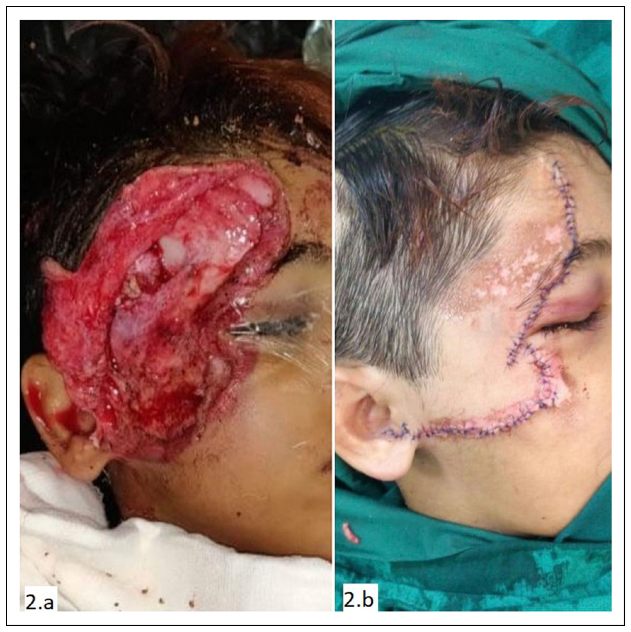
An eight-year-old boy presented to the emergency with a suspected head injury and large facial avulsion over the right upper face with exposed bone (Figure 2A). The child did not have any other major injury. On evaluation, head injury was ruled out. The child was taken to OR for repair of the facial injury (Figure 2B).

Challenges Faced and Steps Taken

  • Contamination: The injury being a road traffic incident, the wound was contaminated with dust and debris. Stone particles and tiny plastic fragments along with grass and soil were found embedded in the wound. The patient presented within 6 hours of injury. In the OR, thorough cleaning was done first with saline to wash away soil and debris. Then scrubbing was done with 4% chlorhexidine gluconate solution and copious saline wash was given. Finally, the wound was painted with a povidone iodine solution as per the institution OR protocol. The wound healed well in the post-operative period; however, the sutures were kept for 7 days instead of the standard 5 days in view of the grossly contaminated wound.
  • Exposed frontal bone: A 1.5 × 2 cm area of the frontal bone was exposed with a small area of periosteal stripping. However, as the reposition of the avulsed flap was adequate to cover this exposed bone, no additional flap was required.
  • Doubtful viability of avulsed skin: The avulsed skin flap had areas of friction burn and also the edges did not have healthy bleeding. However, owing to the excellent blood supply of the face, only minimal marginal debridement was done and tissues were preserved as much as possible. In the post-operative period, the entire skin survived without any necrosis.

Scenario 3.a. Involvement of Specialized Structures: Eyelid

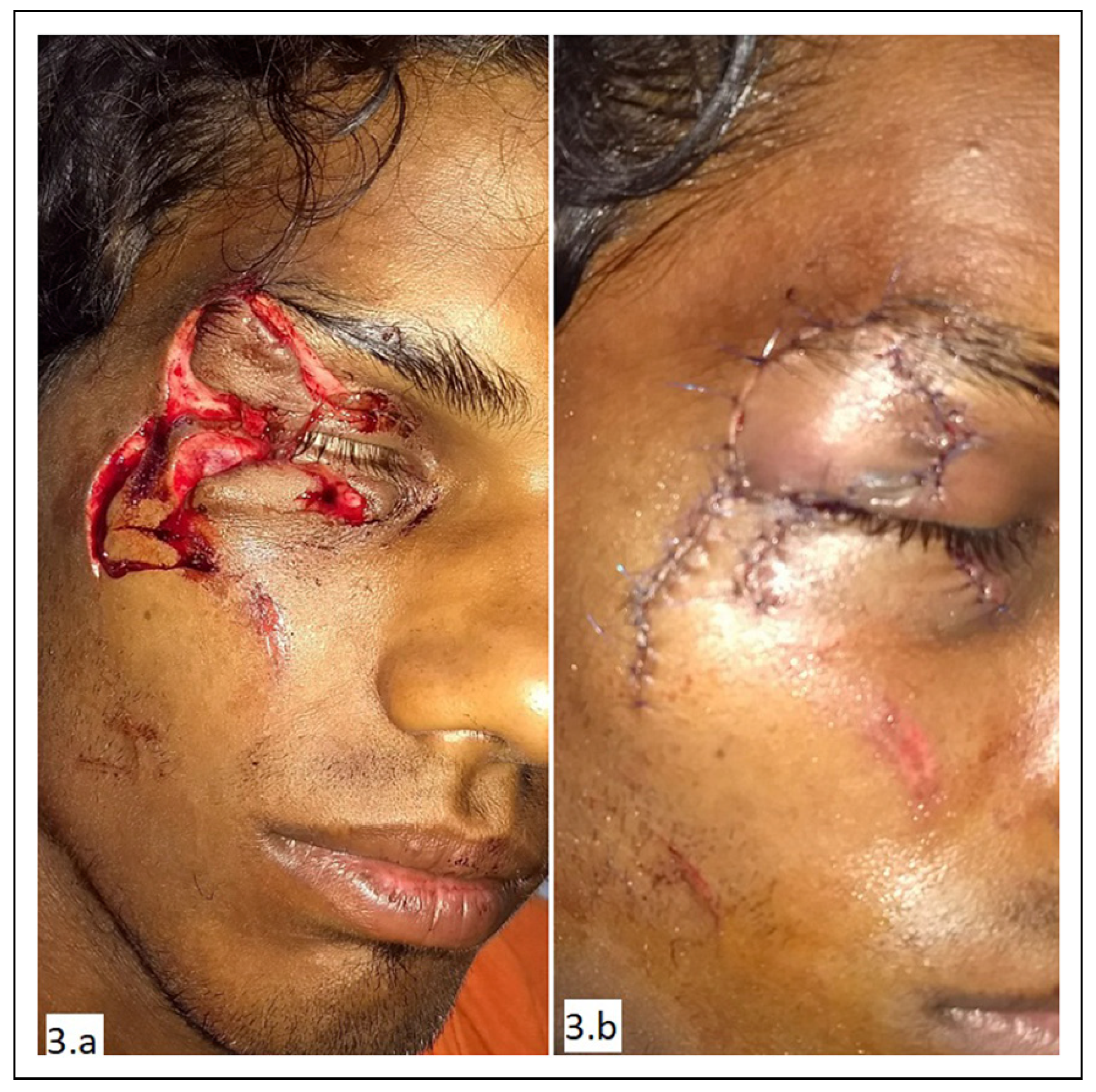
Case #1. A 24-year-old man presented to the emergency department with right eyelid laceration following a physical assault. He had a stellate laceration of the upper eyelid, lower eyelid, and lateral canthus region (zones 1, 2, and 4). The injury involved the outer lamella (skin and orbicularis oculi muscle) (Figure 3A).

Challenges Faced and Steps Taken

  • Involvement of eyebrow: Proper alignment of the eyebrow was done matching the brow hairline to prevent notching of the eyebrow. An exact approximation was done with subcuticular sutures to prevent scar widening and possible future scar alopecia.
  • Involvement of orbicularis oculi: Muscle was approximated with 4-0 vicryl horizontal mattress suture without any overlapping. Any overlapping or gap would cause animation deformity in this readily visible area.
  • Comminuted skin laceration: The laceration was comminuted and had to be matched like the pieces of a jigsaw puzzle to achieve exact approximation. Loupe magnification was used for meticulous repair (Figure 3B).
  • Avulsed skin segments on narrow pedicle: Although skin segments were avulsed with very narrow attachments, no skin was debrided. Owing to the superior blood supply of this region, native tissue was preserved as much as possible.

Case #2

A 32-year-old man was brought to emergency with a facial injury. Head injury was ruled out. The patient had a fullthickness laceration of the left eyelid with exposure of the globe. The laceration extended along the nasolabial crease with full-thickness laceration of the upper lip (Figure 4A).

Challenges Faced and Steps Taken

  • Full thickness eyelid defect with loss of support and exposed globe: The exposure of the globe was an emergency that needed intervention to prevent desiccation and corneal damage. The eyelid defect was meticulously repaired in layers from the deepest to the most superficial layer.
  • Comminuted skin laceration with doubtful vascularity: The lid skin was repaired with 6-0 prolene under loupe magnification. Absolutely no tissue was debrided and discarded from the lid skin.
  • Ruptured tarsal plate with loss of structural support of the lid: The ruptured tarsus was aligned meticulously with 5-0 prolene suture to reconstruct the support framework. Next the orbicularis muscle was approximated without any bunching or overlapping. The muscle closure effectively covered the tarsal plate and the sutures and provided additional support.
  • Conjunctival laceration: The conjunctiva was repaired with 5-0 vicryl inverted sutures to not leave any exposed suture or knot over the corneal side.
  • Involvement of other facial units: Other facial aesthetic units (cheek, nose, and lip) were repaired following the principles of reconstruction of that subunit (Figure 4B).

Scenario 3.b. Involvement of Specialized Structures: Auricle

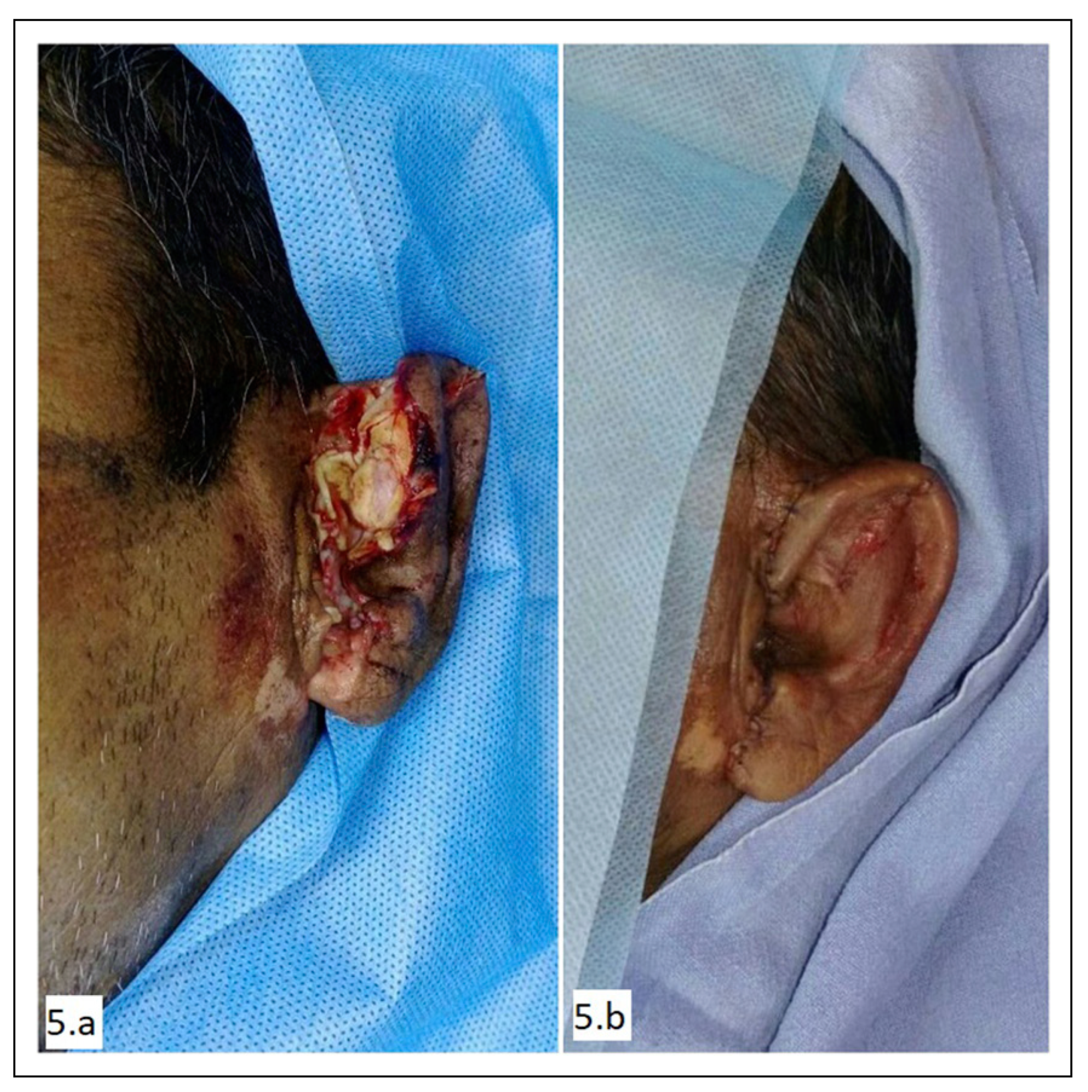
A 40-year-old man presented in emergency with crush and partial avulsion of the left pinna (Figure 5A). The man was a pillion rider on a two-wheeler. He was not wearing a helmet when he fell down and sustained minor scratches over the left upper limb along with injury to the pinna.

Challenges Faced and Steps Taken

  • Crush and avulsion of skin: The overlying lateral surface of the auricle was crushed and avulsed. However, the medial (posterior) surface skin was intact mostly. After minimal debridement, a meticulous approximation of the skin was done (Figure 5B).
  • Exposure and fracture of cartilage: The crushed and damaged cartilage was debrided. All fragments that had an attachment to the posterior skin were left undisturbed. No suture was put through the cartilage. The skin approximation was adequate to cover all the cartilage.
  • Contamination: The gross contamination present was a risk factor for chondritis. Thorough cleaning with chlorhexidine solution along with meticulous debridement was done to decrease the infection load. Systemic antibiotic as well as topical antibiotic cream were used as a prophylaxis.

Scenario 3.c. Involvement of Specialized Structures: Nose

A 26-year-old man presented to emergency with a suspected head injury and facial injury. He was diagnosed with mild head injury managed conservatively by the neurosurgery team.

Challenges Faced and Steps Undertaken

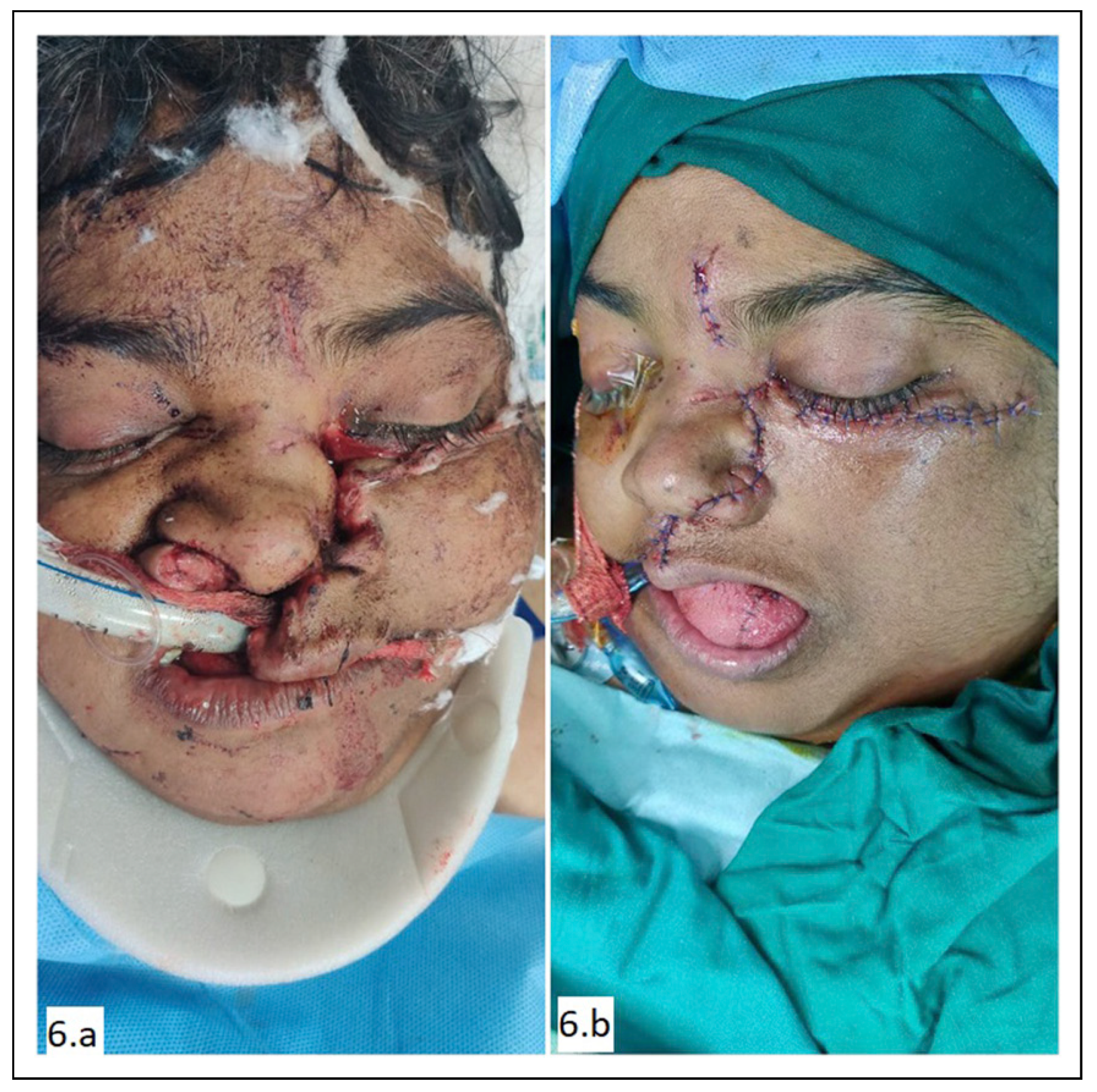
  • Injury to the cartilage: Injury to the upper and lower lateral cartilages as well as the septal cartilage caused loss of structural support to the nose (Figure 6A). The septal cartilage was reduced back in position. No suture was passed through the cartilages. While the immediate outcome was good, this patient had a prolonged ICU stay in view of head injury. On follow-up, there was resorption of septal cartilage support with deformity of the nasal dorsum. The patient is scheduled for rhinoplasty at a later date.
  • Injury to alar margin: Problem with alar margin approximation is due to the thick skin which is adherent to the underlying cartilage. It is difficult to mobilize the skin and therefore unyielding to any sort of tissue rearrangement. A proper approximation is required to prevent notching (Figure 6B).
  • Injury to the nasal mucosa and nasal floor: Nasal mucosa and nasal floor were repaired using 5-0 vicryl sutures.

Scenario 3.d. Involvement of Specialized Structures: Lip

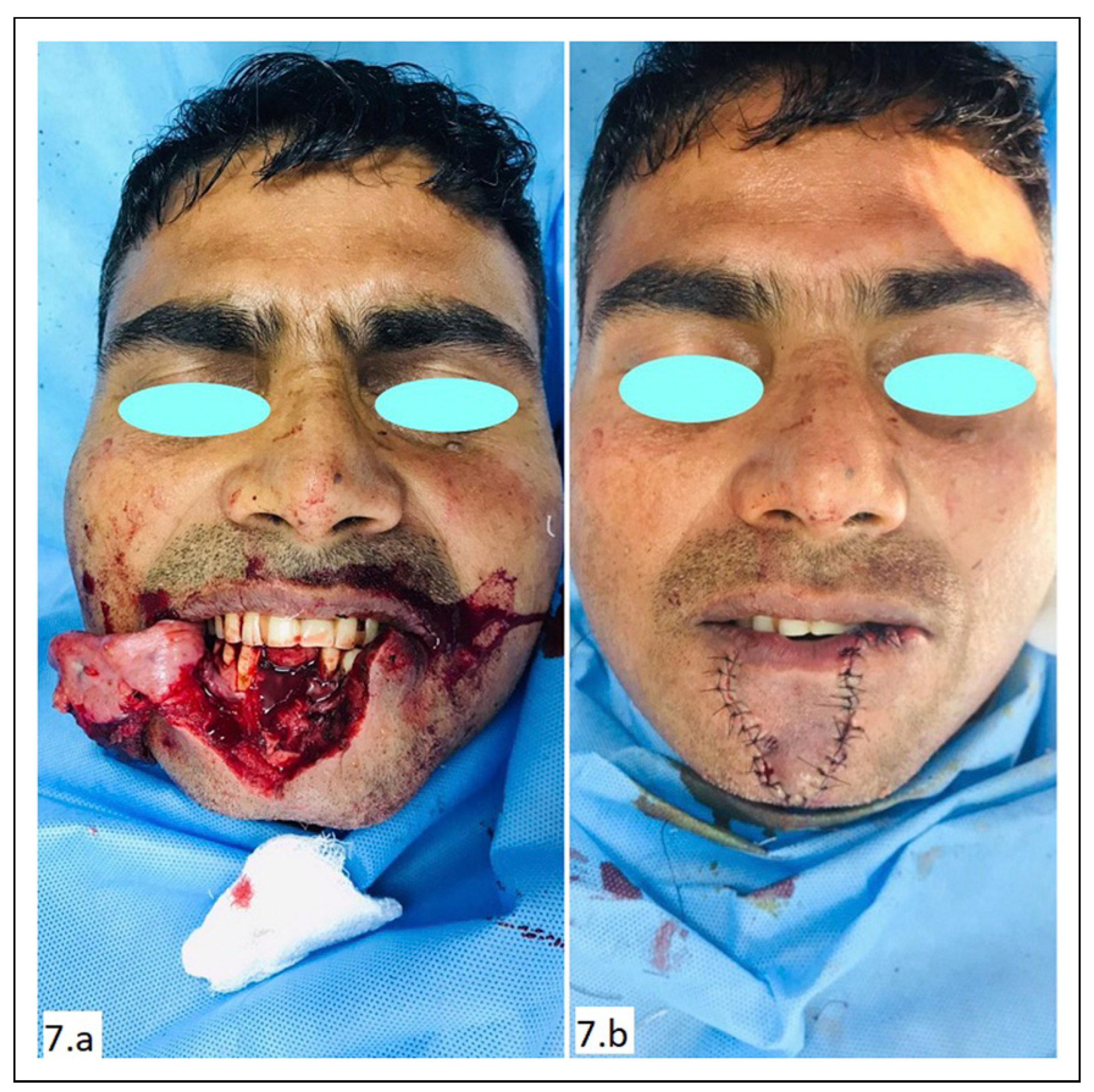
Thirty-year-old man was brought to emergency with near total avulsion of the lower lip (Figure 7A). Additional injury was ruled out.

Challenges Faced and Steps Taken

  • Mucosal breach: Mucosal injury was repaired in a watertight fashion using 4-0 vicryl.
  • Muscle injury: Muscle alignment was very important as any gap present or even any overlapping will be readily visible on animation. As lips are one of the most mobile areas of the body, this becomes very evident during conversation. Muscles were approximated exactly using horizontal mattress sutures with 3-0 vicryl.
  • Vermilion alignment: Vermilion is a specialized feature of the lip that cannot be mimicked by any other tissue. Even 1 mm notching of vermilion is apparent at conversational distance. Therefore, an exact approximation of the vermilion was done using the white roll as a guide. Tattooing of the points was done before infiltrating local anesthetic to prevent distortion (Figure 7B).
  • Vascular issue: One-sided labial artery was found to be intact on exploration which ensured that the entire flap survived.
  • Alveolar fracture with loss of teeth: No immediate intervention was done. The maxillofacial team put the patient under their follow-up for a dental implant at a later date.

Scenario 4: Involvement of Neurovascular Structures

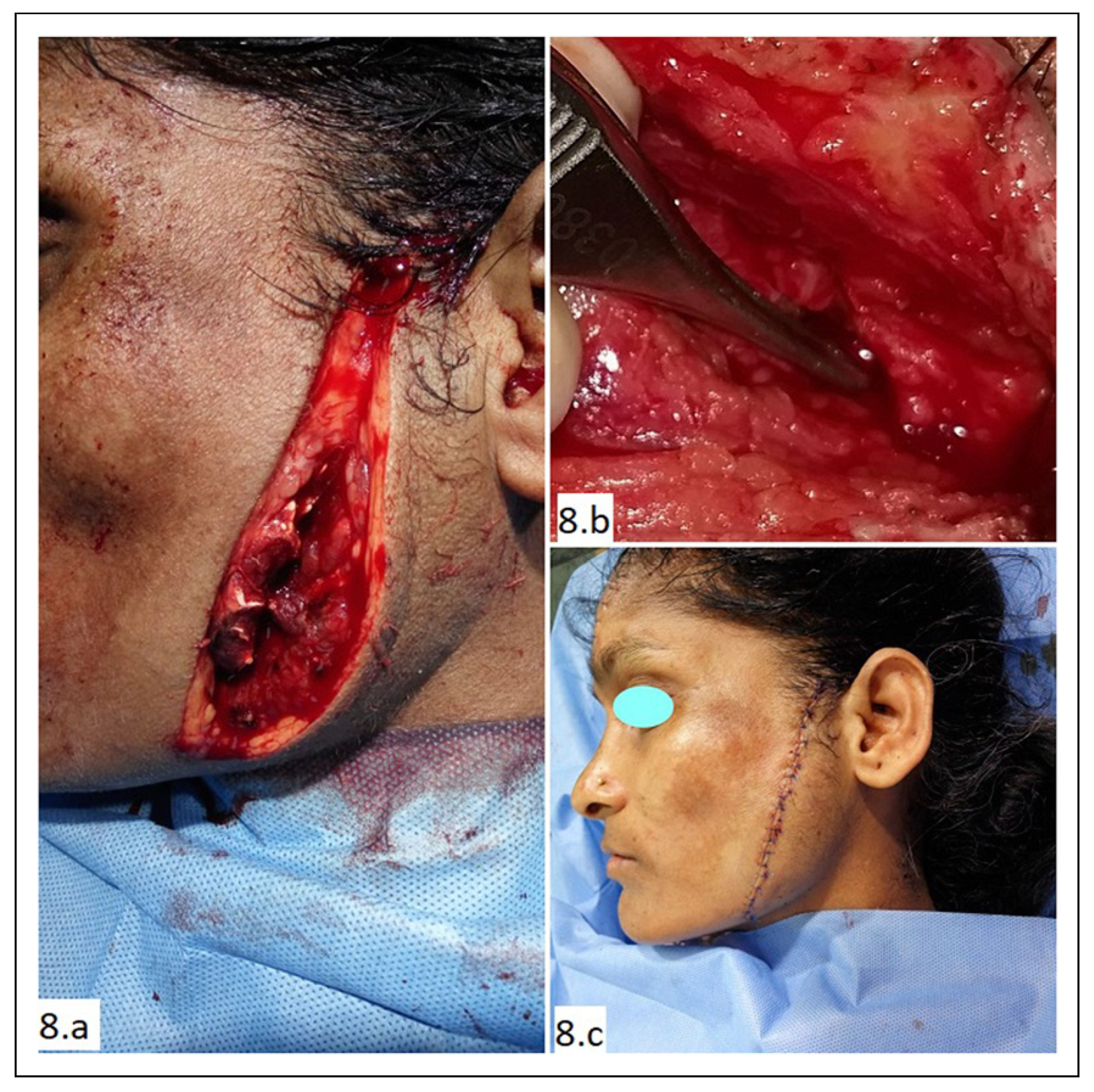
A 29-year-old lady presented with deep laceration over the left parotid region due to attack with a sharp object (Figure 8A). She did not have any other injury. She had mild weakness in puffing the left cheek. On exploration under local anesthesia, the parotid gland was found to be injured and a branch of the facial nerve was found transected (Figure 8B). The facial nerve was repaired under magnification using 9-0 nylon. The parotid capsule was repaired with 4-0 vicryl. The muscle was approximated with 4-0 vicryl, and the skin was repaired with 5-0 prolene (Figure 8C).

Challenges Faced and Steps Taken

  • Injury to the facial nerve: On clinical examination there was only mild weakness of the left buccinator. This could have been missed if thorough exploration was not done. As there is arborization between zygomatic and buccal branches of the facial nerve, any injury in either of these branches may not be easily identifiable.
  • Parotid gland injury: Parotid gland injury poses a risk of parotid fistula and sialocele formation. It was managed by repairing the gland’s capsule and use of an antisialagogues in the postoperative period.

Scenario 5: Involvement of Glands and Ducts

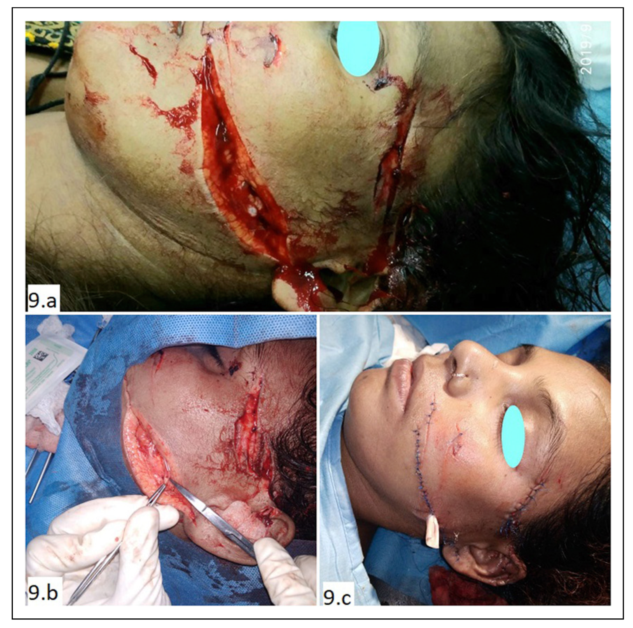
A 23-year-old lady was assaulted with a sharp knife and presented to the emergency department (Figure 9A). She had multiple sharp cuts over the left side of her face and some small lacerations over both hands. On exploration, the parotid duct was found completely transected. It was repaired under magnification using 8-0 nylon over a stent made from the cannula part of an 18G intravenous catheter. The accompanying buccal branch of facial nerve was also repaired using 8-0 nylon. Muscle and skin were then repaired.

Challenges Faced and Steps Taken

  • Identification of parotid duct injury: Identifying a duct injury is often difficult. The typical location of the laceration raised suspicion. The distal end was easily identified by cannulating the parotid duct from its buccal opening using an intravenous cannula. The tip of the cannula was seen coming out through the wound. The proximal end was identified under magnification. Presence of droplets of clear parotid secretion helped in identification of proximal end (Figure 9B).
  • Use of a proper stent: The tube part of an 18G IV cannula was found to be of adequate length and diameter in our case.
  • Long linear laceration: The long straight laceration was broken by placing 2 small z-plasties to make the scar less conspicuous (Figure 9C).

Scenario 6: Laceration in a Patient With Comorbidities

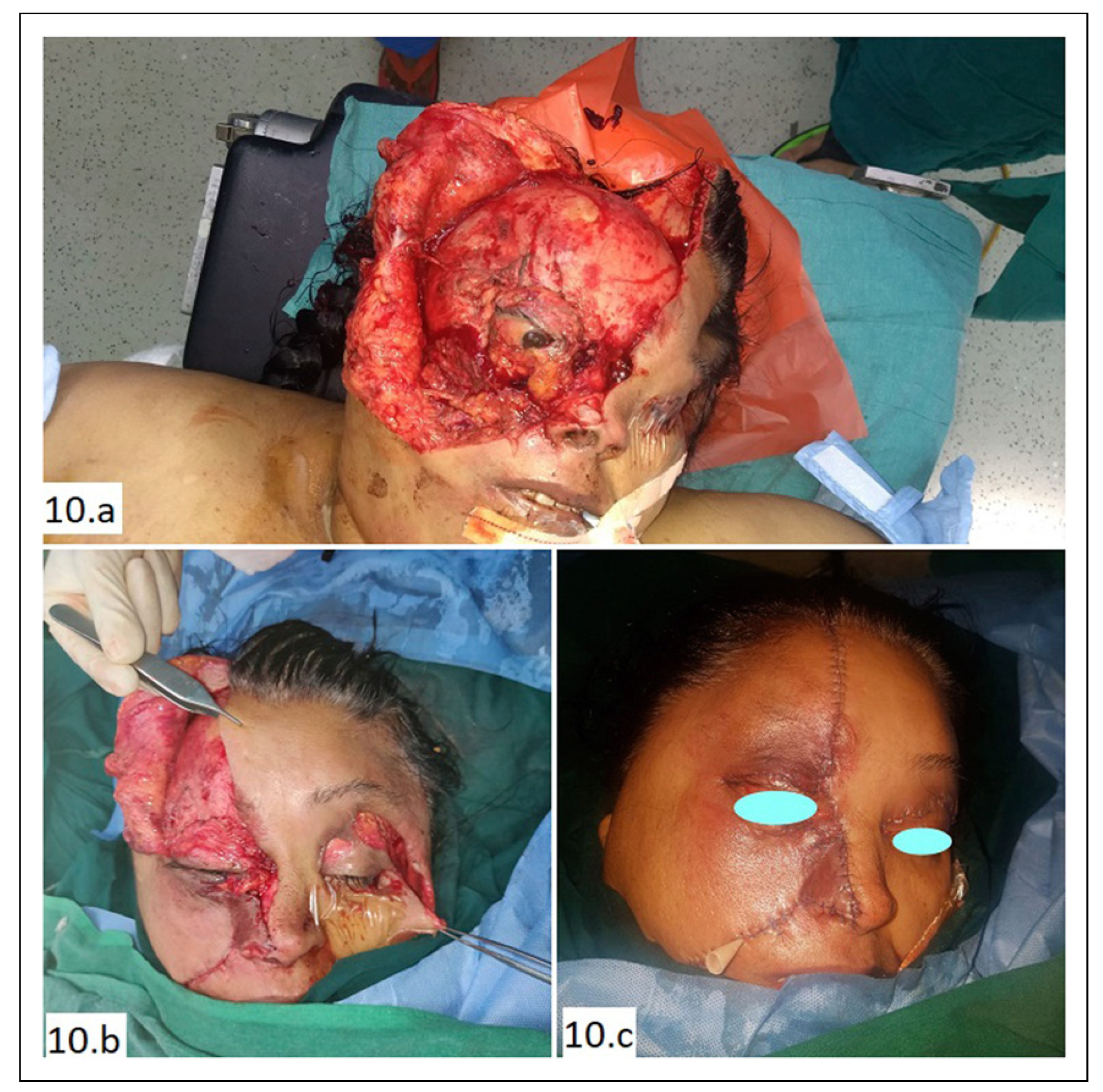
A 62-year-old lady was brought to emergency with extensive facial laceration and avulsion. Both eyelids were injured with exposure of the globe (Figure 10A, B). The mode of injury was fall from the backseat of a two-wheeler. She was not wearing a helmet.

Challenges Faced and Steps Taken

  • Contaminated wound: Thorough lavage of the wound was done using copious saline, chlorhexidine solution and povidone iodine.
  • Multiple co-morbidities:
    • Morbid obesity: Mobilization of the patient in the post-operative period was extremely difficult. She was closely monitored by the dietician and physiotherapist.
    • Diabetes mellitus (poorly controlled): The patient was a diagnosed diabetic on oral hypogylcemic agents for the last 6 years but her blood sugar was poorly controlled. During her admission, endocrinology consultation was taken and insulin injection was started in place of the oral medications. Fivepoint blood sugar monitoring was done and dose adjustment was done accordingly.
    • Hypertension (uncontrolled): She was under irregular medication for hypertension previously. With opinion from internal medicine and cardiology, she was started on appropriate antihypertensive and antilipid medications.
    • Hypothyroidism: She was previously diagnosed as a hypothyroid but was under irregular medication. Levothyroxine was started after endocrinology consultation
    • Depression: Psychiatry consultation was done and she was put on an antidepressant and also followed up by the psychiatrist daily for behavioral therapy.
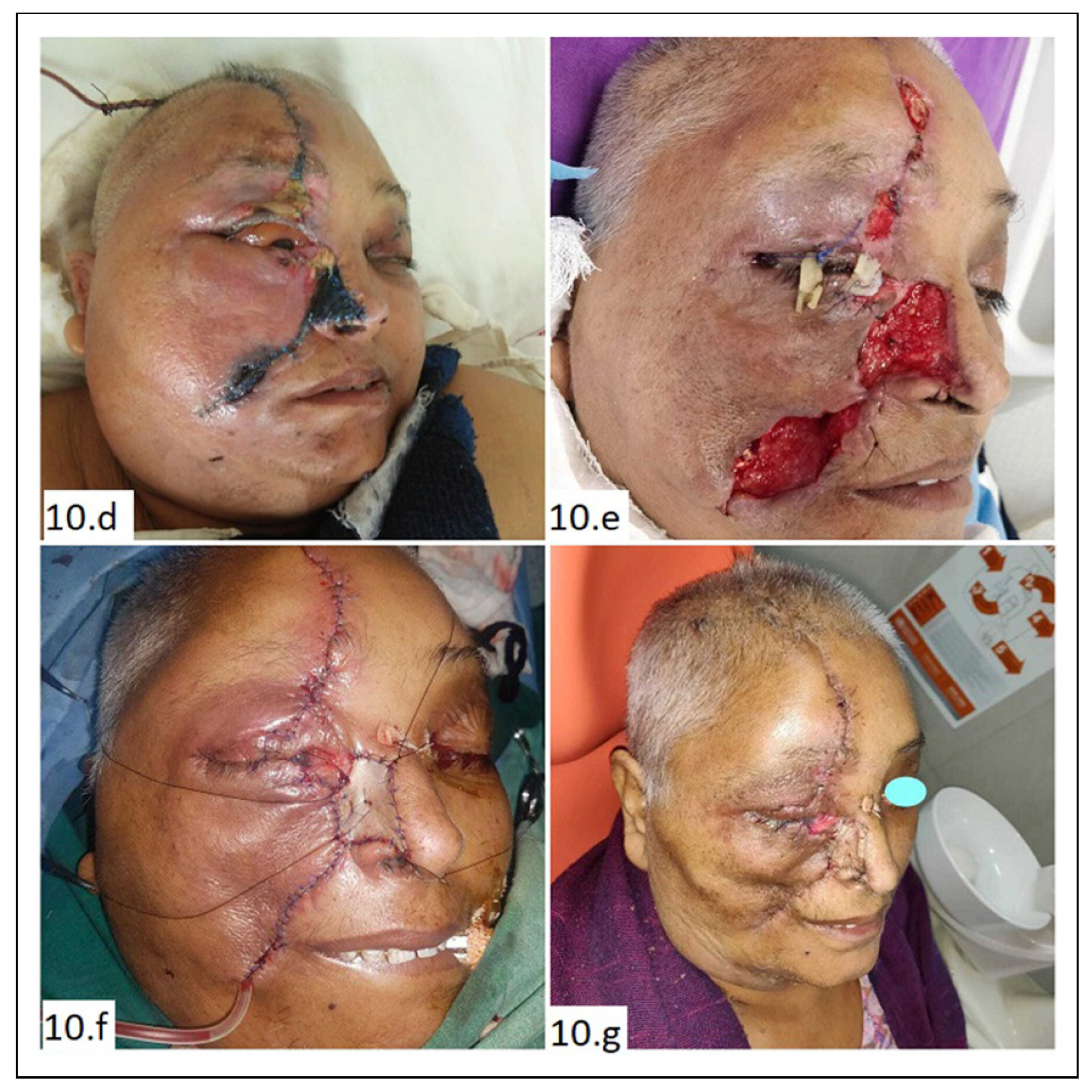
  • Wound infection: She developed cellulitis and an abscess under the reposited flap on post-operative day 2 (Figure 10C, D). It was treated by re-exploration and abscess drainage with copious irrigation. Culture yielded polymicrobial infection. Antibiotics were started according to sensitivity report.
  • Flap necrosis: The tip of the avulsed skin flap covering the lateral nasal wall was necrosed. The margin of the cheek flap was also necrosed. Debridement was done and the raw area was resurfaced with a full thickness skin graft (Figure 10D–G).

Discussion and Literature Review

Scenario 1

Large facial wounds look relatively straightforward for repair. However, they often pose a challenge due to unfavorable scarring in the post-operative period. This is especially true in lacerations that cross aesthetic units of the face or are present on a visible part like cheek, chin, forehead or nasal dorsum. In the Facial Injury Severity Scale (FISS) proposed by Bagheri et al, lacerations more than 10 cm are given one point and found to significantly affect outcome [3]. The first step toward ideal management is thorough lavage of the wound. Lavage with normal saline is often adequate, but certain contaminated wounds often require a more thorough lavage using chlorhexidine gluconate soap solution (4%). Beyond the obvious importance in preventing later wound infection, a thorough lavage helps remove dust and metallic particles. Particles embedded around the deep dermis can result in tattooing which looks conspicuous despite good surgical alignment. Thorough cleansing also allows for proper visualization of any bleeding points and therefore aids in hemostasis [2]. Any obviously necrotic tissue must be debrided. The face has an excellent blood supply, and a conservative approach is advisable when dealing with tissues that have doubtful vascularity but otherwise appear structurally intact. The next important consideration is to break long linear scars by placing z-plasties at select locations. This not only gives a better scar by reducing tension but also prevents future trap door scar formation. Layered closure is another very important preventive measure against future scar formation, as it causes distribution of tension among the different layers; hence, a reduced tension on the final epidermal layer [2].

Scenario 2

Extensive skin avulsion poses 2 challenges to the surgeon.
  • Contamination and a chance of infection.
  • Doubtful vascularity of the avulsed skin segment.
It has been well established that in the face, due to extensive plexus formation of blood vessels in all layers, vascularity is generally robust. Skin flaps with length to breadth ratio of 4:1, and even up to 6: 1 may survive in the face and neck. However, crush injury can complicate by direct tissue injury and indirect damage to blood vessels.
The key points in managing such wounds are to do a thorough debridement and lavage. For lavage, normal saline and chlorhexidine soap solution is generally adequate. Some authors even suggest the use of clean tap water or sterile water [2], while some advocate use of hydrogen peroxide, iodine solution and normal saline to clean large avulsion wounds [4]. Usually in the face the debridement has to be done conservatively but meticulously. The skin margins should be checked for healthy bleeding. Sometimes tissues that look compromised initially may turn out to be viable. So, it is better to err on the side of conservative during debridement. Meticulous reposition of the avulsed skin flaps and layered closure gives the best result.
For loss of skin, it is possible to mobilize and rearrange local tissue in the primary setting to give the best outcome both in terms of form and function. Local flaps always give a better color match and cosmetic outcome than distant tissue. However, in cases where local tissue rearrangement is not possible, primary grafting of the wound gives satisfactory coverage. Kretlow et al advocate local flaps or even primary free flaps in suitable cases, especially for large scalp defects. They mention that local contamination is not a contraindication for free flap coverage in such cases [5].
Hai Gao et al mention that negative pressure wound therapy can be useful in such wounds [4]. Although there is no exact study on avulsed facial wounds, Novelli et al reported satisfactory wound coverage, decreased healing time, and increased patient satisfaction with use of NPWT in complex craniomaxillofacial and cervical wounds [6].
In large avulsions and lacerations, especially that involving the lower face and midface or those with multiple facial injuries, the patient should always be evaluated for brain injury although clinical signs or symptoms may be absent at the outset. Razak et al reported the presence of mild traumatic brain injury in patients with facial lacerations to be as high as 41.4% [7].

Scenario 3.a. Involvement of Specialized Structures: Eyelid

Eyelid defects can be either partial thickness or full thickness. The challenges faced in such defects are loss of tissue, injury to the lacrimal puncta and canaliculi, and injury to conjunctiva in full thickness defects.
In eyelid defects, debridement should be minimal as periocular tissue is seen to recover well even when hardly attached through small bridge tissue [4]. For proper alignment of the margin in full thickness defects, the meibomian gland orifices, the gray line or the lash line can be used as anatomic landmark guides [8]. Alignment of the orbicularis oculi is very important as failure to repair the muscle properly will result in non-contiguous healing which in turn will lead to depressed scars and animation deformity [2].
For full-thickness defects, it is also essential to repair the tarsus in a lamellar fashion using delayed absorbable sutures such as polyglactin. The tarsus should also be the primary tension bearing area of the eyelid repair to avoid unsightly contour defect and notches in the margin [8].
The visualization of orbital fat through the wound suggests a breach in the orbital septum and possible injury to the levator complex. Such presentation warrants exploration and repair of the levator complex [8]. A zone 3 injury (medial canthal region) may also necessitate exploration of the lacrimal canaliculus. If injured, it should be repaired over a stent [5,8].

Scenario 3.b. Involvement of Specialized Structures: Auricle

Involvement of the pinna or auricle poses multiple challenges. In laceration only without tissue loss, thorough debridement of crushed cartilage and skin with a meticulous approximation of skin to cover the entire cartilage framework is necessary [5]. The cartilage framework generally needs no repair if both anterior and posterior skin can be approximated securely without tension. In fact, unnecessary suturing of the cartilage should be avoided as it leads to further devitalization of the cartilage and also provides an entry portal for infection [9]. In cases where there is tissue loss with exposed cartilage, if the defect is small, primary excision of the exposed cartilage in a wedge fashion from the helical rim with primary closure gives the best aesthetic outcome. The resultant shortening of the auricle is generally not much appreciable at conversational distance. If a larger area of cartilage is exposed with intact perichondrium, immediate skin grafting should be done after thorough cleaning to protect the exposed cartilage. In cases where primary skin grafting is not feasible due to patient factors or wound factors, moist occlusive dressing should be done to prevent desiccation [9]. In cases with loss of larger segment of tissue including all of the auricles, an attempt should be made for microsurgical replantation in all cases.
A rare occurrence of perilymph fistula due to stapes footplate injury associated with auricular injury has been reported in literature. A high level of suspicion and referral to an otolaryngologist is necessary in cases of acute trauma with associated hearing loss or problems in balance [10].

Scenario 3.c. Involvement of Specialized Structures: Nose

The nose lies in the center of the face and naturally is the most visible feature in the face. Any scar over the dorsum and tip of the nose is readily visible. The rounded nasal tip also has a propensity for trap-dooring when lacerations are repaired. High velocity trauma leading to nasal bone fracture is a different entity altogether requiring reduction. In cases of nasal injury without nasal bone fracture, the challenges are
  • extensive laceration;
  • injury to the cartilage framework: ULC, LLC, and septal cartilage; and
  • septal hematoma.
Extensive lacerations should be treated like any other part of the face with conservative but meticulous debridement and approximation. The principle of subunit reconstruction is widely followed in the case of nasal defects. It is generally accepted that if greater than 50% of a subunit is compromised, the reminder tissue should also be excised and the entire subunit should be reconstructed as a whole. However, some authors challenge this concept and are in favor of retaining as much native tissue as possible [11]. Special attention must be paid to the alar rim approximation to prevent any notching. Septal hematoma needs to be evacuated and septal cartilage framework needs repair [5].
While most of our cases of nasal injury could be managed satisfactorily this way, some of the cases that looked perfect initially developed nasal deformity later from cartilage resorption or scar contracture and required rhinoplasty to correct it at a later date.

Scenario 3.d. Involvement of Specialized Structures: Lip

Repair of lip laceration is important both from an aesthetic and a functional point of view. Any discrepancy in the vermilion border is readily visible. The choice of anesthesia is very important. Local infiltration may distort the structures and cause a problem with proper alignment. Therefore, a regional block may be the preferred technique [12]. Some prefer to mark the vermilion border with a needle prick that acts as a temporary tattoo [13]. During repair, it is advisable not to take any suture through the white roll. This is for 2 reasons:
  • To not obscure this very prominent part, especially in the upper lip.
  • Suture through this part is shown to have a propensity for persisting inflammation.
Smith brought up the question of whether lip laceration in small children can be closed with glue instead of traditional sutures. While glue has proven to be equally effective as sutures for lacerations in other facial parts, lips prove to be a different entity with all studies excluding lip laceration. This may be due to the possibility of the glue being washed off by saliva and also dehiscence due to high mobility of the lips [14].
In full-thickness laceration, the muscle must be repaired for proper oral competence. Although it has been observed that partial muscle lacerations heal by fibrosis without repair, however, it may give rise to abnormal depression or dimpling during animation. Therefore, points in a lip laceration repair are
  • three-layer closure: mucosa, muscle, and skin;
  • meticulous vermilion approximation; and
  • muscle repair to be done in all cases, either partial or complete muscle transection.

Scenario 4: Involvement of Neurovascular Structures

The main vessels of the face, namely, the facial and the superficial temporal vessels may be injured in facial lacerations. However, due to extensive connections amongst the vessels in the face, ligation of these vessels in an acute setting usually does not cause any adverse effect on overall circulation.
The facial nerve and its branches, whenever found transected at any place within the laceration, should always be repaired. Injury to facial nerve branches is fairly common with deep facial lacerations [15]. Although due to extensive branching and arborization between the zygomatic and buccal branches, injury to any one of these branches may not cause a functional deficit. However, repair of any branch is desirable as it gives a better outcome.
Aggressive and immediate exploration and primary repair in all cases with suspected injury is the standard of care. In cases where there is segmental nerve loss or the neurorrhaphy is under tension, an interposition nerve graft can be used. Usual donor nerves are great auricular or sural nerves [16]. However, some cases may not be amenable to primary repair. In those cases, secondary repair or static or dynamic procedures for facial palsy might be warranted [17]. Patients should always be counseled that the outcome may be variable for different facial nerve branches. Yousef reports suboptimal recovery in the frontal branch of the facial nerve after repair [15].
During recovery, due to complex innervations pattern of the facial musculature and inconsistent spatial orientation of the facial nerve, synkinesis or dyskinesis may occur. This is especially common in repairs done proximal to the stylomastoid foramen [18].

Scenario 5: Involvement of Glands and Ducts

The most commonly injured structure in this category is the parotid gland and duct. Due to its course, it is often injured in lacerations of the mid face. In fact, the most common cause of parotid duct lesion is sharp penetrating injury [19]. Sometimes it is associated with injury to the buccal branch of the facial nerve. A difference of opinion exists regarding whether to surgically repair the duct or manage it conservatively.
In a laceration involving only the parotid gland, generally, it is accepted that over-sewing the gland capsule is sufficient [5]. Any deep injury over the course of the parotid duct should be explored for possible injury to this structure. When identified, any injury should be repaired over a stent. Lewis advocates conservative management of parotid duct injury and reports a 47% healing rate with conservative management. However, complications like salivary fistula (36.8%) and sialocele (21.1%) are also reported [20]. Surgical repair of the duct is advocated to prevent these complications [21,22]. In the absence of a customized stent, any silastic tubing of appropriate caliber and length can be used. We have used an infant feeding tube or even the tube part of an 18G IV cannula successfully in such cases. Various authors have reported using urethral JJ stents, [21] endotracheal tube tubing, [23] and peripheral venous catheters [19] as stents for the parotid duct. Stenting can be done antegrade if transected ends can be identified properly in the laceration, or it can be done retrograde by identifying the papilla opposite the upper second molar. The stent is usually removed by day 10 to day 14. Additionally, antisialagogue medications can be used especially in cases with parotid gland injury [21].

Scenario 6: Laceration in a Patient With Comorbidities

A patient with existing medical conditions is a challenge for any surgical procedure. Repair of even a simple facial laceration may turn into an exhaustive affair in them. In a study by Winstead et al, cardiovascular disease (53.3%) was found to be the major co-morbidity associated with facial trauma caused by a fall [24]. Uncontrolled hypertension caused both increased intraoperative bleeding and also hematoma formation in the post-operative period. Uncontrolled blood sugar directly hampers wound healing and also increases the chance of infection. Obesity, hormonal imbalance, immunosuppression are factors well known to adversely affect the healing process. Aita et al found that the presence of any comorbidity was more likely to need support from other specialties, and those with comorbidity and a FISS score of more than 5 were 6.6 times more likely to need this support [25].

Other Published Guidelines and Proposed Algorithm for the Repair of Complicated Facial Laceration
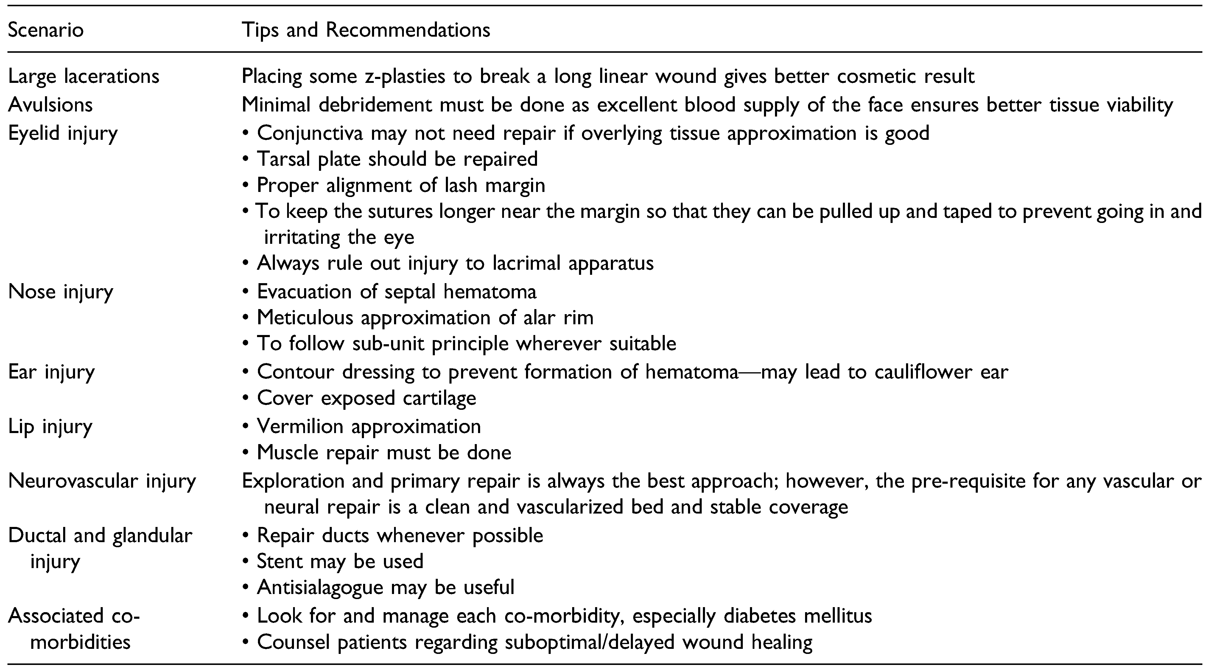
With the above discussion, we realize that complicated facial lacerations are actually a spectrum of conditions that need a protocol-based approach for optimum outcomes. Furthermore, each component of this spectrum demands separate attention. While searching through the available literature, we found some guidelines regarding the management of individual problem areas, like auricle, nose, or lips. However, given the complexity of these injuries, no single guideline exists encompassing the entire spectrum of complicated facial lacerations.
Semer has given a general guideline regarding the use of appropriate suture material according to the area of involvement in case of facial lacerations. The author has recommended non-absorbable sutures, mainly nylon and polypropylene, in sizes ranging from 4-0 to 6-0 for the repair of the skin of different parts of the face. For subcutaneous tissue, muscles, and mucosa of the nose and oral cavity, absorbable sutures like polydioxanone or polyglactin are recommended [26]. Sabatino and Moskovitz gave a generalized guideline for the repair of lacerations of different facial units and Jaffe et al gave basic guidelines for the management of lacerations of the eyelid, lip, ear, and nose for the emergency care physicians [27,28]. The recommendations vary a little according to individual preference or their institutional protocols, but the basic essence remains the same. In the absence of a fixed protocol or algorithm for the management of such injuries, we tried to formulate one based on the recommendations of different authors and adding from our own experience (Chart 1).
The complex anatomy of the face makes the repair of different parts of the face a challenge. While the basic surgical approach remains the same, there are some special points regarding the repair of specialized facial units (Table 1).
Cmtr 16 00007 i001

Conclusion

Complicated facial lacerations pose a multitude of challenges to the surgical team. Protocol-based management of injuries to the different parts and specialized structures of the face can be extremely helpful. Knowledge about the challenges faced in these injuries makes the emergency surgeon better prepared to anticipate and manage them effectively. The role of a specialized facial trauma team consisting of a plastic surgeon, maxillofacial surgeon, and trauma surgeon in the management of such complicated facial injuries cannot be emphasized enough.

Funding

The author(s) received no financial support for the research, authorship, and/or publication of this article.

Declaration of Conflicting Interests

The author(s) declared no potential conflicts of interest with respect to the research, authorship, and/or publication of this article.

References

  1. Yamamoto, R.; Homma, K.; Masuzawa, Y.; et al. Early complications following facial laceration repair performed by emergency physicians after one year of wound closure training. AEM Educ Train. 2018, 2, 259–268. [Google Scholar] [CrossRef] [PubMed]
  2. Badeau, A.; Lahham, S.; Osborn, M. Management of complex facial lacerations in the emergency department. Clin Prac Cases Emerg Med. 2017, 1, 162–165. [Google Scholar] [CrossRef] [PubMed]
  3. Bagheri, S.C.; Dierks, E.J.; Kademani, D.; et al. Application of a facial injury severity scale in craniomaxillofacial trauma. J Oral Maxillofac Surg. 2006, 64, 408–414. [Google Scholar] [CrossRef] [PubMed]
  4. Gao, H.; Wang, W.; Geng, Y.; Shao, Y. Clinical experience in emergency management of severe facial trauma. J Craniofac Surg. 2020, 31, e121–e123. [Google Scholar] [CrossRef] [PubMed]
  5. Kretlow, J.; McKnight, A.; Izaddoost, S. Facial soft tissue trauma. Semin Plast Surg. 2010, 24, 348–356. [Google Scholar] [CrossRef] [PubMed]
  6. Novelli, G.; Daleffe, F.; Birra, G.; et al. Negative pressure wound therapy in complex cranio-maxillofacial and cervical wounds. Int Wound J. 2018, 15, 16–23. [Google Scholar] [CrossRef]
  7. Abdul Razak, N.; Nordin, R.; Abd Rahman, N.; Ramli, R. A retrospective analysis of the relationship between facial injury and mild traumatic brain injury. Dent Traumatol. 2017, 33, 400–405. [Google Scholar] [CrossRef]
  8. Chang, E.L.; Rubin PA, D. Management of complex eyelid lacerations. Int Ophthalmol Clin. 2002, 42, 187–201. [Google Scholar] [CrossRef]
  9. Sharma, K.; Goswami, S.C.; Baruah, D.K. Auricular trauma and its management. Indian J Otolaryngol Head Neck Surg. 2006, 58, 232–234. [Google Scholar] [CrossRef] [PubMed]
  10. Kita, A.; Kim, I.; Ishiyama, G.; Ishiyama, A. Perilymphatic fistula after penetrating ear trauma. Clin Pract Cases Emerg Med. 2019, 3, 115–118. [Google Scholar] [CrossRef]
  11. Rohrich, R.J.; Griffin, J.R.; Ansari, M.; Beran, S.J.; Potter, J.K. Nasal reconstruction? Beyond aesthetic subunits: A 15-year review of 1334 cases. Plast Reconstr Surg. 2004, 114, 1405–1416.discussion 1417-9. [Google Scholar] [CrossRef] [PubMed]
  12. Zide, B.M.; Swift, R. How to block and tackle the face. Plast Reconstr Surg. 1998, 101, 840–851. [Google Scholar] [CrossRef]
  13. James, D. Reliable method for closing lacerations across the vermilion border of the lip. Can Fam Physician. 1998, 44, 47–8. [Google Scholar] [PubMed]
  14. Smith, J.; Maconochie, I. Should we glue lip lacerations in children? Arch Dis Child. 2003, 88, 83–84. [Google Scholar] [CrossRef] [PubMed]
  15. Shafaiee, Y.; Shahbazzadegan, B. Facial nerve laceration and its repair. Trauma Mon. 2016, 21, e22066. [Google Scholar] [CrossRef]
  16. Gordin, E.; Lee, T.S.; Ducic, Y.; Arnaoutakis, D. Facial nerve trauma: Evaluation and considerations in management. Craniomaxillofacial Trauma Reconstr. 2015, 8, 1–13. [Google Scholar] [CrossRef]
  17. White, H.; Rosenthal, E. Static and dynamic repairs of facial nerve injuries. Oral Maxillofac Surg Clin. 2013, 25, 303–312. [Google Scholar] [CrossRef]
  18. Myckatyn, T.M.; Mackinnon, S.E. The surgical management of facial nerve injury. Clin Plast Surg. 2003, 30, 307–318. [Google Scholar] [CrossRef]
  19. Ricci Volpato, L.; Medeiros Paz, A.L.; Caetano, R.S. Borges Reconstruction of the parotid duct. Ann Maxillofac Surg. 2018, 8, 140–142. [Google Scholar] [CrossRef]
  20. Lewis, G.; Knottenbelt, J.D. Parotid duct injury: Is immediate surgical repair necessary? Injury. 1991, 22, 407–409. [Google Scholar] [CrossRef]
  21. Aloosi, S.N.; Khoshnaw, N.; Ali, S.M.; Muhammad, B.A. Surgical management of Stenson’s duct injury by using double J stent urethral catheter. Int J Surg Case Rep. 2015, 17, 75–78. [Google Scholar] [CrossRef] [PubMed]
  22. Lewkowicz, A.A.; Hasson, O.; Nahlieli, O. Traumatic injuries to the parotid gland and duct. J Oral Maxillofac Surg. 2002, 60, 676–680. [Google Scholar] [CrossRef] [PubMed]
  23. Öztürk, M.B.; Barutca, S.A.; Keskin, E.S.; Atik, B. Parotid duct repair with intubation tube: Technical note. Ann Maxillofac Surg. 2017, 7, 129–131. [Google Scholar] [CrossRef] [PubMed]
  24. Winstead, M.L.; Clegg, D.J.; Heidel, R.E.; Ledderhof, N.J.; Gotcher, J.E. Fall-related facial trauma: A retrospective review of fracture patterns and medical comorbidity. J Oral Maxillofac Surg. 2021, 79, 86431209–870. [Google Scholar] [CrossRef]
  25. Aita, T.G.; Pereira Stabile, C.L.; Dezan Garbelini, C.C.; Vitti Stabile, G.A. Can a facial injury severity scale be used to predict the need for surgical intervention and time of hospitalization? J Oral Maxillofac Surg. 2018, 76, e1–1280. [Google Scholar] [CrossRef]
  26. Semer, N. Practical Plastic Surgery for Nonsurgeons; Authors Choice Press: Lincoln, 2007. [Google Scholar]
  27. Sabatino, F.; Moskovitz, J.B. Facial wound management. Emerg Med Clin. 2013, 31, 529–538. [Google Scholar] [CrossRef] [PubMed]
  28. Brown, D.J.; Jaffe, J.E.; Henson, J.K. Advanced laceration management. Emerg Med Clin. 2007, 25, 83–99. [Google Scholar] [CrossRef] [PubMed]
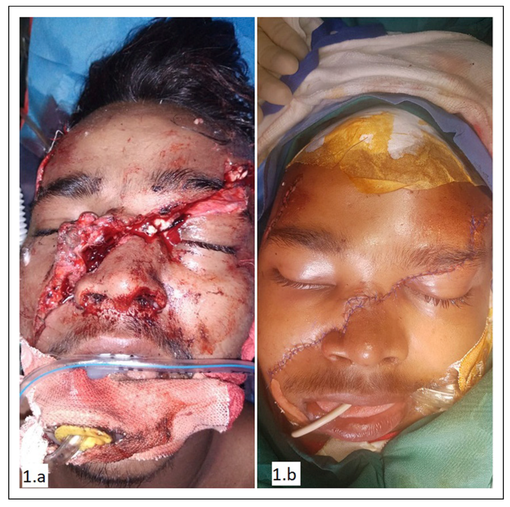
Figure 1. Large laceration and its repair.
Cmtr 16 00007 g001
Figure 2. Avulsion injury and its repair.
Cmtr 16 00007 g002
Figure 3. Partial thickness eyelid laceration and its repair.
Cmtr 16 00007 g003
Figure 4. Full thickness eyelid injury and its repair.
Cmtr 16 00007 g004
Figure 5. Auricle laceration and its repair.
Cmtr 16 00007 g005
Figure 6. Nasal laceration and its repair.
Cmtr 16 00007 g006
Figure 7. Full thickness lip laceration and its repair.
Cmtr 16 00007 g007
Figure 8. Laceration of facial nerve and its repair.
Cmtr 16 00007 g008
Figure 9. Parotid duct injury and its repair.
Cmtr 16 00007 g009
Figure 10. Extensive facial avulsion and immediate post-repair.
Cmtr 16 00007 g010
Figure 10. Cellulitis and flap necrosis, debridement, coverage, and follow-up image of extensive facial avulsion with comorbidity.
Cmtr 16 00007 g011
Table 1. Recommendations for special scenarios in facial laceration repair. Scenario.
Cmtr 16 00007 i001